Functional conservation and divergence of Miscanthus lutarioriparius GT43 gene family in xylan biosynthesis
Bạn đang xem bản rút gọn của tài liệu. Xem và tải ngay bản đầy đủ của tài liệu tại đây (6.6 MB, 19 trang )
Wang et al. BMC Plant Biology (2016) 16:102
DOI 10.1186/s12870-016-0793-5
RESEARCH ARTICLE
Open Access
Functional conservation and divergence of
Miscanthus lutarioriparius GT43 gene family
in xylan biosynthesis
Xiaoyu Wang1,2, Qi Tang1, Xun Zhao1,2, Chunlin Jia3, Xuanwen Yang1, Guo He1, Aimin Wu4, Yingzhen Kong5,
Ruibo Hu1* and Gongke Zhou1*
Abstract
Background: Xylan is the most abundant un-cellulosic polysaccharides of plant cell walls. Much progress in xylan
biosynthesis has been gained in the model plant species Arabidopsis. Two homologous pairs Irregular Xylem 9
(IRX9)/9L and IRX14/14L from glycosyltransferase (GT) family 43 have been proved to play crucial roles in xylan
backbone biosynthesis. However, xylan biosynthesis in grass such as Miscanthus remains poorly understood.
Results: We characterized seven GT43 members in M. lutarioriparius, a promising bioenergy crop. Quantitative
real-time RT-PCR (qRT-PCR) analysis revealed that the expression of MlGT43 genes was ubiquitously detected in the
tissues examined. In-situ hybridization demonstrated that MlGT43A-B and MlGT43F-G were specifically expressed in
sclerenchyma, while MlGT43C-E were expressed in both sclerenchyma and parenchyma. All seven MlGT43 proteins
were localized to Golgi apparatus. Overexpression of MlGT43A-E but not MlGT43F and MlGT43G in Arabidopsis irx9 fully
or partially rescued the mutant defects, including morphological changes, collapsed xylem and increased xylan contents,
whereas overexpression of MlGT43F and MlGT43G but not MlGT43A-E complemented the defects of irx14, indicating that
MlGT43A-E are functional orthologues of IRX9, while MlGT43F and MlGT43G are functional orthologues of IRX14. However,
overexpression of all seven MlGT43 genes could not rescue the mucilage defects of irx14 seeds. Furthermore, transient
transactivation analyses of MlGT43A-E reporters demonstrated that MlGT43A and MlGT43B but not MlGT43C-E were
differentially activated by MlSND1, MlMYB46 or MlVND7.
Conclusion: The results demonstrated that all seven MlGT43s are functionally conserved in xylan biosynthesis during
secondary cell wall formation but diversify in seed coat mucilage xylan biosynthesis. The results obtained provide deeper
insight into xylan biosynthesis in grass, which lay the foundation for genetic modification of grass cell wall components
and structure to better suit for next-generation biofuel production.
Keywords: Miscanthus lutarioriparius, Glycosyltransferase family 43, Xylan biosynthesis, Secondary cell wall, Seed coat
mucilage
Highlight
The functional roles of M. lutarioriparius GT43 family
genes are conserved and diversified in xylan biosynthesis.
Background
Plant cell walls are complex and dynamic structures composed mainly of polysaccharides (cellulose, hemicellulose
* Correspondence: ;
1
Qingdao Institute of Bioenergy and Bioprocess Technology, Key Laboratory
of Biofuels, Qingdao Engineering Research Center of Biomass Resources and
Environment, Chinese Academy of Sciences, Qingdao 266101, PR China
Full list of author information is available at the end of the article
and pectin), phenolic compounds (lignin) and glycoproteins [1]. Xylans are the major hemicellulosic saccharides
in the primary cell walls of grasses and the secondary cell
walls of grasses and dicots, ranking as the second most
abundant polysaccharides in nature [2]. Xylans are mainly
composed of a linear backbone of β-(1,4)-linked D-xylosyl
residues with various sidechains that vary among different
plant species and tissue types [3]. Based on the sidechain
substitutions, xylans can generally be classified as
(methyl)glucuronoxylan (GX), arabinoxylan (AX), and glucuronoarabinoxylan (GAX) [3]. As the major xylan in
© 2016 Wang et al. Open Access This article is distributed under the terms of the Creative Commons Attribution 4.0
International License ( which permits unrestricted use, distribution, and
reproduction in any medium, provided you give appropriate credit to the original author(s) and the source, provide a link to
the Creative Commons license, and indicate if changes were made. The Creative Commons Public Domain Dedication waiver
( applies to the data made available in this article, unless otherwise stated.
Wang et al. BMC Plant Biology (2016) 16:102
dicot plants, GX is usually decorated with α-1,2-linked
glucuronic acid (GlcA) or 4-O-methylglucuronic acid
(MeGlcA), and acetylated at C-2 or C-3 [3, 4]. AX has α1,3-linked arabinose (Ara) sidechains, and presents as
typical hemicellulose components in starchy endosperm
of cereal grains [3]. GAX is the predominant hemicellulose in grass cell walls, and has sidechains of α-1,2 or α1,3-linked arabinose (Ara) and GlcA residues [3]. In
addition, GX in angiosperm and GAX in several gymnosperm species contain a tetrasaccharide sequence [β-D-Xyl(1,3)-α-L-Rha-(1,2) -α-D-GalA-(1,4)-D-Xyl] at the reducing
end [5–7]. However, no such oligosaccharide has yet been
identified for xylans in grasses [8, 9]. It is still in controversy
whether this oligosaccharide functions as a primer or as a
terminator in xylan backbone biosynthesis [10].
Several xylan-related mutants named as irregular
xylem (irx) due to secondary cell wall deficiencies have
been identified in Arabidopsis by reverse genetics approaches [11, 12]. Most of these identified genes encode
putative glycosyltransferases (GT) that are involved in
the biosynthesis of xylan. IRX9/IRX9L and IRX14/
IRX14L from GT43 family as well as IRX10/IRX10L
from GT47 family are responsible for the biosynthesis of
xylan backbone [13–19]. IRX9, IRX10 and IRX14 play
dominant roles in xylan backbone biosynthesis, and mutations in each gene lead to reduced xylan content and
growth defect. By contrast, IRX9L, IRX10L and IRX14L
seem to perform partially redundant roles together with
their close homologues, as loss-function of these genes
have no observable phenotypes and they only partially
complement the phenotypes of irx9, irx10 and irx14 mutants. In addition, double mutations in each gene pairs
dramatically enhance the phenotypes of the single mutant
[13, 14, 18, 19]. However, a recent study proposed that
these gene pairs play equivalent roles in xylan biosynthesis
[20]. Furthermore, two members of DUF579 domaincontaining proteins, IRX15 and IRX15L, are essential for
the normal elongation of xylan backbone [21, 22]. IRX7/
IRX7L from GT47 family, IRX8 and PARVUS from GT8
family are required for the biosynthesis of the reducing
end oligosaccharide [5, 23–26]. Mutations in these genes
lead to almost entirely loss of the tetrasaccharide accompanied with reduced xylan contents, while the xylan backbone elongation activity is not disturbed [5, 23–26].
Recently biochemical and genetic studies have also led to
the identification of several genes that are required for the
sidechain modifications of xylan. For instance, GLUCURONIC ACID SUBSTITUTION OF XYLAN (GUX) 1,
GUX2, GUX4 and GUX5 from GT8 family are proposed to
catalyze the addition of GlcA and MeGlcA sidechains to
GX backbone [20, 27–29]. GLUCURONOXYLAN METHYLTRANSFERASE (GXMT) 1, a DUF579 domain protein,
has been revealed to be responsible for the 4-Omethylation of GlcA residues in GX [30]. In addition,
Page 2 of 19
ESKIMO1/TRICHOME BIREFRINGENCE-LIKE (TBL)
29, a DUF231 domain protein, is required for the Oacetylation of xylan backbone [31, 32]. Moreover, several
XYLAN ARABINOSYLTRANSFERASE (XAT), members
of GT61 family proteins from rice and wheat, are responsible for transferring the Ara residues onto xylan backbone
[33, 34]. XYLOSYL ARABINOSYL SUBSTITUTION OF
XYLAN (XAX) 1, another member from GT61 family in
rice, is involved in transferring the Xyl residues in β-Xylp-(1
→ 2)-α-Araf -(1 → 3) sidechain [34].
Grass xylans have several unique features compared to
those from dicots. GX is the most abundant hemicellulose in dicots, while grass xylans usually contain many
Ara residue substitutions and thus are termed as GAX
or AX [3]. Even though there are clear differences in xylan structure between grasses and dicots, accumulating
evidence implicates that GT43 members are functionally
conserved in xylan biosynthesis between dicots and
monocots. For example, four rice IRX9 orthologues
OsGT43A, OsGT43C, OsGT43E and OsGT43F can fully
or partially rescue the xylan defect phenotype of irx9,
while OsGT43J is able to complement the xylan defect
phenotype of irx14 in Arabidopsis [35, 36]. Three poplar
IRX9 orthologues PtrGT43A, PtrGT43B and PtrGT43E
are capable of rescuing the defects of irx9, whereas the
other two IRX14 orthologues PtrGT43C and PtrGT43D
are able to complement the phenotypes of irx14 [37].
Furthermore, it has been demonstrated that rice and
poplar GT43 family proteins are evolved to retain two
functionally non-redundant groups involved in xylan
backbone biosynthesis [36–38]. Additionally, two GT43
members GhGT43A1 and GhGT43C1 from cotton have
been revealed to be functional orthologues of Arabidopsis IRX9 and IRX14, respectively, and have been shown
to participate in xylan backbone biosynthesis during
fiber development [39].
Miscanthus is a perennial rhizomatous grass with superior characteristics as a bioenergy plant such as high
photosynthetic efficiency, low fertilizer and water demand, wide adaptability and high biomass yield. It has
attracted increasing attention and concern worldwide as
an ideal lignocellulosic feedstock for next-generation
bioenergy production [40–42]. Hemicelluloses account for
29–42 % of the Miscanthus cell walls [43], and the most
abundant hemicellulosic polysaccharide is AX [43, 44],
which is also the typical xylan in grass cell walls [45]. It
has been shown that hemicellulose exerts dominant and
positive effects on biomass digestibility by affecting
cellulose crystallinity after pre-treatment with alkali or
acid [46]. Although much progress has been gained in
the understanding of xylan biosynthesis in the model
plant Arabidopsis thaliana, relatively less is known
about xylan biosynthesis in grasses. To the best of
our knowledge, none of GTs responsible for the
Wang et al. BMC Plant Biology (2016) 16:102
biosynthesis of xylan has been isolated and characterized in Miscanthus as yet.
To provide insight into xylan biosynthesis in Miscanthus, we identified seven GT43 genes in M. lutarioriparius and characterized their functional roles in xylan
biosynthesis. Complementation assay including plant
height, irregular xylem cells in stem cross sections and
xylose content measurements revealed that MlGT43
genes have evolved into two distinct functional groups,
in which MlGT43A-E are orthologous to IRX9, while
MlGT43F and MlGT43G are orthologous to IRX14.
Furthermore, our results indicated that substantial divergence has occurred in the functional roles of MlGT43s
during xylan biosynthesis especially in seed coat mucilage. The results presented deepened our understanding
of xylan biosynthesis in grasses and may lay the foundation for future genetic manipulation of Miscanthus cell
wall structure and components.
Results
Isolation of GT43 genes in M. lutarioriparius
To identify the GT43 family in M. lutarioriparius, the
amino acid sequences of four Arabidopsis GT43 members
Page 3 of 19
were used as query baits to BLAST against the draft genome sequences of M. lutarioriparius, and seven GT43
orthologous genes were identified. Specific primers were
designed and seven candidate genes encoding putative
GT43 proteins designated as MlGT43A to MlGT43G were
obtained by PCR in M. lutarioriparius. As indicated in
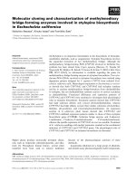
Fig. 1a, all seven proteins had a conserved structure and
ranged in size from 358 to 451 amino acids. Pairwise comparison of the amino acid sequences showed that
MlGT43C and MlGT43D shared the highest sequence
similarity (75.3 %), while MlGT43D and MlGT43G shared
the lowest sequence similarity (43.3 %) (Fig. 1b).
Deduced MlGT43A and MlGT43B amino acid sequences shared the highest sequence identities with Arabidopsis IRX9 (37 and 41 %), and MlGT43C-E shared
relatively higher sequence identities with IRX9L (42, 48
and 53 %) than with IRX14 or IRX14L. By contrast,
MlGT43F and MlGT43G proteins had the highest sequence identities with IRX14 and IRX14L (59 and 37 %)
than with IRX9 (Additional file 1: Table S1).
Furthermore, the gene structure of each MlGT43 was
obtained through the alignment of their coding sequences
and genomic sequences (Fig. 1c). All MlGT43 genes
Fig. 1 Sequence alignment, identities and gene structure of MlGT43. a Sequence alignment of seven MlGT43 proteins. b Sequence identities and
similarities among MlGT43 proteins. The highest and lowest in sequence identity and similarity are outlined. c Gene structure of MlGT43 genes.
Exons and introns are represented by filled boxes and lines, respectively. The sizes of exons and introns are proportional to the scale at bottom
Wang et al. BMC Plant Biology (2016) 16:102
shared very similar gene structure in terms of intron
number and exon length. They all contained three
exons and two introns. In addition, the intron phases
with respect to codons were well conserved among
different MlGT43 genes.
Phylogenetic analysis of GT43 members from
M. lutarioriparius and other plant species
To gain insight into the origin and evolutionary history of
the GT43 family, we further identified GT43 proteins from
nine other currently sequenced genomes that cover a wide
spectrum of plant taxonomic groups including moss (Physcomitrella patens), spikemoss (Selaginella moellendorffii),
the monocot angiosperms (Oryza sativa, Brachypodium
distachyon and Sorghum bicolor), and the dicot angiosperms (Arabidopsis thaliana, Populus trichocarpa, Medicago truncatula and Vitis vinifera). Totally 57 GT43
proteins were identified from these nine plant species
(Additional file 2) and a phylogenetic tree was constructed
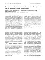
with these GT43 proteins (Fig. 2a). The phylogenetic tree
separated all GT43 proteins into three distinct subfamilies
designated as IRX9, IRX9L and IRX14/IRX14L, which was
similar to the previous studies [13, 38]. The seven GT43
proteins from Miscanthus were classified into the three
subfamilies. MlGT43A and MlGT43B were clustered into
the IRX9 subfamily, MlGT43C-E were classified into the
IRX9L subfamily, while MlGT43F and MlGT43G were distributed into the IRX14/IRX14L subfamily.
The distribution of the three subgroups among the ten
plant species varied within each subfamily (Fig. 2b). It is
noteworthy that the number of GT43 proteins in the
monocot species seems to be higher than that of the
dicot species, at least it is the case for the selected plant
species. For example, there were 10, 10, 10 and 7 members in the monocot species O. sativa, B. distachyon,
S. bicolor and M. lutarioriparius, whereas the number
of GT43 in the dicot species A. thaliana, P. trichocarpa, M. truncatula and V. vinifera were 4, 7, 4 and
4, respectively. In addition, the members of IRX9 and
IRX9L subfamilies in the monocot angiosperms were
generally higher than those of the dicot species. For
instance, the IRX9 subfamily accounted for 40, 40, 40 and
28 % in the monocot species O. sativa, B. distachyon,
S. bicolor and M. lutarioriparius, respectively, whereas
the percentages of the IRX9 subfamily in the dicot
species A. thaliana, P. trichocarpa, M. truncatula and
V. vinifera were 25, 25, 28 and 25 %, respectively.
Noticeably, no IRX9 subfamily members were present
in P. patens and S. moellendorffii.
MlGT43 genes are ubiquitously expressed and have
specific expressions in stem cells
To investigate the expression patterns of MlGT43
genes, we first used the quantitative real-time RT-PCR
Page 4 of 19
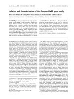
(qRT-PCR) to examine their expressions across seven different tissues. As shown in Fig. 3a, all seven MlGT43
genes were ubiquitously expressed in seven different
tissues examined, but their relative expression levels differed significantly. For example, MlGT43A, MlGT43D and
MlGT43E genes shared similar expression patterns with
predominant expressions in leaf, whereas the expressions
of MlGT43B and MlGT43G genes were relatively lower.
MlGT43C and MlGT43F genes were broadly expressed in
the majority of the tissues, while especially higher expressions were detected in the basal stem. Furthermore, all
MlGT43 genes except MlGT43B exhibited higher expressions in the basal stem than in the upper stem.
To obtain more detailed expression patterns of
MlGT43 genes in specific cell types, we further performed the in situ hybridization analysis to examine
their expressions in the 11th internode of the stem. For
all seven genes, intense hybridization signals were observed in sclerenchyma cells and vascular bundle fiber
cells, the cell types undergoing secondary wall thickening (Fig. 3b-h). Moreover, relatively weak hybridization
signals were also observed for MlGT43C-E in parenchyma cells. By contrast, the control hybridized with
sense probes did not show any signals in vascular bundle
or sclerenchyma cells (Fig. 3g). These results suggest
that MlGT43 genes may participate in diverse plant development processes especially in the secondary cell wall
formation.
MlGT43 members are targeted to Golgi apparatus
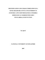
To investigate the subcellular localization of MlGT43
proteins, we constructed fluorescently tagged fusion proteins by fusing Yellow Fluorescent Protein (YFP) to the
C terminus of each MlGT43 protein. The recombinant
constructs were transiently co-expressed in Nicotiana
benthamiana leaf epidermal cells with the Golgi marker
Man49-mCherry [47]. Examination of the fluorescent
signals revealed that seven YFP-tagged MlGT43s all exhibited a punctate distribution, and the pattern perfectly
matched with that of Man49-mCherry (Fig. 4), whereas
the YFP control protein had signals throughout the cytoplasm and the nucleus (data not shown). The colocalization of MlGT43 proteins with the Golgi marker
indicate that MlGT43s are Golgi-localized proteins.
MlGT43 genes rescue the morphological defects of irx9 or
irx14
To reveal whether MlGT43 genes perform the same
functions as IRX9 and IRX14 orthologues in Arabidopsis,
we examined their abilities to rescue the morphological
defects of irx9 and irx14. Due to the severely dwarfed
plant stature and poor fertility of homozygous irx9
plants [5], we used the heterozygous line for the transformation with the 35S:MlGT43s constructs. Positive
Wang et al. BMC Plant Biology (2016) 16:102
Page 5 of 19
Fig. 2 Phylogenetic analysis of GT43 family from Miscanthus and nine other plant species. a Phylogenetic tree of 64 GT43 proteins from ten plant
species. The sequences of 64 GT43 proteins were aligned using ClustalW and their phylogenetic relationship was analyzed using the NeighborJoining method in MEGA 6.0. Numbers at nodes indicate the percentage bootstrap scores and only bootstrap values higher than 50% from 1,000
replicates are shown. MlGT43 proteins are marked with asterisks. b Distribution of the GT43 proteins from selected plant lineages. Pp, Physcomitrella
patens; Sm, Selaginella moellendorffii; At, Arabidopsis thaliana; Pt, Populus trichocarpa; Mt, Medicago truncatula; Vv, Vitis vinifera; Os, Oryza
sativa; Bd, Brachypodium distachyon; Sb, Sorghum bicolor; Ml, Miscanthus lutarioriparius
transgenic lines for each construct were tested for the
presence of MlGT43 genes in homozygous irx9 and
irx14 background by semi-quantitative RT-PCR (Fig. 5a).
Homozygous T2 plants from at least two independent
transformants with higher expressions were used for the
phenotypic analyses.
The growth of the irx9 plants was characterized by the
dwarf stature, smaller rosette size and dark-green leaves
under our growth conditions, which is similar to the
previous reports [5, 12]. Overexpression of MlGT43A-E
genes in irx9 displayed an intermediate growth phenotype between the mutant and the wild type (WT) in
Wang et al. BMC Plant Biology (2016) 16:102
Page 6 of 19
Fig. 3 Expression patterns of MlGT43 genes. a Expression analysis of MlGT43 genes by qRT-PCR. Relative expression levels in seven tissues were
normalized using MlACT11 as the reference gene. For each gene, the tissues with the lowest expression level are set to 1. Data are the means ± SE of
three biological replicates. b In situ localization of MlGT43 genes in Miscanthus stem. Cross-sections of stems were hybridized with digoxigenin-labeled
antisense MlGT43A (b), MlGT43B (c), MlGT43C (d), MlGT43D (e), MlGT43E (f), MlGT43F (g), MlGT43G (h), or sense (i) RNA probes, and the hybridization
signals were detected with alkaline phosphatase-conjugated antibody and were shown as purple color. pv, pitted vessel; x, xylem; ph, phloem;
pa, parenchyma; sc, sclerenchyma. Bar = 100 μm
terms of rosette size and inflorescence height. The rosette diameters of the complemented plants increased by
two- to three-fold, and the inflorescence stems were
two- to four-fold taller compared to the irx9 plants after
four weeks of growth (Fig. 4b, d), suggesting that the irx
phenotype may be partially complemented in these
transformants. By contrast, transformants of MlGT43F
or MlGT43G overexpression in irx9 mutant exhibited a
morphology resembled of the irx9 mutant, indicating
that MlGT43F and MlGT43G were unable to complement the irx9 phenotypes (Fig. 4b, d, f ).
The growth of irx14 mutant did not show any other
obvious phenotypes except for a slight reduction in
plant height compared to WT (Fig. 4c, e) as described
previously [14]. The height of all MlGT43 complemented irx14 plants was indistinguishable from that of
Wang et al. BMC Plant Biology (2016) 16:102
Page 7 of 19
Fig. 4 Subcellular localization of YFP-tagged MlGT43 proteins. YFP-tagged MlGT43 proteins were transiently expressed in leaf epidermal cells of
Nicotiana benthamiana, and their subcellular locations were examined with a laser scanning confocal microscope. The single-plane confocal
micrographs of MlGT43 proteins fused with C-terminal YFP, the Golgi marker Man49-mCherry, differential interference contrast (DIC) image, and
merged YFP and mCherry channels are shown. Note the superimposition of YFP-MlGT43s and Man49-mCherry signals. Bar = 20 μm
irx14 or WT plants, thus it is hard to evaluate the ability of seven MlGT43 genes to complement the irx14
mutant merely judged from their growth phenotypes.
Subsequently, xylem morphology, xylan immunolocalization and cell wall monosaccharide compositions will
be further examined in the transgenic plants to determine the abilities of MlGT43s to complement the irx14
phenotypes.
Microscopic analysis of the secondary cell wall
To demonstrate whether the morphological complementation by MlGT43 genes could be accompanied with the
rescue of xylem morphology, the basal inflorescence
stems of each complemented line were sectioned and
observed by light and transmission electron microscopy.
Toluidine blue O (TBO) staining was performed on
stem sections of WT, irx9, irx14 and complemented
Wang et al. BMC Plant Biology (2016) 16:102
Page 8 of 19
Fig. 5 Expression of seven MlGT43 genes in Arabidopsis irx9 or irx14 mutants. a RT-PCR detection of the MlGT43 transcripts in the complemented
irx9 or irx14 plants. The Arabidopsis UBQ10 gene was used as a reference. b, d, f Phenotype of four-, six- and eight-week-old soil-grown WT, irx9
and MlGT43s complemented irx9 plants. c, e, g Phenotype of four-, six- and eight-week-old soil-grown WT, irx14 and MlGT43s complemented irx14
plants. h Stem height of the WT, irx9 and MlGT43s complemented irx9 plants through 40, 47, 57 days of growth. i Stem height of the WT, irx14
and MlGT43s complemented irx14 plants through 40, 47, 57 days of growth. Data are means ± SD from at least twelve plants for each background.
Two homozygous T3 lines of MlGT43s complemented irx9 or irx14 were used in the analysis
Wang et al. BMC Plant Biology (2016) 16:102
plants to examine the morphology of secondary cell
walls. As shown in Fig. 6, all MlGT43A-E complemented
irx9 plants exhibited dramatically thickened cell walls in
interfascicular fibers compared to irx9. The majority of
xylem vessels in MlGT43A and MlGT43B complemented irx9 plants were characterized by large open round
cells comparable to those in WT plants (Fig. 6C1, D1, L1,
M1). In addition, the xylem vessels of MlGT43C,
MlGT43D or MlGT43E complemented irx9 plants were
usually smaller in size with occasionally irregular shapes,
probably due to the not fully thickened cell walls
Page 9 of 19
compared to WT (Fig. 6 E1-G1, N1-P1). By contrast, overexpression of MlGT43F or MlGT43G in irx9 could not restore the collapsed vessels and the weakly thickened
interfascicular fibers in irx9 (Fig. 6 H1, I1, Q1, R1), which
is in consistency with their growth phenotypes (Fig. 5b, d).
The homozygous irx14 plants also showed collapsed
xylem vessels and thinner secondary cell walls, which is
consistent with the previous study [15]. Overexpression of
either MlGT43F or MlGT43G could almost fully rescue
the irx phenotype of irx14 as witnessed by a relatively less
irregular vessel cells compared to irx14. However, the
Fig. 6 Morphology of xylem and interfascicular fibers of WT, irx9, irx14 and MlGT43 complemented plants. Stems of eight-week-old plants were
sectioned (8 μm-thick) and stained with TBO for examination of the morphology of vessels, xylary fibers and interfascicular fibers. A1-I1, interfasicular
fibers for WT, irx9 and MlGT43 complemented irx9 plants. A2-I2, interfasicular fibers for WT, irx14 and MlGT43 complemented irx14 plants. J1-R1, xylary
fibers and vessels for WT, irx9 and MlGT43 complemented irx9 plants. J2-R2, xylary fibers and vessels for WT, irx14 and MlGT43 complemented irx14
plants. At least two homozygous T3 lines of MlGT43s complemented irx9 or irx14 were used in the analysis. Images for each tissue are set as the same
magnification. Bar = 50 μm
Wang et al. BMC Plant Biology (2016) 16:102
complemented lines still retained relatively thinner cell
walls in both interfascicular fibers and xylem vessels compared to WT (Fig. 6 H2, I2, Q2, R2). By contrast, overexpression of MlGT43A-E in irx14 displayed a collapsed
xylem vessel and thinner fiber cell wall phenotype that
was indistinguishable from the irx14 mutant (Fig. 6 C2G2, L2-P2), indicating that MlGT43A-E genes could not
rescue the defects of irx14.
Transmission electron microscopy confirmed that the
thickness of interfascicular fiber cell walls of the
MlGT43A-E complemented irx9 plants was intermediate
between irx9 and WT (Fig. 7a and Table 1). Meanwhile,
the wall thickness of xylary fibers and vessels in
MlGT43A-E complemented irx9 lines was also significantly increased but not restored to the WT level. By
contrast, the wall thickness of interfascicular fibers,
xylary fibers and vessels of MlGT43F or MlGT43G complemented irx9 plants was similar to that of the irx9
mutant (Fig. 7a and Table 1). The wall thickness of interfascicular fibers, xylary fibers and vessels for MlGT43F
or MlGT43G complemented irx14 plants was intermediate between irx14 and WT, while the wall thickness for
MlGT43A-E complemented irx14 lines was similar to
that of irx14 (Fig. 7b and Table 1). Together, these
results indicate that MlGT43A-E can fully or partially
rescue the irx9 but not the irx14 phenotypes, while
MlGT43F and MlGT43G can complement the irx14 but
not the irx9 defects.
Immunolocalization of xylan in MlGT43s complemented
lines
To investigate whether the phenotypes of the complemented plants are correlated with xylan deposition in secondary cell walls, we performed immunolocalization of xylan
using the xylan-directed monoclonal antibody LM10,
which recognizes unsubstituted or low-substituted xylan
[48], to examine the distribution of xylan in the cell walls.
As indicated in Fig. 8, strong fluorescence signals were
present in the cell walls of interfascicular fibers and xylem
cells in the WT stem, however, relatively weaker signals
were detected in the corresponding tissues of the irx9
plants, although the overall pattern of labeling was unchanged compared with the WT plants (Fig. 8 A1, B1). In
MlGT43A and MlGT43B complemented irx9 lines, the intensity of fluorescence signals was almost restored to the
WT level, and the overall pattern of labeling was almost
identical to that of WT, indicating that the GX content in
interfascicular fibers and xylem cells was nearly restored
to the WT level (Fig. 8 C1, D1). The LM10 signals in the
MlGT43C-E complemented irx9 plants were intermediate
between irx9 and WT plants (Fig. 8 E1-G1). By contrast,
the LM10 signals for MlGT43F and MlGT43G complemented irx9 lines were relatively weaker compared with
the others, and the intensity was comparable to that of the
Page 10 of 19
irx9 mutant (Fig. 8 H1, I1). As for the irx14 background,
the intensity of fluorescence signals of MlGT43F and
MlGT43G complemented lines was comparable to that of
WT in xylem cells and interfascicular fibers (Fig. 8 H2,
I2). By contrast, MlGT43A-E complemented irx14 lines
exhibited nearly equal signal intensity to the irx14 mutant
(Fig. 8 C2-G2). These results indicate that MlGT43A-E
perform a similar biochemical function as IRX9, whereas
MlGT43F and MlGT43G share a conserved biochemical
function with IRX14, thus leading to a restoration of normal xylan synthesis in their complemented plants.
Analysis of cell wall composition
To determine whether the complementation of xylem
morphology and xylan deposition is correlated with the
restoration of chemical composition, we measured the
monosaccharide composition, cellulose and lignin contents of the transgenic lines. Monosaccharide composition
analysis was performed on cell wall preparations from
eight-week-old inflorescence stems of WT, irx9, irx14 and
MlGT43 complemented lines (Fig. 9). The xyl content in
irx14 was decreased by 40 % compared to WT, whereas it
was decreased more dramatically in irx9, with only 21 %
of the WT. The transgenic plants overexpressing
MlGT43A and MlGT43B in irx9 significantly increased
the content of xyl to 73 and 82 % of the WT level, respectively. A modest increase was also observed in the
MlGT43C-E complemented irx9 lines. However, no significant increases in xyl content were observed in
MlGT43F or MlGT43G complemented irx9 lines compared to irx9. Overexpression of MlGT43F and MlGT43G
in irx14 restored the xyl content to 92 and 83 % of the
WT, respectively. The xyl content of MlGT43A-E complemented irx14 plants was individually increased by approximately 5 to 10 % compared to irx14.
In addition, mutations of irx9 and irx14 caused significant reductions in cellulose and lignin contents
compared to WT. Not unexpectedly, overexpression of
MlGT43A-E but not MlGT43F and MlGT43G in irx9 restored the contents of cellulose and lignin almost to the
WT level. Similarly, overexpression of MlGT43F and
MlGT43G but not MlGT43A-E in irx14 recovered the
levels of cellulose and lignin nearly to the WT level
(Additional file 3: Figure S1). These results further indicate that MlGT43A-E but not MlGT43F-G can partially
restore the xylan biosynthesis in irx9, while MlGT43F-G
but not MlGT43A-E are able to rescue the xylan biosynthesis defect in irx14, suggesting that MlGT43A-E are
orthologous to IRX9, while MlGT43F and MlGT43G are
orthologous to IRX14.
Transactivation assay for MlGT43 genes
SND1 (SECONDARY WALL-ASSOCIATED NAC DOMAIN PROTEIN 1), VND7 (VASCULAR-RELATED
Wang et al. BMC Plant Biology (2016) 16:102
Page 11 of 19
Fig. 7 Transmission electron micrographs of stem sections of WT, irx9, irx14 and MlGT43 complemented plants. Stems of eight-week-old plants
were cut into 70 nm-thick sections and observed with transmission electron microscope, indicating increased fiber and vessel wall thickness by
expression of MlGT43 genes. a, Transmission electron micrographs of stem sections of MlGT43 complemented irx9 lines. b, Transmission electron
micrographs of stem sections of MlGT43 complemented irx14 lines. At least two homozygous lines of MlGT43 complemented irx9 or irx14 were
used in the analysis. ve, vessels, xf, xylary fibers. Bar = 5 μm
Wang et al. BMC Plant Biology (2016) 16:102
Page 12 of 19
Table 1 Cell wall thickness of fiber and vessel cells in the stems
of WT, irx9, irx14, and MlGT43s complemented plants
Interfascicular fiber
(μm)
Vessel
(μm)
Xylary fiber
(μm)
WT
1.98 ± 0.11
1.35 ± 0.26
1.46 ± 0.28
irx9
1.15 ± 0.23
0.47 ± 0.10
0.59 ± 0.18
irx9 + MlGT43A
1.66 ± 0.19
1.21 ± 0.10
1.23 ± 0.10
irx9 + MlGT43B
1.68 ± 0.33
1.19 ± 0.14
1.23 ± 0.20
irx9 + MlGT43C
1.62 ± 0.25
0.97 ± 0.05
1.07 ± 0.15
irx9 + MlGT43D
1.36 ± 0.29
0.90 ± 0.08
0.97 ± 0.20
irx9 + MlGT43E
1.40 ± 0.18
0.95 ± 0.19
0.93 ± 0.14
irx9 + MlGT43F
1.26 ± 0.18
0.62 ± 0.14
0.63 ± 0.17
irx9 + MlGT43G
1.23 ± 0.26
0.59 ± 0.12
0.65 ± 0.11
irx14
1.49 ± 0.25
0.98 ± 0.08
1.01 ± 0.22
irx14 + MlGT43A
1.46 ± 0.30
0.97 ± 0.07
1.00 ± 0.19
irx14 + MlGT43B
1.47 ± 0.19
0.93 ± 0.30
0.95 ± 0.10
irx14 + MlGT43C
1.50 ± 0.13
0.96 ± 0.11
0.96 ± 0.15
irx14 + MlGT43D
1.46 ± 0.24
0.95 ± 0.13
0.97 ± 0.17
irx14 + MlGT43E
1.48 ± 0.21
0.97 ± 0.14
0.99 ± 0.13
irx14 + MlGT43F
1.53 ± 0.13
1.04 ± 0.16
1.12 ± 0.12
irx14 + MlGT43G
1.58 ± 0.11
1.10 ± 0.17
1.20 ± 0.11
At least two independent transgenic lines for each construct were used for
measurement. WT, irx9, and irx14 were included for comparison. Eight-weekold plants for each background were used for analysis. Wall thickness was
measured from transmission electron micrographs of fibers and vessels. Data
are means (μm) ± SE from 20 cells
NAC-DOMAIN 7) and MYB46 have been shown to act
as the master switches in the regulatory network of secondary cell wall biosynthesis [49]. To better understand
the underlying regulatory mechanism of MlGT43 genes,
we isolated the orthologues of SND1, VND7 and MYB46
in M. lutarioriparius and analyzed their transactivation
abilities on proMlGT43A-E:GUS reporters using a transient transactivation assay (Fig. 10). The results showed that
MlGT43A was transactivated by MlSND1, MlMYB46a,
MlMYB46b and MlVND7. MlGT43B was also transactivated by MlSND1, MlMYB46a, but not by MlMYB46b
and MlVND7. By contrast, MlGT43C-E were not transactivated by any effectors examined. These results indicate
that MlGT43A and MlGT43B genes are differentially regulated by SND1, MYB46 and VND7 orthologues and there
probably exist other transcriptional factors regulating the
expression of MlGT43C-E genes besides the above effectors examined.
None of MlGT43 genes could rescue the mucilage defects
of irx14 seeds
Since IRX14 has been shown to be responsible for the
synthesis of xylan in seed coat mucilage and mutations
in IRX14 lead to a defect in mucilage cohesiveness property [50, 51], we sought to examine whether MlGT43
genes could rescue the mucilage defect of irx14. The
seeds of MlGT43 complemented lines in irx14 background were examined by ruthenium red staining
(Additional file 4: Figure S2). When seeds were imbibed
in water and subjected to gentle shaking, the seeds of
seven MlGT43 complemented irx14 lines all exhibited a
thin layer of mucilage phenotype similar to that of the
irx14 seeds. By contrast, the WT seeds have a much
thicker mucilage layer tightly attached to the seed. This
result indicated that none of MlGT43 genes could rescue
the mucilage defect of irx14.
We further determined the monosaccharide composition of seed mucilage for each complemented line. The
xyl content was dramatically reduced in irx14 mucilage as
Fig. 8 Immunolocalization of xylan using the monoclonal antibody LM10. Labelling was carried out on 8 μm-thick transverse sections from stem
tissues of eight-week-old plants. A1-I1: xylan immumolocalization in WT, irx9 and MlGT43 complemented irx9 lines. A2-T2: xylan immunolocalization in
WT, irx14 and MlGT43 complemented irx14 lines. Signals were detected with Alexa Fluor488-conjugated secondary antibody and observed with a BX51
fluorescence microscope (OLYMPUS). Bar = 50 μm
Wang et al. BMC Plant Biology (2016) 16:102
Page 13 of 19
Fig. 9 Monosaccharide composition of cell walls isolated from the stems of WT, irx9, irx14 and MlGT43 complemented plants. Cell walls were
prepared from inflorescence stems of eight-week-old plants and their glycosyl compositions were determined by HPLC. Data are means ± SD of
three independent analyses
previously reported [50, 51]. Not surprisingly, the xyl
content in seven complemented lines was comparable to
that of irx14 and not restored to the WT level (Additional
file 5: Figure S3), suggesting that none of MlGT43s could
synthesize the xylan in the seed coat mucilage.
Discussion
Much progress has been gained in xylan biosynthesis
mainly in the model species Arabidopsis. Several
GT43 family proteins have been revealed to participate in xylan backbone biosynthesis in secondary cell
walls [13, 19, 35–38]. By contrast, less knowledge regarding the biosynthesis of xylan is known in grass,
despite that xylan especially arabinoxylan is the major
hemicellulosic components in grass cell walls. In this
study, we identified seven GT43 genes from M. lutarioriparius and revealed that they are functional orthologues of Arabidopsis IRX9 and IRX14. Phylogenetic
analysis of GT43 proteins from nine representative
plant species and Miscanthus revealed that these proteins were classified into three major clades, namely
IRX9, IRX9L and IRX14/IRX14L (Fig. 2). Noteworthy,
our results indicated that no IRX9 orthologues were
present in the lower plant species moss (P. patens)
and spikemoss (S. mellysellia). Moss has been demonstrated to be capable of synthesizing glucuronoxylans
that are structurally similar to those present in the
secondary cell walls of higher plants [52]. The glucuronoxylans are mainly located in primary cell walls in
moss as no mechanical supporting tissues composed
mainly of secondary cell walls have been evolved. As
a basal vascular plant, spikemoss has evolved tissues
containing secondary cell walls. Xylans have been shown
to be one of the most abundant cell wall components in
spikemoss [53]. Since IRX9 has been shown to be mainly
responsible for the biosynthesis of xylans in secondary cell
Wang et al. BMC Plant Biology (2016) 16:102
Page 14 of 19
Fig. 10 Transactivation assay of the MlGT43A-E promoters by MlSND1, MlMYB46a/b or MlVND7. Diagrams indicate the effector and reporter
constructs used for transactivation analysis. The effector constructs contain the MlSND1, MlMYB46a, MlMYB46b or MlVND7 cDNA driven by the 35S
promoter. The reporter constructs consist of the GUS reporter gene driven by the MlGT43A-E promoters. Transactivation ability was represented
by the relative GUS activities. The expression level of the GUS reporter gene in Arabidopsis leaf protoplasts transfected with no effector was used
as a control and was set to 1
walls [13, 19, 20, 35, 38, 54], the absence of xylans in secondary cell walls in moss may partially explain why no
IRX9 orthologues are present in moss genome. Thus, it
seems likely that vascular plants have evolved a specialized
isoform of IRX9, which is responsible for xylan biosynthesis in secondary cell walls. However, this hypothesis
seems somewhat implausible because IRX9 orthologues
are also lacking in spikemoss. Together, these results indicate that the specialization of IRX9 for xylan biosynthesis
in primary and secondary cell walls is not necessary for
the evolution of vascular tissue.
Although the qRT-PCR analysis revealed that MlGT43A
to MlGT43E in M. lutarioriparius exhibited broad expression patterns across the tissues examined, the in situ
hybridization analysis unambiguously indicated that Miscanthus IRX9 orthologues MlGT43A and MlGT43B were
preferentially expressed in cells undergoing secondary wall
thickening, while the IRX9L orthologues MlGT43C-E
were expressed in both parenchymal cells and sclerenchyma cells (Fig. 3). In addition, IRX9 orthologues
MlGT43A and MlGT43B were both transcriptionally
regulated by MlSND1, MlMYB46a or MlVND7, three
candidate transcriptional switches governing secondary
cell wall biosynthesis. By contrast, the Miscanthus IRX9L
orthologues (MlGT43C-E) were not significantly transactivated by these transcription factors (Fig. 10). Similar results were reported for IRX9 orthologues in
Arabidopsis, rice (OsGT43A and OsGT43E) and poplar
(PtrGT43A and PtrGT43B), which were shown to be
highly expressed in tissues with abundant secondary
cell walls [13, 35, 38]. In addition, poplar IRX9 orthologues (PtrGT43A and PtrGT43B) were transcriptionally regulated by PtxtMYB021 (MYB46 orthologue)
and PNAC085 (SND1 orthologue), master transcriptional switches involved in secondary cell wall formation [38]. Together, these results indicated that IRX9
orthologues are mainly involved in secondary cell wall
biosynthesis, and its roles are highly conserved in
angiosperm species.
In addition, the number of GT43 proteins in monocot
species seems to be higher than that of dicot species,
which was mainly due to a significantly expansion of IRX9
Wang et al. BMC Plant Biology (2016) 16:102
and IRX9L members in monocot species (Fig. 2b). In dicots, such as Arabidopsis and poplar, xylan is predominantly deposited in the secondary cell walls, whereas there
is very limited amounts of xylan in the primary cell walls.
By contrast, the monocot species including rice and Miscanthus have abundant amounts of xylan in both primary
and secondary cell walls. This could partially explain why
the number of IRX9 and IRX9L orthologues are overpresented in monocots compared with dicots.
Phylogenetic analysis also indicated that ancestral
IRX9 orthologues emerged after the specification of the
higher plants (Fig. 2a). In addition, IRX9 may possibly
evolve from its IRX9L homologue through the duplication events during the evolutionary process as they share
very high sequence identities [13, 38]. The functional diversification of IRX9 orthologues may be due to their expression specificities and their abilities to respond to the
key transcriptional factors involved in secondary wall
formation (Fig. 10). The different cis-regulatory elements
present in the promoter of Miscanthus IRX9 and IRX9L
orthologues may explain their functional divergences to
some extent (Additional file 6: Table S2). In other words,
Miscanthus IRX9 orthologues may have evolved to gain
some key cis-regulatory elements, which confers their
specific functions in xylan biosynthesis during secondary
cell wall formation.
In Arabidopsis, IRX9 and IRX14 play independent
roles in xylan biosynthesis, since the phenotypes of irx9
mutant cannot be rescued by the overexpression of
IRX14 or IRX14L and vice versa [13, 19]. In addition,
IRX9 and IRX14 are proposed to play dominant roles,
whereas their homologues IRX9L and IRX14L are indicated to play partially redundant or minor roles in xylan
backbone biosynthesis [13, 14, 19]. Contrary to this assumption, a recent study proposed that IRX9L and
IRX14L play equally important roles with IRX9 and
IRX14 in xylan biosynthesis [20]. The seven GT43
orthologues in Miscanthus were classified into three
major subclades namely IRX9, IRX9L and IRX14/IRX14L.
All five Miscanthus IRX9 and IRX9L orthologues
(MlGT43A-E) could nearly fully or partially complement
the phenotypes of irx9, while none of these genes could rescue the phenotypes of irx14. Similarly, two Miscanthus
IRX14 and IRX14L orthologues (MlGT43F and MlGT43G)
were able to rescue the phenotypes of irx14 but not irx9.
These results indicated that GT43 genes have been evolved
into two functional groups in Miscanthus, and the functions between the members in IRX9/IRX9L and IRX14/
IRX14L groups have been diversified substantially. Likewise,
the involvement of two distinctly functional groups of
GT43 genes in xylan biosynthesis seems to be highly conserved in different plant species. For example, the rice
orthologues of IRX9 (OsGT43A and OsGT43E) were able
to rescue the phenotypes of irx9 but were not able to
Page 15 of 19
complement those of irx14. By contrast, the IRX14 orthologue OsGT43J was able to complement the irx14 phenotypes but unable to rescue those of irx9. Similarly, the
poplar IRX9 orthologues (PtrGT43A, PtrGT43B and
PtrGT43E) were able to rescue the xylan defects of irx9 but
could not complement those of irx14, whereas the IRX14
orthologues (PtrGT43C and PtrGT43D) were capable of
rescuing the defects of irx14 but not those of irx9.
Xylans are typically substituted with α-l-Araf residues
at C2- and/or C3-position in arabinoxylans (AX) and
less frequently with GlcpA and/or 4-O-Me-GlcpA sidechains at C2- position in glucuronoarabinoxylans (GAX)
in grasses [3, 4]. AX is the major xylan in Miscanthus
and the degree of Araf substitution positively affects the
lignocellulose saccharification under various pretreatments [44, 45]. AX is also the major xylan of the seed
mucilage in psyllium (Plantago ovata) [55]. During Arabidopsis seed differentiation, the seed coat epidermal
cells synthesize and secrete large amounts of mucilage,
which encapsulated the seed upon imbibition. Although
the Arabidopsis seed coat mucilage are primarily composed of pectic RG I, minor amounts of xylan are also
present in the mucilage and play an important role in
maintaining the structure of seed coat mucilage [50, 51].
Unlike the typical xylan in dicot secondary cell walls,
mucilage xylan has a unique structure with frequent
substitutions with Xyl rather than with GlcA or Ara residues [50, 51]. IRX14 has been revealed to be responsible
for the biosynthesis of xylan in Arabidopsis mucilage
and loss function lead to a mucilage cohesiveness defect
[50, 51]. It is noteworthy that none of the MlGT43 genes
could be able to complement the irx14 mucilage defect
(Additional file 4: Figure S2), suggesting that MlGT43s
could not synthesize the mucilage xylan, which is involved in maintaining the structure of seed coat mucilage (Additional file 5: Figure S3). The reason might due
to the fact that mucilage xylan is structurally different
from that of the stem secondary walls, and the functions of Miscanthus GT43 proteins have diversified
from those of Arabidopsis orthologues during the
evolutionary process. Similarly, there is also lines of
evidence highlighting that mucilage xylan biosynthesis
is diversified in different plant species. For example,
IRX10 but not IRX9 or IRX14 might be responsible
for the synthesis of the xylan backbone in psyllium
mucilage because IRX10 orthologues were highly presented in psyllium mucilage, while relatively very
lower transcripts of IRX9 and IRX14 were detected in
a transcriptome analysis [55].
Conclusion
In this study, we functionally identified seven GT43
genes from M. lutarioriparius. Our results provided the
first line of genetic evidence demonstrating that
Wang et al. BMC Plant Biology (2016) 16:102
Miscanthus has evolved to retain two functionally nonredundant groups of GT43 genes involved in xylan biosynthesis. MlGT43A-E are functional orthologues of
IRX9, while MlGT43F and MlGT43G are functional
orthologues of IRX14. Nevertheless, functional divergence of IRX14 orthologues in M. lutarioriparius has
occurred as none of MlGT43 genes could rescue the
mucilage defects of irx14 seeds. Furthermore, MlGT43A-E
were differentially regulated by SND1, MYB46 or VND7
orthologues, the putative key regulators in secondary cell
wall formation. The results obtained deepen our understanding of xylan biosynthesis in Miscanthus. Understanding how xylan polymers are synthesized may lay a
foundation for the genetic modification of Miscanthus to
be better suited for various economically important applications, including the more efficient utilization of xylan
for biofuel production.
Methods
Plant materials and growth conditions
The M. lutarioriparius used in this study was provided
by Shanghai Institute for Biological Sciences of the
Chinese Academy of Sciences. The plants were clonally
propagated by young rhizomes in greenhouse under 16
h light/8 h dark photoperiod at 25–28 °C.
T-DNA insertion mutants irx9 (SALK_058238) and
irx14 (SALK_038212) were obtained from the Arabidopsis Biological Resource Center (ABRC). Seeds were surface sterilized and sowed on 1/2 MS plate. After
stratified at 4 °C for 3 d, the plates were transferred to
the growth chamber and germinated at 21 °C under 16 h
light/8h dark photoperiod. Homozygous T-DNA insertions were identified by PCR of genomic DNA. The
primers are listed in Additional file 7: Table S3.
RNA isolation and Quantitative real-time RT-PCR
(qRT-PCR) analysis
The total RNA was isolated from root, rhizome, stem,
leaf and sheath of M. lutarioriparius using Trizol reagent (Invitrogen), then treated with RNase-free DNaseI
(Promega) to remove genomic DNA contamination.
First-strand cDNA was synthesized using M-MLV reverse transcriptase (TaKaRa, Japan) according to the
manufacturer’s instructions. The cDNAs were used as
templates for qRT-PCR with gene-specific primers
(Additional file 7: Table S3). The qRT-PCR was carried
out using LightCycler® 480 detection system (Roche)
with SYBR® Premix Ex Taq II (TaKaRa). MlACT11 was
used as an internal control.
Identification of MlGT43 genes
The Arabidopsis GT43 proteins (IRX9, IRX9L, IRX14
and IRX14L) were used as baits to search against the
draft genome sequence of M. lutarioriparius (Lu et al.,
Page 16 of 19
unpublished data). Specific primers were designed to
isolate the full length MlGT43 cDNAs (Additional file 7:
Table S3). The PCR products were purified, cloned
into pMD19-T vector (TIANGEN) and sequenced.
The exon/intron organization was illustrated with Gene
Structure Display Server (GSDS) program (http://
gsds.cbi.pku.edu.cn/) by alignment of the cDNAs with
their corresponding genomic DNA sequences [56].
Phylogenetic analysis of GT43 family from other plant
species
GT43 family protein sequences from nine other species
including moss (P. patens), spikemoss (S. moellendorffii),
monocot angiosperms (O. sativa, B. distachyon and
S. bicolor), and dicot angiosperms (A. thaliana, P. trichocarpa, M. truncatula and V. vinifera) were obtained using
BLASTP search against Phytozome10 database (https://
phytozome.jgi.doe.gov/). Phylogenetic analysis was performed with MEGA6.0 by the Neighbor-Joining (NJ)
method with 1000 bootstrap replicates with default
parameters [57].
In situ mRNA hybridization
For the synthesis of antisense and sense probes, ~200 bp
fragments of MlGT43A-G were amplified by PCR with
their corresponding primers (Additional file 7: Table S3)
and cloned into the pGM-T vector (TIANGEN). The
RNA probes were synthesized with the DIG RNA labelling
kit (Roche) according to the manufacturer’s instructions.
Miscanthus stem segments from the 11th internode were
fixed in FAA solution (70 % ethanol, 5 % formaldehyde
and 5 % acetic acid) at 4 °C overnight, followed by dehydration in gradient ethanol series (10 % increments). The
samples were embedded in paraplast and cut into 8 μmthick sections. The sections were mounted onto slides,
and hybridized with DIG-labeled antisense or sense RNA
probes. Images were captured with the OLYMPUS BX51
microscope.
Subcellular localization
The co-localization of fluorescent protein-tagged
MlGT43A-G with the Golgi marker was examined
using the tobacco leaf transient expression system [58].
The full-length MlGT43 genes without a terminator
codon were amplified and fused with yellow fluorescent
protein (YFP) in pEarleyGate101 vector [59] via LR recombination reactions (Invitrogen). The proteins generated thus encode fusion proteins of MlGT43s with YFP
tagged at the C terminus. After 3 days post coinfiltration of YFP fusion proteins and the Golgi marker
into tobacco leaves, leaf epidermal cells were examined
for yellow fluorescence signal using a FluoView FV1000
Laser Scanning confocal microscope (OLYMPUS)
equipped with 488 nm argon laser.
Wang et al. BMC Plant Biology (2016) 16:102
Overexpression vector construction and complementation
The full-length cDNA sequence of MlGT43s were amplified by PCR and ligated to the pGWC-T as described previously [60]. The products were sequenced and then
transferred into the pEarleyGate 100 vector [59] via LR recombination reaction (Invitrogen) to produce the 35S
CaMV overexpression constructs. The constructs were introduced into Agrobacterium tumefaciens strain EHA105
by electroporation.
For complementation analysis, the overexpression
constructs were transformed into the Arabidopsis irx9
heterozygous or irx14 homozygous mutant via the floral
dip method [61]. Positive T0 and T1 generation plants
were screened by spraying BASTA solution (50 mg/L)
onto one-week-old seedlings in soil. For irx9 complemented lines, transformed seedlings were further genotyped with PCR to verify the homozygous T-DNA
insertions. Homozygous T3 transgenic lines were used
for further analysis.
Microscopy and immunolocalization analysis
Arabidopsis inflorescence stems were taken 0.5 cm
above the rosette of eight-week-old plants. Samples were
fixed in FAA solution, dehydrated via a series of ethanol
gradients, and embedded in paraplast. For light microscopy, 8 μm-thick sections were stained with 0.5 % (w/v)
toluidine blue O (Sigma-Aldrich) for 2 min and rinsed
with water. The sections were photographed with a
BX51 light microscope (OLYMPUS).
For the immunolabelling, sections were incubated with
the LM10 antibody (1/20 dilution) for 2 h, then washed
three times with phosphate-buffered saline, followed
by incubation with rabbit anti-rat Alexa Fluor488conjugated secondary antibody (1/100 dilution) in the
dark for 1 h. Images were captured using a BX51
light microscope (OLYMPUS) equipped with fluorescent light.
For transmission electron microscopy, samples were
embedded in Spurr’s resin. Ultra-thin sections (70 nm)
were viewed by a H-7650 electron microscope (HITACHI). Cell wall thickness was measured in metaxylem
vessels and interfascicular fibres using the software
SmileView (JEOL). For each construct, at least three
transgenic lines with the most severe phenotypes were
examined.
Cell wall monosaccharide composition analysis
To prepare cell-wall alcohol-insoluble residues (AIR),
eight-week-old inflorescence stems from at least 20
independent plants were collected, frozen in liquid
nitrogen, and freeze-dried overnight using a lyophilizer. For monosaccharide composition analysis, AIR
was hydrolyzed in 2 M trifluoroacetic acid for 2 h at
120 °C. The released monosaccharides were derived
Page 17 of 19
by 1-phenyl-3-methyl-5-pyrazolone (PMP) and the derivatives were separated on a Thermo ODS-2 C18 column
(4.6 × 250 mm) connected to a Waters HPLC system.
The absorbance was monitored at 245 nm. Cellulose
content was assayed with the anthrone reagent according to Updegraff [62]. Lignin composition was
determined using the acetyl bromide spectrophotometric method as described [63].
Transcriptional activation analysis
The pBI221 vector was used to produce both effector and
reporter constructs. The MlSND1, MlMYB46a/b and
MlVND7 effector constructs were obtained by PCR using
Miscanthus stem cDNA as the template (Additional file 7:
Table S3). All effector constructs were individually ligated
between the CaMV 35S promoter and the NOS terminator after removing GUS from the pBI221 vector. The
MlGT43A-E promoters were cloned by hiTAIL-PCR [64]
and ligated upstream of the GUS reporter gene after removing the 35S promoter region of pBI221 to create the
reporter constructs.
Ethics approval and consent to participate
Not applicable.
Consent for publication
Not applicable.
Availability of data and materials
The data supporting the results of this article are included as additional files. The MlGT43 gene and promoter sequences were deposited in the Genbank
( under accession
numbers KX082754 to KX082765.
Additional files
Additional file 1: Table S1. Sequence identity and similarity among
seven MlGT43 proteins and their Arabidopsis orthologues. (DOCX 15 kb)
Additional file 2: Protein sequences used for the phylogenetic analysis
of GT43 family. (TXT 27 kb)
Additional file 3: Figure S1. Cellulose and lignin contents in MlGT43
complemented lines. Cell walls were prepared from pooled inflorescence
stems of six independent plants per genotype and used for measurement
of the contents of cellulose (A) and lignin (B). The data are means ± SE of
three independent assays. (TIF 522 kb)
Additional file 4: Figure S2. None of MlGT43 genes could rescue the
mucilage defect of irx14 seeds. Seeds of WT (A), irx14 (B) and MlGT43A-G
complemented irx14 lines (B-I) were stained by ruthenium red with
gentle shaking for 30 min. Bar = 200 μm. (TIF 14007 kb)
Additional file 5: Figure S3. Mucilage weight and monosaccharide
composition of WT, irx14 and MlGT43 complemented irx14 seeds. A,
Mucilage weights from WT, irx14 and MlGT43 complemented irx14 lines.
Water-soluble and adherent mucilage were sequentially extracted with water
and 2 M NaOH. Error bars indicate SD (n = 3). B and C, Monosaccharide
composition of water-soluble and adherent mucilage from WT, irx14 and
MlGT43 complemented irx14 lines. (TIF 5994 kb)
Wang et al. BMC Plant Biology (2016) 16:102
Additional file 6: Table S2. The cis-acting regulatory elements
predicted in the promoter sequences of MlGT43A-E. (DOCX 20 kb)
Additional file 7: Table S3. List of primers used in this study.
(DOCX 18 kb)
Abbreviations
IRX: irregular xylem; GT: glycosyltransferase; qRT-PCR: quantitative realtime RT-PCR; GX: (methyl)glucuronoxylan; AX: arabinoxylan;
GAX: glucuronoarabinoxylan; GlcA: glucuronic acid;
MeGlcA: methylglucuronic acid; Ara: arabinose; GUX: glucuronic acid
substitution of xylan; GXMT: glucuronoxylan methyltransferase;
TBL: trichome birefringence-like; XAT: xylan arabinosyltransferase;
XAX: xylosyl arabinosyl substitution of xylan; Xyl: xylose; CDS: coding
sequence; YFP: yellow fluorescent protein; WT: wild type; TBO: toluidine
blue O; SND1: secondary wall-associated NAC domain protein 1;
VND7: vascular-related NAC-domain 7.
Competing interests
The authors declare that they have no competing interests.
Authors’ contributions
XYW performed gene cloning, in-situ hybridization, qRT-PCR, plant transformation,
histochemical assay, data processing and drafted the manuscript. QT cooperated
with XYW in the histochemical assay, monosaccharide composition analysis,
cellulose and lignin content measurement. XZ assisted in promoter cloning and
transactivation vector construction. CLJ participated in the design of the
study, data processing, and revision of the manuscript. XWY assisted in
plant transformation and phenotypic analysis. GH performed phylogenetic
analysis and sequence alignments. AMW assisted in the conception of the
study, and discussion of the results. YZK assisted in the design of the study,
discussion of the results and revision of the manuscript. RBH participated
in the conception of the study, data analysis, discussion and draft of the
manuscript. GKZ conceived the study, designed the experiment, helped in
interpretation of the results and revision of the manuscript. All authors
have read and approved the final version of the manuscript.
Page 18 of 19
6.
7.
8.
9.
10.
11.
12.
13.
14.
15.
16.
17.
18.
Acknowledgments
This work was supported by the National Key Technology Support
Program of China (2013BAD22B01), the National Natural Science
Foundation of China (31370328 and 31470291), the Youth Innovation
Promotion Association of CAS (2014187), the Taishan Scholar Program of
Shandong (to G. Z.), and the Youth Talent Plan of Chinese Academy of
Agricultural Sciences (to Y. K.).
Author details
1
Qingdao Institute of Bioenergy and Bioprocess Technology, Key Laboratory
of Biofuels, Qingdao Engineering Research Center of Biomass Resources and
Environment, Chinese Academy of Sciences, Qingdao 266101, PR China.
2
University of Chinese Academy of Sciences, Beijing 100049, PR China.
3
Shandong Institute of Agricultural Sustainable Development, Jinan 250100,
PR China. 4State Key Laboratory for Conservation and Utilization of
Subtropical Agrobioresources, South China Agricultural University,
Guangzhou 510642, PR China. 5Tobacco Research Institute of Chinese
Academy of Agricultural Sciences, Key laboratory of Tobacco Genetic
Improvement and Biotechnology, Qingdao 266101, PR China.
19.
20.
21.
22.
23.
Received: 13 January 2016 Accepted: 21 April 2016
24.
References
1. Carpita NC, McCann M. The Cell Wall. In: Buchanan BB, Wilhelm G, Jones RL,
editors. Biochemistry and Molecular Biology of Plants. Rockville, MD:
American Society of Plant Biologists; 2000. p. 52–108.
2. Scheller HV, Ulvskov P. Hemicelluloses. Annu Rev Plant Biol. 2010;61:263–89.
3. Ebringerova’ A, Heinze T. Xylan and xylan derivatives – biopolymers with
valuable properties, 1. Naturally occurring xylans structures, isolation
procedures and properties. Macromol Rapid Commun. 2000;21:542–56.
4. Rennie EA, Scheller HV. Xylan biosynthesis. Curr Opin Biotechnol. 2014;26:100–7.
5. Pena MJ, Zhong R, Zhou GK, Richardson EA, O'Neill MA, Darvill AG,
York WS, Ye ZH. Arabidopsis irregular xylem8 and irregular xylem9:
25.
26.
implications for the complexity of glucuronoxylan biosynthesis. Plant
Cell. 2007;19(2):549–63.
Johansson MH, Samuelson O. Reducing end groups in birch xylan and their
alkaline degradation. Wood Sci Technol. 1977;11:251–63.
Andersson SI, Samuelson O, Ishihara M, Shimizu K. Structure of the reducing
end-groups in spruce xylan. Carbohydrate Res. 1983;111:283–8.
Ratnayake S, Beahan CT, Callahan DL, Bacic A. The reducing end sequence
of wheat endosperm cell wall arabinoxylans. Carbohydr Res. 2014;386:23–32.
Doering A, Lathe R, Persson S. An update on xylan synthesis. Mol Plant.
2012;5(4):769–71.
York WS, O’Neill MA. Biochemical control of xylan biosynthesis - which end
is up? Curr Opin Plant Biol. 2008;11(3):258–65.
Turner SR, Somerville CR. Collapsed xylem phenotype of Arabidopsis
identifies mutants deficient in cellulose deposition in the secondary cell
wall. Plant Cell. 1997;9(5):689–701.
Brown DM, Zeef LAH, Ellis J, Goodacre R, Turner SR. Identification of
novel genes in Arabidopsis involved in secondary cell wall formation
using expression profiling and reverse genetics. Plant Cell.
2005;17:2281–95.
Lee C, Teng Q, Huang W, Zhong R, Ye ZH. The Arabidopsis family
GT43 glycosyltransferases form two functionally nonredundant groups
essential for the elongation of glucuronoxylan backbone. Plant Physiol.
2010;153(2):526–41.
Keppler BD, Showalter AM. IRX14 and IRX14-LIKE, two glycosyl transferases
involved in glucuronoxylan biosynthesis and drought tolerance in
Arabidopsis. Mol Plant. 2010;3(5):834–41.
Brown DM, Goubet F, Wong VW, Goodacre R, Stephens E, Dupree P,
Turner SR. Comparison of five xylan synthesis mutants reveals new
insight into the mechanisms of xylan synthesis. Plant J.
2007;52(6):1154–68.
Brown DM, Zhang Z, Stephens E, Dupree P, Turner SR. Characterization of
IRX10 and IRX10-like reveals an essential role in glucuronoxylan biosynthesis
in Arabidopsis. Plant J. 2009;57(4):732–46.
Lee C, O’Neill MA, Tsumuraya Y, Darvill AG, Ye ZH. The irregular xylem9
mutant is deficient in xylan xylosyltransferase activity. Plant Cell Physiol.
2007;48(11):1624–34.
Wu AM, Rihouey C, Seveno M, Hornblad E, Singh SK, Matsunaga T, Ishii T,
Lerouge P, Marchant A. The Arabidopsis IRX10 and IRX10-LIKE
glycosyltransferases are critical for glucuronoxylan biosynthesis during
secondary cell wall formation. Plant J. 2009;57(4):718–31.
Wu AM, Hornblad E, Voxeur A, Gerber L, Rihouey C, Lerouge P,
Marchant A. Analysis of the Arabidopsis IRX9/IRX9-L and IRX14/IRX14-L
pairs of glycosyltransferase genes reveals critical contributions to
biosynthesis of the hemicellulose glucuronoxylan. Plant Physiol. 2010;
153(2):542–54.
Mortimer JC, Faria-Blanc N, Yu X, Tryfona T, Sorieul M, Ng YZ, Zhang Z, Stott K,
Anders N, Dupree P. An unusual xylan in Arabidopsis primary cell walls is
synthesised by GUX3, IRX9L, IRX10L and IRX14. Plant J. 2015;83(3):413–26.
Jensen JK, Kim H, Cocuron JC, Orler R, Ralph J, Wilkerson CG. The DUF579
domain containing proteins IRX15 and IRX15-L affect xylan synthesis in
Arabidopsis. Plant J. 2011;66(3):387–400.
Brown D, Wightman R, Zhang ZN, Gomez LD, Atanassov I, Bukowski JP,
Tryfona T, McQueen-Mason SJ, Dupree P, Turner S. Arabidopsis genes
IRREGULAR XYLEM (IRX15) and IRX15L encode DUF579-containing proteins
that are essential for normal xylan deposition in the secondary cell wall.
Plant J. 2011;66(3):401–13.
Lee C, Teng Q, Huang W, Zhong R, Ye ZH. The F8H glycosyltransferase is a
functional paralog of FRA8 involved in glucuronoxylan biosynthesis in
Arabidopsis. Plant Cell Physiol. 2009;50(4):812–27.
Lee C, Zhong R, Richardson EA, Himmelsbach DS, McPhail BT, Ye ZH.
The PARVUS gene is expressed in cells undergoing secondary wall
thickening and is essential for glucuronoxylan biosynthesis. Plant Cell
Physiol. 2007;48(12):1659–72.
Zhong R, Pena MJ, Zhou GK, Nairn CJ, Wood-Jones A, Richardson EA,
Morrison WH, Darvill AG, York WS, Ye ZH. Arabidopsis fragile fiber8, which
encodes a putative glucuronyltransferase, is essential for normal secondary
wall synthesis. Plant Cell. 2005;17:3390–408.
Persson S, Caffall KH, Freshour G, Hilley MT, Bauer S, Poindexter P, Hahn MG,
Mohnen D, Somerville C. The Arabidopsis irregular xylem8 mutant is
deficient in glucuronoxylan and homogalacturonan, which are essential for
secondary cell wall integrity. Plant Cell. 2007;19:237–55.
Wang et al. BMC Plant Biology (2016) 16:102
27. Lee C, Teng Q, Zhong R, Ye ZH. Arabidopsis GUX proteins are
glucuronyltransferases responsible for the addition of glucuronic acid
side chains onto xylan. Plant Cell Physiol. 2012;53(7):1204–16.
28. Rennie EA, Hansen SF, Baidoo EE, Hadi MZ, Keasling JD, Scheller HV.
Three members of the Arabidopsis glycosyltransferase family 8 are xylan
glucuronosyltransferases. Plant Physiol. 2012;159(4):1408–17.
29. Bromley JR, Busse-Wicher M, Tryfona T, Mortimer JC, Zhang ZN, Brown DM,
Dupree P. GUX1 and GUX2 glucuronyltransferases decorate distinct
domains of glucuronoxylan with different substitution patterns. Plant J.
2013;74(3):423–34.
30. Urbanowicz BR, Pena MJ, Moniz HA, Moremen KW, York WS. Two
Arabidopsis proteins synthesize acetylated xylan in vitro. Plant J.
2014;80(2):197–206.
31. Xiong G, Cheng K, Pauly M. Xylan O-acetylation impacts xylem
development and enzymatic recalcitrance as indicated by the
Arabidopsis mutant tbl29. Mol Plant. 2013;6(4):1373–5.
32. Yuan Y, Teng Q, Zhong R, Ye ZH. The Arabidopsis DUF231 domaincontaining protein ESK1 mediates 2-O- and 3-O-acetylation of xylosyl
residues in xylan. Plant Cell Physiol. 2013;54(7):1186–99.
33. Anders N, Wilkinson MD, Lovegrove A, Freeman J, Tryfona T, Pellny TK,
Weimar T, Mortimer JC, Stott K, Baker JM, et al. Glycosyl transferases in
family 61 mediate arabinofuranosyl transfer onto xylan in grasses. Proc Natl
Acad Sci U S A. 2012;109(3):989–93.
34. Chiniquy D, Sharma V, Schultink A, Baidoo EE, Rautengarten C, Cheng K,
Carroll A, Ulvskov P, Harholt J, Keasling JD, et al. XAX1 from glycosyltransferase
family 61 mediates xylosyltransfer to rice xylan. Proc Natl Acad Sci U S A.
2012;109(42):17117–22.
35. Chiniquy D, Varanasi P, Oh T, Harholt J, Katnelson J, Singh S, Auer M,
Simmons B, Adams PD, Scheller HV, et al. Three Novel Rice Genes Closely
Related to the Arabidopsis IRX9, IRX9L, and IRX14 Genes and Their Roles in
Xylan Biosynthesis. Front Plant Sci. 2013;4:83.
36. Lee C, Teng Q, Zhong R, Yuan Y, Ye ZH. Functional roles of rice
glycosyltransferase family GT43 in xylan biosynthesis. Plant Signal Behav.
2014;9(1), e27809.
37. Lee C, Teng Q, Zhong R, Ye ZH. Molecular dissection of xylan biosynthesis
during wood formation in poplar. Mol Plant. 2011;4(4):730–47.
38. Ratke C, Pawar PM, Balasubramanian VK, Naumann M, Duncranz ML,
Derba-Maceluch M, Gorzsas A, Endo S, Ezcurra I, Mellerowicz EJ.
Populus GT43 family members group into distinct sets required for
primary and secondary wall xylan biosynthesis and include useful
promoters for wood modification. Plant Biotechnol J.
2015;13(1):26–37.
39. Li L, Huang J, Qin L, Huang Y, Zeng W, Rao Y, Li J, Li X, Xu W. Two cotton
fiber-associated glycosyltransferases, GhGT43A1 and GhGT43C1, function in
hemicellulose glucuronoxylan biosynthesis during plant development.
Physiol Plant. 2014;152(2):367–79.
40. Brosse N, Dufour A, Meng XZ, Sun QN, Ragauskas A. Miscanthus: a fastgrowing crop for biofuels and chemicals production. Biofuel Bioprod Bior.
2012;6:580–98.
41. Yan J, Chen W, Luo FAN, Ma H, Meng A, Li X, Zhu M, Li S, Zhou H,
Zhu W, et al. Variability and adaptability of Miscanthus species
evaluated for energy crop domestication. Glob Change Biol Bioenergy.
2012;4(1):49–60.
42. Lewandowski I, Clifton-Brown JC, Scurlock JMO, Huisman W. Miscanthus:
European experience with a novel energy crop. Biomass Bioenergy.
2000;19(4):209–27.
43. Lygin AV, Upton J, Dohleman FG, Juvik J, Zabotina OA, Widholm JM,
Lozovaya VV. Composition of cell wall phenolics and polysaccharides of
the potential bioenergy crop - Miscanthus. Glob Change Biol Bioenergy.
2011;3:333–45.
44. Li F, Ren S, Zhang W, Xu Z, Xie G, Chen Y, Tu Y, Li Q, Zhou S, Li Y, et al.
Arabinose substitution degree in xylan positively affects lignocellulose
enzymatic digestibility after various NaOH/H2SO4 pretreatments in
Miscanthus. Bioresour Technol. 2013;130:629–37.
45. Kulkarni AR, Pattathil S, Hahn MG, York WS, O’ Neil MA. Comparison of
Arabinoxylan Structure in Bioenergy and Model Grasses. Industrial
Biotechnol. 2012;8(4):222–9.
46. Xu N, Zhang W, Ren S, Liu F, Zhao C, Liao H, Xu Z, Huang J, Li Q, Tu Y, et al.
Hemicelluloses negatively affect lignocellulose crystallinity for high biomass
digestibility under NaOH and H2SO4 pretreatments in Miscanthus.
Biotechnol Biofuels. 2012;5(1):58.
Page 19 of 19
47. Nelson BK, Cai X, Nebenfuhr A. A multicolored set of in vivo organelle
markers for co-localization studies in Arabidopsis and other plants. Plant J.
2007;51(6):1126–36.
48. McCartney L, Marcus SE, Knox JP. Monoclonal antibodies to plant cell wall
xylans and arabinoxylans. J Histochem Cytochem. 2005;53(4):543–6.
49. Wang HZ, Dixon RA. On-off switches for secondary cell wall biosynthesis.
Mol Plant. 2012;5(2):297–303.
50. Hu RB, Li JL, Wang XY, Zhao X, Wang Z, Yang XW, Tang Q, He G, Zhou GK,
Kong YZ. Xylan synthesized by Irregular Xylem 14 (IRX14) maintains the
structure of seed coat mucilage in Arabidopsis. J Exp Bot, accepted 2016
51. Voiniciuc C, Gunl M, Schmidt MH, Usadel B. Highly Branched Xylan Made by
IRREGULAR XYLEM14 and MUCILAGE-RELATED21 Links Mucilage to
Arabidopsis Seeds. Plant Physiol. 2015;169:2481–95.
52. Kulkarni AR, Pena MJ, Avci U, Mazumder K, Urbanowicz BR, Pattathil S, Yin Y,
O'Neill MA, Roberts AW, Hahn MG, et al. The ability of land plants to
synthesize glucuronoxylans predates the evolution of tracheophytes.
Glycobiology. 2012;22(3):439–51.
53. Harholt J, Sorensen I, Fangel J, Roberts A, Willats WG, Scheller HV,
Petersen BL, Banks JA, Ulvskov P. The glycosyltransferase repertoire of
the spikemoss Selaginella moellendorffii and a comparative study of its
cell wall. PLoS One. 2012;7(5), e35846.
54. Lee C, Zhong R, Ye ZH. Biochemical characterization of xylan
xylosyltransferases involved in wood formation in poplar. Plant Signal
Behav. 2012;7(3):332–7.
55. Jensen JK, Johnson N, Wilkerson CG. Discovery of diversity in xylan
biosynthetic genes by transcriptional profiling of a heteroxylan containing
mucilaginous tissue. Front Plant Sci. 2013;4:183.
56. Hu B, Jin J, Guo AY, Zhang H, Luo J, Gao G. GSDS 2.0: an upgraded gene
feature visualization server. Bioinformatics. 2015;31(8):1296–7.
57. Tamura K, Stecher G, Peterson D, Filipski A, Kumar S. MEGA6:
Molecular Evolutionary Genetics Analysis version 6.0. Mol Biol Evol.
2013;30(12):2725–9.
58. Sparkes IA, Runions J, Kearns A, Hawes C. Rapid, transient expression of
fluorescent fusion proteins in tobacco plants and generation of stably
transformed plants. Nat Protoc. 2006;1(4):2019–25.
59. Earley KW, Haag JR, Pontes O, Opper K, Juehne T, Song K, Pikaard CS.
Gateway-compatible vectors for plant functional genomics and proteomics.
Plant J. 2006;45:616–29.
60. Chen QJ, Zhou HM, Chen J, Wang XC. A Gateway-based platform for
multigene plant transformation. Plant Mol Biol. 2006;62(6):927–36.
61. Clough SJ, Bent AF. Floral dip: a simplified method for Agrobacteriummediated transformation of Arabidopsis thaliana. Plant J. 1998;16(6):735–43.
62. Updegraff DM. Semimicro determination of cellulose in biological materials.
Anal Biochem. 1969;32:420–4.
63. Fukushima RS, Hatfield RD. Extraction and isolation of lignin for utilization as
a standard to determine lignin concentration using the acetyl bromide
spectrophotometric method. J Agri Food Chem. 2001;49:3133–9.
64. Liu YG, Chen Y. High-efficiency thermal asymmetric interlaced PCR for
amplification of unknown flanking sequences. BioTechniques. 2007;43(5):649–54.
Submit your next manuscript to BioMed Central
and we will help you at every step:
• We accept pre-submission inquiries
• Our selector tool helps you to find the most relevant journal
• We provide round the clock customer support
• Convenient online submission
• Thorough peer review
• Inclusion in PubMed and all major indexing services
• Maximum visibility for your research
Submit your manuscript at
www.biomedcentral.com/submit