Triterpenoid profiling and functional characterization of the initial genes involved in isoprenoid biosynthesis in neem (Azadirachta indica)
Bạn đang xem bản rút gọn của tài liệu. Xem và tải ngay bản đầy đủ của tài liệu tại đây (1.64 MB, 14 trang )
Pandreka et al. BMC Plant Biology (2015) 15:214
DOI 10.1186/s12870-015-0593-3
RESEARCH ARTICLE
Open Access
Triterpenoid profiling and functional
characterization of the initial genes
involved in isoprenoid biosynthesis in
neem (Azadirachta indica)
Avinash Pandreka1,2†, Devdutta S. Dandekar1†, Saikat Haldar1†, Vairagkar Uttara1, Shinde G. Vijayshree1,
Fayaj A. Mulani1, Thiagarayaselvam Aarthy1 and Hirekodathakallu V. Thulasiram1,2*
Abstract
Background: Neem tree (Azadirachta indica) is one of the richest sources of skeletally diverse triterpenoids and
they are well-known for their broad-spectrum pharmacological and insecticidal properties. However, the abundance
of Neem triterpenoids varies among the tissues. Here, we delineate quantitative profiling of fifteen major triterpenoids
across various tissues including developmental stages of kernel and pericarp, flower, leaf, stem and bark using UPLC-ESI
(+)-HRMS based profiling. Transcriptome analysis was used to identify the initial genes involved in isoprenoid biosynthesis.
Based on transcriptome analysis, two short-chain prenyltransferases and squalene synthase (AiSQS) were cloned and
functionally characterized.
Results: Quantitative profiling revealed differential abundance of both total and individual triterpenoid content across
various tissues. RNA from tissues with high triterpenoid content (fruit, flower and leaf) were pooled to generate 79.08
million paired-end reads using Illumina GA ΙΙ platform. 41,140 transcripts were generated by d e novo assembly.
Transcriptome annotation led to the identification of the putative genes involved in isoprenoid biosynthesis.
Two short-chain prenyltransferases, geranyl diphosphate synthase (AiGDS) and farnesyl diphosphate synthase
(AiFDS) and squalene synthase (AiSQS) were cloned and functionally characterized using transcriptome data.
RT-PCR studies indicated five-fold and ten-fold higher relative expression level of AiSQS in fruits as compared
to leaves and flowers, respectively.
Conclusions: Triterpenoid profiling indicated that there is tissue specific variation in their abundance. The
mature seed kernel and initial stages of pericarp were found to contain the highest amount of limonoids.
Furthermore, a wide diversity of triterpenoids, especially C-seco triterpenoids were observed in kernel as
compared to the other tissues. Pericarp, flower and leaf contained mainly ring-intact triterpenoids. The initial
genes such as AiGDS, AiFDS and AiSQS involved in the isoprenoids biosynthesis have been functionally
characterized. The expression levels of AiFDS and AiSQS were found to be in correlation with the total
triterpenoid content in individual tissues.
Keywords: Azadirachta indica, Triterpenoids, Quantitative profiling, Transcriptome
* Correspondence:
†
Equal contributors
1
Chemical Biology Unit, Division of Organic Chemistry, CSIR-National
Chemical Laboratory, Dr. Homi Bhabha Road, Pune 411008, India
2
CSIR-Institute of Genomics and Integrative Biology, Mall Road, New Delhi
110007, India
© 2015 Pandreka et al. Open Access This article is distributed under the terms of the Creative Commons Attribution
4.0 International License ( which permits unrestricted use, distribution,
and reproduction in any medium, provided you give appropriate credit to the original author(s) and the source,
provide a link to the Creative Commons license, and indicate if changes were made. The Creative Commons Public
Domain Dedication waiver ( applies to the data made available in
this article, unless otherwise stated.
Pandreka et al. BMC Plant Biology (2015) 15:214
Background
Neem tree is one of the richest reserves of secondary
metabolites, mainly tetranortriterpenoids (limonoids),
which are known to be responsible for insecticidal and
wide pharmaceutical activities [1, 2]. Various parts of
this evergreen tree have been used as traditional medicine in day-to-day household remedies from ancient
time. In addition to its therapeutic potential, Neem is
being widely used in eco-friendly commercial pesticides
and agrochemicals [3–5]. Over 150 structurally complex,
highly oxygenated and skeletally diverse tetranortriterpenoids [2] have been isolated and characterized from
different parts of the tree. Depending on the skeletal
modifications, they can be categorized into two groups;
ring-intact (basic) triterpenoids and C-seco triterpenoids
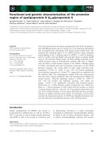
[2, 6]. Ring-intact triterpenoids encompass 4,4,8-trimethyl17-furanylsteroidal skeleton such as azadirone, azadiradione, and gedunin (1-5) type of structures (Fig. 1). C-seco
triterpenoids are generated by the opening and further rearrangements of C-ring thus producing nimbin, salannin and
azadirachtin (6-15) type of skeletons (Fig. 1). Although the
biosynthetic pathway leading to the formation of triterpenoids (Fig. 2a) in Neem plant has been predicted [1, 7]
Page 2 of 14
genes involved in triterpenoid biosynthesis have not been
characterized till date [8].
Secondary metabolites are the final outcome of omics
cascade and their distribution pattern is typical characteristic of every life in nature, which can be considered
as an intrinsic signature of that species. Targeted metabolomics is all about identification and quantification of
known metabolites and their time and space resolved
distribution in a specific biological system [9–13]. Hyphenated mass spectrometry is a powerful and most
utilized analytical technique in metabolomics due to its
high sensitivity, accuracy, resolution, low sample requirement and ability to monitor broad range of metabolites [9, 12–14]. Triterpenoids in Neem are diverse in
skeletal architecture, huge in count and their abundance is highly tissue-specific [1, 2]. Except few discrete
studies [15, 16], there are no systematic investigations
on the tissue- and stage-specific quantitative variation
of Neem triterpenoids. It will be of great importance to
investigate the targeted metabolic profiling of major triterpenoids in Neem plant, which may enlighten the differential tissue specific abundance of skeletally diverse
triterpenoids. Further, correlation of metabolic profiling
Fig. 1 Skeletal diversity of Neem triterpenoids. Basic triterpenoids have azadirone, azadiradione, and gedunin type of skeletons. C- Seco triterpenoids
have nimbin, salannin and azadirachtin type of skeletons
Pandreka et al. BMC Plant Biology (2015) 15:214
Page 3 of 14
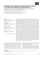
Fig. 2 Predicted triterpenoid biosynthetic pathway, various Neem tissues and their total triterpenoids content in different tissues; (a) Initial genes
involved in triterpenoid biosynthesis. b Different tissues of Neem and physical characteristics of Neem fruits from various stages. c Amount of
triterpenoid extracts obtained from various tissues of Neem
with transcriptome helps in analysis and identification
of genes involved in Neem triterpenoid biosynthesis.
Terpenoid biosynthesis starts with basic building blocks
such as Isopentenyl diphosphate (IPP) and dimethylallyl
diphosphate (DMAPP) which are in turn synthesized
through the mevalonate (MVA) or methylerythritol
phosphate (MEP) pathways [17, 18]. Allylic diphosphate, DMAPP undergoes condensation with one or
more IPP in head-to-tail fashion to produce linear diphosphates such as geranyl diphosphate (C10, GPP),
farnesyl diphosphate (C15, FPP) and geranylgeranyl
diphosphate (C20, GGPP) catalyzed by short-chain
prenyltransferases such as geranyl diphosphate synthase (GDS), farnesyl diphosphate synthase (FDS) and
geranylgeranyl diphosphate synthase (GGDS), respectively
[19–21]. Two molecules of FPP undergo 1-1' head to head
condensation to form squalene via NADPH dependent reduction of presqualene diphosphate intermediate catalyzed by squalene synthase (SQS) [22]. Thus squalene is
the first committed precursor for the biosynthesis of triterpenoids [23]. This molecule is also well known to serve
as a precursor for the primary metabolites such as steroids
required for cell growth and division. Squalene thus acts
as an important intermediate governing the balance
between primary and secondary metabolism. Squalene
undergoes further oxidation to form 2,3-epoxysqualene
mediated by squalene epoxidase, followed by cyclization
catalyzed by triterpene cyclases to form basic triterpene
skeletons [24, 25]. Structural diversity of triterpenoids
arises from the modifications of functional groups and
Pandreka et al. BMC Plant Biology (2015) 15:214
rearrangements on the parental backbone of these triterpenes (Fig. 1) [26].
Short-chain prenyltransferases, such as FDS and SQS
are shown to play key regulatory role in triterpenoid and
phytosterol biosynthesis. To show some instances, when
hairy root culture of Panax ginseng was treated with methyl jasmonate (MJ) to enhance the production of triterpenoids, FDS was up-regulated [27]. Over expression of
mevalonate-5-pyrophosphate decarboxylase and FDS in
Panax ginseng hairy root culture resulted in increased
accumulation of phytosterols and triterepenes [28]. In
Centella asiatica, overexpression of Panax ginseng FDS
resulted in overexpression of dammarenediol synthase
and cycloartenol synthase and when induced with MJ,
enhanced production of triterpenes was observed [29].
Similarly, overexpression of SQS in Panax ginseng,
Eleutherococcus senticosus, Withania coagulans and
Arabidopsis thaliana showed increased production of
phytosterols and triterpenoids [30–33]. Therefore, identification and functional characterization of short-chain prenyltransferases and SQS will assist in understanding of
triterpenoid biosynthesis.
In this study, fifteen major triterpenoids were quantified
in six different Neem tissues including kernel, pericarp,
flower, leaf, stem and bark using UPLC-ESI(+)-HRMS
based targeted profiling. Tissue specific profiling of triterpenoids delineated the variation in the abundance of triterpenoids across various tissues. This information was
further utilized for the selection of tissues for transcriptome analysis followed by identification of initial genes involved in isoprenoid biosynthesis. Amongst the predicted
genes from this pathway, here we report, molecular cloning and functional characterization of full-length geranyl
diphosphate synthase (AiGDS), farnesyl diphosphate synthase (AiFDS) and squalene synthase (AiSQS) from Neem.
Furthermore, using real-time PCR analysis, we showed
that the expression level of one of the important genes in
the pathway, AiSQS correlates with the triterpenoid content in respective tissues (fruit, leaf and flower).
Results and discussion
Tissue specific quantitative profiling of triterpenoids
The levels of individual fifteen triterpenoids (Fig. 1) were
determined in different tissues of Neem including flowers,
leaves, stem, bark, five developmental stages of pericarp
and three stages of kernel (Additional file 1: Figure S5).
The developmental stages of the fruits were classified on
the basis of kernel formation, weight, hardness and colour
(Fig. 2b). The crude mixture of triterpenoids was extracted
from fresh tissues of Neem using solvent partition technique and were analyzed by UPLC-ESI(+)-HRMS in a gradient solvent program of methanol-water. Amount of
crude extract obtained was directly correlated with the triterpenoid content of the corresponding tissue (Fig. 2c).
Page 4 of 14
Quantification of the crude extract revealed that kernel of
stages 4 and 5 contained the highest amounts of triterpenoids (~80 mg/g of the tissue) followed by pericarp of
stages 1, 2 and 3 (~48-66 mg/g). Pericarps of stages 4, 5
and kernel of stage 3 were found to possess comparatively
lower amount of triterpenoids in the range of ~2535 mg/g. Flowers and leaves have been shown to contain 22 and 45 mg/g of triterpenoids (including
chlorophyll and other pigments), while stem and bark
furnished 15 and 10 mg/g of the tissue respectively.
Standard graphs were prepared for each of the fifteen
isolated triterpenoids within the concentration range of
0.04 to 0.003 mg/mL with injection volume 5 μL in
UPLC-ESI(+)-HRMS (Additional file 1: Figure S4). They
were further utilized for the quantification of individual
molecules in the extracts of different tissues of Neem by
correlating with the area under respective peaks of
extracted ion chromatograms (Additional file 1: Figures
S2 and S3). The quantitative level of individual fifteen triterpenoids across various tissues of Neem has been represented in Additional file 1: Figures S3 and S6. Among the
fifteen triterpenoids under investigation, azadirachtin A
(14), a well-studied Neem triterpenoid was found to be
highly abundant in seed kernels, especially in the stages 4
and 5 (~3.6 mg/g of the tissue). Pericarp, flowers and
leaves showed 100-500 fold lower levels (~0.004-0.04 mg/
g) of azadirachtin A as compared to the kernel, whereas
bark and stem contained negligible quantities (≤0.005 mg/
g, 1000 fold lesser than seed kernel). Similar distribution
was observed with the levels of azadirachtin B (15). Highest level of azadirachtin B was observed in kernel of stages
4 and 5 (0.5-0.6 mg/g), whereas pericarp and flowers
showed 100-150 fold lesser amounts in comparison.
Stem and bark were found to possess negligible levels
(<0.005 mg/g, 1000 fold lesser than seed kernel) of azadirachtin B. Salannin (9) showed highest levels in kernel of
stages 4 and 5 (1.2-1.4 mg/g). Salannin content was 4 fold
less (~0.3 mg/g of the tissue) in stem as compared to that
in kernel. Salannin content in bark was ~0.04 mg/g which
was 35 fold lesser in comparison to seed kernel. Flowers,
leaves and pericarp showed negligible levels of salannin
(≤0.02 mg/g). Highest percentage of 3-deacetylsalannin
(10) was observed in kernel of stages 4, 5 and stem with
0.01 mg/g of the tissue. Other tissues showed traceable
amounts of 3-deacetylsalannin. Nimbin (6) was mainly
present in kernels in the range of 0.1-0.2 mg/g and in negligible quantities in other tissues. 6-Deacetylnimbin (7)
was found to be present in kernel of stages 4, 5 and leaves
(0.08-0.23 mg/g). Nimbinene (12) and 6-deacetylnimbinene
(13), two pentanortriterpenoids exhibited similar pattern of
distribution across different tissues. Highest level was observed in seed kernels of stages 4, 5 and stem within the
range of 0.15-0.25 mg/g. Flowers and leaves showed minor
quantity (0.02-0.06 mg/g), whereas bark and pericarps
Pandreka et al. BMC Plant Biology (2015) 15:214
exhibited negligible level. Nimbanal (8) was present in
higher level in kernel of stages 4, 5 and stem (0.05-0.10 mg/
g) and traceable levels were observed in other parts. Salannol acetate (11) was found to be abundant in seed kernels
and stem with ~0.15 mg/g and in other tissues in minor
amounts. Ring-intact triterpenoids (basic limonoids) such
as azadirone, azadiradione, epoxyazadiradione and gedunin
were found to be present at higher levels in pericarps. Azadiradione (3) showed highest level (3.0-8.0 mg/g) in all five
developmental stages of pericarps especially in the stages 2
and 3 (7.0-8.0 mg/g), during which the seed kernel formation is about to start. These levels were about 100-200 fold
higher than that in seed kernels (kernel stage 4 and 5) and
flowers (0.01-0.05 mg/g). Other tissues contained negligible
amounts of it (<0.001 mg/g). Similarly, epoxyazadiradione
(4) showed 400-500 folds higher level in pericarps (9.012.0 mg/g; in stages 2 and 3) in comparison to that in the
seed kernels (0.01-0.04 mg/g) and 50 folds higher than in
flowers (~0.20 mg/g). Azadirone (1) was also found to be
most abundant in all the developmental stages of pericarps
(0.3-0.7 mg/g) especially in the stages 2 and 3 (0.6-0.7 mg/
g) and flowers (0.5 mg/g). Leaves showed very less quantity
(~0.08 mg/g) of 1 whereas other tissues contained traceable amounts (<0.001 mg/g). Gedunin (5), a potent anticarcinogenic triterpenoid was abundantly present in
pericarps, especially in the stages 2 and 3 (~1.0 mg/g).
Negligible amount of 5 was present in other tissues
(<0.002 mg/g). Nimocinol (2), 6α-hydroxy derivative of
azadirone was observed to be abundant in leaves
(2.9 mg/g), 15 fold higher than flowers (0.18 mg/g)
and 50-150 times higher than pericarps (0.02-0.08 mg/g).
Other tissues such as kernel, bark and stem showed very
less amount of nimocinol (<0.001 mg/g).
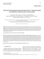
Metabolic profiling data (Fig. 3 and Additional file 1:
Figure S6) depicted the kernel to be rich in quantity and
Page 5 of 14
diversity of triterpenoids especially C-seco triterpenoids
of azadirachtin (14, 15), salannin (9, 11), nimbin (6, 7,
8) and nimbinene (12, 13) skeletons. However, pericarps
were found to be rich in triterpenoids mainly consisting
of ring-intact (basic) structures such as azadirone (1),
azadiradione (3), epoxyazadiradione (4) and gedunin (5).
Flowers and leaves showed relatively lower levels of
triterpenoids and mostly of ring-intact skeletons (1, 2, 3,
4). Stem and bark contained very low levels of triterpenoids; majorly C-seco metabolites of salannin (9, 11) and
nimbinene (12, 13) type. In essence, profiling data revealed C-seco triterpenoids (6-10) to be the major constituents of triterpenoid pool from seed kernel, stem and
bark whereas ring-intact skeletons (1-5) were observed to
be major metabolites of the triterpenoid content obtained
from pericarp, flower and leaf.
Transcriptome analysis
For extensive coverage, RNA isolated from triterpenoid
rich tissues such as fruit stage 4, leaves and flowers were
pooled and used for transcriptome sequencing. A total
of 79,079,412 (79.08 million) paired-end reads each of
72 bp length were generated by Illumina GA II platform.
71,537,895 (90.46 %) high quality reads were obtained
with more than 20 phred score and reads of low quality
were trimmed and used for further analysis. Total
27,390 contigs were generated using Velvet with a hash
length of 41. These contigs were given as input for
Oases to generate 41,140 transcripts. The average length
of transcripts obtained was 1331 bp and the N50 length
was 1953 bp (Table 1).
All the transcripts were submitted to Blastx against
non-redundant database available at NCBI with an Evalue cutoff of 10-5, where, a total of 32,856 (79.8 %)
transcripts were annotated (Fig. 4a). Pathway annotation
Fig. 3 Quantitative abundance of major triterpenoids in different tissues of Neem. Basic and C-seco triterpenoids are highly abundant in Pericarp
and Kernel respectively as compared to other tissues
Pandreka et al. BMC Plant Biology (2015) 15:214
Page 6 of 14
Table 1 Summary of transcriptome sequencing and assembly
Total number of reads
79079412
Total number of HQ reads
71537895
Hash length
41
Contigs generated
27390
Average contig length
897.431
N50 length of contigs
1479
Transcripts generated
41140
N50 length of transcripts
1953
Number of reads assembled
68871778
was carried out by KAAS (KEGG Automatic Annotation
Server) with Arabidopsis thaliana (thale cress) and Oryza
sativa japonica (Japanese rice) as the reference database.
Out of the 41,140 transcripts only 6281 transcripts were
assigned 2749 unique KO numbers, which covered 223
pathways (Fig. 4b). Virtual ribosome, a web based server,
was used for finding the Open Reading Frame (ORF) of
transcripts. 27,368 transcripts had an ORF with length
more than 99 amino acids and 67 transcripts without any
ORF (Fig. 4c). The peptide sequences of transcripts
with length more than 99 amino acids were submitted
to Pfam analysis. 18,807 transcripts were assigned different Pfam IDs. A total of 3467 different Pfam IDs
were assigned to the transcripts (Fig. 4d). Based on
transcriptome annotation, all the genes involved in triterpenoid back-bone biosynthesis from isoprene units
(MVA pathway and MEP pathway) to triterpene cyclase
were found (Additional file 1: Table S1). A total of 134 transcripts predicted as cytochrome P450 monooxygenases and
two transcripts as cytochrome P450 reductases were identified. Based on BLAST results, with reference to Arabidopsis
thaliana cytochrome P450, Neem CYP450s were classified
into 39 families and 78 subfamilies, out of which most of
the CYP450 belonged to CYP71 family. Seven transcripts
were related to plant steroid biosynthesis and six transcripts
related to triterpenoid biosynthesis were predicted
(Additional file 1: Table S1). Recently, Neem draft genome and transcriptome of fruit, stem, leaf and flower
[34], and suppression subtractive hybridization of transcripts between fruit mesocarp and endocarp [35] have
been reported. However, there are no reports regarding
functional characterization of the genes involved in Neem
triterpenoid biosynthesis. To further explore this pathway,
two short-chain prenyltranferases and squalene synthase
were selected for functional characterization based on the
transcriptome data.
Heterologous expression and functional characterization
of short-chain prenyltransferases (AiGDS and AiFDS)
Short-chain prenyltransferases function at the branching
point of terpenoid metabolism and play regulatory role
Fig. 4 Functional annotation of transcriptome; (a) Based on Blastx analysis 80 % (32,856) transcripts had homologous proteins in NCBI nr database. b
Based on KAAS analysis only 15.2 % (6281) transcripts were assigned 2749 KO numbers. c Based on virtual ribosome analysis 66.5 % (27,368) transcripts
had ORF region length more than 100 amino acids and 0.001 % (67) Transcripts did not show ORF region. d Based on Pfam analysis 69.1 % (18,907)
transcripts were assigned Pfam IDs
Pandreka et al. BMC Plant Biology (2015) 15:214
in the distribution of isoprene units into various terpenoids
biosynthesis. In total, 12 short-chain prenyltranferases from
Neem transcriptome were identified (Additional file 1:
Table S1). Based on functional annotation studies, two geranyl diphosphate synthases (GDS), nine putative geranylgeranyl diphosphate synthases (GGDS) and one farnesyl
diphosphate synthase (FDS) were identified. Sequence analysis using BLAST indicated that Neem_transcript_10912
was a homomeric GDS and Neem_transcript_10001 could
be the smaller subunit of heteromeric GDS. TargetP analysis showed that both of these genes are localized in
the mitochondria (Additional file 1: Table S5). For
further study, Neem_transcript_10912 (AiGDS) and
Neem_transcript_25722 (AiFDS) were selected for cloning
and functional characterization.
The ORF of AiGDS [GenBank: KM108315] was 1263 bp,
which coded for a protein of 420 amino acids with theoretical molecular weight and calculated pI as 46.1 kDa and
6.33, respectively. AiGDS had maximum identity with several plant characterized homomeric GDSs such as 90 %
identity to homomeric GDS from Citrus sinensis [GenBank:
CAC16851] [36], 86 % identity to GDS from Mangifera
indica [GenBank: AFJ52721] [37] and 76 % identity to GDS
from Catharanthus roseus [GenBank: AGL91647] [38]. The
percentage identity matrix of AiGDS with other plant
homomeric GDS and heteromeric GDS larger subunits indicated that AiGDS possesses 71 % to 89 % identity with
homomeric GDS (Additional file 1: Table S2). The multiple
sequence alignment of AiGDS consisted of two aspartate
rich motifs DDX(2-4)D and DDXXD which are highly conserved motifs in prenyltransferases and involved in substrate and metal ion binding (Additional file 1: Figure S7).
CxxxC motifs were not observed in AiGDS, which play a
key role in the interaction of heteromeric GDS [39]. The
ORF of AiGDS was cloned into pET32a expression vector
having an N-terminal thioredoxin domain and subsequently expressed in BL21 (DE3) cells. However recombinant AiGDS protein was found in inclusion bodies. To
enhance solubility, AiGDS cloned construct was transformed into Lemo 21 (DE3) cells [40] and expression was
carried out. Recombinant AiGDS protein remained solely
in the insoluble portion in the pellet. Eventually we were
able to obtain soluble active AiGDS by re-suspending the
pellets in lysis buffer, then drop-wise addition of 0.1 M
NaOH until pH 11.0 with constant swirling on ice till the
solution became clear. The pH was then reduced to 7.0
using 0.1 M HCl under similar conditions [41]. The resulting solution was centrifuged at 10,000 × g and subjected to
SDS-PAGE analyses (Additional file 1: Figure S11A). The
AiGDS was found to be in soluble form in the supernatant,
which was subjected to purification by Ni-NTA affinity
chromatography. The recombinant protein was over 94 %
pure as analysed by SDS-PAGE (Additional file 1: Figure
S11A). Purified recombinant AiGDS was incubated with
Page 7 of 14
equimolar concentration of IPP and DMAPP followed by
treatment with alkaline phosphatase to hydrolyze the diphosphate esters to their corresponding alcohols. The extracted assay mixture was analyzed by GC-MS and the
products formed were confirmed by comparing the retention time and coinjection studies with standard geraniol
(Fig. 5a). GC-MS analyses of the extracts of alkaline phosphatase treated assay mixture of AiGDS with GPP/FPP and
IPP indicated that AiGDS failed to synthesize chain elongation products FPP (C15) or GGPP (C20) suggesting that
AiGDS can catalyse the chain elongation reaction to produce GPP (C10) as sole enzymatic product.
AiFDS [GenBank: KM10831] ORF of 1029 bp length
was found to be encoding for a protein of 342 amino
acids. The theoretical molecular weight and pI for this
polypeptide were 39.5 kDa and 5.59 respectively. The sequence comparison of AiFDS exhibited 83 % identity
with FDS from Mangifera indica [GenBank: AFJ52720]
[37], 82 % identity with that from Santalum album
[GenBank: AGV01244.1] and 81 % identity with FDS
from Catharanthus roseus [GenBank: ADO95193.1]
[42]. The multiple sequence alignment of AiFDS consisted of two aspartate rich motifs DDX(2-4)D and
DDXXD (Additional file 1: Figure S8) which were
highly conserved motifs in prenyltransferases. AiFDS
was cloned into pET32a expression vector. The cloned
construct was transformed into BL21 (DE3) cells and
expressed. AiFDS was obtained as soluble form and
purified by Ni-NTA affinity column chromatography.
The recombinant protein was over 98 % pure as analyzed by SDS-PAGE (Additional file 1: Figure S11B).
Buffers used for AiGDS and AiFDS protein purification are given in Addition file 1: Table S4. The purified
short-chain prenyltransferase was incubated with DMAPP/
GPP and IPP followed by treatment with alkaline phosphatase. GC-MS analyses of the assay extracts indicated the formation of FPP which was further confirmed by comparing
the retention time, mass fragmentation pattern and coinjection studies with standard (E,E)-farnesol (Fig. 5b). Further
GC-MS analysis of alkaline phosphatase treated assay mixture of AiFDS with FPP and IPP did not show formation of
geranylgeraniol indicating that AiFDS catalyses the chain
elongation reaction to produce FPP as the sole enzymatic
product.
Heterologous expression and functional characterization
of squalene synthase (AiSQS)
An ORF of 1176 bp encoding a polypeptide of 396 amino
acids was identified as AiSQS [GenBank: JQ327160]. The
theoretical pI of protein was found to be 8.18 and molecular weight of 44 kDa. The amino acid sequence of AiSQS
shared 86 % identity with squalene synthase from
Diospyros kaki [GenBank: ACN69082], 85 % identity
with Camellia oleifera [GenBank: AGB05603], 84 %
Pandreka et al. BMC Plant Biology (2015) 15:214
Page 8 of 14
Fig. 5 Total ion chromatograms (TICs) of AiGDS, AiFDS and AiSQS assays and relative expression level of AiSQS; (a) TICs of AiGDS assays; (1)
Standard Nerol, (2) Standard geraniol, (3) Co-injection of standard nerol and geraniol, (4) Substrate control, (5) Enzyme control, (6) AiGDS enzyme
assay with IPP and DMAPP as substrates, (7) Co-injection of standard geraniol with AiGDS enzyme assay extract. b TICs of AiFDS assays; (1)
Standard (E,E)-farnesol, (2) IPP and DMAPP substrate control, (3) Enzyme control, (4) AiFDS enzyme assay with IPP and DMAPP as substrates, (5)
Co-injection of standard (E,E)-farnesol and extract of AiFDS enzyme assay with IPP and DMAPP as substrates, (6) Extract of AiFDS enzyme assay
with GPP and IPP as substrates. c TICs of AiSQS assays; (1) Standard squalene, (2) Substrate control, (3) Enzyme control, (4) Extract of full length
AiSQS enzyme assay with FPP as substrate and NADPH as co-factor, (5) Co-injection of standard squalene and AiSQS enzyme assay extract, (6)
Extract of truncated AiSQS enzyme assay with FPP as substrate and NADPH as co-factor and (7) Co-injection of standard squalene and truncated
AiSQS enzyme assay extract
identity with Euphorbia tirucalli [GenBank: BAH23428]
and 84 % identity with that from Glycyrrhiza glabra
[GenBank: BAA13084.1]. Eukaryotic SQSs have four conserved regions and are important for catalysis as indicated
by biochemical characterization of site-directed mutants
and crystal structure of human squalene synthase [43]
(Additional file 1: Figure S9). The aspartate rich motifs
found in region 1 and 3 are involved in binding of the
diphosphate moiety of FPP via bridging Mg2+ ions. Careful
analysis of AiSQS sequence with TMHMM program
showed the presence of transmembrane motif YNTTM
IIMLFIILAIIFAYLSAN at the C-terminus. Although transmembrane domain exhibits low level of sequence homology with other SQS enzymes, this domain is highly
hydrophobic and consistent with the putative endoplasmic
reticulum anchoring function.
Squalene synthase has been characterized previously
from human [43], rodents [44, 45], plants [46–48],
protozoa [49] and fungi [50]. All these SQS enzymes
were obtained in soluble form by deletion of a putative Cterminal membrane-spanning motif [51]. In the present
study we have cloned the full-length ORF of AiSQS, as
well as a truncated AiSQS by deletion of 15 amino acids
from N-terminal and 63 amino acids from the C-terminal
end into pRSET-C and pET28c vectors respectively. The
truncated AiSQS was transformed into BL21 (DE3) cells,
expressed and purified by subjecting to Ni-NTA affinity
column chromatography. Purified truncated AiSQS was
analyzed by SDS-PAGE which showed a single band
(>90 % purity) at ~35 kDa, consistent with the predicted
molecular mass for the (His)6-tagged enzyme (Additional
file 1: Figure S11D).
The full-length recombinant AiSQS protein was
expressed in BL21 star (DE3) cells. Majority of the protein
was found to be insoluble (Additional file 1: Figure S11C).
Lee and Poulter observed that adding glycerol to the lysis
and purification buffers helped in solubilization of the insoluble T. elonatus BP-1 SQS [52]. Induced cell pellets
were disrupted in lysis buffer containing 50 % (v/v) glycerol and 1 % CHAPS. The glycerol concentration in
cell lysate obtained was reduced to 20 % (v/v) by adding
lysis buffer (without glycerol). This lysate was subjected
to Ni-NTA affinity column chromatography. The purified full length AiSQS, when analyzed by SDS-PAGE,
exhibited a single band (90 % purity) at approximately
44 kDa, consistent with the predicted molecular mass
for the (His)6-tagged enzyme (Additional file 1: Figure
S11C). Purified proteins were flash-frozen in liquid nitrogen and stored at -80 °C until further use. Buffers
used for AiSQS full length and truncated protein purification are given in Addition file 1: Table S4.
GC-MS analyses of the assay extracts of full length
and truncated AiSQS with FPP in the presence of
NADPH indicated the formation of squalene. The formation of squalene was further confirmed by comparing
the retention time, mass fragmentation pattern and coinjection studies with standard squalene (Fig. 5c). This
confirms that AiSQS catalyzes the condensation of two
Pandreka et al. BMC Plant Biology (2015) 15:214
molecules of farnesyl diphosphate (FPP) to form squalene through a NADPH-dependent rearrangement of
C1′-2-3-linked triterpene intermediate, presqualene diphosphate [52].
Real time PCR analysis
To determine the role of short-chain prenyl diphosphate synthases and squalene synthase in triterpenoid
biosynthesis, real time PCR analysis of the Neem_
transcript_10001 (smaller subunit of heteromeric geranyl
diphosphate synthase), AiGDS, AiFDS, and AiSQS was
carried out.
AiSQS is the first committed enzyme involved in
triterpene biosynthesis in Neem. Real time PCR was
carried out for AiSQS from flowers, leaves and fruit
and normalized with 18S rRNA expression level.
Neem fruit showed fivefold higher expression level in
comparison with the leaves and tenfold higher relative
expression level than flowers (Fig. 6d). The results
Page 9 of 14
were in correlation with profiling of triterpenoids from
different tissues. Neem fruits as a whole, not only showed
structurally diverse triterpenoids but also showed very
high levels of these metabolites. On the other hand,
flowers and leaves exhibited lesser skeletal diversity and
quantity of abundant triterpenoids. Squalene is the precursor of primary metabolites such as membrane sterols and
steroid hormones required for cell division and growth.
Also, it serves as precursor for triterpenoids found in
Neem, which assign squalene, a crucial branch point between primary and secondary metabolism. Transgenic
Panax ginseng overexpressing squalene synthase has previously shown to produce higher levels of triterpene and
phytosterols than wild type strains which depict the key
role of intracellular squalene flux between primary and
secondary metabolism [31]. High expression levels of
AiSQS in fruits indicated considerable amount of squalene
flux might get diverted towards triterpenoids formation in
Neem fruits.
Fig. 6 Real-time PCR analysis. a Neem_transcript_10001 showed very high expression in flower. b AiGDS was highly expressed in leaf. c AiFDS
has higher expression level in seeds. d Relative expression levels of AiSQS was very high in seeds as compared to other tissues. Error bars
represents standard error
Pandreka et al. BMC Plant Biology (2015) 15:214
AiFDS (Fig. 6c), compared to other tissues, showed very
high expression levels in seeds. Similar expression patterns
of AiFDS and AiSQS suggest that both these genes could
be involved in triterpenoid biosynthesis. On the contrary,
AiGDS (Fig. 6b) and Neem_transcript_10001 (Fig. 6a)
showed very high expression in leaf and flower, respectively, compared to other tissues. These results indicate
that AiGDS may not be involved in triterpene biosynthesis
in Neem.
Phylogenetic analysis
Neighbour joining phylogenetic tree was constructed
based on the deduced amino acid sequences of AiGDS,
AiFDS and AiSQS with corresponding enzymes from
different organisms, which were retrieved from the
NCBI GenBank database (Additional file 1: Figure S10).
The degree of relatedness correlated well with the amino
acid similarity among the plant proteins, which indicated
AiGDS, AiFDS and AiSQS belonged to the clade of plant
kingdom. These enzymes from Neem were classified
into one cluster revealing their closest evolutionary relationships with the plant group.
Page 10 of 14
54, 6] and described briefly in Additional file 1. For extraction, HPLC grade solvents were purchased from
Sigma (St. Louis, MO, USA). For UPLC-ESI(+)-MS experiments LC-MS grade solvents were procured from
Avantor Performance Materials, JT Baker (PA, USA).
SuperScript® III First-Strand Synthesis System (Invitrogen)
was used for cDNA synthesis. For PCR amplification,
AccuPrime™ (Invitrogen) polymerase was used. For Restriction digestion, NEW ENGLAND BioLabs®inc(NEB) restriction enzymes were used. Gel extraction of restricted
product and vector were carried out by GenElute™ Gel Extraction Kit from Sigma. T4 DNA ligase from Invitrogen
was used for ligation. TOP10 cells (Invitrogen) were used
for cloning. Lemo21 (DE3) cells (NEB), BL21 (DE3) cells
(NEB) and BL21 Star (DE3) cells (Invitrogen) were used as
expression cells. Ni-NTA agarose (Invitrogen) was used
for protein purification. Enzyme assay samples were analyzed on Agilent 7890A GC coupled with 5975C mass detector. Geraniol, nerol, (E,E)-farnesol, squalene standards
were purchased from Sigma Aldrich. IPP, FPP, GPP, and
DMAPP were synthesized as reported previously [55, 56].
Extraction of total triterpenoids
Conclusions
Due to immense significance of Neem as a wonder tree
and known to synthesize biologically and commercially
important triterpenoids having highly complex carbon
skeleton with diverse functional groups, it is of great
interest to study their biosynthetic pathway. Levels of
total triterpenoid and fifteen major individual triterpenoids were quantified in various tissues of the Neem
plant. Tissue specific variation in the abundance of triterpenoids has been observed. The mature seed kernel
and pericarp of initial stages were found to contain the
highest amount of triterpenoids. Furthermore, a wide diversity of triterpenoids, especially C-seco triterpenoids
were observed in kernel as compared to the other tissues.
Pericarp, flower and leaf contained mainly ring-intact triterpenoids. From transcriptome analysis, short-chain prenyl
trasnferases, squalene synthase, squalene expoxidase, triterpene synthases and putative cytochrome P450 genes were
predicted. The genes involved in the initial steps of isoprenoid biosynthesis, such as AiGDS, AiFDS and AiSQS were
cloned and functionally characterized. Furthermore, AiFDS
and AiSQS expression levels were found to be nicely correlating with the triterpenoids content of various tissues of
Neem.
Methods
Materials and chemicals
Neem tissues for the profiling of triterpenoids were
collected from Pune region, Maharashtra, India in the
period March to May. Fifteen reference triterpenoids
were isolated and characterized as reported earlier [53,
Fresh Neem tissues (0.5 g) were extracted with methanol
(10 mL × 3), by continuous stirring for 3 h. The pooled
methanol layer after concentration under reduced pressure at 50 °C was partitioned between ethyl acetate
(20 mL) and water (20 mL). The organic layer was separated, passed through anhydrous sodium sulphate and
concentrated under similar conditions to obtain the crude
triterpenoid extract. Extraction of individual tissues was
performed in triplicates.
UPLC-ESI(+)-HRMS profiling of triterpenoid extract
For triterpenoids profiling, UPLC-ESI(+)-HRMS runs were
performed on Q Exactive Orbitrap associated with Accela
1250 pump (Thermo Scientific, MA, USA). Mixture of triterpenoids were dissolved in a known volume of methanol
(concentration ~0.2 mg/mL), centrifuged to remove the
suspended particles and injected (10 μL) in UPLC-ESI(+)HRMS (Additional file 1: Figure S5). Samples were resolved
through Acquity BEH C18 UPLC column (2.1 × 100 mm)
of particle size 1.7 μM with a flow rate of 0.3 mL/min and
gradient solvent program of 35 min (0.0 min, 40 % methanol/water; 5.0 min, 50.0 % methanol/water; 10.0 min, 60 %
methanol/water; 25.0 min, 65 % methanol/water; 30.0 min,
90 % methanol/water; 32.0 min, 90 % methanol/water;
34.0 min, 40 % methanol/water; 35.0 min, 40 % methanol/
water). 0.1 % LC-MS grade formic acid was also added to
water (mobile phase). Profiling experiments were performed in ESI-positive ion mode using the tune method as
follows: sheath gas (nitrogen) flow rate 45 units, auxiliary
gas (nitrogen) flow rate 10 units, sweep gas (nitrogen) flow
rate 2 units, spray voltage (|KV|) 3.60, spray current (μA)
Pandreka et al. BMC Plant Biology (2015) 15:214
Page 11 of 14
3.70, capillary temperature 320 °C, s-lens RF level 50, heater
temperature 350 °C. ESI(+)-HRMS data were recorded in
full scan mode within the mass range m/z 100 to 1000. Profiling data were analyzed through Thermo Xcalibur software. Retention times (Rt) and extracted ions for the
individual studied triterpenoids have been listed in Table 2.
UPLC-ESI(+)-HRMS chromatograms for individual standard triterpenoids and their corresponding ESI(+)-HRMS
spectra have been provided in Additional file 1: Figures S2
and S3. R version 3.1.2 was used for generating heatmap.
Transcriptome analysis
Total RNA was isolated using Spectrum Plant total
RNA isolation kit (Sigma-Aldrich). Equal quantity of
RNA from each tissue was mixed. Transcriptome library was constructed using TruSeq RNA Sample
Preparation Guide (Illumina). Quality of the prepared
library was analyzed by running an aliquot on High
Sensitivity Bioanalyzer Chip (Agilent). 79,079,412 paired
end raw reads were generated with the length of 72 bp by
Illumina GA ΙΙ analyzer. De novo assembly was carried out
by Velvet (version- 1.1.05) with hash length 41 [57]. A total
of 27,390 contigs were generated with average contig length
of 897 and N50 value of 1479. These contigs were then
submitted to Oases (version- 0.2.01) to generate a total of
41,140 transcripts [58]. Neem transcripts were submitted to
Blastx against non-redundant database available at NCBI
with E-value cutoff of 10-5. Pathway annotation was done
by bidirectional best hit method of KAAS (KEGG Automatic Annotation Server. />with Arabidopsis thaliana (thale cress) and Oryza sativa japonica (Japanese rice) as the reference database [59].
Table 2 Retention times (Rt), extracted ions and corresponding
molecular fragments for the studied triterpenoids
Triterpenoid
Extracted ion (m/z)
Fragment
Azadirachtin A (14)
Rt (min)
7.4
703
[M + H-H2O]+
Azadirachtin B (15)
8.6
645
[M + H-H2O]+
Salannin (9)
21.3
597
[M + H]+
3-Deacetylsalannin (10)
18.5
555
[M + H]+
Nimbin (6)
18.0
541
[M + H]+
6-Deacetylnimbin (7)
14.7
499
[M + H]+
Nimbinene (12)
20.7
483
[M + H]+
6-Deacetylnimbinene (13)
16.5
441
[M + H]+
Nimbanal (8)
17.8
511
[M + H]+
Salannol acetate (11)
28.4
599
[M + H]+
Azadiradione (3)
16.3
451
[M + H]+
Epoxyazadiradione (4)
27.4
467
[M + H]+
Azadirone (1)
31.7
437
[M + H]+
Gedunin (5)
20.4
483
[M + H]+
Nimocinol (2)
29.9
453
[M + H]+
Virtual ribosome, ( />Ribosome/) a web based server, was used for deducing the
ORFs of these transcripts [60]. The peptide sequences of
transcripts with length more than 99 amino acids were
submitted to batch search of Pfam ( />search#tabview=tab1) [61].
Cloning and characterization of AiGDS, AiFDS and AiSQS
The Neem seed RNA was used for the synthesis of cDNA
using SuperScript® III First-Strand Synthesis System
(Invitrogen). Full length primers for AiGDS and AiFDS
ORFs were designed using their transcripts as a template
(Additional file 1: Table S3). Synthesized cDNA was used
for PCR reaction using AccuPrime (Invitrogen). PCR
products were cloned into pET32a expression vector using
respective cloning sites. Full length and truncated primers
for AiSQS were designed from Neem_transcript_33869
(Additional file 1: Table S3). PCR products were cloned into
pCR Blunt vector. Further, the ORF was digested with
EcoRI and the resulting fragment was ligated into pRSET-C
vector for full length AiSQS and pET28c for truncated
AiSQS. The expression of the recombinant plasmids containing AiGDS, AiFDS, truncated AiSQS were carried out
in BL21 (DE3) cells except full length AiSQS, which was
expressed in BL21 Star (DE3) cells.
Initially, AiGDS and full length AiSQS were found in
inclusion bodies. Expression of AiGDS in Lemo 21
(DE3) cells did not show any improvement in the solubility. To obtain the soluble AiGDS protein, the pellet
obtained after crude lysate centrifugation at 10,000 × g
was resuspended in lysis buffer, pH was increased to
11.0 with 0.1 M NaOH and then reduced to 7.0 with
0.1 M HCl (pH adjustment was done on ice with continuous stirring). The resulting solution was centrifuged
at 10,000 × g for 10 min at 4 °C [41]. The supernatant
containing AiGDS protein was purified over Ni-NTA affinity chromatography by following user manual.
Purification of full length AiSQS was attempted under
denaturing conditions in 50 mM Tris buffer containing
6 M guanidium hydrochloride as well as 8 M urea as denaturing agents. Refolding was attempted by stepwise
slow removal of denaturants under dialysis. However,
the protein obtained was not catalytically active. Purification under native conditions using buffer combinations of HEPES, TRIS, MOPS with non-ionic detergents
like T ween 20, Triton X-100 also did not yield sufficient
amount of soluble protein. A considerable amount of
protein was found in soluble fractions using 50 % glycerol
and 1 % CHAPS in Phosphate buffer. All the recombinant
proteins were purified by Ni-NTA affinity chromatography.
Buffers used for recombinant protein purifications were
given in Addition file 1: Table S4. Protein estimation was
performed by Bradford assay [62] and the protein purity
was analyzed on SDS-PAGE (Additional file 1: Figure S11).
Pandreka et al. BMC Plant Biology (2015) 15:214
Enzyme assays for AiGDS and AiFDS were performed
in HEPES buffer with DMAPP (100 μM)/GPP (100 μM)
and IPP (100-200 μM) as substrates. 100 μM FPP was
used as substrate for full length and truncated AiSQS
with 1 mM NADPH as cofactor. The reaction mixtures
were incubated at 30 °C for 2 h. AiSQS assay reaction
was quenched by adding 1 M sodium hydroxide. For
AiGDS and AiFDS assays, alkaline phosphatase (6 U)
was added and further incubated at 37 °C for 1 h. Reaction mixtures were extracted thrice using n-hexane.
Samples were concentrated with a stream of dry nitrogen and analysed by GC-MS on 30 m × 0.25 mm ×
0.25 μm capillary columns (HP-5 and HP-5 MS, J & W
Scientific). Functional characterization of AiFDS and
AiGDS was carried out on GC-MS using the program:
70 °C for 1 min, 5 °C/min rise till 150 °C, 10 °C/min rise
till 270 °C and hold for 5 min (Program 1). For the functional characterization of AiSQS, the program used was:
initial temperature of 150 °C for 2 min followed by increase in temperature to 320 °C at the rate of 10 °C/min
and hold at 320 °C for 11 min (Program 2). Product formation was confirmed by co-injection with authentic
standards and comparing the mass fragmentation pattern and retention time (Fig. 5).
RT-PCR analysis
Real time PCR was carried out using Super Script III
platinum SYBR green one-step qRT-PCR kit (Invitrogen,
USA). In brief, for AiSQS quantification, 100 ng of
DNase treated total RNA was added with AiSQS primers
and for 18S intrinsic control, 18S primers were used
(Additional file 1: Table S3). cDNA synthesis and PCR
were carried out in a single tube reaction. cDNA synthesis was performed at 50 °C for 5 min followed by denaturation at 95 °C for 5 min and subsequent 40 cycles
of denaturation step at 95 °C for 3 s, combined annealing and extension step at 60 °C for 30 s per cycle.
Quantification of AiGDS, AiFDS and Neem_Transcript_
10001, was performed as follows: Initial cDNA synthesis
was performed at 50 °C for 20 min, followed by 95 °C for
5 min, 40 cycles of 95 °C for 10 s and 60 °C for 30 s.
GAPDH primers were used as an endogenous control to
normalize the expression levels between different tissues.
Threshold (Ct) values were obtained and ΔCt was calculated as Ct target gene – Ct endogenous reference gene.
Relative fold difference was calculated using 2ΔCt. Experiments were carried out using three biological replicates
with five technical replicates each.
Page 12 of 14
analyses with 1000 replicates were also conducted in order
to obtain confidence levels for the branches.
Availability of supporting data
The Illumina RNA-seq data generated from pooled RNA
from leaves, fruits and flowers of Azadirachta indica are
available in the NCBI SRA ( />Traces/sra) with accession SRR2145149.
Additional file
Additional file 1: Methods 1. Isolation of Neem triterpenoids from
seed kernel and pericarp. Methods 2. Characterization of purified Neem
triterpenoids. Figure S1. TLC profile of crude extracts and purified
triterpenoids (developed in 70 % ethyl acetate in n-hexane for twice).
Figure S2. UPLC-ESI(+)-quadrupole/orbitrap-MS extracted ion
chromatograms of the fifteen pure triterpenoids from Neem.
Chromatograms have been arranged in the order of
increasing retention time. Figure S3. ESI(+)-quadrupole/orbitrap-MS
spectra of the fifteen pure triterpenoids from Neem. Figure S4.
Standard graphs for the purified triterpenoids prepared in UPLC-ESI(+)-quadrupole/orbitrap-MS; concentration range 0.040-0.003 mg/mL, injection
volume 5 μL. Figure S5. Representative UPLC-ESI(+)-quadrupole/
orbitrap-MS chromatograms of various Neem tissue extracts (× denotes
non-triterpenoids with molecular mass less than 350). Figure S6.
Quantitative abundance of individual triterpenoids in different tissues
of Neem. Figure S7. Multiple sequence alignment of A. indica
geranyl diphosphate synthases (AiGDS). Figure S8. Multiple sequence
alignment of A. indica farnesyl diphoshate synthase (AiFDS). Figure S9.
Multiple sequence alignment of A. indica Squalene synthase (AiSQS); Amino
acid sequence alignment of C. annuum (CaSQS, AAD20626), N. tabacum
(NtSQS, AAB08578), A. indica (AiSQS, AFJ15526), L. japonicas (LjSQS,
BAC56854), G. max (GmSQS, NP_001236365), P. vulgaris (PvSQS, AHA84150).
The solid lines indicate four highly conserved regions 1, 2, 3 and 4 which
are considered to be the catalytic sites of squalene synthases. Figure S10.
Phylogenetic analysis of AiGDS, AiFDS and AiSQS. Figure S11. Purification
of recombinant AiGDS, AiFDS and AiSQS. Table S1. Predicted genes for
Triterpenoid back bone biosynthesis. Table S2. Present Identity Matrix of
AiGDS with plant homomeric GDS and heteromeric GDS Larger subunits.
Table S3. Primers and vectors used for cloning of AiGDS, AiFDS and AiSQS
and RT-PCR primers of 18S rRNA, GAPDH, Neem_transcript_10001, AiGDS
and AiSQS. Table S4. Buffers used for AiGDS, AiFDS and AiSQS protein
purification. Table S5. TargetP analysis Neem_transcript_10912 (AiGDS)
and Neem_Transcript_10001. (DOCX 3387 kb)
Abbreviations
MVA: Mevalonate pathway; MEP: Methylerythritol phosphate pathway;
GPP: Geranyl diphosphate; FPP: Farnesyl diphosphate; GGPP: Geranylgeranyl
diphosphate; GDS: Geranyl diphosphate synthase; FDS: Farnesyl diphosphate
synthase; GGDS: Geranylgeranyl diphosphate synthase; SQS: Squalene
synthase; MJ: Methyl jasmonate; ORF: Open Reading Frame.
Competing interests
The authors declare that they have no competing interests.
Phylogenetic analysis
Authors’ contributions
SH, FAM and AT carried out isolation and characterization of metabolites
and tissue specific quantitative profiling of triterpenoids. AP performed
transcriptome analysis. DSD, UV, VGS and AP carried out the cloning and
characterization of genes. HVT has conceptualized, supervised and acted as
overall study director. All authors have read and approved the final
manuscript.
Reference protein sequences were obtained from GenBank
database. Sequences were aligned using ClustalW using default parameters [63]. Neighbour joining tree was constructed with MEGA version 6.06 software [64]. Bootstrap
Acknowledgements
AP, DSD, SH and AT acknowledge UGC New Delhi, ICMR New Delhi, CSIR
New Delhi and DBT New Delhi, respectively, for their fellowship. This work is
supported by CSIR-New Delhi sponsored network projects (HCP0002,
Pandreka et al. BMC Plant Biology (2015) 15:214
CSC0106 and CSC0130). Authors thank Dr. Dhanashekaran Shanmugam for
helping in analyzing the transcriptome data.
Footnotes
This work is dedicated to Dr. Vidya Gupta, Biochemical Sciences Division,
CSIR-NCL, Pune, on the occasion of her 60th birthday.
Received: 9 February 2015 Accepted: 13 August 2015
References
1. Champagne DE, Koul O, Isman MB, Scudder GGE, Towers GHN. Biologicalactivity of limonoids from the rutales. Phytochemistry. 1992;31(2):377–94.
2. Tan QG, Luo XD. Meliaceous limonoids: chemistry and biological activities.
Chem Rev. 2011;111(11):7437–522.
3. Jacobson M. Focus on Phytochemical Insecticides: The Neem Tree. Boca
Raton: CRC Press; 1988.
4. Morgan ED. Azadirachtin, a scientific gold mine. Bioorg Med Chem.
2009;17(12):4096–105.
5. Schmutterer H. The Neem Tree: Source of Unique Natural Products for
Integrated Pest Management, Medicine, Industry and Other Purposes.
Weinheim, Germany: VCH; 1995.
6. Haldar S, Phapale PB, Kolet SP, Thulasiram HV. Expedient preparative
isolation, quantification and characterization of limonoids from Neem fruits.
Anal Methods. 2013;5(20):5386–91.
7. Siddiqui S, Siddiqui BS, Faizi S, Mahmood T. Tetracyclic triterpenoids and
their derivatives from Azadirachta indica. J Nat Prod. 1988;51(1):30–43.
8. Ekong DEU, Ibiyemi SA, Olagbemi EO. The meliacins (limonoids).
Biosynthesis of nimbolide in the leaves of Azadirachta indica. J Chem Soc
Chem Commun. 1971;18:1117–8.
9. Ellis DI, Goodacre R. Metabolomics-assisted synthetic biology. Curr Opin
Biotechnol. 2011;23(1):22–8.
10. Hall RD. Plant metabolomics: from holistic hope, to hype, to hot topic. New
Phytol. 2006;169(3):453–68.
11. Hollywood K, Brison DR, Goodacre R. Metabolomics: Current technologies
and future trends. Proteomics. 2006;6(17):4716–23.
12. Kueger S, Steinhauser D, Willmitzer L, Giavalisco P. High-resolution plant
metabolomics: from mass spectral features to metabolites and from whole-cell
analysis to subcellular metabolite distributions. Plant J. 2012;70(1):39–50.
13. Nguyen Q-T, Merlo ME, Medema MH, Jankevics A, Breitling R, Takano E.
Metabolomics methods for the synthetic biology of secondary metabolism.
FEBS Lett. 2012;586(15):2177–83.
14. Gika HG, Theodoridis GA, Plumb RS, Wilson ID. Current practice of liquid
chromatography-mass spectrometry in metabolomics and metabonomics.
J Pharmaceut Biomed. 2013;87:12–25.
15. Johnson S, Morgan ED, Peiris CN. Development of the major triterpenoids
and oil in the fruit and seeds of Neem (Azadirachta indica). Ann Bot London. 1996;78(3):383–8.
16. Sidhu OP, Kumar V, Behl HM. Variability in triterpenoids (nimbin and salanin)
composition of neem among different provenances of India. Ind Crop Prod.
2004;19(1):69–75.
17. Kuzuyama T. Mevalonate and nonmevalonate pathways for the biosynthesis
of isoprene units. Biosci Biotechnol Biochem. 2002;66(8):1619–27.
18. Vranova E, Coman D, Gruissem W. Network analysis of the MVA and MEP
pathways for isoprenoid synthesis. Annu Rev Plant Biol. 2013;64:665–700.
19. Dewick PM. The biosynthesis of C5-C25 terpenoid compounds. Nat Prod
Rep. 2002;19(2):181–222.
20. Thulasiram HV, Erickson HK, Poulter CD. A common mechanism for
branching, cyclopropanation, and cyclobutanation reactions in the
isoprenoid biosynthetic pathway. J Am Chem Soc. 2008;130(6):1966–71.
21. Thulasiram HV, Poulter CD. Farnesyl diphosphate synthase: the art of
compromise between substrate selectivity and stereoselectivity. J Am Chem
Soc. 2006;128(49):15819–23.
22. Thulasiram HV, Erickson HK, Poulter CD. Chimeras of two isoprenoid
synthases catalyze all four coupling reactions in isoprenoid biosynthesis.
Science. 2007;316(5821):73–6.
23. Hill RA, Connolly JD. Triterpenoids. Nat Prod Rep. 2013;29(7):780–818.
24. Phillips DR, Rasbery JM, Bartel B, Matsuda SP. Biosynthetic diversity in plant
triterpene cyclization. Curr Opin Plant Biol. 2006;9(3):305–14.
25. Xu R, Fazio GC, Matsuda SPT. On the origins of triterpenoid skeletal
diversity. Phytochemistry. 2004;65(3):261–91.
Page 13 of 14
26. Roy A, Saraf S. Limonoids: overview of significant bioactive triterpenes
distributed in plants kingdom. Biol Pharm Bull. 2006;29(2):191–201.
27. Kim OT, Bang KH, Jung SJ, Kim YC, Hyun DY, Kim SH, et al. Molecular
characterization of ginseng farnesyl diphosphate synthase gene and its
up-regulation by methyl jasmonate. Biologia Plantarum. 2010;54(1):47–53.
28. Kim Y-K, Kim YB, Uddin MR, Lee S, Kim S-U, Park SU. Enhanced triterpene
accumulation in Panax ginseng hairy roots overexpressing mevalonate-5pyrophosphate decarboxylase and farnesyl pyrophosphate synthase. ACS
Synth Biol. 2014;3(10):773–9.
29. Kim OT, Kim SH, Ohyama K, Muranaka T, Choi YE, Lee HY, et al.
Upregulation of phytosterol and triterpene biosynthesis in Centella asiatica
hairy roots overexpressed ginseng farnesyl diphosphate synthase. Plant Cell
Rep. 2010;29(4):403–11.
30. Johnson EE, Jetter R, Wasteneys G. Rapid induction of the triterpenoid
pathway in Arabidopsis thaliana mesophyll protoplasts. Biotechnol Lett.
2014;36(4):855–8.
31. Lee MH, Jeong JH, Seo JW, Shin CG, Kim YS, In JG, et al. Enhanced
triterpene and phytosterol biosynthesis in Panax ginseng overexpressing
squalene synthase gene. Plant Cell Physiol. 2004;45(8):976–84.
32. Mirjalili MH, Moyano E, Bonfill M, Cusido RM, Palazon J. Overexpression of
the Arabidopsis thaliana squalene synthase gene in Withania coagulans
hairy root cultures. Biologia Plantarum. 2011;55(2):357–60.
33. Seo JW, Jeong JH, Shin CG, Lo SC, Han SS, Yu KW, et al. Overexpression of
squalene synthase in Eleutherococcus senticosus increases phytosterol and
triterpene accumulation. Phytochemistry. 2005;66(8):869–77.
34. Krishnan NM, Pattnaik S, Jain P, Gaur P, Choudhary R, Vaidyanathan S, et al. A
draft of the genome and four transcriptomes of a medicinal and pesticidal
angiosperm Azadirachta indica. BMC Genomics. 2012;13(464):1471–2164.
35. Narnoliya LK, Rajakani R, Sangwan NS, Gupta V, Sangwan RS. Comparative
transcripts profiling of fruit mesocarp and endocarp relevant to secondary
metabolism by suppression subtractive hybridization in Azadirachta indica
(neem). Mol Biol Rep. 2014;41(5):3147–62.
36. Bouvier F, Suire C, d'Harlingue A, Backhaus RA, Camara B. Molecular cloning
of geranyl diphosphate synthase and compartmentation of monoterpene
synthesis in plant cells. Plant J. 2000;24(2):241–52.
37. Kulkarni R, Pandit S, Chidley H, Nagel R, Schmidt A, Gershenzon J, et al.
Characterization of three novel isoprenyl diphosphate synthases from the
terpenoid rich mango fruit. Plant Physiol Biochem. 2013;71:121–31.
38. Rai A, Smita SS, Singh AK, Shanker K, Nagegowda DA. Heteromeric and
homomeric geranyl diphosphate synthases from Catharanthus roseus
and their role in monoterpene indole alkaloid biosynthesis. Mol Plant.
2013;6(5):1531–49.
39. Wang G, Dixon RA. Heterodimeric geranyl(geranyl)diphosphate synthase
from hop (Humulus lupulus) and the evolution of monoterpene
biosynthesis. Proc Natl Acad Sci U S A. 2009;106(24):9914–9.
40. Wagner S, Klepsch MM, Schlegel S, Appel A, Draheim R, Tarry M, et al.
Tuning Escherichia coli for membrane protein overexpression. Proc Natl
Acad Sci U S A. 2008;105(38):14371–6.
41. Gennadios HA, Gonzalez V, Di Costanzo L, Li A, Yu F, Miller DJ, et al. Crystal
structure of (+)-delta-cadinene synthase from Gossypium arboreum and
evolutionary divergence of metal binding motifs for catalysis. Biochemistry.
2009;48(26):6175–83.
42. Thabet I, Guirimand G, Courdavault V, Papon N, Godet S, Dutilleul C, et al.
The subcellular localization of periwinkle farnesyl diphosphate synthase
provides insight into the role of peroxisome in isoprenoid biosynthesis.
J Plant Physiol. 2011;168(17):2110–6.
43. Pandit J, Danley DE, Schulte GK, Mazzalupo S, Pauly TA, Hayward CM, et al.
Crystal structure of human squalene synthase. A key enzyme in cholesterol
biosynthesis. J Biol Chem. 2000;275(39):30610–7.
44. Inoue T, Osumi T, Hata S. Molecular cloning and functional expression
of a cDNA for mouse squalene synthase. Biochim Biophys Acta.
1995;1260(1):49–54.
45. McKenzie TL, Jiang G, Straubhaar JR, Conrad DG, Shechter I. Molecular
cloning, expression, and characterization of the cDNA for the rat hepatic
squalene synthase. J Biol Chem. 1992;267(30):21368–74.
46. Gupta N, Sharma P, Santosh Kumar RJ, Vishwakarma RK, Khan BM.
Functional characterization and differential expression studies of squalene
synthase from Withania somnifera. Mol Biol Rep. 2012;39(9):8803–12.
47. Uchida H, Yamashita H, Kajikawa M, Ohyama K, Nakayachi O, Sugiyama R, et
al. Cloning and characterization of a squalene synthase gene from a
petroleum plant, Euphorbia tirucalli L. Planta. 2009;229(6):1243–52.
Pandreka et al. BMC Plant Biology (2015) 15:214
Page 14 of 14
48. Kribii R, Arro M, Del Arco A, Gonzalez V, Balcells L, Delourme D, et al.
Cloning and characterization of the Arabidopsis thaliana SQS1 gene
encoding squalene synthase-involvement of the C-terminal region of the
enzyme in the channeling of squalene through the sterol pathway. Eur J
Biochem. 1997;249(1):61–9.
49. Bhargava P, Kumar K, Chaudhaery SS, Saxena AK, Roy U. Cloning,
overexpression and characterization of Leishmania donovani squalene
synthase. FEMS Microbiol Lett. 2010;311(1):82–92.
50. Zhang D, Jennings SM, Robinson GW, Poulter CD. Yeast squalene synthase:
expression, purification, and characterization of soluble recombinant
enzyme. Arch Biochem Biophys. 1993;304(1):133–43.
51. Thompson JF, Danley DE, Mazzalupo S, Milos PM, Lira ME, Harwood Jr HJ.
Truncation of human squalene synthase yields active, crystallizable protein.
Arch Biochem Biophys. 1998;350(2):283–90.
52. Lee S, Poulter CD. Cloning, solubilization, and characterization of
squalene synthase from Thermosynechococcus elongatus BP-1. J
Bacteriol. 2008;190(11):3808–16.
53. Alam A, Haldar S, Thulasiram HV, Kumar R, Goyal M, Iqbal MS, et al. Novel
anti-inflammatory activity of epoxyazadiradione against macrophage
migration inhibitory factor: Inhibition of tautomerase and proinflammatory
activities of macrophage migration inhibitory factor. J Biol Chem.
2012;287(29):24844–61.
54. Haldar S, Mulani FA, Aarthy T, Dandekar DS, Thulasiram HV. Expedient
preparative isolation and tandem mass spectrometric characterization of
C-seco triterpenoids from Neem oil. J Chromatogr A. 2014;2014(31):1–14.
55. Davisson VJ, Woodside AB, Neal TR, Stremler KE, Muehlbacher M, Poulter
CD. Phosphorylation of isoprenoid alcohols. J Org Chem. 1986;51(25):4768–79.
56. Jo Davisson V, Woodside AB, Dale Poulter C, John H. Law HCR. Synthesis of
allylic and homoallylic isoprenoid pyrophosphates. Method Enzymol.
1985;110:130-44.
57. Zerbino DR, Birney E. Velvet: algorithms for de novo short read assembly
using de Bruijn graphs. Genome Res. 2008;18(5):821–9.
58. Schulz MH, Zerbino DR, Vingron M, Birney E. Oases: robust de novo
RNA-seq assembly across the dynamic range of expression levels.
Bioinformatics. 2012;28(8):1086–92.
59. Moriya Y, Itoh M, Okuda S, Yoshizawa AC, Kanehisa M. KAAS: an automatic
genome annotation and pathway reconstruction server. Nucleic Acids Res.
2007;35:W182–5.
60. Wernersson R. Virtual Ribosome - a comprehensive DNA translation tool
with support for integration of sequence feature annotation. Nucleic Acids
Res. 2006;34:W385–8.
61. Finn RD, Bateman A, Clements J, Coggill P, Eberhardt RY, Eddy SR, et al.
Pfam: the protein families database. Nucleic Acids Res. 2012;42(D1):D222–30.
62. Bradford MM. A rapid and sensitive method for the quantitation of
microgram quantities of protein utilizing the principle of protein-dye
binding. Anal Biochem. 1976;72:248–54.
63. Larkin MA, Blackshields G, Brown NP, Chenna R, McGettigan PA, McWilliam
H, et al. Clustal W and Clustal X version 2.0. Bioinformatics. 2007;23(21):2947–8.
64. Tamura K, Stecher G, Peterson D, Filipski A, Kumar S. MEGA6: molecular
evolutionary genetics analysis version 6.0. Mol Biol Evol. 2013;30(12):2725–9.
Submit your next manuscript to BioMed Central
and take full advantage of:
• Convenient online submission
• Thorough peer review
• No space constraints or color figure charges
• Immediate publication on acceptance
• Inclusion in PubMed, CAS, Scopus and Google Scholar
• Research which is freely available for redistribution
Submit your manuscript at
www.biomedcentral.com/submit