Genome-wide identification of mitogen-activated protein kinase gene family in Gossypium raimondii and the function of their corresponding orthologs in tetraploid cultivated cotton
Bạn đang xem bản rút gọn của tài liệu. Xem và tải ngay bản đầy đủ của tài liệu tại đây (3.58 MB, 17 trang )
Zhang et al. BMC Plant Biology 2014, 14:345
/>
RESEARCH ARTICLE
Open Access
Genome-wide identification of mitogen-activated
protein kinase gene family in Gossypium raimondii
and the function of their corresponding orthologs
in tetraploid cultivated cotton
Xueying Zhang, Liman Wang, Xiaoyang Xu, Caiping Cai and Wangzhen Guo*
Abstract
Background: Mitogen-activated protein kinase (MAPK) cascades play a crucial role in plant growth and development
as well as biotic and abiotic stress responses. Knowledge about the MAPK gene family in cotton is limited, and
systematic investigation of MAPK family proteins has not been reported.
Results: By performing a bioinformatics homology search, we identified 28 putative MAPK genes in the
Gossypium raimondii genome. These MAPK members were anchored onto 11 chromosomes in G. raimondii,
with uneven distribution. Phylogenetic analysis showed that the MAPK candidates could be classified into the
four known A, B, C and D groups, with more MAPKs containing the TEY phosphorylation site (18 members)
than the TDY motif (10 members). Furthermore, 21 cDNA sequences of MAPKs with complete open reading
frames (ORFs) were identified in G. hirsutum via PCR-based approaches, including 13 novel MAPKs and eight
with homologs reported previously in tetraploid cotton. The expression patterns of 23 MAPK genes reveal
their important roles in diverse functions in cotton, in both various developmental stages of vegetative and
reproductive growth and in the stress response. Using a reverse genetics approach based on tobacco rattle
virus-induced gene silencing (TRV-VIGS), we further verified that MPK9, MPK13 and MPK25 confer resistance
to defoliating isolates of Verticillium dahliae in cotton. Silencing of MPK9, MPK13 and MPK25 can significantly
enhance cotton susceptibility to this pathogen.
Conclusions: This study presents a comprehensive identification of 28 mitogen-activated protein kinase genes in
G. raimondii. Their phylogenetic relationships, transcript expression patterns and responses to various stressors were
verified. This study provides the first systematic analysis of MAPKs in cotton, improving our understanding of
defense responses in general and laying the foundation for future crop improvement using MAPKs.
Keywords: Mitogen-activated protein (MAP) kinase, Phylogenetic analysis, Signal molecules, Stress, qRT-PCR,
TRV-VIGS, Cotton
* Correspondence:
State Key Laboratory of Crop Genetics & Germplasm Enhancement, Hybrid
Cotton R & D Engineering Research Center, MOE, Nanjing Agricultural
University, Nanjing 210095, Jiangsu Province, P. R. China
© 2014 Zhang et al.; licensee BioMed Central Ltd. This is an Open Access article distributed under the terms of the Creative
Commons Attribution License ( which permits unrestricted use, distribution, and
reproduction in any medium, provided the original work is properly credited. The Creative Commons Public Domain
Dedication waiver ( applies to the data made available in this article,
unless otherwise stated.
Zhang et al. BMC Plant Biology 2014, 14:345
/>
Background
Stressors including salinity, limited water availability, extreme temperatures and fungal pathogens severely limit
crop productivity [1]. Cotton is the world’s most important natural textile fiber and a significant oilseed crop.
Four cultivated cotton species have been domesticated
independently, including the tetraploids G. hirsutum L.
(AD)1 and G. barbadense L. (AD)2, and diploids G. herbaceum L. (A1) and G. arboreum L. (A2). Among these,
allotetraploid Upland cotton has significant advantages
including high yield potential and adaptability to diverse
environments, accounting for >95% of worldwide cotton
production (National Cotton Council, 2012, http://www.
cotton.org/econ/cropinfo/index.cfm). One of the major
ways to sustain increases in cotton production in many regions of the world affected by abiotic and biotic stresses
involves mining key genes for stress tolerance improvement. Protein phosphorylation and dephosphorylation are
major defense mechanisms for controlling cellular functions in response to external signals. The mitogen-activated
protein kinase (MAPK) cascade is one of the universal signaling pathways involved in responses to external stimuli
[2-6]. MAPK cascades are composed of three sequentially
activated kinase, i.e., MAP kinase kinase kinase (MAPKKK),
MAP kinase kinase (MAPKK) and MAP kinase (MAPK)
[7]. MAPKs are a specific class of serine/threonine protein
kinases. As the last component of the MAPKKK-MAPKKMAPK cascade, MAPK plays crucial roles in signal
transduction of extracellular stimuli in eukaryotes by
phosphorylating various downstream targets [8-10].
According to amino acid sequencing, MAPK contains
11 domains (I–XI) that are necessary for the catalytic
function of serine/threonine protein kinase, and domains
VII and VIII of MAPKs are well conserved [11]. MAPKs
carry either a Thr-Glu-Tyr (TEY) or Thr-Asp-Tyr (TDY)
phosphorylation motif at the active site, which can be
classified into four major groups (A, B, C and D) based
on the presence of TDY and TEY motifs [12].
Recently, a number of studies employing molecular
and biochemical approaches have revealed that plant
MAPKs play an important role in responses to a broad
variety of biotic and abiotic stresses including wounding,
pathogen infection, temperature, drought and salinity stress
as well as plant hormones [5,13,14]. Utilizing genome-wide
scans, the MAPK gene family has been systematically investigated in Arabidopsis [12], tomato [15], tobacco [16], wheat
[17], rice [18] and soybean [19]. In Arabidopsis, MPK3,
MPK4 and MPK6 are involved in stress responses, and both
MPK3 and MPK6 are dependent on salicylic acid signaling
[7]. In addition, MPK4 and MPK6 in Arabidopsis are also
related to the cold stress response [20]. Several studies on
MAPKs have been reported in cotton. GhMPK2 and
GbMPK3 are upregulated by diverse abiotic stresses and
likely play a role in drought and oxidative stress tolerance
Page 2 of 17
[21,22]. GhMPK6 plays an important role in abscisic
acid -induced catalase1 expression and H2O2 production
[23], while GhMPK6a negatively regulates osmotic stress
and bacterial infection [24]. Two additional MAPKs,
GhMPK7 and GhMPK16, are involved in plant defense responses and the regulation of certain components of multiple stress-signaling pathways [25,26]. Nevertheless, our
knowledge of the MAPK gene family in cotton is limited.
The completion of the genome-sequencing project for
G. raimondii has made it possible for the first time to
identify MAPK family members in Gossypium species on
a genome-wide scale. In this study, we identified 28
putative MAPK genes in the G. raimondii genome and
analyzed their sequence phylogeny, genomic structure,
chromosomal location and adaptive evolution. Our data,
combined with sequence data from G. raimondii (http://
www.phytozome.net) and ESTs from different cotton
species in the NCBI databases (.
gov/dbEST/), led to the identification of 21 cDNA sequences of MAPKs with complete ORFs in G. hirsutum
via PCR-based approaches, including 13 novel MAPKs
and eight with homologs reported previously in tetraploid cotton. We investigated the temporal and spatial
expression profiles of MAPK genes in different tissues
and in response to different hormone, temperature and
stress treatments in tetraploid cultivated cotton species.
Furthermore, we verified the functional roles of three
MAPKs that are significantly induced by Verticillium
dahlia in response to cotton V. dahliae resistance. This
study opens up the possibility of exploring the use of
MAPKs to improve stress tolerance in future cottonbreeding programs.
Results
Genome-wide identification of MAPK genes and their
chromosomal distribution
To identify MAPK genes from G. raimondii, HMMER
software version 3.0 [27] and the Pfam protein families
database with the MAPK domain (PF00069) [28] were
used to screen the G. raimondii genomic database
() [29]. Furthermore, we used
20 Arabidopsis MAPK protein sequences as direct queries
to screen the potential MAPKs. These predicted GrMAPK
sequences were confirmed by FGENESH (http://www.
softberry.com/berry.phtml) and the conserved protein
domains in their sequences were analyzed by ExPASy proteomics Server ( [30]. After
extensive bioinformatics analysis of the G. raimondii genome databases, a total of 28 MAPK genes were identified.
In addition, we anchored expressed sequence tag (EST) sequences for four cotton species, Gossypium hirsutum
(Gh), G. barbadense (Gb), G. arboreum (Ga) and G. raimondii (Gr), which we downloaded from the GenBank
EST database ( We
Zhang et al. BMC Plant Biology 2014, 14:345
/>
found that 611 ESTs, including 68 from G. raimondii, 422
from G. hirsutum, 51 from G. barbadense and 70 from
G. arboreum matched these MAPK members with at least
one EST hit (e ≤ −10). These MAPK genes were predicted
to encode proteins 366 to 628 amino acids in length, with
putative molecular weights ranging from 42.35 to 71.5
KDa and pIs ranging from 5.13 to 9.32.
To elucidate the chromosomal distribution of these
MAPK genes, we integrated 13 scaffolds of the G. raimondii genome (named Chr01 to Chr13) from Paterson
et al. [31] with a previously reported high-density interspecific genetic map of allotetraploid cultivated cotton
species [32]. The collinearity between the genetic map
and the cotton D5 genome revealed homologs between
13 Dt chromosomes in tetraploid cotton species and 13
scaffolds of G. raimondii. We reordered the 13 scaffolds
of G. raimondii according to the corresponding D1 to
D13 chromosomes in tetraploid cotton species [32]. As a
result, 28 candidate MAPK genes were matched to 11
scaffolds of the D5 genome, except for corresponding
chromosomes D6 and D13. We designated MPK1 to
MPK28 based on the order of the homologs on chromosomes (Figure 1). The chromosomal distribution pattern
of these MAPK genes is non-random. For example, five
MAPKs are found on D2, while four MAPKs each are
found on D5 and D12. The remaining members are also
localized to different chromosomes: three MAPKs each
are present on D3 and D11; two MAPKs each are
present on D1, D7 and D10; only one MAPK is present
on D4, D8 and D9, respectively. Information about
the MAPK genes, including their gene names, origins,
chromosome locations, isoelectric points (pIs), molecular weights (MWs) and subcellular localizations,
are shown in Additional file 1: Table S1.
Classification, structure and variation of MAPK genes in
Gossypium raimondii
Alignment of GrMAPK amino acid sequences revealed
that all of the GrMAPK proteins contain 11 domains
(I–XI; Figure 2). TEY or TDY motifs of GrMAPKs are
located in the activation loop between kinase subdomain
Page 3 of 17
VII and VIII. All GrMAPK protein sequences contain
four types of special subdomains, including the active
site, ATP binding site, substrate-binding site and activation loop (A-loop). Phylogenetic analysis indicated that
GrMAPK could be divided into four major groups (A, B,
C and D), with five members in group A, seven in group
B, six in group C and 10 in group D. GrMAPKs in subgroup A, B, C possess a Thy-Glu-Tyr (TEY) and a short
C-terminus containing a common docking (CD) domain
consisting of the sequence [LHY]Dxx[DE]EpxC, whereas
those of subgroup D possess a Thr-Asp-Tyr (TDY) activation domain, without a CD domain but with a relatively
long C-terminal region.
Analysis of exon/intron structures further revealed
the classification of the GrMAPK family (Figure 3).
GrMAPKs in groups A and B exhibit a highly conserved
distribution of exons and introns consisting of six exons
of conserved length and five introns of variable sizes.
Each MAPK in group C contains only two similarly
sized exons, except that GrMPK25 has a shorter intron.
Compared with these three highly conserved groups,
MAPKs in group D show a complex distribution of exons
and introns; GrMPK2 and GrMPK7 have 10 exons,
GrMPK22 and GrMPK28 have 11 exons, while the others
are composed of nine exons.
The phylogenetic relationships of MAPK genes have
been systematically investigated in Arabidopsis [12], tomato [15], tobacco [16], wheat [17], rice [18] and soybean
[19]. Here, to examine the evolutionary relationships of
MAPK members in G. raimondii and other species, 20
MAPK genes in Arabidopsis, 38 in G. max, 17 in O. sative
and 28 in G. raimondii were individually selected to
construct an unrooted tree based on the alignment of the
full MAPK amino acid sequences using the Maximum
likelihood method via MEGA5.1 [33]. The information
for MAPK genes from different species was showed in
Additional file 2: Table S2.
Phylogenetic analysis indicated that all of the MAPKs
could be classified into the A, B, C, D and E groups
(Figure 4). Interestingly, more MAPK members from
Arabidopsis, G. max and G. raimondii contain the TEY
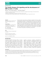
Figure 1 Chromosomal distribution of MAPK genes in G. raimondii. The chromosome numbers are indicated at the top of each bar. The
chromosome numbers from D1 to D5, and D7 to D12 were consistent with our newly-updated interspecific genetic map in allotetraploid
cultivated cotton species reported recently (Zhao et al. [32]), and the scaffolds name from G. raimindii genome was showed in the bracket. Lines
were drawn to connect duplicated genes. The nomaclature of MAPKs were based on the order of the chromosomes in G. raimondii.
Zhang et al. BMC Plant Biology 2014, 14:345
/>
Page 4 of 17
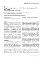
Figure 2 Comparison of the amino acid sequences of GrMAPKs. Roman numerals indicate regions containing the 11 domains (I–XI) found in
the cotton PK subdomains. The A-Loop, CD-domain and phosphorylation-activation motif (TEY and TDY) are indicated with red boxes.
phosphorylation site than the TDY motif. There are 12
AtMAPKs, 18 GmMAPKs and 18 GrMAPKs containing
the TEY motif, whereas eight AtMAPKs, 14 GmMAPKs
and 10 GrMAPKs belong to the TDY groups, with an
exception of six GmMAPKs containing the TQY motif.
By contrast, the rice genome contains more MAPKs
with the TDY phosphorylation site than the TEY motif;
11 OsMAPKs have the TDY motif but only seven contain the TEY motif. These results indicate that MAPKs
containing the TEY motif might play more important
roles in dicot plants than MAPKs containing the TDY
motif. The orthlogous relationship among MAPK genes
in G. raimondii, Arabidopsis, O. sativa and G. max was
showed in Additional file 3: Table S3.
Recent studies have shown that the G. raimondii genome has undergone at least two rounds of genome-wide
duplication [29]. To understand the expansion mechanism
of the G. raimondii MAPK gene family, we investigated
tandem and segmental duplication events of MAPK gene
family members on the 11 chromosomes by genome synteny analysis. As shown in Figure 1, 19 paralogs in 28
G. raimondii MAPKs were identified, including 18 segmental duplication events between chromosomes and one
tandem duplication event within the same chromosome
(GrMPK16 and GrMPK17). Furthermore, these paralogs
are clustered together in the phylogenic tree and share
similar exon-intron structures. These results indicate that
segmental duplication events have played a significant role
in MAPK gene expansion in the G. raimondii genome.
Cloning and expression analysis of MAPK genes in G.
hirsutum acc. TM-1
Based on predicted sequence information, we performed
PCR cloning of MAPK genes by designing gene-specific
Zhang et al. BMC Plant Biology 2014, 14:345
/>
Page 5 of 17
Figure 3 Intron and exon organization of G. raimondii MAPK genes (GrMPKs). Introns and exons are represented by black lines and colored
boxes, respectively. GrMPKs were grouped according to phylogenetic classification. Phylogenetic analysis was done using the ML method with
1,000 resampling replicates. Bootstrap values (%) based on 1000 replicates are indicated beside the nodes.
primers (Additional file 4: Table S4) and amplifying the
transcripts of given tissues of G. hirsutum acc. TM-1.
We ultimately obtained 21 MAPK cDNA sequences with
complete ORFs (GenBank accession Nos: KM190106KM190126), including 13 novel MAPKs and eight with
homologs that had been reported previously, with seven in
Upland cotton and one in Sea Island cotton (Additional
file 1: Table S1). Other seven genes with partial cDNA sequences were also identified.
To explore the possible physiological functions of
MAPKs, we designed gene-specific qRT-PCR primers
(Additional file 4: Table S4) to elucidate the expression
levels of MAPK genes in tetraploid cotton. In total, we
detected the expression patterns of 23 MAPK genes in
different tissues and organs of G. hirsutum acc. TM-1,
including roots, stems, leaves, petals, anthers, ovules and
fibers at three different developmental stages (0 days postanthesis [dpa], 10 dpa and 21 dpa). As shown in Figure 5,
MAPKs from different groups showed diverse expression
patterns in different tissues and organs, with partial overlap observed in a range of physiological processes. In detail, expression pattern of individual gene for each tissue/
organ tested was showed in Additional file 5: Figure S1.
First, five genes, including MPK3, MPK6, MPK9 and
MPK13 in group A and MPK8 in group C, were predominantly expressed in both vegetative and reproductive
organs, with the highest expression observed for MPK8 in
all tissues and organs examined. Second, MPK16 and
MPK27 (in group B) showed preferential expression in
vegetative organs; MPK16 was ubiquitously expressed in
all organs and preferentially expressed in roots, while
MPK27 showed that the highest expression levels in leaf
tissues, with 10-fold higher expression in leaves than in
other organs. Third, nine genes were predominantly
expressed in reproductive organs. Of these, four genes, including MPK10 and MPK12 in group B and MPK22 and
MPK24 in group D, had the highest expression levels in
fiber tissues, and five genes, including MPK14 in group C
and MPK2, MPK7, MPK15 and MPK28 in group D, were
preferentially expressed in anthers, petals or both. Two
additional genes, MPK5 and MPK25 (in group C) were
expressed moderately in reproductive organs, with preferential expression in fibers at different developmental
stages. Fourth, five genes, i.e., MPK18 in group B, MPK20
and MPK23 in group C, and MPK11 and MPK19 in group
D, showed very low levels of expression in all tested tissues
and organs. These results indicate that MAPK genes from
the same or different groups showed differential but overlapping expression patterns in different tissues, suggesting
that genes belonging to the same group may have diverse
functions, whereas MAPK genes from different groups
may share the same function.
Zhang et al. BMC Plant Biology 2014, 14:345
/>
Page 6 of 17
Figure 4 Phylogenetic relationships of MAPK family genes from G. raimondii, A. thaliana, O. sativa, and G. max. Amino acid sequences
were aligned using ClustalX software and subjected to phylogenetic analysis using the ML method with 1,000 resampling replicates. Bootstrap
values (%) based on 1000 replicates are indicated beside the nodes. GrMAPKs are highlighted in red and the other MAPKs from A. thaliana, O.
sativa and G. max are shown in different colors.
Expression profiles of MAPKs in response to various
stress-related signals
To investigate the roles of MAPK genes under various
stress-related stimuli, we performed qRT-PCR to detect
the differences in their expression abundance after
exposure to three stress-related signaling compounds
(abscisic acid [ABA], salicylic acid [SA], jasmonic acid
[JA]) or an oxidative stress inducer [H2O2]). A total of
23 of the MAPKs were induced by at least one of four
inducers, implying that MAPKs play important roles in
signaling pathways. Among these, ten were simultaneously induced and accumulated at higher levels after all
four treatments; ten were induced by three inducers; one
gene by two inducers and two genes by only one of the
four inducers (Figure 6). For further details, expression
pattern of individual gene under each treatment was
showed in Additional file 6: Figure S2.
Under JA conditions, 23 MAPKs were induced significantly. However, MAPKs from the four groups showed
differently altered expression patterns. Transcript levels
of genes in group A and B and most in group C
significantly increased, reaching a peak at 8 h after treatment, while those of MPK14 and MPK20 (in group C)
significantly increased and reached two peaks at 2 h and
4 h, respectively. The expression levels of the other
MAPK genes in group D significantly increased, quickly
reaching a peak at different time points.
Twenty one MAPK genes were significantly upregulated
after H2O2 treatment. In addition, MPK6 was induced,
and its expression reached two peak values at 8 and 12 h,
respectively. The other genes were significantly upregulated, reaching their highest levels at 10 h of treatment,
including three genes in group A, five in group B, five in
group C, and seven in group D. Fifteen MAPK genes, including three in group A, two in group B, four in group C,
and six in group D, were significantly upregulated after
ABA treatment, with diverse expression patterns. Finally,
fifteen MAPK genes were significantly upregulated after
SA treatment. Of these, six MAPKs were induced, including four in group A, one each in group C and D, with a
peak observed at 6 or 8 h, while three genes each in group
B, C, and D reached peak values at other time points.
Zhang et al. BMC Plant Biology 2014, 14:345
/>
Page 7 of 17
Figure 5 Real-time qRT-PCR analysis of MAPK genes in different tissues and organs in G. hirsutum acc. TM-1. A total of eight cotton
tissues (root; stem; leaf; petal; anther; ovule at 0 day post anthesis (DPA); fiber at 10 DPA; and fiber at 21 DPA) were sampled to analyze. Differences in
gene expression intensities are shown in colors indicated in the scale. Phylogenetic analysis was done using the ML method with 1,000 resampling
replicates. Bootstrap values (%) based on 1000 replicates are indicated beside the nodes.
Expression profiles of MAPKs in response to abiotic stress
To investigate the roles of MAPK genes under various
abiotic stress conditions, we performed qRT-PCR to
detect the differences in their expression after five
stress treatments (salinity, drought, cold, heat and
wounding). As shown in Figure 7, the transcript levels
of 22 MAPK genes significantly increased after NaCl
treatment. In addition to MPK5, MPK9 and MPK14,
MAPK genes in groups A, B and C were induced and
accumulated at 4 h. All members of group D were also
induced, but their expression patterns were diverse.
The detailed information for expression pattern of individual genes under each treatment was showed in
Additional file 7: Figure S3.
Eleven MAPK genes were significantly induced under
drought treatment, including two in group B, four in
group C, and five in group D. In addition, 23 MAPK
genes were induced and highly expressed after low
temperature treatment (4°C), with diverse expression
pattern. Except for MPK3, all of the MAPK genes were
induced and expressed at high levels. Moreover, 21
MAPK genes were induced and highly expressed upon
exposure to high temperature conditions. Of these, two
genes each in groups B and C and five in group D were
significantly upregulated and reached a peak at 10 h
after treatment, while other twelve genes were induced
and reached peak values at other time points. Finally, 21
MAPK genes were induced and upregulated when the
Zhang et al. BMC Plant Biology 2014, 14:345
/>
Page 8 of 17
Figure 6 Relative expression of G. hirsutum MAPK genes under stress-related signal treatments. The data are presented in clusters using
the fold-change (E/C) of relative expression for all MAPK genes in response to stress-siganl treatments (Experiment), in comparison to their
respective controls (Control). Red and blue colors represent increased or decreased expression levels, respectively, in comparison to controls.
The stress-related signals included JA, H2O2, ABA and SA, respectively.
seedling leaves were cut with scissors. Of these, three in
group A, five each in groups B and C, and eight in group
D were significantly induced and reached peak values at
different time points.
In total, the 23 detected MAPK genes were widely
induced by all types of abiotic stress (Table 1). Among
these genes, eight were induced and expressed at higher
levels under all five abiotic stress treatments. Thirteen
and two MAPK genes were induced by four and three
abiotic stresses, respectively. These expression patterns
suggest that MAPK genes carry out multiple physiological functions to help the plant adapt to various complex environmental challenges.
Paralogs of MAPKs show diverse expression patterns
To investigate whether these duplicated paralog pairs
were with the same expression patterns, we compared
their expression profiles in different organs and under
different stress treatments (Table 2). In organs, only the
correlation coefficient between MPK8 and MPK14 was
greater than 0.5, indicating a positive correlation and
similar expression patterns between these two genes.
However, other pairs had no clear positive or negative
correlation. Notably, the correlation coefficient between
MPK20 and MPK25 was lower than −0.5, suggesting
distinctly different expression patterns between these
two genes. Comparison analysis indicated that paralogs
Figure 7 Relative expression of G. hirsutum MAPK genes under different stress treatments. The data are presented in clusters using the
fold-change (E/C) of relative expression for all MAPK genes in response to different treatments (Experiment), in comparison to their respective
controls (Control). Red and blue colors represent increased or decreased expression levels, respectively, in comparison to controls. The stressors
included NaCl, PEG, 4°C, 37°C, and wounding treatment, respectively.
Zhang et al. BMC Plant Biology 2014, 14:345
/>
Page 9 of 17
Table 1 Expression profiles of MAPK genes under different stress treatments in cotton
Gene
Group
Signalling molecules
Environmental stress factors
JA (100uM)
H2O2 (10 mM)
ABA (100uM)
SA (10 mM)
Salt (200 mM)
PEG6000 (20%)
4°C
37°C
Wounding
MPK10
Group B
**
**
D
D
**
D
**
**
**
MPK27
Group B
**
**
**
D
**
**
**
**
**
MPK16
Group B
**
**
*
**
**
*
**
**
**
MPK18
Group B
**
**
-
**
**
**
**
**
**
MPK12
Group B
**
**
**
**
**
D
**
**
**
MPK3
Group A
**
**
*
**
**
-
**
**
*
MPK6
Group A
**
**
**
**
**
D
**
**
**
MPK9
Group A
**
**
**
**
**
D
**
**
**
MPK13
Group A
**
**
**
**
**
D
**
**
**
MPK5
Group C
**
**
**
**
-
-
**
**
**
MPK14
Group C
**
D
D
D
**
**
**
**
D
MPK25
Group C
**
**
**
**
**
**
**
**
**
MPK23
Group C
**
**
*
**
**
**
**
**
**
MPK8
Group C
**
**
**
**
**
*
**
**
**
MPK20
Group C
**
**
**
*
**
**
**
**
**
MPK2
Group D
**
**
**
**
**
**
**
**
**
MPK7
Group D
**
**
**
**
**
**
**
*
**
MPK22
Group D
**
**
*
**
**
-
**
**
**
MPK28
Group D
**
**
**
**
**
**
**
**
**
MPK11
Group D
**
**
**
*
**
**
**
/
**
MPK19
Group D
**
*
*
D
**
D
**
**
**
MPK15
Group D
**
**
**
*
**
**
**
**
**
MPK24
Group D
**
**
**
*
**
D
**
**
**
Note: For hormone treatments, the leaves of seedlings were harvested at 0, 0.5, 1, 2, 4, 6, 8, 10, 12 and 24 h after treatment;
For the environmental stress factor treatments, the leaves of seedlings were harvested at 0, 0.25, 0.5, 1, 2, 4, 6, 8, 10, 12 and 24 h after treatment;
“**” and “*” indicate significant difference at P < 0.01 and P < 0.05, respectively;
“-” represents no change and weak upregulation; “D” represents significant reduction in MAPK gene expression after treatment;
“/” represents absent data. The Student’s t-test was performed between treated samples and untreated samples.
of MAPKs from the same ancestor showed differential
expression in different tissues and organs, implying that
these genes evolved via gene duplication followed by expressional divergence.
Furthermore, correlation analysis indicated that there
were eight paralogs involved in stress-related signals and
seven in abiotic stress with values greater than 0.5, implying positively correlated expression between paralogs
under stress. Unlike MPK2-MPK7, MPK14-MPK25 and
MPK22-MPK28, four paralogs, i.e., MPK8-MPK20, MPK8MPK23, MPK9-MPK13 and MPK16-MPK27, showed clear
positive correlations under both stress-related signal and
abiotic stress treatment, and other seven paralogs showed
positive correlations under one or two stress conditions.
Taken together, these results suggest that MAPKs may have
retained functional conservation after gene duplication to
help plants cope with different stresses, acting as the main
contributors to wide adaptation during the cotton evolutionary process.
Potential functional roles of three MAPK genes in
Verticillium dahliae resistance, as determined by TRV-VIGS
Three MAPKs, including MPK9, MPK13 and MPK25,
were significantly induced after Verticillium dahliae inoculation (Figure 8a). The transcript levels of MPK9 and
MPK13 significantly increased, with the highest peak observed at 24 h of treatment. MPK25 was significantly
downregulated in response to inoculation after 24 h and
48 h, and its expression recovered to high levels at 96 h
post-inoculation.
Virus-induced gene silencing (VIGS) has been successfully used in cotton [34-36]. To further investigate the
function of MPK9, MPK13 and MPK25 in V. dahliae
resistance, we constructed recombinant viruses to silence endogenous genes in cotton, producing constructs
TRV2:MPK9, TRV2:MPK13 and TRV2:MPK25, with
TRV1-TRV2 for the mock treatment. To validate the
reliability of VIGS in cotton, we silenced an indicator
gene, CLA1 (CLOROPLASTOS ALTERADOS 1, encoding
Zhang et al. BMC Plant Biology 2014, 14:345
/>
Page 10 of 17
Table 2 Pearson correlation coefficients of the expression profiles of paralogous pairs
Gene1
Gene2
Similarity
Correlation coefficient*
Correlation coefficient
Correlation coefficient
(organs)
(hormone)
(abiotic stress)
0.26
MPK2
MPK7
86.73%
0.24
0.40
MPK5
MPK14
93.28%
−0.04
0.63
0.27
MPK8
MPK14
84.51%
0.86
0.60
0.10
MPK8
MPK20
93.48%
0.34
0.51
0.56
MPK8
MPK23
91.85%
0.15
0.89
0.84
MPK8
MPK25
85.87%
−0.39
0.26
0.69
MPK9
MPK13
92.27%
−0.46
0.71
0.73
MPK10
MPK27
90.37%
−0.15
0.83
−0.03
MPK10
MPK16
86.63%
0.03
0.66
−0.06
MPK14
MPK25
80.65%
−0.30
0.14
0.07
MPK16
MPK27
84.96%
−0.26
0.83
0.64
MPK20
MPK23
92.39%
−0.49
0.33
0.67
MPK20
MPK25
86.96%
−0.61
−0.06
0.53
MPK22
MPK28
77.28%
−0.09
−0.07
0.23
*Correlation coefficient: r > 0.5: positive correlation, showed in bold type; 0 < r < 0.5: no clear positive correlation; −0.5 < r < 0: no clear negative correlation; r < −0.5:
negative correlation.
1-deoxy-D-xylulose-5- phosphate synthase), producing
plants with a photobleached phenotype. At least 15 plants
were infiltrated per construct at 8 days post-emergence,
and untreated plants were grown in the same environment
without syringe treatment. Two weeks later, all treated individuals infiltrated with TRV2-CLA1 showed highly uniform
bleaching in newly emerged leaves (Figure 8b). Real-time
quantitative PCR confirmed that untreated and mocktreated plants showed the same and high expression levels
of MPK9, MPK13 and MPK25. However, the transcripts of
these three genes exhibited strong silencing in infiltrated
TRV2:MPK9, TRV2:MPK13 and TRV2: MPK25 plants
(P < 0.01)(Figure 8c).
We inoculated cotton seedlings using dip-infection
with liquid containing 1 × 10⁷ V. dahliae spores. Two
weeks later, spontaneous lesions in stems and yellow leaf
veins were found in target gene-silenced plants. Four
weeks later, the true leaves of diseased plants exhibited
wilting (Figure 9a). In general, the control plants seldom
exhibited leaf wilting, with average diseased leaf: healthy
leaf ratios of approximately 30%. However, 69.3% of the
MPK9-silenced plants were severely infected by V. dahlia,
which was similar to the results observed in susceptible
control plants (G. hirsutum cv. Junmian 1, with the percentage of diseased plants at 76.8%). Furthermore, 63.75%
of the MPK13-silenced plants showed a severe wilting
phenotype, and 54% of the MPK25-silenced plants exhibited wilting symptoms on leaves when infected with
V. dahlia (Figure 9b). These results demonstrate that silencing of MPK9, MPK13 and MPK25 compromises the
resistance of cotton to this pathogen, and gene-silenced
plants exhibited more wilting and etiolated leaves than the
vector control plants with P < 0.01 significance. In summary, MPK9, MPK13 and MPK25 are important components of resistance to V. dahlia infection in cotton.
Discussion
Characterization of MAPKs in G. raimondii and evolution
of MAPK genes
Based on the genome scans of several plant genomes,
MAPK family genes have been systematically investigated in Arabidopsis [12], tomato [15], tobacco [16],
wheat [17], rice [18] and soybean [19]. In the current
study, a total of 28 MAPKs from G. raimondii were
identified. These MAPKs were classified into four
groups (A, B, C and D) according to their phylogenetic
clades, which were similar to those reported in
Arabidopsis and O. sativa [18,37]. We also found that all
MAPK proteins contain 11 domains (I–XI; Figure 1),
and TEY or TDY motifs of MAPKs are located in the activation loop between kinase subdomain VII and VIII, as
described previously [12,38]. The subgroup of A, B and
C possesses a Thr-Glu-Tyr (TEY) domain and a short
C-terminus containing a common docking (CD) domain
that consists of the sequence[LHY]Dxx[DE]EpxC, whereas
those of subgroup D possess a Thr-Asp-Tyr(TDY) activation domain, without the CD domain but with a relatively
long C-terminal region, which is also consistent with
previous reports [39,40]. Previous studies, such as reports
in Arabidopsis, tobacco, tomato and rice, focused on TEY
MAPKs [2]. Interestingly, Arabidopsis, G. max and
G. raimondii contain more MAPKs with the TEY
phosphorylation site than the TDY motif. By contrast,
the O. sativa genome contains more MAPKs with the
Zhang et al. BMC Plant Biology 2014, 14:345
/>
Page 11 of 17
Figure 8 Expression patterns of three MAPK genes induced by V. dahliae and VIGS analysis. (a) Q-PCR analysis of the expression of three
MAPK genes after inoculation by V. dahliae. The error bars were calculated based on three biological replicates using standard deviation. “*”: significant
difference (P < 0.05); “**”: significant difference (p < 0.01). (b) Phenotypes after TRV-VIGS silencing of three MAPK genes and GhCLA1. After two weeks
post-treatment with TRV1 and TRV2/TRV2-MPKs, the treated plants exhibited normal growth. TRV1- and TRV-GhCLA1-treated plants exhibited a
photobleaching phenotype. (c) Gene expression of MPK9, MPK13 and MPK25 in silenced and control plant leaves by Q-PCR analysis; The error bars
were calculated based on three biological replicates using standard deviation. “*”: significant difference (P < 0.05); “**”: significant difference (p < 0.01).
The cotton histone 3 (AF026714) was used as the reference gene.
TDY phosphorylation site than the TEY motif. We
propose that in dicot plants, MAPKs with the TEY
motif play more important roles than MAPKs with the
TDY motif.
Phylogenetic analysis between cotton and other plants
revealed that GrMAPKs in A, B and C might come from
the same ancestor, while D MPAK genes might be paralogous products. The distribution of GrMPAKs is nonrandom, which is similar to the result of a previous
study [41]. Based on phylogenetic tree analysis, we found
that a large number of MAPKs belong to subgroup D,
which is similar to reports in Arabidopsis, rice and poplar [12,18,41,42]. These results support the previous suggestion that subgroup D expanded before and after the
monocot/dicot split [41]. Comparison of exon-intron
structures indicated that the A, B and C groups share a
similar number of exons and the lengths of exons are
more conserved than those of introns. However, members in the D group have more exons and the lengths of
these exons and introns are diverse. Synteny analysis of
the G. raimondii genome indicated that the MAPK family mainly resulted from segmental duplication.
Expression patterns of MAPKs imply their functional
divergence during plant development and growth
Previous studies have demonstrated that some MAPK
genes exhibit tissue specificity in various plants such as
Arabidopsis, tobacco, poplar, Brachypodium distachyon,
wheat and Brassica [17,37,43-46]. RsMPK2 was detected
in vegetative and reproductive organs, with different expression patterns [47]. TaMAPK13 is expressed in all tissue, but TaMPK15 is only expressed at low levels in
flowers [17].
In the current study, we observed differential expression
patterns of MAPKs in vegetative organs (roots, stems and
leaves) and reproductive organs (anthers, petals and fiber
tissues at different developmental stages). Five genes were
constitutively expressed at high levels in both vegetative
Zhang et al. BMC Plant Biology 2014, 14:345
/>
Page 12 of 17
Figure 9 Silencing of MPK9, MPK13 and MPK25 enhances plant susceptibility to Verticillium dahlia infection. Individual genes of the
cotton cultivar Hai7124 were first silenced by VIGS, and the plants were then inoculated with V. dahliae suspension spores at a concentration
of 1 × 10⁷/mL. (a) Plant phenotypes at 28 days after V. dahlia inoculation. (b) Percentage of diseased leaves after V. dahlia inoculation. The
percentage of diseased leaves was scored. The experiments were repeated using 15 plants per treatment. The error bars were calculated
based on three biological replicates using standard deviation.
and reproductive organs. Two genes were expressed at
higher levels in vegetative organs, and the remaining genes
were expressed at higher levels in reproductive organs. A
recent study demonstrated that AtMPK4 plays an important role in meiotic cytokinesis during pollen development
[48]. PsMPK3 is involved in fruit set, which is activated by
gibberellins and cytokinins [49]. SlMPK3 is expressed
at markedly high levels in stamens [15]. The tissue- or
organ-specific MAPK expression patterns observed in the
current study indicate their functional divergence during
plant development and growth. Interestingly, four MAPK
genes, MPK3, MPK6, MPK9, and MPK13 in Group A and
MPK8 in Group C with higher expression in almost all
tested tissues were worth to be further studied for actual
function.
Varied expression of MAPKs in response to stress-related
signals and abiotic stressors
Abiotic and biotic stresses such as cold, drought and
pathogens seriously affect cotton growth and yield, and
studies have focused on the molecular mechanisms
underlying the response to these stresses in cotton. To
date, an increasing number of studies have shown that
MAPKs can regulate plant development, growth and responses to abiotic/biotic stress. In cotton, GhMPK2 and
GbMPK3 are upregulated by diverse abiotic stresses and
likely play a role in drought and oxidative stress
tolerance [21,22]. GhMPK6 plays an important role in
ABA-induced CAT1 expression and H2O2 production
[23], whereas GhMPK6a negatively regulates responses
to osmotic stress and bacterial infection [24]. GhMPK7
and GhMPK16 are involved in plant defense responses
and the regulation of certain components of multiple
stress-signaling pathways [25,26]. Extensive studies
have revealed that MAPKs are not only involved in abiotic stress and biotic responses but also in plant development and hormonal signaling. Vlot and coworkers
(2009) suggested that SA can regulate responses to biotrophic pathogens and systemic acquired resistance,
while JA mediates responses to necrotrophs [50]. Recent studies have shown that ABA is involved in salinity and drought responses [51]. H2O2 can induce
oxidative bursts or the accumulation of reactive oxygen
species (ROS) in plant cells. ROS may contribute to
resistance by directly killing the invading pathogen or
activating cell wall crosslinking and lignification and
Zhang et al. BMC Plant Biology 2014, 14:345
/>
subsequently strengthening the cell wall to help confine pathogen infection [52].
Systematic analyses of the expression patterns of MAPK
genes under stress-related signal treatment showed that
ten of 23 (43.5%) MAPK genes were induced by four
inducers. Ten (43.5%) and one (4.3%) of these genes were
induced by three and two inducers, respectively. Two
(8.7%) genes were induced by only one of the four inducers. Accumulating evidence has shown that systemic
defense responses in plants are controlled by the mutually
antagonistic hormones JA and SA. Our data indicate that
all MAPK genes were induced under JA treatment, with
15 were simultaneously induced by SA. This finding implies that these genes are coregulated by JA and SA and
highlights the notion that MAPK genes might play key
roles in plant defense responses. Furthermore, the gene
expression patterns under abiotic stress show that eight of
23 (34.8%) MAPK genes were upregulated by five abiotic
stressors. Thirteen (56.5%) and two (8.7%) were upregulated by four or three stressors, respectively. The present
results further demonstrate that MAPKs are involved in
the response to environmental stress in cotton.
Previous reports have shown that each hormone signaling pathway contributes to an interactive network
that coordinates responses to different stresses [53]. In
addition, abiotic stress-regulated genes act either in an
ABA-dependent or ABA-independent manner, SA can
regulate responses to biotrophic pathogens and systemic
acquired resistance. Here, among 15 ABA-regulated
MAPK genes, 10 were also regulated by SA. Further, our
data indicate that eight MAPK genes were upregulated
by five stressors and also induced by JA and H2O2.
Among these, five genes were simultaneously induced by
four stress-related signals. The widespread induction of
MAPK genes in response to diverse stressors and hormones suggests that MAPK genes play a significant role
in hormone signaling pathways during stress tolerance.
Gene duplication is followed by functional diversification. A comparison of the expression patterns of paralogous genes demonstrated that most, but not all, of these
genes showed similar responses towards various hormone
and abiotic treatments. For instance, MPK9-MPK13,
MPK16-MPK27, MPK8-MPK20 and MPK8-MPK23 paralogs showed similar responses to both hormone and
abiotic treatments. The paralogs MPK5-MPK14, MPK8MPK14, MPK10-MPK16 and MPK10-MPK27 showed
similar expression patterns in response to hormones, and
the paralogs MPK8-MPK25, MPK20-MPK23 and MPK20MPK25 showed similar expression patterns in response to
abiotic stresses. Our results suggest that these pairs of
MAPKs share similar functions under abiotic and/or hormone treatment, respectively. An examination of the overall
transcription patterns suggests that gene duplication resulted in partially overlapping functions; the redundant
Page 13 of 17
functions of MAPK genes may be beneficial for protecting
the cell from various stress conditions. On the other hand,
paralogs of MAPK genes also showed an interesting pattern
of functional divergence in different organs and tissues, implying that MAPK genes may play a crucial role in driving
evolutionary novelty and adaptation to new environments.
MPK9, MPK13 and MPK25 are required for resistance
against Verticillium dahliae in cotton
Gene expression patterns are usually an indicator of
gene functions. In the current study, we found that
MAPK genes were generally responsive to biotic and
abiotic stress treatments, suggesting that they play important roles in responses to environmental stress and
pathogens. Verticillium wilt is a serious disease that significantly affects the yield and quality of cotton.
Accumulating evidence demonstrates that the MAPK
cascade plays an important role in the regulation of
pathogen-induced defenses. AtMPK3/AtMPK6 are activated by pathogens and regulate the pathogen defense
response pathway, and AtMPK3/AtMPK6 are involved
in abiotic stress response (salt, drought, cold, wounding)
and hormone signal pathways [54,55]. OsMPK5 is also
induced by abiotic stresses, pathogen infection and ABA
treatment [56]. In cotton, GhMPK6a, GhMPK7 and
GhMPK16 are involved in the pathogen-resistance response [24-26]. Here, we found that MPK9, MPK13 and
MPK25 were significantly upregulated in cotton roots
after inoculation with V. dahliae, and these three genes
were upregulated in leaves after exposure to JA, H2O2,
ABA and SA. We speculate that MPK9, MPK13 and
MPK25 are involved in regulating the pathogen response. Using VIGS technology [34-36], we further investigated the function of MPK9, MPK13 and MPK25 in
V. dahliae resistance. Statistical analysis showed that silencing of whether MPK9, MPK13 or MPK25 by VIGS
increased significantly the susceptibility of cotton to
V. dahliae. Compared with the two other genes, plants harboring a silenced MPK9 gene were more severely infected
by V. dahlia, implying that MPK9 plays an important role
in V. dahlia resistance in cotton.
Conclusions
A total of 28 GrMAPKs were identified based on the
genome sequence of G. raimondii, and 21 cDNA sequences of MAPKs with complete ORFs were cloned
from G. hirsutum. Phylogenetic tree and motif analysis
showed that GrMAPKs could be classified into four
groups, comparable to those in Arabidopsis, O. sativa
and G. max. Most MAPKs showed different temporal
and spatial expression patterns in vegetative and reproductive organs, and crosstalk occurred under biotic/abiotic stress and stress-related signal treatment. VIGS
analysis indicated that MPK9, MPK13 and MPK25 are
Zhang et al. BMC Plant Biology 2014, 14:345
/>
important components in cotton resistance to V. dahliae
infection. Our work provides a reference for systematically elucidating the important roles of MAPKs in cotton
growth, development and responses to abiotic and biotic
stresses and for effectively utilizing MAPKs in cotton
stress tolerance breeding.
Methods
Prediction, mapping and analysis of the MAPK gene
family
Genes and proteins annotated in G. raimondii were
downloaded from . HMMER
software version 3.0 [27] and the Pfam protein family
database with the MAPK domain (PF00069) [28] were
used as a query to screen the G. raimondii genomic
database. Expressed sequence tag (EST) sequences for
four cotton species, Gossypium hirsutum(Gh), G. barbadense (Gb), G. arboreum (Ga) and G. raimondii (Gr),
were downloaded from the GenBank EST database
( />Mapping of MAPK genes was performed using
MapInspect ( />mapinspect.html). The exon/intron structures of individual GrMAPK genes were determined by aligning
the cDNA sequences to their corresponding genomic
DNA sequences.
Conserved domain detection and subcellular location
predication
The programs INTERPROSCAN, SMART, MOTIF and
PLANTSP were employed to detect conserved domains.
If a given protein sequence contained the MAP Kinase
signature, PK domain and ATP-binding domain, it was
regarded as a candidate member of the cotton MAPK
family. The subcellular localization of each GrMAPK
was analyzed using CELLO v2.5 server (e.
nctu.edu.tw/) [57].
Page 14 of 17
conditions. Petals and anthers were sampled on the day
of flowering, and ovules and fibers were excised from
developing flower buds or bolls on selected days post
anthesis (dpa). Roots, stems and leaves were collected
from two-week-old seedlings. The materials were quickfrozen in liquid nitrogen and stored at −70°C before use.
G. hirsutum L. cv. Jinmian 19, which exhibits high tolerance to abiotic stress, was used for the abiotic stress
treatments. Cotton seedlings (G. hirsutum L. cv. Jinmian
19) were grown in a growth chamber under greenhouse
conditions at 28°C under a 16 h light/8 h dark cycle.
Three-week-old cotton seedlings were used for the following treatments. For signaling substance treatments,
leaves were sprayed with 100 μM JA, 100 μM ABA,
100 mM SA or 10 mM H2O2 (ddH2O as a solvent control). For the salt and drought treatments, the roots of
cotton seedlings were irrigated with 200 mM NaCl and
20% PEG, respectively (ddH2O as a mock control). For
temperature stress treatments, the seedlings were placed
in a growth chamber at a high temperature (37°C) or a
low temperature (4°C; 28°C as a mock). Seedling leaves
were cut with scissors for wound treatment. The leaves
were harvested at the appropriate time points as indicated (triplicate samples were collected at each time
point [n = 3 seedlings]), frozen in liquid nitrogen and
stored at −70°C for further analysis.
G. barbadense L. cv. Hai7124, which exhibits Verticillium resistance, was used for fungal pathogen (V. dahliae) inoculation. For pathogen treatment, the roots of
Hai7124 seedlings were dipped in V. dahliae strain V991
conidial suspensions containing 107 spores mL−1. The
roots were harvested at the appropriate time points,
quick-frozen in liquid nitrogen and stored at −70°C before use. G. hirsutum L. cv. Junmian 1, which is susceptible to Verticillium, was used as a susceptible plant
control.
RNA isolation and real-time PCR analysis
Sequence alignments and phylogenetic construction
Multiple sequence alignments of the MAPK domain
with 28 amino acids were performed using ClustalX
(ver.1.83) [58], and a phylogenetic tree was constructed
by the Maximum likelihood (ML) method in MEGA
5.1 (www.megasoftware.net) [33]; the bootstrap test of
phylogeny was performed with 1,000 replications. In
addition, the amino acid sequences of MAPKs from four
plants (Arabidopsis, O. sativa, G. max and G. raimondii)
were initially aligned and used to construct phylogenetic
trees.
Plant materials and treatments
G. hirsutum L. acc TM-1, a genetic standard line of
Upland cotton, was used for tissue/organ expression
analysis. The plants were cultivated under normal field
Total RNA was extracted from cotton seedling leaves
using the CTAB-acidic phenol extraction method
[59]. RNA was then treated with DNase I (Invitrogen,
to remove genomic DNA,
and 2 μg of total RNA was used for first-strand cDNA
synthesis. The primer pairs used for real-time PCR were
designed using Beacon Designer 7.0 according to cotton
MAPK gene sequences. The amplified fragment lengths
were between 75 bp and 200 bp, and the annealing
temperature was between 58°C and 60°C. The cotton
histone3 (AF024716) gene was used as the reference
gene [60].
The amplification reactions of real-time PCR were performed on an ABI 7500 Real Time PCR System (Applied
Biosystems, USA) using SYBR Green (Bio-Rad, USA)
with three replicates. The amplification parameters were
Zhang et al. BMC Plant Biology 2014, 14:345
/>
as follows: denaturation at 95°C for 10 min, 40 cycles of
denaturation at 95°C for 15 s, annealing between 58°C
and 60°C for 15 s, extension at 72°C for 15 s. For the
melting curve stage, the default settings were chosen.
The expression levels of MAPK genes were calculated
according to Livak and Schmittgen [61].
Cloning of MAPK genes from G. hirsutum acc. TM-1
Based on the predicted sequences, gene-specific primers
were designed to obtain the complete coding sequences.
The primer pairs for all genes and the optimal melting
temperature are listed in Additional file 4: Table S4; the
transcripts from various tissues of G. hirsutum acc. TM-1
were used for amplification. Standard PCR reactions were
performed using High-fidelity ExTaq DNA Polymerase
(TaKaRa Biotechnology [Dalian] Co., Ltd., China). The
PCR products were cloned into the pMD18-T Vector
(TaKaRa) according to the manufacturer’s instructions and
sequenced from plasmid DNA templates. At least six
clones per gene were randomly picked and sequenced.
The cDNA sequences of the MAPK genes were determined using alignment analysis with their corresponding
sequences obtained from bioinformatic analysis.
Construction of VIGS vectors and agro-infiltration
The pTRV1 and pTRV2 VIGS vectors were kindly
provided by Dr. Libo Shan of Texas A&M University
(College Station, TX, USA). The constructs contained
the following fragments: TRV2:MPK9, a 501-bp fragment of an MPK9 cDNA fragment that corresponds to
base positions 47–538 bp; TRV2: MPK13, a 391-bp fragment of an MPK13 cDNA fragment that corresponds to
base positions 43–434 bp and TRV2:MPK25, a 392-bp
fragment of an MPK25 cDNA fragment that corresponds to base positions 58–440 bp. These fragments
were amplified by PCR from TM-1 cDNA using primers
with XbaI/XhoI enzyme sites for TRV2:MPK9 and
XbaI/SacI sites for both TRV2:MPK13 and TRV2:
MPK25, for insertion into TRV2, respectively. The primer pairs used for the construction of VIGS vectors harboring the three MAPK genes are listed in Additional
file 4: Table S4. The control vector pTRV2-GhCLA1 was
the kind gift of Dr. Xinyu Wang, Nanjing Agricultural
University.
Plasmids containing TRV1, TRV2, TRV2:MPK9, TRV2:
MPK13 and TRV2:MPK25 were individually introduced
into Agrobacterium tumefaciens strain GV3101. Agrobacterium cultures carrying the recombinant TRV vectors
were grown overnight at 28°C in LB medium containing
the antibiotics 50 μg/mL kanamycin and 25 μg/mL rifampicin. The cultures were then inoculated into 50 mL LB
medium (50 μg/mL kanamycin, 25 μg/mL rifampicin) at a
concentration of 1:100 and cultured, with shaking, to
an OD of 0.5 at 28°C. The cells were pelleted by
Page 15 of 17
centrifugation at 1,180 × g at room temperature for 5 min
and resuspended in infiltration media (10 mM MgCl2,
10 mM MES and 200 μM acetosyringone).
The cell suspensions were incubated at room
temperature for 3 h and then Agrobacterium GV3101
carrying TRV1 and TRV2:MPK9/13/25 was infiltrated
into two fully expanded cotyledons of eight-day-old
cotton seedlings (Hai7124) using a needleless 1 mL
syringe at a 1:1 ratio, with Junmian-1 serving as a susceptible control. For mock treatment and the technical
control, the same plants were infiltrated with a 1:1
mixture of Agrobacterium carrying TRV1 and TRV2 or
TRV1 and TRV2:CLA1, respectively. The plants were
grown at 23/22°C (day/night) in a growth chamber
with a 16 h light/8 h dark cycle for four weeks before
they were used for the assays. Untreated plants were
grown under the same conditions but were not wounded.
VIGS experiments were repeated at least three times with
more than 16 plants for each construct per repeat.
Pathogen inoculation
The defoliating isolate V991 of V. dahliae was grown on
potato dextrose agar for 4 d at room temperature (25°C)
and then incubated in Czapek’s medium at 25°C for 5 d.
The spore suspensions were prepared at 1 × 10⁷ conidia
mL−1 for inoculation of cotton seedlings by dip-infection.
Additional files
Additional file 1: Table S1. Genome-wide bioinformatic analysis of
MAPK genes in Gossypium.
Additional file 2: Table S2. The information for MAPK genes from
different species used in phylogenetic analysis.
Additional file 3: Table S3. The orthlogous relationship among MAPKs
in Arabidopsis, O. sativa, G. max, and G. raimondii.
Additional file 4: Table S4. Oligonucleotide primers used in this study.
Additional file 5: Figure S1. Expression patterns of the 23 MAPK genes
in various tissues in cotton by quantitative real time PCR analysis. 1: root;
2: stem; 3: leaf; 4: petal; 5: anther; 6: ovule at 0 day post anthesis (DPA); 7:
fiber at 10 DPA; 8: fiber at 21 DPA. The Y-axis indicates relative expression
levels and the X-axis indicates different tissues. The error bars were
calculated based on three biological replicates using standard deviation.
Additional file 6: Figure S2. Expression patterns of MAPK genes under
stress-related signal treatments (a, JA; b, H2O2; c, ABA; d, SA). The expression
levels data were presented as the mean fold by comparing treated samples
with controls. The Y-axis indicates relative expression levels and the X-axis
indicates the hours of stress-related signal treatments. The error bars were
calculated based on three biological replicates using standard deviation. “*”:
significant difference (P < 0.05); “**”: significant difference (p < 0.01).
Additional file 7: Figure S3. Expression patterns of MAPK genes under
stress treatments (a, NaCl; b, PEG; c, 4°C; d, 37°C; e, wounding). The expression
levels data were presented as the mean fold by comparing experiments and
controls samples. The Y-axis indicates relative expression levels and the X-axis
indicates the hours of stress treatments. The error bars were calculated based
on three biological replicates using standard deviation. “*”: significant
difference (P < 0.05); “**”: significant difference (p < 0.01).
Competing interests
The authors declare that they have no competing interests.
Zhang et al. BMC Plant Biology 2014, 14:345
/>
Authors’ contributions
Experiments were designed by WZG. Experiments were performed by XYZ,
LMW, XYX and CPC. XYZ and WZG drafted the manuscript and WZG revised
the manuscript. All authors read and approved the final manuscript.
Acknowledgements
This program was financially supported in part by National Natural Science
Foundation of China (31171590), the National Transgenic Program
(2011ZX08005-004), Jiangsu Agriculture Science and Technology Innovation
Fund (CX(14)2065), and projects funded by PAPD-JHEI and JCIC-MCP.
Supporting data
GrMAPKs were identified from the genome of G. raimondii downloaded from
(Additional file 1: Table S1). MAPK genes (from
Arabidopsis, O. sativa, G. max) used in phylogenetic analyses can be found
with gene ID and protein sequences in Additional file 2: Table S2. Multiple
sequence alignment and phylogenetic tree are available from TreeBASE
( />Received: 15 July 2014 Accepted: 20 November 2014
References
1. Nakashima K, Ito Y, Yamaguchi-Shinozaki K: Transcriptional regulatory
networks in response to abiotic stresses in Arabidopsis and grasses.
Plant Physiol 2009, 149(1):88–95.
2. Nakagami H, Pitzschke A, Hirt H: Emerging MAP kinase pathways in plant
stress signalling. Trends Plant Sci 2005, 10(7):339–346.
3. Pitzschke A, Schikora A, Hirt H: MAPK cascade signalling networks in plant
defence. Curr Opin Plant Biol 2009, 12(4):421–426.
4. Romeis T: Protein kinases in the plant defense response. Curr Opin Plant
Biol 2001, 4(5):407–414.
5. Tena G, Asai T, Chiu WL, Sheen J: Plant mitogen-activated protein kinase
signaling cascades. Curr Opin Plant Biol 2001, 4(5):392–400.
6. Zhang S, Klessig DF: MAPK cascades in plant defense signaling. Trends Plant
Sci 2001, 6(11):520–527.
7. Jonak C, Ökrész L, Bögre L, Hirt H: Complexity, cross talk and integration
of plant MAP kinase signalling. Curr Opin Plant Biol 2002, 5(5):415–424.
8. Larade K, Storey KB: Analysis of signal transduction pathways during
anoxia exposure in a marine snail: a role for p38 MAP kinase and
downstream signaling cascades. Comp Biochem Physiol B 2006,
143(1):85–91.
9. Rohila JS, Yang Y: Rice mitogen‐activated protein kinase gene family
and its role in biotic and abiotic stress response. J Integr Plant Biol 2007,
49(6):751–759.
10. Fiil BK, Petersen K, Petersen M, Mundy J: Gene regulation by MAP kinase
cascades. Curr Opin Plant Biol 2009, 12(5):615–621.
11. Hirt H: Multiple roles of MAP kinases in plant signal transduction.
Trends Plant Sci 1997, 2(1):11–15.
12. Ichimura K, Shinozaki K, Tena G, Sheen J, Henry Y, Champion A, Kreis M,
Zhang S, Hirt H, Wilson C: Mitogen-activated protein kinase cascades in
plants: a new nomenclature. Trends Plant Sci 2002, 7(7):301–308.
13. Lee SK, Kim BG, Kwon TR, Jeong MJ, Park SR, Lee JW, Byun MO, Kwon HB,
Matthews BF, Hong CB, Park SC: Overexpression of the mitogen-activated
protein kinase gene OsMAPK33 enhances sensitivity to salt stress in rice
(Oryza sativa L.). J Biosci 2011, 36(1):139–151.
14. Ichimura K, Mizoguchi T, Yoshida R, Yuasa T, Shinozaki K: Various abiotic
stresses rapidly activate Arabidopsis MAP kinases ATMPK4 and ATMPK6.
Plant J 2000, 24(5):655–665.
15. Kong F, Wang J, Cheng L, Liu S, Wu J, Peng Z, Lu G: Genome-wide analysis
of the mitogen-activated protein kinase gene family in Solanum
lycopersicum. Gene 2012, 499(1):108–120.
16. Zhang X, Cheng T, Wang G, Yan Y, Xia Q: Cloning and evolutionary
analysis of tobacco MAPK gene family. Mol Biol Rep 2013, 40(2):1407–1415.
17. Lian WW, Tang YM, Gao SQ, Zhang Z, Zhao X, Zhao CP: Phylogenetic
analysis and expression patterns of the MAPK gene family in wheat
(Triticum aestivum L.). J Integr Agr 2012, 11(8):1227–1235.
18. Reyna NS, Yang Y: Molecular analysis of the rice MAP kinase gene family
in relation to Magnaporthe grisea infection. Mol Plant Microbe In 2006,
19(5):530–540.
Page 16 of 17
19. Neupane A, Nepal MP, Piya S, Subramanian S, Rohila JS, Reese RN,
Benson BV: Identification, nomenclature, and evolutionary relationships
of mitogen-activated protein kinase (MAPK) genes in soybean. Evol Bioinform
2013, 9:363–386.
20. Teige M, Scheikl E, Eulgem T, Doczi R, Ichimura K, Shinozaki K, Dangl JL,
Hirt H: The MKK2 pathway mediates cold and salt stress signaling in
Arabidopsis. Mol Cell 2004, 15(1):141–152.
21. Long L, Gao W, Xu L, Liu M, Luo X, He X, Yang X, Zhang X, Zhu L: GbMPK3,
a mitogen-activated protein kinase from cotton, enhances drought and
oxidative stress tolerance in tobacco. Plant Cell Tiss Org 2013, 116(2):153–162.
22. Zhang L, Xi D, Li S, Gao Z, Zhao S, Shi J, Wu C, Guo X: A cotton group C
MAP kinase gene, GhMPK2, positively regulates salt and drought
tolerance in tobacco. Plant Mol Biol 2011, 77(1–2):17–31.
23. Luo J, Zhao LL, Gong SY, Sun X, Li P, Qin LX, Zhou Y, Xu WL, Li XB:
A cotton mitogen-activated protein kinase (GhMPK6) is involved in
ABA-induced CAT1 expression and H 2O2 production. J Genet Genomics
2011, 38(11):557–565.
24. Li Y, Zhang L, Wang X, Zhang W, Hao L, Chu X, Guo X: Cotton GhMPK6a
negatively regulates osmotic tolerance and bacterial infection in
transgenic Nicotiana benthamiana, and plays a pivotal role in
development. FEBS J 2013, 280(20):5128–5144.
25. Shi J, Zhang L, An H, Wu C, Guo X: GhMPK16, a novel stress-responsive
group D MAPK gene from cotton, is involved in disease resistance and
drought sensitivity. BMC Mol Biol 2011, 12(1):22.
26. Shi J, An HL, Zhang L, Gao Z, Guo XQ: GhMPK7, a novel multiple
stress-responsive cotton group C MAPK gene, has a role in broad
spectrum disease resistance and plant development. Plant Mol Biol
2010, 74(1–2):17.
27. Eddy SR: Accelerated profile HMM searches. PLoS Comput Biol 2011,
7(10):e1002195.
28. Finn RD, Bateman A, Clements J, Coggill P, Eberhardt RY, Eddy SR, Heger A,
Hetherington K, Holm L, Mistry J: Pfam: the protein families database.
Nucleic Acids Res 2014, 42(D1):D222–D230.
29. Wang K, Wang Z, Li F, Ye W, Wang J, Song G, Yue Z, Cong L, Shang H,
Zhu S, Zou C, Li Q, Yuan Y, Lu C, Wei H, Gou C, Zheng Z, Yin Y, Zhang X,
Liu K, Wang B, Song C, Shi N, Kohel RJ, Percy RG, Yu JZ, Zhu YX, Wang J,
Yu S: The draft genome of a diploid cotton Gossypium raimondii.
Nat Genet 2012, 44(10):1098–1103.
30. Gasteiger E, Gattiker A, Hoogland C, Ivanyi I, Appel RD, Bairoch A: ExPASy:
the proteomics server for in-depth protein knowledge and analysis.
Nucleic Acids Res 2003, 31(13):3784–3788.
31. Paterson AH, Wendel JF, Gundlach H, Guo H, Jenkins J, Jin D, Llewellyn D,
Showmaker KC, Shu S, Udall J: Repeated polyploidization of Gossypium
genomes and the evolution of spinnable cotton fibres. Nature 2012,
492(7429):423–427.
32. Zhao L, Yuanda L, Caiping C, Xiangchao T, Xiangdong C, Wei Z, Hao D,
Xiuhua G, Wangzhen G: Toward allotetraploid cotton genome assembly:
integration of a high-density molecular genetic linkage map with DNA
sequence information. BMC Genomics 2012, 13(1):539.
33. Tamura K, Peterson D, Peterson N, Stecher G, Nei M, Kumar S: MEGA5:
molecular evolutionary genetics analysis using maximum likelihood,
evolutionary distance, and maximum parsimony methods. Mol Biol Evol
2011, 28(10):2731–2739.
34. Gao X, Wheeler T, Li Z, Kenerley CM, He P, Shan L: Silencing GhNDR1 and
GhMKK2 compromises cotton resistance to Verticillium wilt. Plant J 2011,
66(2):293–305.
35. Idris AM, Tuttle JR, Robertson D, Haigler CH, Brown JK: Differential Cotton
leaf crumple virus-VIGS-mediated gene silencing and viral genome
localization in different Gossypium hirsutum genetic backgrounds.
Physiol Mol Plant Pathol 2010, 75(1–2):13–22.
36. Tuttle JR, Idris AM, Brown JK, Haigler CH, Robertson D: Geminivirusmediated gene silencing from Cotton leaf crumple virus is enhanced by
low temperature in cotton. Plant Physiol 2008, 148(1):41–50.
37. Nicole MC, Hamel LP, Morency MJ, Beaudoin N, Ellis BE, Seguin A:
MAP-ping genomic organization and organ-specific expression profiles of
poplar MAP kinases and MAP kinase kinases. BMC Genomics 2006, 7(1):223.
38. Janitza P, Ullrich KK, Quint M: Toward a comprehensive phylogenetic
reconstruction of the evolutionary history of mitogen-activated protein
kinases in the plant kingdom. Front Plant Sci 2012, 3:271.
39. Baedwell A, Abdollahi M, Bardwell L: Docking sites on mitogen-activated
protein kinase (MAPK) kinases, MAPK phosphatases and the Elk-1
Zhang et al. BMC Plant Biology 2014, 14:345
/>
40.
41.
42.
43.
44.
45.
46.
47.
48.
49.
50.
51.
52.
53.
54.
55.
56.
57.
58.
59.
60.
61.
transcription factor compete for MAPK binding and are crucial for
enzymic activity. Biochem J 2003, 370:1077–1085.
Tanoue T, Adachi M, Moriguchi T, Nishida E: A conserved docking motif in
MAP kinases common to substrates, activators and regulators. Nat Cell
Biol 2000, 2(2):110–116.
Hamel LP, Nicole MC, Sritubtim S, Morency MJ, Ellis M, Ehlting J, Beaudoin N,
Barbazuk B, Klessig D, Lee J: Ancient signals: comparative genomics of plant
MAPK and MAPKK gene families. Trends Plant Sci 2006, 11(4):192–198.
Liu Q, Xue Q: Computational identification and phylogenetic analysis of
the MAPK gene family in Oryza sativa. Plant Physiol Bioch 2007, 45(1):6–14.
Liang W, Yang B, Yu BJ, Zhou Z, Li C, Jia M, Sun Y, Zhang Y, Wu F, Zhang H:
Identification and analysis of MKK and MPK gene families in canola
(Brassica napus L.). BMC Genomics 2013, 14(1):392.
Chen L, Hu W, Tan S, Wang M, Ma Z, Zhou S, Deng X, Zhang Y, Huang C,
Yang G: Genome-wide identification and analysis of MAPK and MAPKK
gene families in Brachypodium distachyon. PLoS One 2012, 7(10):e46744.
Jammes F, Song C, Shin D, Munemasa S, Takeda K, Gu D, Cho D, Lee S,
Giordo R, Sritubtim S, Leonhardt N, Ellis BE, Murata Y, Kwak JM: MAP
kinases MPK9 and MPK12 are preferentially expressed in guard cells and
positively regulate ROS-mediated ABA signaling. Proc Natl Acad Sci U S A
2009, 106(48):20520–20525.
Voronin V, Aionesei T, Limmongkon A, Barinova I, Touraev A, Laurière C,
Coronado M-J, Testillano PS, Risueño M-C, Heberle-Bors E: The MAP kinase
kinase NtMEK2 is involved in tobacco pollen germination. Febs Lett 2004,
560(1):86–90.
Liu Y, Li X, Tan H, Liu M, Zhao X, Wang J: Molecular characterization of
RsMPK2, a C1 subgroup mitogen-activated protein kinase in the desert
plant Reaumuria soongorica. Plant Physiol Bioch 2010, 48(10):836–844.
Zeng Q, Chen JG, Ellis BE: AtMPK4 is required for male‐specific meiotic
cytokinesis in Arabidopsis. Plant J 2011, 67(5):895–906.
Beck M, Komis G, Ziemann A, Menzel D, Šamaj J: Mitogen‐activated protein
kinase 4 is involved in the regulation of mitotic and cytokinetic microtubule
transitions in Arabidopsis thaliana. New Phytol 2011, 189(4):1069–1083.
Vlot AC, Dempsey DA, Klessig DF: Salicylic acid, a multifaceted hormone
to combat disease. Annu Rev Phytopathol 2009, 47:177–206.
Ton J, Flors V, Mauch-Mani B: The multifaceted role of ABA in disease
resistance. Trends Plant Sci 2009, 14(6):310–317.
O’Brien JA, Daudi A, Butt VS, Bolwell GP: Reactive oxygen species and their
role in plant defence and cell wall metabolism. Planta 2012, 236(3):765–779.
Devoto A, Turner JG: Regulation of jasmonate-mediated plant responses
in Arabidopsis. Ann Bot 2003, 92(3):329–337.
Sinha AK, Jaggi M, Raghuram B, Tuteja N: Mitogen-activated protein kinase
signaling in plants under abiotic stress. Plant Signal Behav 2011, 6(2):196–203.
Ahlfors R, Macioszek V, Rudd J, Brosche M, Schlichting R, Scheel D, Kangasjarvi
J: Stress hormone-independent activation and nuclear translocation of
mitogen-activated protein kinases in Arabidopsis thaliana during ozone
exposure. Plant J 2004, 40(4):512–522.
Xiong L, Yang Y: Disease resistance and abiotic stress tolerance in rice
are inversely modulated by an abscisic acid-inducible mitogen-activated
protein kinase. Plant Cell 2003, 15(3):745–759.
Yu CS, Lin CJ, Hwang JK: Predicting subcellular localization of proteins for
Gram‐negative bacteria by support vector machines based on n‐peptide
compositions. Protein Sci 2004, 13(5):1402–1406.
Higgins DG, Thompson JD, Gibson TJ: [22] Using CLUSTAL for multiple
sequence alignments. Methods Enzymol 1996, 266:383–402.
Jiang J, Zhang T: Extraction of total RNA in cotton tissues with CTAB-acidic
phenolic method. Cotton Sci 2003, 15(3):166–167.
Xu YH, Wang JW, Wang S, Wang JY, Chen XY: Characterization of GaWRKY1,
a cotton transcription factor that regulates the sesquiterpene synthase gene
(+)-δ-cadinene synthase-A. Plant Physiol 2004, 135(1):507–515.
Livak KJ, Schmittgen TD: Analysis of relative gene expression data using realtime quantitative PCR and the 2− ΔΔCT method. Methods 2001, 25(4):402–408.
doi:10.1186/s12870-014-0345-9
Cite this article as: Zhang et al.: Genome-wide identification of
mitogen-activated protein kinase gene family in Gossypium raimondii
and the function of their corresponding orthologs in tetraploid
cultivated cotton. BMC Plant Biology 2014 14:345.
Page 17 of 17
Submit your next manuscript to BioMed Central
and take full advantage of:
• Convenient online submission
• Thorough peer review
• No space constraints or color figure charges
• Immediate publication on acceptance
• Inclusion in PubMed, CAS, Scopus and Google Scholar
• Research which is freely available for redistribution
Submit your manuscript at
www.biomedcentral.com/submit