Functional analysis of Flavonoid 3′,5′-hydroxylase from Tea plant (Camellia sinensis): Critical role in the accumulation of catechins
Bạn đang xem bản rút gọn của tài liệu. Xem và tải ngay bản đầy đủ của tài liệu tại đây (2.14 MB, 14 trang )
Wang et al. BMC Plant Biology 2014, 14:347
/>
RESEARCH ARTICLE
Open Access
Functional analysis of Flavonoid 3′,5′-hydroxylase
from Tea plant (Camellia sinensis): critical role in
the accumulation of catechins
Yun-Sheng Wang1,2? , Yu-Jiao Xu2? , Li-Ping Gao2? , Oliver Yu3,4, Xin-Zhen Wang2, Xiu-Juan He1, Xiao-Lan Jiang1,
Ya-Jun Liu1,2 and Tao Xia1*
Abstract
Background: Flavonoid 3′,5′-hydroxylase (F3′5′H), an important branch point enzyme in tea plant flavan-3-ol
synthesis, belongs to the CYP75A subfamily and catalyzes the conversion of flavones, flavanones, dihydroflavonols
and flavonols into 3′,4′,5′-hydroxylated derivatives. However, whether B-ring hydroxylation occurs at the level of
flavanones and/or dihydroflavonols, in vivo remains unknown.
Results: The Camellia sinensis F3′5′H (CsF3′5′H) gene was isolated from tea cDNA library. Expression pattern analysis
revealed that CsF3′5′H expression was tissue specific, very high in the buds and extremely low in the roots. CsF3′5′
H expression was enhanced by light and sucrose. Over-expression of CsF3′5′H produced new-delphinidin derivatives, and increased the cyanidin derivative content of corollas of transgenic tobacco plants, resulting in the deeper
transgenic plant flower color. Heterologous expressions of CsF3′5′H in yeast were carried out to demonstrate the
function of CsF3′5′H enzyme in vitro. Heterologous expression of the modified CsF3′5′H (CsF3′5′H gene fused with
Vitis vinifera signal peptide, FSI) revealed that 4′-hydroxylated flavanone (naringenin, N) is the optimum substrate for
CsF3′5′H, and was efficiently converted into both 3′4′- and 3′4′5′-forms. The ratio of 3′4′5′- to 3′4′-hydroxylated
products in FSI transgenic cells was significantly higher than VvF3′5′H cells.
Conclusions: CsF3′5′H is a key controller of tri-hydroxyl flavan-3-ol synthesis in tea plants, which can effectively
convert 4′-hydroxylated flavanone into 3′4′5′- and/or 3′4′-hydroxylated products. These findings provide
animportant basis for further studies of flavonoid biosynthesis in tea plants. Such studies would help accelerate
flavonoid metabolic engineering in order to increase B-ring tri-hydroxyl product yields.
Keywords: Camellia sinensis, Flavonoid 3′5′-hydroxylase, Functional analysis, Heterologous expression, Catechins
Background
Flavonoids are polyphenol antioxidants found naturally in
plants, which possess key pharmacological activities, including antioxidant, antimutagenic, anticarcinogenic, and
antibacterial properties [1]. Flavonoids in most higher
plants can be divided into six major subgroups: chalcones,
flavones, flavonols, flavan-3-ols (catechins), anthocyanins,
and proanthocyanins (PAs, also called condensed tannins,
flavan-3-ol and flavan-3,4-diol polymers) [2].
* Correspondence:
?
Equal contributors
1
Key Laboratory of Tea Biochemistry and Biotechnology, Ministry of
Education in China, Anhui Agricultural University, Hefei, Anhui, China
Full list of author information is available at the end of the article
The structure of the flavonoid B ring is the primary
determinant of the antioxidant activity of flavonoids [3],
and flavonoids can be divided into three subclasses
according to the hydroxylation pattern of their B-ring,
including B-ring 4′-hydroxylated, 3′4′-dihydroxylated,
and 3′4′5′-trihydroxylated compounds. The number of
hydroxyl groups on the B-ring affects the capacity to inhibit lipid peroxidation [4,5]. For instance, Liu and Yang
reported that the antioxidant activity of epigallocatechin3-gallate (EGCG) is greater than that over epigallocatechin
(ECG) at concentrations of up to 100 mg ? L −1 [6].
In the flavonoid biosynthesis pathway, the hydroxylation
pattern of the B-ring is determined by two cytochrome
P450-dependent monooxygenases (P450s): flavonoid 3′-
? 2014 Wang et al.; licensee BioMed Central Ltd. This is an Open Access article distributed under the terms of the Creative
Commons Attribution License ( which permits unrestricted use, distribution, and
reproduction in any medium, provided the original work is properly credited. The Creative Commons Public Domain
Dedication waiver ( applies to the data made available in this article,
unless otherwise stated.
Wang et al. BMC Plant Biology 2014, 14:347
/>
hydroxylase (F3′H) and flavonoid 3′,5′-hydroxylase (F3′5′
H). Hydroxylation of the 5′-position by F3′5′H is a particularly important step, which determines the B-ring trihydroxyl flavonoid end-product (EGCG or delphinidin)
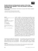
formed in plants, as illustrated in Figure 1.
F3′5′Hs have been previously cloned and functionally
analyzed from multiple plants, including grape (Vitis vinifera) [7,8], petunia (Petunia hybrida), snapdragon
(Antirrhinum majus) [9], Cineraria (Pericallis hybrida)
[10], tomato (Solanum lycopersicum) [11], big leaf periwinkle (Vinca major) [12], and potato (Solanum tuberosum) [13]. Through heterologous expression in transgenic
plants and yeasts, F3′5′Hs were shown to hydroxylate a
broad range of flavonoid substrates, including naringenin
(N), dihydrokaempferol (DHK), kaempferol (K) and apigenin [8,14]. However, optimum substrates for these enzymes remain to be determined.
Tea (Camellia sinensis) is an important commercial crop,
the leaves of which can be processed into popular nonalcoholic beverages. Because of the high flavonoid content, epidemiological and pathological studies have suggested that
tea consumption may potentially be protective against human cancers [15,16] and high blood pressure [17], and contribute to weight reduction [18]. The total concentration of
flavonoid compounds is around 12? 24% of tea leaf dry
mass [19]. We have previously shown that catechins are
among the most abundant flavonoids in tea leaves, followed
by proanthocyanidins (PAs), flavonols, flavones and anthocyanins (Figure 1A) [20,21]. In recent years, some of the flavonoid structural and regulatory genes have been cloned,
and functions of these genes have been investigated [22-25].
While 4′-hydroxylated catechins are very rare or undetectable in tea leaves [22], 3′4′5′-trihydroxylated catechins (gallocatechin (GC), EGC, and EGCG), are the most
abundant flavonoids in young leaves and the stem, with
significantly higher concentrations than 3′4′-dihydroxylated catechins (catechin (C), epicatechin (EC) and ECG)
(Figure 1B). Therefore, characterizing the pattern of Bring hydroxylation is clearly a valuable contribution to the
understanding of flavonoid biosynthesis in tea plants.
However, it has not yet been possible to prepare active
membrane-bound F3′5′H enzymes from Camellia sinensis, and it is still unclear whether B-ring hydroxylation occurs at the level of flavanones and/or dihydroflavonols,
in vivo. Aiming to analyze the in vivo expression pattern
of CsF3′5′H and to characterize the function of this gene
in vitro, we isolated the CsF3′5′H gene from tea cDNA
library. We found that CsF3′5′H was highly expressed in
the bud, but little or no CsF3′5′H was detected in the
root. CsF3′5′H expression was enhanced by light and sucrose treatment, and over-expression of CsF3′5′H resulted
in production of delphinidin derivatives, producing redder
flowers in transgenic tobacco plants, in comparison to
with wild type. Heterologous expression of modified
Page 2 of 14
CsF3′5′H in yeast revealed that 4′-hydroxylated flavanone
(naringenin, N) is the optimum substrate for CsF3′5′H,
and the ratio of 3′4′5′- to 3′4′-hydroxylated products in
the modified CsF3′5′H transgenic cells was significantly
higher than in VvF3′5′H cells.
Results
Isolation and characterization of the CsF3′5′H gene
The CsF3′5′H gene (NCBI cDNA accession number:
DQ194358, protein number: ABA40923) was successfully cloned from the cDNA library of the 3rd tea leaf,
and encoded 510 amino acid residues. A BLAST search
(NCBI) performed with the coding sequence revealed
83, 82 and 81% identity with Cyclamen persicum
(ACX37698), Cyclamen graecum (BAJ08041) and Vitis
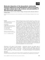
vinifera (XP_003632212) genes, respectively. The phylogenetic tree (Figure 2) was generated using protein sequences from several plant F3′5′H and F3′H enzymes
retrieved from the NCBI database. The tree demonstrated that F3′Hs and F3′5′Hs were grouped in
CYP75B and CYP75A clusters, respectively. CsF3′5′H
was grouped into the CYP75A subfamilies, and most
closely related to the F3′5′H enzymes of Cyclamen persicum, Cyclamen graecum and Vitis vinifera.
Expression pattern of CsF3′5′H in tea
The expression pattern of CsF3′5′H in tea was detected
by qRT-PCR. The GADPH gene (accession number:
FS952640), expected to show a constitutive expression
pattern, was used as control [21]. CsF3′5′H expression
was tissue specific, expressed highly in leaves and stem
(Figure 3A), with transcripts peaking in the buds. We also
assessed substrate specificity of crude extracts from tea
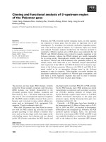
leaves, measuring hydroxylation of N and Dihydroquercetin (DHQ), which yielded Eriodictyol (E) and Dihydromyricetin (DHM), respectively (Figure 4). The enzyme
activities of these crude extracts were 0.072 and 0.023
pcat ? g −1 protein, respectively. Surprisingly hydroxylation
of N did not yield, 3′4′5′-hydroxylated product (5, 7, 3′,
4′, 5′-pentahydroxyflavanone, P).
Interestingly, CsF3′5′H transcripts were barely detected in the root, and the monomer and polymer of 3′
4′ -dihydroxylated catechins (EC and ECG), but no 3′ 4′
5′-trihydroxylated catechins, accumulated in Camellia
sinensis roots [21], indicating that extremely low CsF3′5′
H expression might directly lead to absence of B-ring trihydroxyl catechins in the root.
We used tissue culture seedlings, developed from the
embryo of tea-seeds, to assess the direct influence of light
and sucrose on CsF3′5′H expression. CsF3′5′H expression levels in light-exposed and sucrose-induced seedlings
were significantly increased by 22.69 and 3.00-fold, respectively (Figure 3B), indicating that CsF3′5′H expression can be efficiently induced by light and sucrose.
Wang et al. BMC Plant Biology 2014, 14:347
/>
Figure 1 (See legend on next page.)
Page 3 of 14
Wang et al. BMC Plant Biology 2014, 14:347
/>
Page 4 of 14
(See figure on previous page.)
Figure 1 Biosynthesis pathway and end-product accumulation of flavonoids in camellia sinensis. (A) Biosynthesis pathway of flavonoids.
CHS, chalcone synthase; CHI, chalcone isomerase; F3H, flavanone 3-hydroxylase; F3′H, flavonoid 3′-hydroxylase; F3′5′H, flavonoid 3′,5′-hydroxylase;
DFR, dihydroflavonol 4-reductase; FLS, flavonol synthase; LAR, leucoanthocyanidin reductase; ANS, anthocyanidin synthase; ANR, anthocyanidin
reductase; UFGT, UDP-glycose flavonoid glycosyltransferase; UGGT, UDP-glucose galloyl-1-O-β-D-glucosyltransferase; ECGT, epicatechins:
1-O-galloyl-β-D-glucose O-galloyltransferase; (B) Relative quantity of different flavonoid compounds. The data for relative quantity of different
flavonoid compounds were quoted from Jiang (Jiang XL, 2013).
Functional analysis of the CsF3′5′H gene in
Nicotiana tabacum
The vector for constitutive expression of the 35S:CsF3′
5′H gene was introduced into Tobacco ? G28? (Nicotiana
tabacum ? G28? ), which lacks F3′5′H genes and has pink
flowers [26]. About 20 independent transgenic tobacco
plants were obtained. Most flowers from the transgenic
plants exhibited a clear color change from pale pink of
the host to magenta (Figure 5A).
The expression of CsF3′5′H in several transgenic lines
with magenta flowers was detected by qRT-PCR, with βactin (accession number: EU938079) used as reference gene
(Figure 5B, E), and we found varying levels of CsF3′5′H
gene expression in Glyphosate-resistant transgenic tobaccos. To investigate whether the flavonoid biosynthesis pathway was affected by over-expression of CsF3′5′H, the
flavonoid pathway genes (CHS (chalcone synthase, accession number: AF311783), CHI (chalcone isomerase, accession number: KJ730247), F3H (flavanone 3-hydroxylase,
accession number: AF036093), F3′H (flavonoid 3′-hydroxylase, accession number: KF856279), DFR (dihydroflavonol
4-reductase, accession number: EF421430), FLS (flavonol
synthase, accession number: DQ435530), ANS (anthocyanidin synthase, accession number: JQ866631), ANR (anthocyanidin reductase, accession number: XM_009786976),
UFGT (UDP-glycose flavonoid glycosyltransferase, accession number: GQ395697)) from Nicotiana tabacum were
examined by qRT-PCR in wild type (G28) and transgenic
lines. The expression levels of CHS, F3H, ANS, ANR, UFGT
genes in transgenic lines significantly increased in comparison to the wild type and vector control (Figure 5E), suggesting that expression of these genes was stimulated by the
over-expression of CsF3′5′H in transgenic lines.
The level of glycosylated flavonoids in flowers was
assessed by reverse phase HPLC and LC-MS. 3′,5′-Hydroxylated flavonol glacoside (myricetin-3-O-rutinoside,
MYR) was detected in the petals of the transgenic lines,
but not in wild-type tobaccos (G28). However, the concentration of MYR in the flowers was too low to quantify (Figure 5C).
Petal pigments were extracted and chemically converted to anthocyanidins, for anglicizing the anthocyanin
components by reverse phase HPLC. Petals expressing
the CsF3′5′H gene contained a novel 3′,5′-hydroxylated
anthocyanidin (delphinin, DEL) and increased cyaniding
(CYA) derivative content. The ratio of delphinin to total
anthocyanin compounds in transgenic tobacco plants
reached a maximum of 31.09% (line-1, Figure 5D), and
the average anthocyanin concentration in the petals of
transgenic tobaccos was 1.51-fold higher than in wild-
Figure 2 Phylogenetic tree for a selection of F3′5′H protein. Phylogenetic tree based on amino acid sequences of F3′Hs and F3′5′Hs in various
plant species from the NCBI web page. Accession numbers are displayed in the figure. Bootstrap values (1,000 replicates) are shown at nodes.
Wang et al. BMC Plant Biology 2014, 14:347
/>
Page 5 of 14
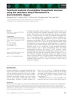
Figure 3 Expression of CsF3′5′H in different tea tissues. (A) Relative expression of CsF3′5′H in different tea tissues analyzed by qRT-PCR and
Semi-quantitative RT PCR for CsF3′5′H and GAPDH in different tea tissues. (B) Relative expression of CsF3′5′H in different light and sucrose conditions
analyzed by qRT-PCR and Semi-quantitative RT PCR for GAPDH and CsF3′5′H in different light and sucrose conditions. The data represent the mean ? SD
from three independent measurements. The different letters (a, b, c, d) and *indicated the significant level at P < 0.05.
type plants, suggesting that CsF3′5′H encodes a protein
with B-ring 3′, 5′-hydroxylation function, and that anthocyanin synthesis can be stimulated by CsF3′5′H overexpression in transgenic lines.
Heterologous CsF3′5′H expression in yeast
Figure 4 HPLC chromatograms of flavanones or
dihydroflavonols formation in CsF3′5′H assays with tea leaf
enzyme. (A) Reaction assay with substrate N of heat-denatured
protein in the control treatment (the crude enzyme extract were
heated to 100?C to inactivate enzyme activities); (B) Reaction assay
with substrate N of the crude enzyme extract from the leave of tea;
(C) Reaction assay with substrate DHQ of heat-denatured protein in
the control treatment; (D) Reaction assay with substrate DHQ of the
crude enzyme extract from the leave of tea.
The yeast strain Saccharomyces cerevisiae WAT11, engineered to over-express the Arabidopsis thaliana P450
reductase [27], is a suitable heterologous host for P450
expression [11,28]. A pYES-DEST 52a CsF3′5′H vector
was transformed into WAT11. However, these transgenic cells did not produce functional F3′5H protein
(Figure 6), so the codon optimized yeast CsF3′5′H sequence (yCsF3′5′H) was designed and transformed into
WAT11, resulting in only minimal activity of approximately 0.9 pkat ? L −1 culture, with N as substrate.
Transgenic cells, harboring the Vitis vinifera F3′5′H
(VvF3′5′H, NCBI cDNA accession number: XM_003632
164) gene, achieved a high overall F3′5′H activity of 48.00
pkat ? L −1 culture with N as substrate. With the predicted
signal peptide, both F3′5′Hs were translated into precursor proteins and delivered to the ER. We hypothesized
that imperfect recognition of the Camellia sinensis signal
peptide might account for low expression levels detected
in Saccharomyces cerevisiae cells, and tested this hypothesis by fusing CsF3′5′H with VvF3′5′H at three different
points of the sequence based on amino acid sequence
homology (Figure 6A). The 5′-sequences of yCsF3′5′H
were replaced by VvF3′5′H at 55 Aa (Fusion sequence I,
FSI), 153 Aa (Fusion sequence II, FSII), 308 Aa (Fusion sequence III, FSIII) respectively.
Vitis vinifera sequences were fused to yCsF3′5′H and
cloned into the plasmid pYES-DEST 52a for transformation of WAT11 cells. The cells containing FSI, replaced
at signal and leader peptide region, led to high F3′5′H activity in the range of 39.26 pkat ? L −1 culture, a significant
Wang et al. BMC Plant Biology 2014, 14:347
/>
Page 6 of 14
Figure 5 Flower color after overexpression of CsF3′5′H and qRT-PCR of transgenic tobacco plants. (A) Tobacco flowers of wild-type (CK)
and CsF3′5′H transgenes (Line 1). (B) Tobacco flowers of wild-type (CK), CsF3′5′H transgenes (Line 1, 3, 9 and 15)and qRT-PCR for CsF3′5′H in
flowers from CK and transgenic lines. (C) HPLC chromatograms of anthocyanidins (at 530 nm) and flavonol glacosides (at 340 nm) in tobacco
flowers from CK and Line 1 (1: DEL; 2: CYA; 3: quercetin-3-O-rutinoside, 4: kaempferol-3-O-rutinoside). (D) Concentration of anthocyanidins in
tobacco flowers from CK, CsF3′5′H transgenes (Line 1, 3 9, and 15) and vector control. The data represent the mean ? SD from three independent
measurements. (E) qRT-PCR for flavonoid-related genes in tobacco flowers from CK, CsF3′5′H transgenes (Line 1, Line3 Line 9) and vector
control. *indicated the significant level at P < 0.05. #indicated the significant level compared between every detected lines versus CK (wild type
and vector control).
increase in comparison with the reference construct
(yCsF3′5′H) (Figure 6B). These results indicated that
CsF3′5′H signal peptide might be imperfectly recognized
in Saccharomyces cerevisiae cells. The cells transformed
with FSII also resulted in F3′5′H activity, albeit significantly less (in the range of 12.37 pkat ? L −1 culture). Generally, overall activities of chimeras are often low, e.g. most
chimeras between limonene 3-hydroxylase and limonene
6-hydroxylase achieve no, or less than 5% of that of wild
type [29]. Unexpectedly, F3′5′H activity was undetected in
cells transformed with FSIII. In comparison to VvF3′5′H,
the FSIII fusion gene was only altered at the 3′-terminal
sequence.
Finally, we assessed the substrate specificity of cells
expressing FSI and VvF3′5′H. Based on previous findings
and other intermediate compounds in the catechin
synthesis pathway, we assessed catalysis of N, E, K, Quercetin (Q), DHK, DHQ, pelargonidin (PEL), CYA and C
(Figure 7, Additional file 1: Figure S1, Table 1). WAT11
cells transformed with pYES-DEST 52a vector were used
as controls. As observed with VvF3′5′H, FSI preferred Bring 4′-hydroxylated compounds (including N, K and
DHK) to 3′, 4′-hydroxylated compounds (including E, Q
and DHQ). No activity was detected with PEL, CYA and
C as substrates, in both transgenic cells. Both proteins displayed highest activities with N and significant activities
with K and DHK, yielding 3′4′- and 3′4′5′-forms as
products. Interestingly, for FSI with N as substrate, the
ratio of 3′4′5′- to 3′4′-hydroxylated products (2.07:1) was
significantly higher than for VvF3′5′H (0.98:1).
Microsomes from WAT11 cells transformed by pYESDEST 52a -FSI and -VvF3′5′H were assayed for NADPH-
Wang et al. BMC Plant Biology 2014, 14:347
/>
Page 7 of 14
Figure 6 Optimization of CsF3′5′H expression in Saccharomyces cerevisiae ? WAT11? . (A) Comparison of amino acid sequences encoded by
CsF3′5′H and VvF3′5′H proteins. The boxed regions represent fusion between CsF3′5′H and VvF3′5′H. (B) Primary structure schemes of expressed
CsF3′5′H sequence variants and resulting expression strength expressed as CsF3′5′H activity ? L −1 culture. Enzyme activity was expressed as
pKat ? L −1 culture. The data represent the mean ? SD from three independent measurements.
dependent flavonoid 3′, 5′-hydroxylation with N, K and
DHK as substrates. No activity was detected with microsomes from the control, pYES-DEST 52a-transformed
cells. In contrast the Km values of the microsome extracted
from FSI-transformed cells, with N, K, and DHK as substrates, were 3.22, 4.33, and 3.26 μM, respectively (Table 2,
Figure 8), indicating that N might be the optimum substrate for the CsF3′5′H enzyme. FSI achieved significantly
higher Km values than VvF3′5′H with K and DHK as
substrates, but lower Km values with N. However, the max
reaction rates (Vmax) for FSI and VvF3′5′H with N as substrate were significantly lower than the values with K and
DHK as substrates.
Discussion
The role of CsF3′5′H in catechin formation in tea leaves
All flavonoids are hydroxylated at the 4′ position of the
B-ring. B-ring hydroxylation patterns determie the color
of anthocyanins and thus have been extensively investigated in ornamental plants for color engineering. The
F3′5′H gene is commonly known as the blue gene [30]
and previous studies have shown that F3′5′H catalyzes
the hydroxylation at the 3′ and 5′ positions of flavonoids
to determine the hydroxylation pattern of the B-ring [28].
Flavonoids are important secondary metabolites in tea
and account for 18 to 36% of the dry weight of fresh
leaves and tender stem. 3′,4′,5′-trihydroxylated flavan-3-
Wang et al. BMC Plant Biology 2014, 14:347
/>
Page 8 of 14
Figure 7 HPLC chromatograms of products from pYES-dest52-FS and pYES-dest52-VvF3′5′H with flavanones, flavonols and
dihydroflavonols as substrates. HPLC chromatograms of products from pYES-dest52- FSI with N (2), E (5), K (8), Q (11), DHK (14) and DHQ (17)
as substrates; HPLC chromatograms of products from pYES-dest52-VvF3′5′H with N (3), E (6), K (9), Q (12), DHK (15) and DHQ (18) as substrates;
HPLC chromatograms of products from control treatment with N (1), E (4), K (7), Q (10), DHK (13) and DHQ (16) as substrates.
Table 1 Accepted substrates and enzyme activity units for F3′5′H
Substrate
Modified-CsF3′5′H (FSI)
VvF3′5′H
3′-Hydroxyla-tion
product (pKat ? L −1)
Naringenin
12.79 ? 0.11
5′-Hydroxylat-ion
product (pKat ? L −1)
26.47 ? 1.08
Eriodictyol
4.58 ? 0.39
23.73 ? 0.85
2.84 ? 0.76
5.53 ? 0.44
0.58 ? 0.27
?
Dihydro-kaempferol
?
2.69 ? 0.48
0.08 ? 0.03
?
?
?
?
Antho-cyanin
?
?
?
Dihydro-flavonol
0.05 ? 0.08
?
Cyanidin
Catechin
Flavonol
0.36 ? 0.19
3.42 ? 0.54
Dihydro-quercetin
Flavanone
0.84 ? 0.13
8.54 ? 0.40
Quercetin
Epicatechin
24.27 ? 0.70
5′-Hydroxylat-ion
product (pKat ? L −1)
0.13 ? 0.79
Kaempferol
Pelargonidin
Class
3′-Hydroxylat-ion
product (pKat ? L −1)
?
Flavan-3-ols
?
Enzyme activity was expressed as pKat ? L −1 culture. The data represent mean ? SD from three independent measurements. ?
indicates results below the detection limit.
Wang et al. BMC Plant Biology 2014, 14:347
/>
Page 9 of 14
Table 2 Comparison of steady-state kinetic parameters for cinnamate 4-hydroxylation in yeast microsomes
Substrate
Modified-CsF3′5′H (FSI)
Km (μM)
Naringenin
3.22 ? 0.31
Vmax (pM ? min −1 ?mg −1 Microsome)
124.49 ? 10.11
VvF3′5′H
Km (μM)
2.03 ? 0.34
Class
Vmax (pM ?min −1 ?mg −1 Microsome)
183.00 ? 11.02
Flavanone
Kaempferol
4.33 ? 0.19
306.00 ? 7.89
5.30 ? 0.71
327.00 ? 12.02
Flavonol
Dihydro-kaempferol
3.26 ? 0.25
219.00 ? 9.37
3.74 ? 0.54
220.18 ? 8.79
Dihydro-flavonol
Experiments were carried out in 50 mM phosphate buffer pH 7.0 at 28?C for 30 min. Protein concentration of F3′5′H -transformed yeast microsomes was 0.1 mg/ml in
the reaction system. The data represent the mean ? SD from three independent measurements.
ols (catechins) are the most abundant flavonoids in tea
leaves, present at significantly higher levels than 3′ 4′dihydroxylated catechins. Catechins anabolic and catabolic processes are dynamic and subject to complex
regulatory control, but the link between F3′5′H gene activity and relative catechin content is not well understood, due to the lack of easily assessable reporters.
Herein, we demonstrated that the CsF3′5′H gene is
highly expressed in the leaves and stem, but expressed at
extremely low levels in the root, as previously reported
[25]. We have previously shown that the tea plant root
lacks tri-hydroxyl groups in B-ring flavonols and flavan3-ols, indicating that CsF3′5′H participates in the control of tri-hydroxyl flavan-3-ols synthesis in tea plant.
We found that CsF3′5′H gene transcripts peaked in
the bud, and we were unable to explain the role of
CsF3′5′H in the accumulation of leaf end-products. The
content of most flavonoids such as galloylated catechins,
PAs, and anthocyanidin were highest in the bud or first
leaf and declined gradually with the leaf development
[21]. These findings indicated that F3′5′H expression
was closely associated with the accumulation of endproducts of flavonoids in tea leaves.
CsF3′5′H expression was significantly increased after
seven days of treatment with light or sucrose, indicated
that CsF3′5′H expression can be efficiently induced by
light and sucrose. Cloning analysis revealed that the CsF3′
5′H gene promoter contains several light-responsive
Figure 8 Concentration dependence of F3′5′H observed in yeast microsomes. F3′5′H-containing microsomes originating from transformed
pYES-dest52-FSI and -VvF3′5′H cells were incubated in 50 mM phosphate buffer pH 7.0 at 28?C. The solid line represents the result of a
multi-iterative fitting of experimental data using the Michaelis-Menten equation. Insert: Michaelis-Menten double-reciprocal plot. The data
represent the mean ? SD from three independent measurements.
Wang et al. BMC Plant Biology 2014, 14:347
/>
promoter elements (not shown), further indicating that
light might be a key factor in the control of CsF3′5′H
transcription.
Anthocyanidin accumulation in CsF3′5′H transgenic tobacco
The main anthocyanin in the wild-type tobacco corolla
is cyanidin [31]. As shown above, most transgenic plant
flower petals contained delphinins. Interestingly, the
cyanidin and delphinin content was significantly higher
in transgenic tobacco plants than wild type plants, indicating that CsF3′5′H performs both 3′,5′- and 3′
-hydroxylation in vivo, in agreement with results of
heterologous expression of F3′5′Hs in Pericallis ?
hybrida [10], Senecio cruentus [32], Antirrhinum kelloggii [9], and Solanum lycopersicum [11]. However, the
hydroxylation pattern of the B-ring cannot be elucidated
by tobacco transgenic experiments. Flavonoid pathway,
which is a complex metabolic network in plants, starts
with general phenylpropanoid metabolism and leads to a
myriad of end-products. The enzymes of flavonoid biosynthesis are likely to function as multienzyme complexes,
which facilitate the direct transfer, or channeling of active
sites [33]. Therefore, the overall concentrations of the intermediates, including free flavanones and flavanols, are
extremely low in vivo [2].
CsF3′5′H transgenic tobacco plants produced deeper
and redder flowers than wild-type plants. The qRT-PCR
results indicated that the flavonoid pathway genes, including CHS, F3H, ANS, ANR, UFGT, could be stimulated by CsF3′5′H over-expression in transgenic lines.
F3′5′H, a crucial microsomal cytochrome P450 enzyme
in these pathways, may serve to anchor the complexes
to the microsme membrane [33]. Therefore, our results
indicate that over-expression of CsF3′5′H may stimulate
metabolic flux toward anthocyanin products in tobacco
petals by formatting more enzyme complexes.
The transgenic lines, however, did not produce blue
flowers in this study. These findings demonstrated that
blue flowers are not necessarily generated only by controlling the anthocyanin content [34]. Indeed, previous
studies have reported that anthocyanidin content, copigments, metal ion type and concentration, pH of vacuoles, anthocyanin localization and shapes of surface cells
all contribute to the final flower color [35]. However, our
findings do reveal a clear impact of CsF3′5′H gene on
flower phenotype. This gene might therefore be applied to
molecular design of flower color in ornamental plants.
Heterologous expression of CsF3′5′H in yeast
Heterologous expressions of CsF3′5′H in yeast were carried out to further confirm the catalytic position of
CsF3′5′H enzyme in flavonoid pathways. To our knowledge, the Camellia sinensis F3′5′H gene has not been
previously successfully expressed in yeast. For effective
Page 10 of 14
expression of CsF3′5′H in yeast, a codon optimized
yeast CsF3′5′H sequence (yCsF3′5′H) was designed, but
only minor activity was detected. Generally, the presence
of an N-terminal signal peptide can translocate P450
proteins into the endoplasmic reticulum (ER). We further optimized yCsF3′5′H by replacing the N-terminal
sequence with a signal peptide from the VvF3′5′H gene.
Fortunately, transgenic cells expressing the fusion F3′5′H
gene exhibited high F3′5′H activity, indicating that the
signal peptide of CsF3′5′H might be imperfectly recognized in S. cerevisiae cells. Unexpectedly, another fusion
gene (FSIII), only altered at the 3′-terminal sequence in
comparison to VvF3′5′H, not achieving detectable F3′5′H
activity. These results suggested that the region of F3′5′H
conferring enzymatic activity might be located at the Cterminal of F3′5′H. Indeed, previous reports have suggested that the functional difference between F3′H and
F3′5′H is determined by the C-terminal end [36].
F3′5′Hs have been shown to hydroxylate a broad range
of flavonoid substrates, including N, DHK, K and apigenin,
possibly allowing the formation of 3′,4′- and 3′,4′,5′-hydroxylated flavonoids. However, the optimum substrate
for the F3′5′H enzymes needs to be further defined,
in vivo and in vitro. F3′5′H enzymes from Catharanthus
roseus and Petunia x hybrida have achieved highest activities with naringenin and apigenin [37], and N and DHK
are equally hydroxylated by Osteospermum hybrida F3′5′
H, whereas F3′H from Gerbera hybrida exhibits a clear
substrate preference for N [36]. In contrast, the F3′5′H
gene from tomato (Solanum lycopersicum) has a preference for naringenin, with a Km value of 1.20 μM [11].
To assess substrate specificity of the modified CsF3′5′
H (FSI), flavanones (N and E), flavonols (K and Q), dihydroflavonols (DHK and DHQ), anthocyanins (PEL and
CYA) and catechin (C) were selected as substrates. 4′hydroxylated flavanone (N) was the optimum substrate
for the CsF3′5′H enzyme, and was effectively converted
to both 3′4′- and 3′4′5′-forms. Interestingly, with N as
substrate in FSI transgenic cells, the ratio of 3′4′5′- to
3′4′-hydroxylated products was significantly higher than
in VvF3′5′H cells. Modified CsF3′5′H genes could thus
tailor flavonoid metabolism, enhancing the yields of specific B-ring tri-hydroxyl products.
The broad substrate acceptance is consistent with the
possibility that multiple paths lead to the same intermediates, and that competition could occur in vivo. The
substrates used in vivo are mostly not yet precisely identified [37]. We also detected the B-ring hydroxyl reaction
patterns of total enzyme extract from tea leaves. Interesting, with N as a substrate, the 3′4′5′-hydroxylated
flavanone product (P) was undetected and only the 3′4′hydroxylated product (E) was detected. It is not known
whether the product P might be efficiently transformed
into other end-products or the product E synthesized by
Wang et al. BMC Plant Biology 2014, 14:347
/>
another P450 gene (F3′H). Therefore, further analysis
should be carried out to further describe the mechanism
underlying B-ring hydroxylation in vivo.
Conclusions
In this study, the CsF3′5′H gene was isolated from the
tea cDNA library. Phylogenetic analyses revealed that
the Camellia sinensis F3′5′H gene belongs to the
CYP75A subfamily. qRT-PCR analysis indicated CsF3′5′
H is highly expressed in the bud, but very little is
expressed in the root. Over-expression of CsF3′5′H
resulted in production of new delphinidin derivatives in
the corollas of transgenic tobacco plants, increased the
content of cyanidin derivatives and produced deeper
and redder flowers in transgenic plants. Heterologous
expressions of CsF3′5′H in yeast were carried out to
demonstrate the function of CsF3′5′H enzyme in vitro.
Heterologous expression of the modified yCsF3′5′H
(FSI) in yeast revealed the 4′-hydroxylated flavanone
naringenin to be the optimum substrate for the CsF3′
5′H enzyme; naringenin was effectively converted into
both 3′4′- and 3′4′5′-forms. Importantly, the ratio of
3′4′5′- to 3′4′-hydroxylated products was significantly
higher in modified yCsF3′5′H transgenic cells than in
VvF3′5′H cells. The findings reported here provide a
basis for better understanding of the role of Camellia
sinensis F3′5′H in B-ring hydroxylation of flavonoids,
in vitro and in vivo.
Methods
Plant materials
Samples of Camellia sinensis cv. Shucazao (Variety Approval number: CHN20022008), were obtained from the
experimental tea garden of Anhui Agricultural University in Hefei, China (north latitude 31.86, east longitude
117.27, altitude 20 m above mean sea level). Leaves were
collected at five different stages (bud, 1st leaf, 2nd leaf,
and 3rd leaf, older leaf ), stem and root, snap frozen in liquid nitrogen and stored at −80?C.
Healthy tissue culture seedlings were used for light
and sucrose induction experiments. Seedlings were cultured in normal light? dark cycle (light/dark: 14 h/10 h)
in N6 medium containing 3% sucrose, and subcultured
every 20 days by transferring about 5 g (fresh weight) to
fresh medium. Six separate culture flasks were selected
from the light and sucrose treatments. For light treatment, plates were exposed to 50 ? 5 μmolm−2 s−1 light
(Cool white, 55 W, Philips, Netherlands) for 7 days, and
culture flasks covered with aluminum foil were used as full
darkness controls. For sucrose treatment, the seedlings
were subcultured in the previously described medium or
the previously described medium containing additional
90 mM/L sucrose for 7 days. Total RNA was isolated from
leaves for quantitative real time polymerase chain reaction
Page 11 of 14
(qRT-PCR) in three independent experiments. The morphology of tea seedlings were captured with a Cannon
600D camera (Cannon, Japan).
The yeast strain (Saccharomyces cerevisiae cv. WAT11)
and the tobacco variety (Nicotiana tabacum cv. G28),
were kindly provided by Conagen Inc (Bedford, MA,
USA) and University of Science and Technology of China
(Hefei, Anhui, China), respectively.
End-to-end PCR
The CsF3′5′H gene from the NCBI database was subjected to standard end-to-end PCR reactions, with the
primers designed according to the cDNA sequence (synthesized by Invitrogen, Shanghai, China; Additional file 2:
Table S1). The cDNA strands for end-to-end PCR were
synthesized with Phusion? High-Fidelity DNA Polymerase
(New England Biolabs, USA). PCR products were gel purified using the MiniBEST Agarose Gel Extraction Kit
(Takara, DaLian, China), ligated into a pMD18-T vector,
and transformed into E. coli DH5α competent cells for sequencing. The results were assembled using DNAMAN 7
software (Lynnon, Canada). Briefly, end-to-end PCR was
performed under the following conditions: 98?C for 30 s,
30 cycles at 98?C for 30 s, 58?C for 10 s, 72?C for 40 s, and
a final extension at 72?C for 10 min.
Validation of expression by qRT-PCR
Total RNA was isolated from Camellia sinensis organs
with RNAiso Plus (Takara, DaLian, China) and RNAisomate for Plant Tissue (Takara, DaLian, China), according
to the manufacturers? instructions.
All primers were blasted against the NCBI database to
guarantee specificity. Values were normalized against the
expression levels of the housekeeping gene glyceraldehyde3-phosphate dehydrogenase (GAPDH) in tea plant [21]
and actin in tobacco [38]. The first strand cDNA samples
for qRT-PCR were synthesized with the PrimeScript? RT
reagent Kit (Takara, DaLian, China). The PCR mixture
contained cDNA template (approximately 0.01 μg/μL),
10 μL SYBR Green PCR Master Mix (Takara), and 200
nmolL−1 of each gene-specific primer in a final volume of
20 μL. Real-time PCR was performed using a CFX96?
optical reaction module (Bio-Rad, USA) as follows: 95?C
for 30 s, followed by 40 cycles at 95?C for 5 s and 60?C
for 30 s (58?C for 30 s for root) in 96-well optical reaction plates. The amplification specificity was verified
by melting curve analysis (55? 95?C ). Data were expressed as mean value of three replicates, normalized
against the expression levels of GAPDH or actin.
The relative expression was derived by the 2-ΔΔCt
method. △CT = CT, target -CT, internal standard, −△△CT = −(△CT,
target -△CT, control), where CT, target and CT, internal standard are
cycle threshold (CT) values for targets and housekeeping
genes, respectively.
Wang et al. BMC Plant Biology 2014, 14:347
/>
Transformation of tobacco plants with CsF3′5′H
transgenes
The Gateway? Cloning System was used to construct the
vectors provided by Prof Xiang [39] of the University of
Science and Technology of China. CsF3′5′H PCR products were obtained by end-to-end PCR and ligated into
pMD18-T vectors. The CsF3′5′H - pMD18-T plasmids
were amplified in E. coli strain DH5α and used as PCR
templates. The PCR primer pairs for linking the attB
adaptors are listed in Additional file 2: Table S1. PCR
products were purified, transferred to pMD18-T and
confirmed by sequencing. The correct plasmid was
cloned into the entry vector pDONR207 by Gateway? BP
Clonase? Enzyme mix according to the manufacturer ? s
instructions (Invitrogen, USA). The resulting entry
pDONR207- clones were selected on gentamycin plates
and validated by restriction enzyme digestion. Entry vectors were then transferred into the Gateway plant transformation destination vector pCB2004 using Gateway?
LR Clonase? (Invitrogen, USA). Recombinant colonies
pCB2004-CsF3′5′H and control pCB2004 vectors were
selected on kanamycin plates and validated by restriction
enzyme digestion, followed by transformation into
EHA105 by electroporation at 2500 V for about 5.5 ms.
A single colony containing each target construct was
confirmed by PCR and used for genetic transformation of
tobacco. EHA105-pCB2004-CsF3′5′H and EHA105empty pCB2004 were inoculated in liquid LB medium
containing 50 mg/L kanamycin and 50 mg/L spectinomycin. Cells were allowed to grow in the dark at 28?C, for
20? 22 h at 200 rpm to OD600 = 0.6, then pelleted by centrifugation (6000 rpm, 10 min) followed by two washing
steps with liquid MS medium containing 100 μmol/L acetosyringone (Sigma, R40456). The leaf disc approach was
used for tobacco transformation, with 25 mg/L phosphinothricin selection [40].
Construction of the yeast strain Saccharomyces cerevisiae
? WAT11? vector for CsF3′5′H expression
PCR products of VvF3′5′H, FS, FSII, FSIII were obtained
by end-to-end PCR, gel purified, and ligated into
pENTR? /TEV/D-TOPO vectors using Top cloning
(pENTR? /TEV/D-TOPO? Cloning Kits, Invitrogen, USA).
Then, the entry vectors pENTR-VvF3′5′H, pENTR-CzyF3′
5′H-1, pENTR-CzyF3′5′H-2, and pENTR-CzyF3′5′H-3
were cloned into the destination vector pYES-dest52 using
Gateway? LR Clonase ? enzyme (Invitrogen, USA). The
resulting pYES-dest52-VvF3′5′H, pYES-dest52-FSI, pYESdest52-FSII, and pYES-dest52-FSIII were transformed into
Saccharomyces cerevisiae WAT11 with Frozon-EZ yeast
Transformation II? (Zymo Research, USA).
Yeast cells were propagated at 28?C for 12 h in 10 ml
SD-U liquid medium containing 20 g/l glucose, by inoculation of a single colony from a SGlu plate. The
Page 12 of 14
thalli collected were transferred into 10 ml SD-U
medium containing 20 g/l galactose, and grown at 28?C
for 5 h.
For substrate specificity experiments, N, E, DHK, DHQ,
K, and Q were separately added into the yeast culture to a
final concentration of 5 μM, and incubated at 28?C for
10 h. Reactions were terminated by sonication for 15 min
and addition of ethyl acetate. Products from each reaction
were extracted three times with 10 ml ethyl acetate, evaporated and re-dissolved in 150 μl methanol for HPLC analysis at 280? 370 nm.
Microsome preparation
Protein synthesis was indiced in the yeast culture by the
addition of galactose and the microsomal yeast fraction
was prepared with MgCl2 as described by Olsen et al.
[11]. Protein quantities were estimated according to the
Bradford method. The microsome was dissolved in 1.0
to 1.5 ml TEG (30% glycerol in 50 mM Tris? HCl with
1 mM EDTA) on ice. All buffers/solutions and centrifuge were pre-cooled to 4?C.
Enzyme extraction from Camellia sinensis
About 2 g of tea leaves were homogenized under liquid nitrogen, and total protein was extracted with 0.1 molL−1
phosphate-buffered saline (PBS, pH 7.4) containing an
equivalent amount of PVPP, then centrifuged at 15000 g
for 10 min at 4?C. The supernatants were used to assess
F3′5′H activity. Protein concentrations of enzyme extract
were determined by spectrometric analysis using Coomassie Brilliant Blue G-250.
Enzyme assays
All enzyme assays were carried out in phosphate buffer.
In the multi-enzyme incorporative reaction system, the
F3′5′H assay solution was incubated at 28?C for 30 min
(for microsomes) or 1 h (for crude enzyme extract) in
100 mM phosphate buffer (pH 7.0) containing 1 mM
NADPH, 1? 300 μM substrates. Enzyme reactions were
terminated by adding ethyl acetate. Products from each reaction were extracted three times with an equal volume
ethyl acetate, evaporated and re-dissolved in 500 μl methanol for HPLC analysis at 280? 370 nm.
Flavonoid pigment preparation and analyses
Anthocyanin aglycones were extracted with 1.6 ml methanol containing 20% water from about 500 mg frozen tobacco flowers. After centrifugation at 6,000 g at 4?C for
5 min, supernatants were extracted three times by equal
volume of ethyl acetate, and the extracts were added to
1/3 volume of 4 M HCl aqueous solution for acidhydrolysis by heat treatment at 90?C for 1 h. Hydrolysates were tested by HPLC at 530 nm.
Wang et al. BMC Plant Biology 2014, 14:347
/>
HPLC and MS analyses
Mass spectra were acquired using the electrospray
ionization in the negative ionization modes at fragmentation voltages of 175 V over the range of m/z 100 to
2000 on the UPLC-QQQ-MS/MS (Waters 2478, Waters
Instruments) with drying gas flow of 12 L min−1, a drying gas temperature of 350?C, a nebulizer pressure of
35 psi, and capillary voltages of 3500 V.
The HPLC consisted of a quaternary pump with a vacuum degasser, thermostatted column compartment, autosampler and diode array detector (DAD). A Phenomenex
Synergi 4u Fusion-RP80 column (5 μm, 250*4.6 mm) was
used at a flow rate of 1.0 mL min−1. The column oven
temperature was set at 25?C. The mobile phase consisted of
1% acetic acid in water (A) and 100% acetonitrile (B). The
gradient increased linearly from 0 to 10% B (v/v) at 5 min,
to 15% B at 15 min, 40% B at 20 min, 60% B at 22 min, and
maintained at 10% B to 25 min. The DAD was set at 280
and 340 nm for real-time monitoring of the peak intensities. Ultraviolet (UV) spectra were recorded continuously
from 200 to 600 nm for plant component identification.
Among the standards used, N, E, P, DHK, DHQ, and
DHM were quantified at 280 nm, whereas K, Q, and myricetin (M) were quantified at 365 nm. All products, except
P, were identified and quantified by mass spectrums (MS)
and peak area compared with standards. Since standard
samples of 5, 7, 3′, 4′, 5′-pentahydroxyflavanone were unavailable, P was identified with LC-MS, and its relative
concentration was quantified using E as the molar equivalent. All samples were run in triplicate for both quantitation and multivariate statistical analysis.
Bioinformatics and statistical analyses
The phylogenetic tree was constructed using protein sequences from several plant F3′5′H, F3′H, and Cinnamic
acid 4-hydroxylase (C4H) enzymes retrieved from the
NCBI database by ClustalW of MEGA5 (accession
numbers are given in the phylogenetic tree, Figure 2).
The phylogenetic tree was constructed according to the
neighbor-joining method. Branches corresponding to
partitions reproduced in less than 50% bootstrap replicates were collapsed. The evolutionary distances were
computed using the p-distance method. Evolutionary
analyses were conducted in MEGA5 (web page: http://
www.megasoftware.net/).
Data were presented as the mean ? SD of three independent measurements. The statistical significance of differences
between groups was determined with Student? s t-test using
SPSS software (SPSS, Chicago, IL, USA). P < 0.05 was considered statistically significant.
Supporting data
The data set(s) supporting the results of this article
is (are) included within the article (and its additional
Page 13 of 14
file(s)). The cDNA and protein sequences from several
plant F3′5′H, F3′H and C4H enzymes retrieved from
the NCBI web page ( />
Additional files
Additional file 1: Figure S1. UPLC-QQQ-MS analysis of products from
pYES-dest52-FS assayed with different substrates. (A) MS analysis of E;
(B) MS analysis of P; (C) MS analysis of Q; (D) MS analysis of M; (E) MS
analysis of DHQ; (F) MS analysis of DHM.
Additional file 2: Table S1. Sequences of primers used for cloning,
fusion, and expression analysis of F3′5′H.
Abbreviations
ANR: Anthocyanidin reductase; ANS: Anthocyanidin synthase; C: Catechin;
C4H: Cinnamic acid 4-hydroxylase; CHI: Chalcone isomerase; CHS: Chalcone
synthase; DFR: Dihydroflavonol 4-reductase; DHK: Dihydrokaempferol;
DHM: Dihydromyricetin; DHQ: Dihydroquercetin; E: Eriodictyol; F3H: Flavanone
3-hydroxylase; F3′H: Flavonoid 3′-hydroxylase; F3′5′H: Flavonoid 3′,5′-hydroxylase;
FLS: Flavonol synthase; K: Kaempferol; LAR: Leucoanthocyanidin reductase;
M: Myricetin; N: Naringenin; P: 5, 7, 3′, 4′, 5′-pentahydroxyflavanone; Q: Quercetin;
UFGT: UDP-glycose flavonoid glycosyltransferase.
Competing interests
The authors declare that they have no competing interests.
Authors? contributions
WYS conceived of the study, carried out all the experiments and drafted the
manuscript. XYJ carried out the enzyme analysis and helped to draft the
manuscript. GLP performed the bioinformatics and statistical analysis. YO
helped to set up and conduct the biological experiments and revised of
manuscript. WXZ carried out the yeast and tobacco transformation. HXJ
prepared the yeast microsome. JXL performed the UPLC-QQQ-MS analysis.
LYJ prepared the RNA. TX conceived of the study, coordinated and helped
to draft the manuscript. All authors read and approved the final manuscript.
Acknowledgements
We thank Conagen Inc for the generous gift of the yeast strain
Saccharomyces cerevisiae WAT11, and the Chengbi Xiang Lab. (University of
Science and Technology of China, Hefei city, China) for excellent assistance
with the F3′5′H expression in tobacco leaves. This work was supported by
the National Natural Science Foundation of China (nos. 31170647, 31170282,
31270730, 31470689), Science and Technology Projects of Anhui Province,
China (Project 13Z03012), Chinese National 863 Project (nos. 2013AA102801)
and The Biology Key Subject Construction of Anhui.
Author details
1
Key Laboratory of Tea Biochemistry and Biotechnology, Ministry of
Education in China, Anhui Agricultural University, Hefei, Anhui, China. 2School
of Life Science, Anhui Agricultural University, Hefei, Anhui, China. 3Conagen
Inc, 15 DeAngelo Dr, Bedford, MA 01730, USA. 4Wuxi NewWay, 401 Xing
Yuan Bei Road, Wuxi, Jiangsu, China.
Received: 16 May 2014 Accepted: 24 November 2014
References
1. Cabrera C, Artacho R, Gimenez R: Beneficial effects of green tea-a review.
J Am Coll Nutr 2006, 25(2):79? 99.
2. Winkel-Shirley B: Flavonoid biosynthesis. A colorful model for genetics,
biochemistry, cell biology, and biotechnology. Plant Physiol 2001,
126(2):485? 493.
3. Sekher Pannala A, Chan TS, O? Brien PJ, Rice-Evans CA: Flavonoid B-ring
chemistry and antioxidant activity: fast reaction kinetics. Biochem Biophys
Res Commun 2001, 282(5):1161? 1168.
4. Lee ER, Kang GH, Cho SG: Effect of flavonoids on human health: old
subjects but new challenges. Recent Pat Biotechnol 2007, 1(2):139? 150.
Wang et al. BMC Plant Biology 2014, 14:347
/>
5.
6.
7.
8.
9.
10.
11.
12.
13.
14.
15.
16.
17.
18.
19.
20.
21.
22.
23.
Seeram NP, Nair MG: Inhibition of lipid peroxidation and structureactivity-related studies of the dietary constituents anthocyanins,
anthocyanidins, and catechins. J Agric Food Chem 2002, 50(19):5308? 5312.
Liu TT, Yang TS: Effects of water-soluble natural antioxidants on
photosensitized oxidation of conjugated linoleic acid in an oil-in-water
emulsion system. J Food Sci 2008, 73(4):C256? C261.
Castellarin SD, Di Gaspero G, Marconi R, Nonis A, Peterlunger E, Paillard S,
Adam-Blondon A-F, Testolin R: Colour variation in red grapevines (Vitis
vinifera L.): genomic organisation, expression of flavonoid 3′-hydroxylase,
flavonoid 3′, 5′-hydroxylase genes and related metabolite profiling of red
cyanidin-/blue delphinidin-based anthocyanins in berry skin.
BMC Genomics 2006, 7(1):12.
Falginella L, Castellarin SD, Testolin R, Gambetta GA, Morgante M, Di
Gaspero G: Expansion and subfunctionalisation of flavonoid 3′, 5′hydroxylases in the grapevine lineage. BMC Genomics 2010, 11(1):562.
Ishiguro K, Taniguchi M, Tanaka Y: Functional analysis of Antirrhinum
kelloggii flavonoid 3′-hydroxylase and flavonoid 3′,5′-hydroxylase genes;
critical role in flower color and evolution in the genus Antirrhinum.
J Plant Res 2012, 125(3):451? 456.
Sun Y, Huang H, Meng L, Hu K, Dai SL: Isolation and functional analysis of
a homolog of flavonoid 3′,5′-hydroxylase gene from Pericallis x hybrida.
Physiol Plant 2013, 149(2):151? 159.
Olsen KM, Hehn A, Jugde H, Slimestad R, Larbat R, Bourgaud F, Lillo C:
Identification and characterisation of CYP75A31, a new flavonoid
3′5′-hydroxylase, isolated from Solanum lycopersicum. BMC Plant Biol
2010, 10:21.
Mori S, Kobayashi H, Hoshi Y, Kondo M, Nakano M: Heterologous expression
of the flavonoid 3′,5′-hydroxylase gene of Vinca major alters flower color in
transgenic Petunia hybrida. Plant Cell Rep 2004, 22(6):415? 421.
De Jong WS, Eannetta NT, De Jong DM, Bodis M: Candidate gene analysis
of anthocyanin pigmentation loci in the Solanaceae. Theor Appl Genet
2004, 108(3):423? 432.
DE Vetten N, Ter Horst J, van Schaik H-P, de Boer A, Mol J, Koes R: A cytochrome
b5 is required for full activity of flavonoid 3′, 5′-hydroxylase, a cytochrome
P450 involved in the formation of blue flower colors. Proc Natl Acad Sci 1999,
96(2):778? 783.
Nakachi K, Suemasu K, Suga K, Takeo T, Imai K, Higashi Y: Influence of
drinking green tea on breast cancer malignancy among Japanese
patients. Jpn J Cancer Res 1998, 89(3):254? 261.
Sasazuki S, Tamakoshi A, Matsuo K, Ito H, Wakai K, Nagata C, Mizoue T,
Tanaka K, Tsuji I, Inoue M, Tsugane S, Research Group for the Development
Evaluation of Cancer Prevention Strategies in Japan: Green tea
consumption and gastric cancer risk: an evaluation based on a
systematic review of epidemiologic evidence among the Japanese
population. Jpn J Clin Oncol 2012, 42(4):335? 346.
Hodgson JM, Croft KD, Woodman RJ, Puddey IB, Fuchs D, Draijer R,
Lukoshkova E, Head GA: Black tea lowers the rate of blood pressure
variation: a randomized controlled trial. Am J Clin Nutr 2013, 97(5):943? 950.
Auvichayapat P, Prapochanung M, Tunkamnerdthai O, Sripanidkulchai BO,
Auvichayapat N, Thinkhamrop B, Kunhasura S, Wongpratoom S, Sinawat S,
Hongprapas P: Effectiveness of green tea on weight reduction in obese
Thais: a randomized, controlled trial. Physiol Behav 2008, 93(3):486? 491.
Ho C-T, Lin J-K, Shahidi F: Tea and tea Products: Chemistry and
Health-Promoting Properties. New York: CRC Press; 2008.
Wang Y, Gao L, Shan Y, Liu Y, Tian Y, Xia T: Influence of shade on
flavonoid biosynthesis in tea (Camellia sinensis (L.) O. Kuntze). Sci Hortic
2012, 141:7? 16.
Jiang X, Liu Y, Li W, Zhao L, Meng F, Wang Y, Tan H, Yang H, Wei C, Wan X,
Gao L, Xia T: Tissue-specific, development-dependent phenolic
compounds accumulation profile and gene expression pattern in tea
plant [Camellia sinensis]. PLoS ONE 2013, 8(4):e62315.
Punyasiri PA, Abeysinghe IS, Kumar V, Treutter D, Duy D, Gosch C, Martens
S, Forkmann G, Fischer TC: Flavonoid biosynthesis in the tea plant
Camellia sinensis: properties of enzymes of the prominent epicatechin
and catechin pathways. Arch Biochem Biophys 2004, 431(1):22? 30.
Singh K, Rani A, Kumar S, Sood P, Mahajan M, Yadav SK, Singh B, Ahuja PS:
An early gene of the flavonoid pathway, flavanone 3-hydroxylase,
exhibits a positive relationship with the concentration of catechins in
tea (Camellia sinensis). Tree Physiol 2008, 28(9):1349? 1356.
Page 14 of 14
24. Eungwanichayapant PD, Popluechai S: Accumulation of catechins in tea in
relation to accumulation of mRNA from genes involved in catechin
biosynthesis. Plant Physiol Biochem 2009, 47(2):94? 97.
25. Ashihara H, Deng WW, Mullen W, Crozier A: Distribution and biosynthesis
of flavan-3-ols in Camellia sinensis seedlings and expression of genes
encoding biosynthetic enzymes. Phytochemistry 2010, 71(5? 6):559? 566.
26. Shimada Y, Nakano-Shimada R, Ohbayashi M, Okinaka Y, Kiyokawa S, Kikuchi Y:
Expression of chimeric P450 genes encoding flavonoid-3′, 5′-hydroxylase in
transgenic tobacco and petunia plants(1). FEBS Lett 1999, 461(3):241? 245.
27. Pompon D, Louerat B, Bronine A, Urban P: Yeast expression of animal and
plant P450s in optimized redox environments. Methods Enzymol 1996,
272:51? 64.
28. Seitz C, Eder C, Deiml B, Kellner S, Martens S, Forkmann G: Cloning,
functional identification and sequence analysis of flavonoid
3′-hydroxylase and flavonoid 3′,5′-hydroxylase cDNAs reveals independent
evolution of flavonoid 3′,5′-hydroxylase in the Asteraceae family.
Plant Mol Biol 2006, 61(3):365? 381.
29. Schalk M, Croteau R: A single amino acid substitution (F363I) converts the
regiochemistry of the spearmint (−)-limonene hydroxylase from a C6- to
a C3-hydroxylase. Proc Natl Acad Sci U S A 2000, 97(22):11948? 11953.
30. Holton TA, Brugliera F, Lester DR, Tanaka Y, Hyland CD, Menting JG, Lu C-Y,
Farcy E, Stevenson TW, Cornish EC: Cloning and Expression of Cytochrome
P450 Genes Controlling Flower Colour; 1993.
31. Aharoni A, De Vos CH, Wein M, Sun Z, Greco R, Kroon A, Mol JN, O? Connell AP:
The strawberry FaMYB1 transcription factor suppresses anthocyanin and
flavonol accumulation in transgenic tobacco. Plant J 2001, 28(3):319? 332.
32. He H, Ke H, Keting H, Qiaoyan X, Silan D: Flower colour modification of
chrysanthemum by suppression of F3′H and overexpression of the
exogenous Senecio cruentus F3′5′H gene. PLoS ONE 2013, 8(11):e74395.
33. Winkel-Shirley B: Evidence for enzyme complexes in the phenylpropanoid
and flavonoid pathways. Physiol Plantarum 1999, 107(1):142? 149.
34. Okinaka Y, Shimada Y, Nakano-Shimada R, Ohbayashi M, Kiyokawa S, Kikuchi
Y: Selective accumulation of delphinidin derivatives in tobacco using a
putative flavonoid 3′,5′-hydroxylase cDNA from Campanula medium.
Biosci Biotechnol Biochem 2003, 67(1):161? 165.
35. Tanaka Y, Brugliera F, Chandler S: Recent progress of flower colour
modification by biotechnology. Int J Mol Sci 2009, 10(12):5350? 5369.
36. Seitz C, Ameres S, Forkmann G: Identification of the molecular basis for
the functional difference between flavonoid 3′-hydroxylase and
flavonoid 3′,5′-hydroxylase. FEBS Lett 2007, 581(18):3429? 3434.
37. Kaltenbach M, Schr?der G, Schmelzer E, Lutz V, Schr?der J: Flavonoid
hydroxylase from Catharanthus roseus: cDNA, heterologous expression,
enzyme properties and cell-type specific expression in plants. Plant J
1999, 19(2):183? 193.
38. Pang Y, Peel GJ, Wright E, Wang Z, Dixon RA: Early steps in
proanthocyanidin biosynthesis in the model legume Medicago
truncatula. Plant Physiol 2007, 145(3):601? 615.
39. Lei ZY, Zhao P, Cao MJ, Cui R, Chen X, Xiong LZ, Zhang QF, Oliver DJ, Xiang
CB: High-throughput binary vectors for plant gene function analysis.
J Integr Plant Biol 2007, 49(4):556? 567.
40. Clough SJ, Bent AF: Floral dip: a simplified method for Agrobacteriummediated transformation of Arabidopsis thaliana. Plant J 1998,
16(6):735? 743.
doi:10.1186/s12870-014-0347-7
Cite this article as: Wang et al.: Functional analysis of Flavonoid
3′,5′-hydroxylase from Tea plant (Camellia sinensis): critical role in the
accumulation of catechins. BMC Plant Biology 2014 14:347.