Natural rice rhizospheric microbes suppress rice blast infections
Bạn đang xem bản rút gọn của tài liệu. Xem và tải ngay bản đầy đủ của tài liệu tại đây (1.38 MB, 17 trang )
Spence et al. BMC Plant Biology 2014, 14:130
/>
RESEARCH ARTICLE
Open Access
Natural rice rhizospheric microbes suppress rice
blast infections
Carla Spence1,2†, Emily Alff2,3†, Cameron Johnson4, Cassandra Ramos4, Nicole Donofrio3,
Venkatesan Sundaresan4 and Harsh Bais2,3*
Abstract
Background: The natural interactions between plant roots and their rhizospheric microbiome are vital to plant
fitness, modulating both growth promotion and disease suppression. In rice (Oryza sativa), a globally important
food crop, as much as 30% of yields are lost due to blast disease caused by fungal pathogen Magnaporthe oryzae.
Capitalizing on the abilities of naturally occurring rice soil bacteria to reduce M. oryzae infections could provide a
sustainable solution to reduce the amount of crops lost to blast disease.
Results: Naturally occurring root-associated rhizospheric bacteria were isolated from California field grown rice
plants (M-104), eleven of which were taxonomically identified by16S rRNA gene sequencing and fatty acid methyl
ester (FAME) analysis. Bacterial isolates were tested for biocontrol activity against the devastating foliar rice fungal
pathogen, M. oryzae pathovar 70–15. In vitro, a Pseudomonas isolate, EA105, displayed antibiosis through reducing
appressoria formation by nearly 90% as well as directly inhibiting fungal growth by 76%. Although hydrogen
cyanide (HCN) is a volatile commonly produced by biocontrol pseudomonads, the activity of EA105 seems to be
independent of its HCN production. During in planta experiments, EA105 reduced the number of blast lesions
formed by 33% and Pantoea agglomerans isolate, EA106 by 46%. Our data also show both EA105 and EA106 trigger
jasmonic acid (JA) and ethylene (ET) dependent induced systemic resistance (ISR) response in rice.
Conclusions: Out of 11 bacteria isolated from rice soil, pseudomonad EA105 most effectively inhibited the growth
and appressoria formation of M. oryzae through a mechanism that is independent of cyanide production. In
addition to direct antagonism, EA105 also appears to trigger ISR in rice plants through a mechanism that is
dependent on JA and ET signaling, ultimately resulting in fewer blast lesions. The application of native bacteria as
biocontrol agents in combination with current disease protection strategies could aid in global food security.
Keywords: Rice, Blast, Magnaporthe oryzae, Psuedomonas, Hydrogen cyanide (HCN), Biocontrol, Induced systemic resistance
Background
With a burgeoning world population, food security and
crop protection are of utmost importance. One of the
most important staple food crops is rice, which over 3.5
billion people are dependent on for daily energy consumption. Rice blast disease, caused by the wide-spread
foliar fungal pathogen Magnaporthe oryzae, occurs in
more than 85 countries and causes devastating crop loss.
Each year this disease destroys enough rice to feed an
* Correspondence:
†
Equal contributors
2
Delaware Biotechnology Institute, Newark, USA
3
Department of Plant and Soil Sciences, University of Delaware, Newark, USA
Full list of author information is available at the end of the article
estimated 60 million people [1] and, unfortunately, there
are currently no effective means to provide lasting, adequate control of the pathogen.
Current low cost protection strategies include planting
of uninfected seeds, limiting nitrogen fertilizers, perpetual
field flooding, and post-harvest burning of plant remains [2]; however, these strategies can neither eliminate infections nor resolve situations when a field does
become infected. Rice varieties with genetic resistance
to rice blast, for example, a cultivar carrying the Pi-ta
R-gene are effective in initiating a gene-for-gene interaction with the corresponding M. oryzae avirulence (AVR)
gene and conferring resistance; yet the pathogen rapidly
overcomes plant-encoded resistance [3,4]. Chemical pesticides offer marginal protection from the disease, yet pose
© 2014 Spence et al.; licensee BioMed Central Ltd. This is an Open Access article distributed under the terms of the Creative
Commons Attribution License ( which permits unrestricted use, distribution, and
reproduction in any medium, provided the original work is properly credited. The Creative Commons Public Domain
Dedication waiver ( applies to the data made available in this article,
unless otherwise stated.
Spence et al. BMC Plant Biology 2014, 14:130
/>
environmental risks and may put non-pathogenic organisms, including humans, at risk [5]. Thus, the control
strategies currently employed are limited in effectiveness
and may lead to further problems. An alternative means of
crop protection would be through the use of biological
control agents (BCA).
An effort is underway to describe the microbiome that
associates with plants and their impact on plant health
and productivity. As with the gut microflora in humans,
rhizospheric microbial communities aid in nutrient acquisition and control soil pathogens through competition
for nutrients and production of antimicrobials [6]. Some
gram-negative Pseudomonas species are well-studied
biocontrol bacteria that have been shown to produce a
number of antimicrobial secondary metabolites [7]. These
include but are not limited to phenazines [8], hydrogen
cyanide [9,10], 2,4-diacetylphloroglucinol [11], pyrrolnitrin
[12], and pyoluteorin [13], as well as the cyclic lipopeptides tensin [14] and viscosinamide [15]. The most
well studied Gram-positive biocontrol bacteria are
within the genus Bacillus, and have been shown to
produce low molecular weight surfactins with antifungal activity [16] as well as antifungal lipopeptides
called kurstakins [17].
BCA also help protect plants against foliar pathogens by
altering of host immunity for quicker defense responses.
This induced systemic resistance (ISR) response occurs
through root to shoot long distance intra-plant signaling,
priming the plants to better resist pathogen attack [18]. In
most cases ISR depends on jasmonic acid (JA) and ethylene (ET) plant signaling and not salicylic acid (SA) signaling as seen with systemic acquired resistance [19]. Priming
occurs when the plant recognizes microbial cell components, secretions, or volatiles [20]. Upon attack by a
pathogen, primed plants have more rapid cellular defense
responses [21]. This is due to increased accumulation of
inactive transcription factors as a response to microbial
colonization, that are then activated during pathogen attack, creating enhanced expression of defense genes [22].
Pseudomonas fluorescens strain WCS417r was the first
bacterium documented to induce a systemic response in
carnation (Dianthus caryophyllus L.) allowing it to be
more resistant to Fusarium wilt [23].
Schroth et al. [24] described how plants grown in
certain soils are less prone to disease. These diseasesuppressive soils can occur naturally due to their
physiochemical properties promoting colonization of
biological control (hereafter biocontrol) microbes, or
can be established through plant recruitment of beneficial microbes to the roots, regardless of soil type, when
under biotic stress. For example, Arabidopsis thaliana
infection by the foliar bacterial pathogen Pseudomonas
syringae pv tomato DC3000 (hereafter DC3000) induces
root secretion of L-malic acid, which attracts the beneficial
Page 2 of 17
rhizobacterium Bacillus subtilis FB17 to the roots [25,26].
FB17 then triggers the expression of defense-related genes
in A. thaliana leaves, including pathogenesis-related protein PR1 and plant defensin PDF1.2, reducing DC3000
growth and disease incidence [25,26].
Understanding and manipulating natural associations
between rice plants and their rhizospheric communities,
in combination with current disease control strategies,
would be a comprehensive and effective way to reduce
infection and increase food production. The objective of
this study is to isolate and characterize naturally occurring and closely associated rhizospheric rice bacteria in
order to identify possible biocontrol bacteria for M. oryzae.
The bacteria and bacteria-derived components could
then be used as fungal suppressors. We have identified
a Pseudomonas isolate, EA105, which appears to inhibit
M. oryzae through direct antagonism as well as through
the induction of systemic resistance in rice.
Results
Isolation and identification of rhizobacteria
Rhizospheric soil samples from California field-grown
M-104 rice plants were sequenced for bacterial 16S
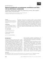
rDNA and distributions of the phyla (Figure 1) and genera
(Additional file 1: Figure S1) of bacteria present in the
soil samples were determined. There were 8 to 10 phyla
(among Acidobacteria, Actinobacteria, Bacteroidetes,
Cyanobacteria, Firmicutes, Gemmatimonadetes, Nitrospira, Planctomycetes, Proteobacteria, Verrucomicrobia)
that were considered abundant for the 2008 and 2009
data respectively (Figure 1). For these, the 16S rRNA sequences each individually make up greater than 1% of
the total. Apart from the Proteobacteria that make up
44% and 50% of the 16S sequences, the second-most
abundant phylum was Acidobacteria making up 24%
and 30% of the sequences in the 2008 and 2009 samples
respectively. Other phyla making up greater than 4% of
the sequences were Actinobacteria, Bacteroidetes and
Firmicutes. At the rank of genera, the top 1% of sequences
(99th percentile) were comprised of Acidobacteria subdivisions Gp1, Gp3, Gp4, and Gp6, and also Nitrosospira, a
member of the Betaproteobacteria (Additional file 1:
Figure S1). From the same soil samples, naturally occurring root-associated and root-bound rhizospheric bacteria
were isolated (Table 1). Strains labeled EA101-EA108 were
isolated on TY agar, and strains labeled EA201-EA202
were isolated on LB agar. One bacterium, labeled EA303,
was isolated using Chlorobium plating (CP) agar plates
with benzoate as the sole carbon source. A total of eleven
isolates were taxonomically identified by fatty acid methyl
ester (FAME) analysis and their identities were further
confirmed using 16S rRNA gene sequencing (Table 1).
Six out of the 11 isolates belonged to the class Gammaproteobacteria, and of these, 5 were of the genus
Spence et al. BMC Plant Biology 2014, 14:130
/>
Page 3 of 17
Figure 1 Relative abundance (frequency) of the major bacterial phyla present in the rice rhizosphere microbial community recorder
over two-years. The frequencies shown were obtained via classification of 16S rDNA sequences corresponding to a total of 654 and 630 clones,
for 2008 and 2009 respectively.
Table 1 Identification of rice soil isolates. List of rhizospheric bacteria isolated from rhizosphere of O. sativa cultivar
M-104 and identified by 16S rRNA gene sequencing and fatty acid methyl ester (FAME) analysis
Genus
Speciesa
Similarity Index
Confidence Level
Strain Label
Pseudomonas
Corrugata
0.761
Species inconclusive
EA104
Root associated
Chlororaphis
0.598
Genus
EA105
Root
Chlororaphis
0.77
Species
EA107
Root
Putida
0.785
Species
EA108
Root
-
0.232
No match*
EA303
Root associated
Pantoea
Agglomerans
0.896
Dyadobacter
-
Species
EA106
Root
Genus*
EA202
Root associated
Pedobacter
Heparinus
0.682
Species
EA101
Root associated
Chryseobacterium
Balustinum
0.776
Species
EA102
Root associated
Rhodococcus
Rubripertincta
0.807
Species
EA103
Root associated
Arthrobacter
Oxydans
0.758
Species
EA201
Root associated
a
Closest match in MIDI library as determined by FAME analysis.
- Inconclusive match.
*Genus solely determined by 16S rRNA gene sequencing.
Spence et al. BMC Plant Biology 2014, 14:130
/>
Page 4 of 17
Pseudomonas. This may be due to their ability to be cultured and their natural abundance in the soil environment, including the rhizosphere.
In vitro antifungal properties of rice rhizospheric bacterial
isolates
The effect of naturally associated rice rhizobacteria (see
Table 1) on growth and development of M. oryzae strain
70–15 was assessed using petri dish assays. A diffusible
assay evaluated the effect, if any, of bacterial-derived diffusible compounds on M. oryzae 70–15 (hereafter 70–15)
without direct contact. The two microbes could communicate and interact through both volatile compounds and
diffusible compounds. All isolates were tested and five
Pseudomonas isolates (EA104, EA105, EA107, EA108, and
EA303) showed significant inhibition of 70–15 growth
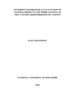
(Figure 2A). The most dramatic effect was seen by the
Pseudomonas isolate EA105, inhibiting fungal growth by
65% after 5 days, relative to the control (Figure 2A).
Bacterial volatiles have been receiving increasing attention
for their roles not only as odors, but as phytostimulators,
antimicrobials, and compounds involved in inducing a
systemic resistance response as well [27-29]. To examine whether volatile antifungal metabolites were playing
a role in the observed hindering of 70–15 growth, a
volatile (compartment) plate assay was performed using
petri dishes that were divided into four quadrants. M.
oryzae and rice bacterial isolates were placed in opposite
compartments where they shared the same headspace, yet
there was no exchange of diffusible compounds. Any
inhibition observed was therefore due to volatile compounds. All of the Pseudomonas isolates significantly
reduced growth to about the same degree as seen in
direct plates, except for EA105, whose inhibition effect
was reduced in compartment plates (Figure 2A). Bacterial
motility allows for a number of beneficial activities, including acquiring more nutrients, maneuvering away from
toxic substances, and colonizing in optimal environments
[29]. EA105 is able to spread across plates quickly through
swimming and swarming (Additional file 2: Figure S2) and
restriction to one quadrant of a plate could have contributed to the reduction in inhibition. A similar reduction in
A
70
a
Diffusible
Volatile
60
b
abc
bcd
% Inhibition
50
40
cde
30
cde
cde
defg
defg
ef
efgh
efghi
20
bc
bcd
efghi
fghij
10
0
ghij
ij
ghij
hij
ghij
ij ij
hij
j ij
-10
EA101 EA102 EA103 EA104 EA105 EA106 EA107 EA108 EA201 EA202 EA303 CHAO CHA77
B
Control
EA105
EA303
CHAO
CHA77
Diffusible
Volatile
Figure 2 Inhibition of M. oryzae vegetative growth by rice soil isolates. A) Antimicrobial assay showing the degree of inhibition of M. oryzae
70–15 by naturally isolated rice rhizobacteria as well as P. fluorescens CHAO and cyanide mutant CHA77. Error bars indicate standard error.
Different letters indicate statistically significant differences between treatments (Tukey’s HSD). B) Representative images of the fungal inhibitory
effect seen when 70–15 was exposed to bacterial diffusible and volatile compounds (diffusible plates), or solely through volatile compounds
(volatile plates).
Spence et al. BMC Plant Biology 2014, 14:130
/>
Page 5 of 17
EA105’s inhibitory activity was seen when EA105 was
grown on CM agar instead of LB agar, and in liquid culture as opposed to agar (Additional file 3: Table S1).
To see if metabolically active cells are needed for the
direct antagonism exhibited by EA105, a control experiment was performed using the same diffusible assay setup, except heat killed EA105 cells or the spent media
(cell-free supernatant) were used in place of live cells.
Neither the heat killed cells nor the spent media showed
any significant effect on fungal growth (Additional file 4:
Figure S3A), indicating that active cells are needed
for fungal inhibition. To further examine the nature of
EA105-derived inhibition, M. oryzae 70–15 plugs were
taken from plates where 70–15 had been exposed to
EA105 (inhibited) and were subcultured onto fresh CM
agar. When no longer exposed to the bacteria, 70–15
grew normally (Additional file 4: Figure S3B), indicating
the fungistatic nature of EA105.
One frequently reported toxin produced by some
pseudomonad species is hydrogen cyanide (HCN), which
binds to cytrochome c oxidase and blocks cellular respiration [30]. HCN can exist in both a gaseous or aqueous
state, suggesting that it can be released by the bacteria
as a volatile, as well as secreted into the media. Therefore,
we tested the tolerance of 70–15 to a known cyanide (CN)
producer, Pseudomonas fluorescens CHAO [31], and its
HCN production negative mutant, P. fluorescens CHA77
[32]. In diffusible plates, CHAO significantly reduced
fungal growth by 46% (Figure 2A); however, this was
not as drastic of an inhibition effect as seen by EA105.
CHA77 also significantly reduced fungal growth, but only
by 22% directly and 10% through volatiles (Figure 2A).
Since many of the known pseudomonads, including
P. fluorescens strain CHAO [31], produce CN as a
major antimicrobial component, bacterial CN production in stationary phase culture supernatants of all rice
isolates was quantified using the Lazar Model LIS146CN-CM micro cyanide ion electrode [33]. As controls, we also measured CN generated by P. fluorescens
CHAO and CHA77. EA105 produced around 500 μM
cyanide after 24 hours of incubation, while EA303 and
CHAO produced around 700 μM (Figure 3A). As expected, CN production was severely diminished in
CHA77, which has a disrupted CN biosynthesis operon
(Figure 3A). Even though EA105 produces less cyanide,
it inhibits M. oryzae vegetative growth more than
A
1200
a
Cyanide (μM)
1000
800
a
b
600
c
400
d
200
e
e
e
e
de
de
e
de
e
0
B
M. oryzae 70-15
M. oryzae guy11
80.0
80.0
a a
70.0
a a
60.0
50.0
40.0
EA105
30.0
D5
20.0
10.0
% Inhibition
% Inhibition
70.0
a
a
b
b
60.0
50.0
40.0
EA105
30.0
D5
20.0
10.0
0.0
0.0
Diffusible
Volatile
Diffusible
Volatile
Figure 3 Cyanide production by rice isolates and activity of cyanide mutant D5 against M. oryzae. A) Bacterial cyanide production of all
rice isolates, D5, CHAO, and CHA77 was measured after 24 hour incubation using the Lazar Model LIS-146CNCM micro cyanide ion electrode.
Different letters indicate statistical significance (Tukey’s HSD). B) Antimicrobial assay against M. oryzae strain 70–15 and its parental strain guy11
with EA105 and its cyanide deficient mutant, D5. Different letters indicate statistical significance (Tukey’s HSD).
Spence et al. BMC Plant Biology 2014, 14:130
/>
Page 6 of 17
CHAO, indicating the involvement of other antifungal
metabolites.
Additionally, a HCN biosynthetic mutant, D5, was
created in EA105 in which the hcnABC operon involved
in CN synthesis was disrupted and CN generation was
diminished (Figure 3A). The two plate-based bioassays
were utilized to evaluate the importance of CN in EA105
antibiosis against 70–15. Our data show that EA105 and
the D5 mutant attenuate the growth of 70–15 and guy11
to a similar degree under both diffusible and volatile
assays (Figure 3B). CHAO’s cyanide deficient mutant,
CHA77, shows a drastic reduction in ability to inhibit
M. oryzae (Figure 2A), while EA105’s cyanide deficient
mutant, D5, only shows minimal reduction in antifungal
activity, suggesting that EA105 and CHAO have different
mechanisms of antibiosis. This also indicates that the
restriction of M. oryzae growth by EA105 is mainly independent of CN, and requires an unidentified bacteriaderived compound.
Both organic and inorganic volatile compounds produced by bacteria have been shown to provide biocontrol
activity against plant pathogens [34,35]. To determine
whether the antifungal activity seen by EA105 volatiles
Appressorial Formation (%)
A
100
are due to organic or inorganic compounds, or both,
the volatile (compartment) plate design was used. As
previously described, M. oryzae 70–15 and the bacteria
were placed in opposite compartments; however, the
two remaining compartments were filled with activated
charcoal/carbon, which will adsorb organic bacterial
volatiles. The plates amended with activated charcoal
showed normal fungal growth and no inhibition through
bacterial volatile compounds (Additional file 5: Figure S4).
This implies that the active antifungal volatiles are organic compound(s), and henceforth referred to as volatile organic compounds (VOCs).
In addition to the effect rhizobacterial isolates have on
vegetative growth, these bacteria also affect development
of conidia into a specialized infection structure called
the appressorium. During pathogenesis, a penetration
peg develops at the tip of the appressoria, which enables
physical puncturing of the plant cuticle and infection of
the host [36]. EA105 inhibited 70–15 appressorial formation by nearly 90% compared to the control; while a
known biocontrol strain of P. fluorescens, CHAO, inhibited about 60% through direct treatment (Figure 4A). An
unexpected observation was that both cyanide mutants,
Diffusible
a
a
90
80
70
b
60
c
50
40
30
20
d
10
e
0
CONTROL
EA105
Appressorial Formation (%)
B
D5
CHAO
CHA77
DH5a
c
a
a
a
D5
CHAO
CHA77
DH5α
Volatiles
100
90
80
70
60
50
40
30
20
10
0
a
b
Control
EA105
Figure 4 Inhibition of M. oryzae appressoria after bacterial treatment. Effect of bacteria on M. oryzae 70–15 appressorial formation through
A) direct bacterial treatment, or through B) indirect (or volatile) bacterial treatment. Germinated conidia were incubated in a 50uL drop with
bacterial treatment (EA105, cyanide mutant D5, CHAO, cyanide mutant CHA77, or E. coli DH5α) or placed in a drop next to the bacterial
treatment for the indirect assay. Error bars represent standard deviation. Different letters indicate a significant difference (Tukey’s HSD).
Spence et al. BMC Plant Biology 2014, 14:130
/>
Page 7 of 17
D5 and CHA77, inhibited appressorial formation slightly
more than their cyanide-producing counterparts, EA105
and CHAO, respectively. Although it has not been
shown in fungi, there is evidence that sub-lethal concentrations of cyanide can trigger defense mechanisms in
nematodes [37]. Through indirect treatment, CHAO
completely failed to inhibit appressorial formation while
EA105 was still able to reduce appressorial formation by
about 20% (Figure 4B). This indicates that volatile compounds may be involved in the inhibition of vegetative
growth as well as in the reduction of appressorial formation in the case of EA105.
To gain a better understanding of the effectiveness of
EA105’s antimicrobial potential against diverse phytopathogens, EA105 was tested against a variety of naturally
isolated pathogens. Both EA105 and CHAO inhibited
other phytopathogens to a similar and lesser degree than
M. oryzae; however EA105 was able to restrict M. oryzae
growth to a significantly greater degree than CHAO
(Figure 5). This suggests the antimicrobial activity seen
by EA105 is more specific and effective against a rice
pathogen compared to other non-specific pathogens.
a concentration of 270 μM over 24 hours based on commercial standards (Additional file 6: Table S2; Additional
file 7: Figure S5A). Past antimicrobial studies with 1undecene shows it has no effect on Sclerotinia sclerotiorum [34] and a small effect on Fusarium culmorum
[38]. S-methyl thioesters were also identified in the
volatile profile of EA105, producing around 30 μM in
24 hours (Additional file 6: Table S2; Additional file 7:
Figure S5A). Antifungal activity against 70–15 by these
compounds was examined and no significant growth reduction was seen at biologically relevant concentrations
(Additional file 7: Figure S5B-C), suggesting these compounds are not the bioactive volatiles produced by EA105
as an antifungal.
Although not directly correlated to vegetative growth
reduction, we were interested to see if EA105-derived
thiol-esters could reduce virulence; therefore the effect
on 70–15 conidial germination and ability to form appressorium was examined post EA105 treatment. Even
though a large effect was not seen, there was significant
reduction in appressorial formation by all compounds at
100 μM concentration (Additional file 8: Table S3).
Characterization of antifungal metabolites from EA105
EA105 treatment to rice roots primes resistance against
M. oryzae
Volatile organic compounds (VOCs) produced by EA105
were identified using solid-phase microextraction-gas chromatography mass-spectrometry (SPME-GC-MS) (Table 2).
The most abundant peak in the headspace profile of
EA105 was identified as 1-undecene, being produced at
Induced systemic resistance (ISR) is elicited by plant
growth promoting rhizobacteria (PGPR) and results in
increased disease resistance in plants. Our data previously showed that EA105 directly inhibits fungal growth
Figure 5 Activity of EA105 against naturally isolated phytopathogens. Inhibition of naturally isolated phytopathogens by EA105 and CHAO
in comparison to M. oryzae. With the exception of lab strain F. oxysporum FO5, all pathogens were isolated from infected plants or soil, and
acquired from Nancy Gregory at the University of Delaware. Error bars represent standard error. Asterisks indicate significant differences between
EA105 and CHAO treatment (Student’s t-test, p < 0.05).
Spence et al. BMC Plant Biology 2014, 14:130
/>
Table 2 List of volatile organic compounds (VOCs)
identified in Pseudomonas isolate EA105 headspace by
GC-MS
RT (min)
Compound
Alcohols
14.07
2-Undecanol
Hydrocarbons
7.28
Cyclopropane, 1-methyl-2-pentyl-
10.77
1,4-Octadiene
10.89
1-Undecene
12.42
1-Dodecene
13.71
Cyclodecene
13.91
1-Tridecene
Ketones
13.94
2-Undecanone
16.67
2-Tridecanone
S-containing compounds
3.72
Methyl thiolacetate
4.5
Dimethyl disulfide
5.54
S-methyl propanethioate
8.27
S-methyl 3-methylbutanethioate
by the production of an antifungal compound. Next, we
tested if EA105 could also suppress M. oryzae indirectly
by inducing changes in the host plant. Three-week old
roots of soil-grown rice cv. Maratelli (highly susceptible
to M. oryzae) were root inoculated with rhizobacteria and
after 24 hours, the plants were challenged with M. oryzae
70–15 spores. In addition to EA105, rice isolates EA106,
a Pantoea agglomerans, and EA201, an Arthrobacter
oxydans, were also tested (see Table 1). Strikingly, the
plants whose roots had been pretreated, or ‘primed’, with
EA105 and EA106 showed a significantly reduced number
of blast lesions (P ≤ 0.0087 and 0.0003, respectively),
as compared to the plants receiving no pretreatment
(Figure 6). Interestingly, pretreatment with a previously
characterized direct antagonist of M. oryzae, P. fluorescence CHAO [39], conferred no protection against disease
formation on the leaves (Figure 6). Although it has previously been reported that CHAO induces ISR in Arabidopsis thaliana [40], rice is a non-native host of CHAO, being
originally isolated from Swiss soils suppressive to black
root rot [41]. These results clearly support the hypothesis
that root colonization by EA105 and EA106 induces
plant-encoded mechanisms which prime rice for foliar
attack by M. oryzae, enhancing a defense response
which leads to reduction of M. oryzae infection on the
aerial portion of the plant.
To further explore the mechanism by which isolates
EA105 and EA106 reduce lesions through a plantmediated mechanism, the expression of several key ISR
Page 8 of 17
genes were examined in rice at 24 hours post bacterial
treatment. As a control, we also examined the effect of
CHAO, which does not reduce the number M. oryzae
lesions on rice plants. With EA105 or EA106 treatment,
there was significant up-regulation of the JA responsive
genes, JAR1 and WRKY30, while CHAO treatment down
regulated these genes. Similarly, ET responsive genes,
EIL1 and ERF1, were also up-regulated with EA105 and
EA106 treatment, but to a significantly lesser extent with
CHAO treatment (Figure 7). A positive control with JA
(50 μM) treatment also induced JAR1 and WRKY30 (data
not shown). There was only slight induction of SA responsive genes PR1 and WRKY77 with the bacterial treatments
(Figure 7C). The SA responsive genes were also induced
by SA treatment (1 mM) (data not shown). Of the 6
genes examined, expression patterns were similar between EA105 and EA106 treatments for all genes except
PR1. In rice treated with EA106, there was a significantly stronger induction of PR1 than in rice plants
treated with EA105. The data suggest that EA105 induces a JA and ET dependent ISR that may protect
plants against M. oryzae.
Discussion
In order to make a significant impact on global food security, a biocontrol solution to rice blast disease must be
developed that is both effective and sustainable while reducing or eliminating the need for synthetic chemical
fungicides. We have found microbes from the rice rhizosphere that attenuate M. oryzae in vitro and in planta.
Most notable is P. chlororaphis strain EA105, which has
demonstrated the ability to severely restrict the growth
of rice pathogen M. oryzae, and is therefore a strong
candidate for a novel biocontrol agent against rice blast
disease. Previously, P. chlororaphis isolates have been
shown to be agriculturally important in the biocontrol of
several plant pathogens including Sclerotinia sclerotiorum
[42], Rhizoctonia cerealis [43], Seiridium cardinale [44],
and Leptosphaeria maculans [45]. To our knowledge, this
is the first report of P. chlororaphis reducing rice blast
symptoms. In contrast to chemical fungicides, biocontrol bacteria produce a mixture of antifungal compounds
which can fluctuate based on environmental cues [46].
The fungistatic activity of EA105 could lead to a longerterm, more effective strategy for reducing rice blast disease
than current chemical fungicides, which exert stronger selective pressure for M. oryzae to develop resistance. Furthermore, as living organisms, these biocontrol microbes
are continuing to evolve with their rhizospheric neighbors
ensuring a more sustainable solution.
To gain a better understanding of the composition
and diversity of the rice rhizospheric soil, we used a
metagenomic approach to examine the phyla and genera
that naturally inhabit this niche. Distribution of phyla was
Spence et al. BMC Plant Biology 2014, 14:130
/>
Page 9 of 17
A
None/
Gel
B
Mock/
70-15
EA105/
70-15
EA106/
70-15
EA201/
70-15
60
CHAO/
70-15
A
No. lesions per leaf
A
50
AB
40
BC
C
30
20
10
0
Mock
EA105
EA106
EA201
Root Treatment
CHAO
Figure 6 The effect of rhizobacterial priming on rice blast lesion formation. Spores were sprayed on 3-week old whole plants 24 hour after
being root primed with mock, EA105, EA106, EA201 or CHAO suspension. A) Representative leaf segments of mock or rhizobacterial primed
plants. B) The average number of lesions formed on the second youngest leaf of O. sativa cv. Maratelli. Error bars indicate standard error. Means
with the same letter do not differ significantly (Tukey’s HSD).
consistent across growing seasons, with the two predominant phyla being Acidobacteria and Proteobacteria. Acidobacteria have only recently been discovered and the vast
majority are currently unculturable. However, their abundance in soil has been documented, and they may be playing a crucial role in the rhizosphere that has yet to be
determined [47]. Proteobacteria is a very broad phylum,
encompassing a variety of bacteria, including Pseudomonads which are gamma-proteobacteria [48].
Evidence shows that stress to the aerial portions of
plants can stimulate rhizo-deposition of chemo-attractants
to enhance colonization by rhizobacteria [26,27]. Effective
plant defense may be due to an ability of the host plant
to modulate the composition of root exudates, attracting
microbes which can trigger plant resistance. The recruitment of beneficial microbes can also alter physiological
functions in plants to resist aerial pathogens [49]. Although M. oryzae is most commonly a foliar pathogen,
it also has the ability to infect roots [50,51] and is
closely related to other root pathogens such as M. poae,
M. rhizophila, and Gaeumannomyces graminis [51].
Root infection by M. oryzae is often followed by dispersal to the shoots and traditional blast lesion formation
[51]. Therefore, the direct antifungal activity of EA105
against M. oryzae could have ecologically relevant implications in preventing blast infections.
Our data reveal that treatment of soil-grown rice
plants with EA105 activates basal resistance mechanisms
against 70–15 in planta. The precise mechanism by
which rice rhizospheric microbes induce physiological
effects on the host (rice) is not known, although some of
these changes are modulated through the signaling of
Spence et al. BMC Plant Biology 2014, 14:130
/>
Page 10 of 17
A
Fold Change
100
10
A
a
A
a
EIL1
ERF1
b
B
1
EA105
B
EA106
CHAO
10
Fold Change
A
a
A a
B b
1
JAR1
WRKY30
0.1
EA105
EA106
CHAO
10
A
Fold Change
C
PR1
B
WRKY77
a
a
B
a
1
EA105
EA106
CHAO
Figure 7 Expression of defense related genes in rice plants treated with rhizobacteria EA105 and EA106. Roots of aseptically grown rice
plants were treated with EA105 or EA106. Leaf samples were collected at 24 hrs post treatment and the expression of genes involved in A) ethylene,
B) jasmonic acid (JA), or C) salicylic acid (SA) signaling was examined. Error bars indicate standard error. Means with the same letter do not
differ significantly (Tukey’s HSD).
small molecules such as salicylic acid (SA), jasmonic acid
(JA), or ethylene (ETH) [52]. The pathogenesis related,
or PR, genes such as PR1 and WRKY77 are SA responsive [53] and are up-regulated during pathogen infection,
ultimately triggering a defense response and reducing
disease symptoms [54]. However, beneficial rhizobacteria
such as P. fluorescens WCS374r have been shown to
stimulate a defense response which induces resistance in
rice to M. oryzae, but is completely independent of SA
signaling [55]. Similar to this finding, our gene expression data suggest that EA105 triggers ISR in rice through
a mechanism that involves both JA and ETH and to a
Spence et al. BMC Plant Biology 2014, 14:130
/>
lesser extent SA signaling. The JA responsive genes JAR1
and WRKY30 are crucial to JA signaling and are required
for the stimulation of ISR in A. thaliana as well as rice
[56,57] and both of these genes were highly expressed
24 hours after EA105 and EA106 treatment but not with
CHAO treatment. We saw similar up-regulation of the
ethylene responsive genes EIL1 and ERF1, which have also
been implicated in ISR signaling and reduction in disease
susceptibility [58]. Moreover, we demonstrate the ability of
EA105 to severely restrict mycelial growth of 70–15 and
almost completely halt appressorium formation on abiotic
hydrophobic surfaces. This suggests that the beneficial
microbiome of rice could attenuate the virulence of rice
blast through multiple mechanisms; therefore, manipulation of the rhizosphere is a valuable and comprehensive
manner in which to target biotic stresses.
Biocontrol agents are currently employed to control
rice pathogens that cause fungal sheath blight [59-63]
and a subset of fungal pathogens that cause rice blast
[64,65]. With a few exceptions [66,67] the biocontrol
agents tested were not isolated from rice, as compared
to the bacterial strain EA105, which was isolated from
the rice rhizosphere. We speculate that a microbe which
is confirmed to associate with field grown rice roots,
such as EA105, may have better implications for rice
protection compared to unrelated biocontrol isolates
due to its ability to compete and survive in the rice
rhizosphere. Previous studies have shown a relative of
Psuedomonas, Delftia tsuruhatensis, to directly inhibit
M. oryzae and also reduce lesions in rice by about 50%,
however the mechanism of lesion reduction has not
been examined [66]. Isolates from the rice and millet
rhizospheres, including 13 Bacilli and 6 Psuedomonads,
did show direct inhibition and lesion reduction of Setaria
blast, on the host plant Foxtail millet (Setaria italica L)
though these isolates were not tested in rice [68]. There
have also been reports of naturally isolated rice rhizobacteria reducing blast in aerobically grown rice in Brazil,
though the isolates have not been identified and the mechanism by which they induce resistance has not yet been
examined [69]. Similarly, Naureen et al. investigated
multiple isolates from bulk soil and the rice rhizosphere
for their direct antagonism against M. oryzae and their
ability to reduce lesions in planta, but the mechanisms
underlying these activities have not yet been explored.
Five of the isolates examined were Pseudomonas sp. but
these 5 isolates were from bulk soil rather than the rice
rhizosphere [67]. Two isolates from the rhizosphere of
Lupinus hispanicus, Pseudomonas fluorescens Aur 6 and
Chryseobacterium balustinum Aur 9, showed the ability
to reduce blast severity and increase rice production
when co-inoculated [70] however, these isolates were
not originally isolated from the rice rhizosphere and the
way in which they reduce lesions has not yet been
Page 11 of 17
described. De Vleesschauwer et al. [55], thoroughly examined the way in which P. fluroescens WCS374r induces resistance in rice, independent of SA signaling,
and mediated through the ETH and octadecanoid pathways. Strain WCS374r is a spontaneous rifampycin mutant of lab strain WCS374 [55]. De Vleesschauwer et al.
provide valuable insight into the mechanisms underlying
ISR against M. oryzae, and we have shown that a natural
rice isolate, EA105, shows parallels in its ability to trigger
ETH signaling while minimally impacting SA signaling.
We have, in a way, combined these stories to investigate
how a natural rice isolate works in reducing blast both
through direct and plant-mediated mechanisms.
Shimoi et al. [71], examined a novel mechanism of blast
reduction by selectively isolating phyllospheric microbes
from rice, including one P. geniculata strain, which catabolizes collagen and gelatin. Some of these microbes were
able to reduce blast symptoms when co-inoculated onto
rice leaves, presumably by disrupting the adhesion of
the spore tip mucilage and extracellular matrix from the
leaf surface, preventing proper attachment by M. oryzae
[71]. It would be interesting to test such a method in
combination with a root-associated microbe such as
EA105, which can induce resistance through plant based
signaling.
Thorough groundwork has been laid in testing methods
for introducing biocontrol bacteria to plants. Talc-based
powder applications of P. fluorescens to rice seeds followed
by foliar sprays on rice shoots have resulted in the most
effective reduction of blast symptoms [72]. The survival of
two strains of P. fluorescens was examined in 3 cultivars of
rice, and bacterial treatment of seeds resulted in persistence of the bacteria throughout the 110 day experiment
[65]. However, the mode by which these two strains were
reducing blast symptoms has not been elucidated and
appears to differ from the mechanism used by EA105.
While we noted elevated JA and ET signaling with
minimal effect on SA, these two Pseudomonas isolates
resulted in elevated SA levels in rice [73].
To our knowledge, this is the first report of a Pseudomonas chlororaphis isolate which can protect against rice
blast, and this isolate shows two distinct mechanisms of
action- direct antifungal activity and induction of resistance in the host. Beyond showing the ability of EA105 to
inhibit vegetative growth of M. oryzae, we also show an
ability to reduce M. oryzae pathogenesis by inhibiting
appressoria formation. Interestingly, the activity of EA105
is largely independent of cyanide production, despite cyanide commonly being associated with biocontrol activity in
Psuedomonads.
Microbes are essential for animal health and immunity,
and there are compelling reasons to believe that rootassociated microbes are equally important to plants as
they are to animals. Plant roots encounter diverse
Spence et al. BMC Plant Biology 2014, 14:130
/>
microbial populations in soil and generate a unique ecological niche for microbes by the secretion of resources
into the rhizosphere. These rhizospheric resources are
limited in abundance, and some microbes have evolved
antimicrobial traits to reduce competition from other
microbes and to bolster the health of their plant host.
However, we lack a clear understanding of the contribution conferred by individual microbial strains within a
microbiome to plant growth and protection. Since biocontrol has proven to be a successful approach to crop
protection, more efforts are needed to identify potential
biocontrol agents from the diverse pool of rhizospheric
bacteria and to understand the mechanisms by which
they positively influence plant productivity.
Conclusions
Eleven bacteria were isolated from rhizospheric rice soil
and identified. Isolate EA105, Psuedomonas chlororaphis,
showed the strongest biocontrol potential against blast
pathogen M. oryzae. EA105 reduced mycelial growth,
and almost completed halted appressoria formation in
M. oryzae. A HCN mutant in EA105, D5, showed similar
antagonistic abilities against M. oryzae, indicating a mechanism of action which is independent of HCN. Isolate
EA105 as well as Pantoea agglomerans EA106 were able
to reduce the number of blast lesions in rice, when roots
were pre-treated with the bacteria prior to infection with
M. oryzae. The response elicited in rice by EA105 and
EA106 is mediated through JA and ET signaling. Isolate
EA105 was the only isolate which was effective both as a
direct antagonist to M. oryzae as well as an elicitor of the
ISR response in rice. Isolate EA105 shows promise as a
potentially valuable biocontrol agent to reduce crop losses
from blast disease. The resulting increase in rice yields
could have a tremendous impact on global food security.
Materials and methods
DNA extraction from rhizospheric soil and processing
Field grown rice plants were harvested for root associated microbial DNA for cloning and sequencing of 16S
rRNA sequences. The majority of the aerial part of the
rice plants was removed and a clump of soil encompassing the root ball was retained for processing. Individual
roots from single plants were processed one at a time
until sufficient root material was obtained for this plant.
A single complete root, considered untouched during
harvest, was excised from the middle of the root ball.
Excess soil was removed from the root using gloved
hands until only tightly bound soil remained. The root
was then added to 30 ml of PBS buffer (pH 7.0). Further
roots from the same plant were added until volume of
roots collected approximated 12 ml. Roots in PBS buffer
were vortexed, and about 16 ml of the root wash soil
suspension (rice rhizosphere soil) was spun down and
Page 12 of 17
the pellets stored at −80 C until DNA extraction.
Microbial DNA was extracted from 0.25 to 1 gram
of rhizospheric soil using the MoBio UltraClean Soil
DNA Isolation Kit with use of the maximum yield
'Alternative Protocol'. Amplification of 16S rDNA was
performed using the primers 27 F(AGAGTTTGATCCT
GGCTCAG) and 1492R (GGTTACCTTGTTACGACTT).
The sequences were screened of possible chimeras using
Mallard [74] and then passing sequences classified against
the taxonomic reference set available from the Ribosomal
Database Project (RDP) resource ( />Specifically, the sequences were classified using the java
based RDP Naïve Bayesian rRNA Classifier Version 2.1
[75] with the taxonomic reference set RDP 10.18 [76]. The
R package ggplot2 [77] was used to generate the barplots
depicting taxonomic composition. The amplified product
was gel purified, and cloned using the Topo TA vector.
Colonies with inserts were purified, and the insert DNA
sequences were obtained by Sanger sequencing.
Isolation and identification of rhizobacteria
Natural rhizobacteria were isolated from root-associated
soil and roots of M-104 rice plants, a temperate japonica
cultivar widely grown in California. M-104 roots were
harvested and the soil adhering to the root was removed
using a sterile spatula and collected as the root-associated
soil sample. The root was then rinsed, crushed and
processed as the root sample, which included endophytic bacteria as well as tightly bound root bacteria.
The samples were suspended in sterile water (0.1 g/ml)
and serial dilutions were dispensed on LB [78], TY [79],
or CP + benzoate [80] agar plates. They were incubated
for 48 hours at 30°C and single colonies were selected
based on morphology and re-streaked on fresh agar plates.
Isolate identification was initiated by sequencing the 16S
rDNA using colony PCR and the universal primers 27 F
(AGAGTTTGATCCTGGCTCAG) and 1492R (GGTTAC
CTTGTTACGACTT). Taxonomic assignments were determined using the Ribosomal Database Project (RDP)
website classifier. Further identification was done by
MIDI, Inc (midi-inc.com) through a fatty acid methyl
ester (FAME) analysis. A similarity (SIM) index of 1.000
means an exact species match determined by fatty acid
make-up. The lower the SIM index, the more varied the
fatty acid content. SIM Index cutoff of 0.600 was used
to determine confident species match, unless otherwise
noted.
Plant materials and growth conditions
Oryza sativa ‘M-104’ seeds were a gift from Dr. Thomas
Tai (University of California-Davis). The seeds were dry
planted in a Davis field where rice had been previously
grown for several years. The field was flooded soon after
emergence, and the roots were harvested for sampling at
Spence et al. BMC Plant Biology 2014, 14:130
/>
Page 13 of 17
about 1 month after planting. O. sativa ‘Maratelli’, a susceptible variety to blast fungus M. oryzae strain 70–15
was used for the studies. All plants were grown in a
growth chamber with a daily cycle of 16 hr light (28°C,
80% RH), and 8 hr dark (26°C, 60% RH).
on swimming plates (5 g/L NaCl, 10 g/L tryptone, and
0.03% (w/v) agarose), and swarming plates (8 g/L nutrient
broth, 5 g/L glucose, with 0.5% (wt/vol) agar and after
incubation at 30°C the diameter of bacterial growth was
measured.
In vitro antibiosis assay
Measurement of cyanide
Two experimental designs were created using petri dishes
to determine the antagonistic activity of bacterial isolates.
First is the diffusible assay, whereby sterile petri dishes
were filled with autoclaved complete media (CM) agar,
consisting of 10 g sucrose, 6 g yeast extract, 6 g casaminoacids, 15 g agar, and 1 ml Aspergillus nidulans trace elements in 1 L water. Five mm plugs of M. oryzae 70–15 or
guy11 mycelia were placed 4 cm from 5 μl of 5 × 10⁵ bacterial cells. The plates were sealed with parafilm and put
in the dark in a 25°C incubator. Photographs were taken
after 5 days and the diameter of the mycelium growing
out from the plug was measured using ImageJ software.
Percentage (%) inhibition was calculated by the formula: %
inhibition = ([C – T) × 100]/C), where C = fungal diameter (cm) in the control plate, and T = fungal diameter
(cm) in the bacterial treated plates. Three biological replicates were performed and an average was taken. Second,
the volatile (compartment) assay used compartmentalized
petri dishes where the bacteria were grown on LB agar or
LB liquid and M. oryzae was grown on CM agar in separate compartments. Three biological replicates were performed and an average was taken. The activated charcoal
assay used the same experimental design as the volatile
assay, except the remaining two compartments were each
filled with 1 g of activated charcoal (Darco®, 20–40 mesh
particle size, granular, Aldrich, Milwaukee, WI) wrapped
in KimWipes. Two biological replicates were performed
and an average was taken. For the heat killed and spent
media assay, bacterial isolate EA105 was grown overnight
in 10 mL of LB liquid in a 50 mL falcon tubes and optical
density at 600 nm (OD600) was measured. The culture was
either placed in a 65°C water bath for 24 hours, or spun
down (centrifuged for 8 minutes at 4000 rpm) and the
supernatant passed through and 0.45 μm filter (Millipore,
Billerica, MA). Sterile filter discs were placed on CM agar
plates 4 cm away from a 5 mm plug of M. oryzae 70–15.
The filter discs were inoculated with 50 μl of LB liquid,
50 μl of EA105 heat-killed cells, or 50 μl of EA105 supernatant (cell-free spent media). Two biological replicates
were performed and an average was taken. All fungal diameters were measured using ImageJ, and % inhibition
was calculated as described above.
Cyanide production in bacterial culture supernatant was
measured using the Lazar Model LIS-146CNCM micro
cyanide ion electrode from Lazar Research Laboratories,
Inc. Bacterial cultures were grown in LB for 24 hours
shaking at 200 rpm at 30°C. Optical density at 600 nm
(OD600) was recorded. The cells were centrifuged (8 minutes at 4000 rpm) and supernatant was taken for measurement. The electrode was conditioned prior to use, and
rinsed with 70% ethanol then water between each sample
reading. Two biological replicates were performed.
Bacterial motility
To evaluate the bacterial motility, swimming and swarming assays were performed with rice isolates as per the
published protocol [81]. Briefly, bacterial stabs were placed
Construction of cyanide mutant D5
The D5 mutant was constructed using the Targetron gene
knockout system (Sigma-Aldrich) to disrupt a region of
the hydrogen cyanide biosynthetic operon that encompassed both the hcnB and hcnC genes. Primers for the
insertion sites of the group II intron were chosen by a
Sigma-Aldrich computer algorithm based on an input
sequence from the hcnBC genes. These primers (IBS,
EBS1d, and EBS2) as well as the EBS universal primer
were used to amplify the intron template. The resulting
amplicon was purified using the QiaQuick PCR purification kit (Qiagen), double digested with HindIII and
BsrGI, and then ligated into the linear pACD4K-C vector using T4 DNA ligase and 2X Rapid ligation buffer
(Promega) with a 1:2 molar ratio of vector to insert DNA.
Transformation was performed according to Targetron’s
suggestions, with exception of the heat shock being extended to 60 seconds, the recovery period being extended
to 3 hours, and the incubation temperature being at 30°C.
Induction of the group II intron insertion using IPTG was
performed as per the Targetron protocol. Potential transformants were selected using colony PCR and absence
of cyanide production was confirmed using the LIS-146
Micro Cyanide probe (Lazar Research Laboratories).
Solid-phase microextraction-gas chromatography
mass-spectrometry (SPME-GC-MS)
Volatile metabolites produced by EA105 were extracted
using an SPME fused silica fiber coated with 65 μm of
polydimethylsiloxane/divinylbenzene (Sigma-Adrich). EA105
was grown on LB agar for 2 days and then the fiber was
exposed for 24 hours to the headspace above EA105. The
fiber was then manually injected into an Agilent 6890 GC
with a 5973 N MS detector (Agilent Technologies), installed with a HP-5MS capillary column (30 m × 0.25 mm,
0.5 μm) and a flame ionization detector. Inlet temperature
Spence et al. BMC Plant Biology 2014, 14:130
/>
was 250°C. Oven conditions started at 40°C for 2 min,
ramped at 10°C/min to 250°C, and held for 2 min. VOCs
were identified using the mass spectral library (NIST).
Standard curves of the identified compounds were created
using commercially available compounds. They were
diluted in methanol and 2 μl was injected into the GC.
The concentration of the volatiles produced was determined by comparing peak heights of the EA105 profile
to the standard curve. Four biological replicates were
performed.
Spore germination and appressoria formation
Plastic coverslips were sterilized with ethanol and used
as hydrophobic surfaces for the conidiospores. M. oryzae
70–15 spores grown on oatmeal agar for 10 days were
suspended in water and filtered through Miracloth. For
S-methyl thioester treatments, a 100 mM stock of the
compounds in 100% methanol was used, and compared
to a control treatment with the same final amount of
methanol. For cyanide treatments, potassium cyanide
was dissolved in 35 mM KOH to make a 100 mM stock,
which was further diluted in water. A 1:1 (v:v) solution
of spores plus compound were made with a final concentration of 105 spores/ml in compound concentrations
ranging from 1–500 μM. For bacterial treatments, a final
concentration of OD600 = 0.02 (~1×10⁷ cells/mL) was
used. Five plastic coverslips were placed into a petri dish
containing a wet filter disc in the center to maintain humidity. A 50 μL drop of treated spores was placed on
each coverslip. For indirect bacterial treatment, a drop
of bacterial cells was placed next to each coverslip and a
50 uL drop of untreated spores was placed on the coverslip. Petri dishes were parafilmed and placed in the dark
at room temperature. Percent germination was determined at 3 hours post treatment and percent appressorium formation was determined 24 hours post treatment
using the Zeiss Axioscope2 upright light microscope.
Five images were taken at different locations on each
coverslip for a total of 25 images per treatment. Percentage germination was calculated by counting the number
of germinated spores and the total number of spores in
the images. Percentage appressorium formation was determined by counting the number of germinated conidia
which had produced an appressorium. Three biological
replicates were examined following the protocol described above.
Page 14 of 17
root primed with 2 mL of the rhizobacterial suspension
per plant. Eight replicates were used per treatment. Mock
plants were treated with 2 mL of sterile water. After
24 hours, the shoots (stems and leaves) of each plant were
sprayed with 1 milliliter of M. oryzae strain 70–15 at a
concentration of 105 spores per mL. Ten-day old spores
were suspended in sterile water, filtered through Miracloth, and counted using a hemocytometer. Spores were
adjusted to a concentration of 1×10⁵ spores/mL water and
a 1:10 (v:v) of 0.2% gelatin was added to the suspension.
Plants were sprayed inside of plastic bags containing
wet paper towels using an artist’s air brush, sealed to
maintain humidity, and covered with plastic bins for
24 hours of darkness. As a precautionary measure,
pathogen-inoculated plants were transferred to separate
growth chambers and grown in identical growth conditions as the other treatment groups. Photographs of
leaves were taken after 1 week and the number of lesions on the second youngest leaf was counted using
the image analysis program ImageJ to facilitate accurate
scoring. Four biological replicates were performed.
To test gene expression changes in rice, M-104 seeds
were sterilized and germinated in petri dishes. At 7 days
post germination, seedlings were transferred to clear,
sterile boxes containing 50 mL of Hoagland’s liquid
medium. The pH of the medium was maintained at 5.7.
At 14 days post germination, the liquid medium was inoculated with bacteria which had been washed in water,
to a final concentration of 106 cells/mL. At 24 hours
post treatment, leaf tissue was frozen in liquid nitrogen
and RNA was extracted using the Bio Basic EZ-10 Spin
Column Plant RNA Mini-Prep Kit. RNA was treated
with Turbo DNAse (Ambion) and the High Capacity
cDNA Reverse Transcription Kit (Ambion) was used to
synthesize cDNA, using 500 ng of RNA. PCR was carried
out using standard Taq Polymerase (New England
Biolabs). Primers to test for SA responsive genes PR1 and
WRKY77, JA responsive genes JAR1 and WRKY30, and
ETH responsive genes EIL1 and ERF1 were designed using
Primer Blast (NCBI) of Nipponbare gene sequences, and
are listed in SOM Additional file 9: Table S4. PCR products were run on a 1.4% agarose gel, stained with ethidium
bromide, and imaged using an Alpha Imager system. Band
intensities were quantified using ImageJ. A ubiquitin control was used to normalize all samples. Each biological
replicate was pooled from 9 plants, and there were 3 biological replicates per treatment.
Evaluation of rhizobacterial-mediated ISR
Rhizobacterial isolates were grown overnight in LB at
30°C shaking at 200 rpm. Cells were spun down by centrifugation (8 minutes at 4000 rpm) and the supernatant
discarded. Cells were washed in sterile water twice, then
resuspended to an OD600 of 0.5 (~2.5×10⁸ cells/mL).
Three- week old soil-grown Maratelli rice plants were
Statistical analysis
The statistical software JMP 10 was used to analyze data.
To compare across treatments, the Tukey’s HSD test
was used and results were considered to be statistically
different when p < 0.05.
Spence et al. BMC Plant Biology 2014, 14:130
/>
Additional files
Additional file 1: Figure S1. Relative abundance (frequency) of the
major bacterial genera in the rice rhizosphere microbial community
recorded over a two-year period. The frequencies shown were obtained
via classification of 16S rDNA sequences corresponding to a total of 654
and 630 clones, for 2008 and 2009 respectively.
Additional file 2: Figure S2. Swimming and swarming motility of
Pseudomonas isolates. Cells were grown on motility plates for 24 hours
as described by Rashid & Kornberg (81). Means comparisons for all pairs
were done using Tukey-Kramer HSD statistical test, where means with
the same letter do not differ significantly (n=3). Treatments were
compared within swarming plates, and within swimming plates.
Additional file 3: Table S1. Comparison of fungal inhibition elicited by
EA105 grown on direct or compartment plates and on agar or in liquid.
Additional file 4: Figure S3. Growth of M. oryzae treated with heat
killed cells and growth after inhibition by EA105. A) Effect of heat killed
cells and cell-free spent media on fungal inhibition. A 50 μl drop of
either heat killed EA105 cells or EA105 cell-free spent media was placed 4
cm from M. oryzae 70-15 and 70-15 diameters were measured after three
days. Error bars indicate standard deviation. There was no significant
difference between the control and treatments using Student’s t-test and
a p-value of <0.05. B) Recovery of M. oryzae 70-15 growth after exposure
to EA105 volatiles. Fungal plugs were replated onto fresh CM agar after
previously being exposed to antifungal volatiles produced by the
Pseudomonas isolate EA105. Fungal diameter was measure after three
days, and normal growth was observed. There was no significant
difference between the control and previously exposed 70-15. Error bars
indicate standard error.
Additional file 5: Figure S4. Activity of volatile compounds produced
by bacteria in the presence of activated charcoal. Inhibitory effect
through bacterial volatiles was abolished in the presence of activated
charcoal. Error bars indicate standard deviation. Means with the same
letter do not differ significantly as per Student’s t-test, p<0.05. Capital
letters were used for plates without activated charcoal, and lower case
letters were used for plates amended with activated charcoal.
Additional file 6: Table S2. Concentration at which volatile metabolites
are being produced by EA105.
Additional file 7: Figure S5. Inhibition of M. oryzae by S methyl
thioesters and 1-undecene. A) Standard curves used to calculate
biological concentrations of volatiles produced by EA105. Commercially
available compounds were diluted in methanol (S-methyl thiopropioante,
S-methyl thioisovalerate), or chloroform (1-undecene) and injected into a
GC-MS for analysis. B) Growth of M. oryzae 70-15 after 5 days on plates
containing different concentrations of S-methyl thioesters in the media.
Significant inhibition occurred by 1 mM for all except S-methyl
thioisovalerate (Student’s t-test, p<0.05) Error bars indicate standard error.
C) Growth of M. oryzae 70-15 after 5 days on plates containing different
concentrations of 1-undecene in the media. Significant inhibition
occurred by 5 mM 1-undecene (Student’s t-test, p<0.05). Error bars
indicate standard error.
Additional file 8: Table S3. Effect of treating spores with thiol-esters
on germination and ability to form appresoria.
Additional file 9: Table S4. Primer sequences used for RT-PCR gene
expression in rice cv. M-104.
Competing interests
The authors declare that they have no competing interests.
Authors’ contributions
CS and EA isolated soil bacteria and carried out the inhibition assays. CS
carried out the construction and testing of the cyanide mutant, the
appressorial assays, and the gene expression assays. EA carried out the
GC-MS experiments and tested the resulting compounds. CR maintained rice
plants and collected rhizospheric soil samples and performed the 16S
sequencing. The bioinformatic analysis of the 16S sequences was performed
by CJ. CS, EA, and HB drafted the manuscript. HB conceived the study and
Page 15 of 17
HP, VS, and ND participated in its design and coordination. All authors read
and approved the final manuscript.
Acknowledgements
H.P.B. and V.S. acknowledge the support from NSF Award PGPR-0923806. We
would like to thank Dr. Rovshan Mahmudov for his assistance using the
GC-MS. Additionally, we would like to thank Nancy Gregory for donating the
naturally isolated phytopathogens and Adam Draper for his assistance with
the inhibition experiments involving these strains. Lastly, we would like to
thank Dr. Thomas Hanson for his advice and guidance.
Author details
Department of Biological Sciences, University of Delaware, Newark, USA.
2
Delaware Biotechnology Institute, Newark, USA. 3Department of Plant and
Soil Sciences, University of Delaware, Newark, USA. 4Department of Plant
Biology, University of California, Davis, USA.
1
Received: 13 January 2014 Accepted: 28 April 2014
Published: 13 May 2014
References
1. Zeigler RS, Leong SA, Teng PS: Rice blast disease. Rice Blast Disease 1994,
1–626.
2. Skamnioti P, Gurr SJ: Against the grain: safeguarding rice from rice blast
disease. Trends Biotechnol 2009, 27:141–150.
3. Bonman JM, Khush GS, Nelson RJ: Breeding rice for resistance to pests.
Annu Rev Phytopathol 1992, 30:507–528.
4. Chuma I, Isobe C, Hotta Y, Ibaragi K, Futamata N, Kusaba M, Yoshida K,
Terauchi R, Fujita Y, Nakayashiki H, Valent B, Tosa Y: Multiple translocation
of the AVR-Pita effector gene among chromosomes of the rice blast
fungus Magnaporthe oryzae and related species. PLoS Pathogens 2011,
7:e1002147.
5. Aktar MW, Sengupta D, Chowdhury A: Impact of pesticides use in
agriculture: their benefits and hazards. Interdisciplinary Toxicol 2009,
2:1–12.
6. Lugtenberg B, Kamilova F: Plant-growth-promoting rhizobacteria. Annu
Rev Microbiol 2009, 63:541–556.
7. Silby MW, Winstanley C, Godfrey SA, Levy SB, Jackson RW: Pseudomonas
genomes: diverse and adaptable. FEMS Microbiol Rev 2011, 35:652–680.
8. Thomashow LS, Weller DM: Role of a phenazine antibiotic from
Pseudomonas fluorescens in biological control of Gaeumannomyces
graminis var. tritici. J Bacteriol 1988, 170:3499–3508.
9. Voisard C, Keel C, Haas D, Defago G: Cyanide production by Pseudomonas
fluorescens helps auppress black root-rot of tobacco under gnotobiotic
conditions. EMBO Journal 1989, 8:351–358.
10. Rudrappa T, Splaine RE, Biedrzycki ML, Bais HP: Cyanogenic pseudomonads
influence multitrophic interactions in the rhizosphere. PloS One 2008,
3:1–11.
11. Raaijmakers JM, Weller DM, Thomashow LS: Frequency of antibioticproducing Pseudomonas spp. in natural environments. Appl Environ
Microbiol 1997, 63:881–887.
12. Howell CR, Stipanovic RD: Control of Rhizoctonia solani on cotton
seedlings with Pseudomonas fluorescens and with an antibiotic
produced by the bacterium. Phytopathol 1979, 69:480–482.
13. Howell CR, Stipanovic RD: Suppression of Pythium ultimum-induced
damping-off of cotton seedlings by Pseudomonas fluorescens and its
antibiotic, pyoluteorin. Phytopathol 1980, 70:712–715.
14. Nielsen TH, Thrane C, Christophersen C, Anthoni U, Sørensen J: Structure,
production characteristics and fungal antagonism of tensin – a new
antifungal cyclic lipopeptide from Pseudomonas fluorescens strain
96.578. J Appl Microbiol 2000, 89:992–1001.
15. Nielsen TH, Christophersen C, Anthoni U, Sørensen J: Viscosinamide, a new
cyclic depsipeptide with surfactant and antifungal properties produced
by Pseudomonas fluorescens DR54. J Appl Microbiol 1999, 87:80–90.
16. Vitullo D, Di Pietro A, Romano A, Lanzotti V, Lima G: Role of new bacterial
surfactins in the antifungal interaction between Bacillus
amyloliquefaciens and Fusarium oxysporum. Plant Pathol 2012,
61:689–699.
17. Bechet M, Caradec T, Hussein W, Abderrahmani A, Chollet M, Leclere V,
Dubois T, Lereclus D, Pupin M, Jacques P: Structure, biosynthesis, and
Spence et al. BMC Plant Biology 2014, 14:130
/>
18.
19.
20.
21.
22.
23.
24.
25.
26.
27.
28.
29.
30.
31.
32.
33.
34.
35.
36.
37.
38.
39.
40.
41.
properties of kustakins, nonribosomal lipopeptides from Bacillus spp.
Appl Microbiol Biotechnol 2012, 95:593–600.
Van Loon LC: Plant responses to plant growth-promoting bacteria. Eur J
Plant Pathol 2007, 119:243–254.
Van der Ent S, Van Wees SCM, Pieterse CMJ: Jasmonate signaling in plant
interactions with resistance-inducing beneficial microbes. Phytochemistry
2009, 70:1581–1588.
Ryu C, Farag MA, Hu C, Reddy MS, Kloepper JW, Paré PW: Bacterial volatiles
induce systemic resistance in Arabidopsis. Plant Physiol 2004, 134:1017–1026.
Van Wees SCM, Van der Ent S, Pieterse CMJ: Plant immune responses
triggered by beneficial microbes. Curr Opin Plant Biol 2008, 11:443–448.
Pozo MJ, Van Der Ent S, Van Loon LC, Pieterse CMJ: Transcription factor
MYC2 is involved in priming for enhanced defense during rhizobacteriainduced systemic resistance in Arabidopsis thaliana. New Phytol 2008,
180:511–523.
Van Peer R, Niemann GJ, Schippers B: Induced resistance and phytoalexin
accumulation in biological control of Fusarium wilt of carnation by
Pseudomonas sp. strain WCS417r. Phytopathol 1991, 81:728–734.
Schroth MN, Hancock JG: Disease-suppressive soil and root-colonizing
bacteria. Science 1982, 216:1376–1381.
Lakshmanan V, Kitto SL, Caplan JL, Hsueh Y-H, Kearns DB, Wu Y-s, Bais HP:
Microbe-asoociated molecular patterns (MAMPs)-triggered root
responses mediate beneficial rhizobacterial recruitment in Arabidopsis.
Plant Physiol 2012, doi:10.1104/pp.112.200386.
Rudrappa T, Czymmek KJ, Paré PW, Bais HP: Root-secreted malic acid
recruits beneficial soil bacteria. Plant Physiol 2008, 148:1547–1556.
Haas D, Keel C: Regulation of antibiotic production in root-colonizing
Pseudomonas spp. and relevance for biological control of plant disease.
Annu Rev Phytopathol 2003, 41:117–153.
Rudrappa T, Biedrzycki ML, Kunjeti SG, Donofrio NM, Czymmek KJ, Paré PW,
Bais HP: The rhizobacterial elicitor acetoin induces systemic resistance in
Arabidopsis thaliana. Communicative & Integrative Biology 2010, 3:1–9.
Ryu CM, Farag MA, Hu CH, Reddy MS, Wie HX, Paré PW, Kloepper JW:
Bacterial volatiles promote growth of Arabidopsis. Proc Natl Acad Sci
U.S.A. 2003, 100:4927–32.
Blumer C, Haas D: Mechanism, regulation, and ecological role of bacterial
cyanide biosynthesis. Arch Microbiol 2000, 173:170–177.
Pessi G, Haas D: Transcriptional control of the hydrogen cyanide
biosynthetic genes hcnABC by the anaerobic regulator ANR and the
quorum-sensing regulators LasR and RhlR in Pseudomonas aeruginosa.
J Bacteriol 2000, 182:6940–6949.
Laville J, Blumer C, Von Schroetter C, Gaia V, Défago G, Keel C, Haas D:
Characterization of the hcnABC gene cluster encoding hydrogen
cyanide synthase and anaerobic regulation by ANR in the strictly aerobic
biocontrol agent Pseudomonas fluorescens CHA0. J Bacteriol 1998,
180:3187–3196.
Zlosnik JE, Williams HD: Methods for assaying cyanide in bacterial culture
supernatant. Let Appl Microbiol 2004, 38:360–365.
Fernando WGD, Ramarathnam R, Krishnamoorthy AS, Savchuk SC:
Identification and use of potential bacterial organic antifungal volatiles
in biocontrol. Soil Biol Biochem 2005, 37:955–964.
Howell CR, Beier RC, Stipanovic RD: Production of ammonia by
Enterobacter cloacae and its possible role in the biological control of
Pythium preemergence damping-off by the bacterium. Phytopathology
1988, 78:1075–1078.
Wilson R, Talbot NJ: Under pressure: investigating the biology of plant
infection by Magnaporthe oryzae. Nature Rev Microbiol 2009, 7:185–95.
Neidig N, Paul RJ, Scheu S, Jousset A: Secondary metabolites of
Pseudomonas fluorescens CHA0 drive complex non-trophic interactions
with bacterivorous nematodes. Microb Ecol 2011, 61:853–859.
Kai M, Haustein M, Molina F, Petri A, Scholz B, Piechulla B: Bacterial volatiles
and their action potential. Appl Microbiol Biot 2009, 81:1001–1012.
de Werra P, Péchy-Tarr M, Keel C, Maurhofer M: Role of gluconic acid
production in the regulation of biocontrol traits of Pseudomonas
fluorescens CHA0. Appl Environ Microbiol 2009, 75:4162–4174.
Iavicoli A, Boutet E, Buchala A, Métraux JP: Induced Systemic Resistance in
Arabidopsis thaliana in Response to Root Inoculation with Pseudomonas
fluorescens CHA0. Mol Plant Microbe In 2003, 6:851–858.
Stutz E, Defago G, Kern H: Naturally-occurring fluorescent pseudomonads
involved in suppression of black root-rot of tobacco. Phytopathology
1986, 76:181–185.
Page 16 of 17
42. Berry CL, Nandi M, Manuel J, Brassinga AKC, Fernando WGD, Loewen PC, de
Kievit TR: Characterization of the Pseudomonas sp DF41 quorum sensing
locus and its role in fungal antagonism. Biological Control 2014, 69:82–89.
43. Jiao Z, Wu N, Hale L, Wu W, Wu D, Guo Y: Characterisation of
Pseudomonas chlororaphis subsp aurantiaca strain Pa40 with the ability
to control wheat sharp eyespot disease. Annals of Applied Biology 2013,
163(3):444–453.
44. Raio A, Puopolo G, Cimmino A, Danti R, Della Rocca G, Evidente A: Biocontrol
of cypress canker by the phenazine producer Pseudomonas chlororaphis
subsp aureofaciens strain M71. Biological Control 2011, 58(2):133–138.
45. Ramarathnam R, Fernando WGD, de Kievit T: The role of antibiosis and
induced systemic resistance, mediated by strains of Pseudomonas
chlororaphis, Bacillus cereus and B. amyloliquefaciens, in controlling
blackleg disease of canola. Biocontrol 2011, 56(2):225–235.
46. Hoitink HAJ, Boehm MJ: Biocontrol within the context of soil microbial
communities: a substrate-dependent phenomenon. Ann Rev Phytopathol
1999, 37:427–446.
47. da Rocha UN, van Overbeek L, van Elsas JD: Exploration of hitherto-uncultured
bacteria from the rhizosphere. Fems Microbiol Ecol 2009, 69:313–328.
48. Rudramurthy SM, Chakrabarti A, Geertsen E, Mouton JW, Meis JF: In vitro
activity of isavuconazole against 208 Aspergillus flavus isolates in
comparison with 7 other antifungal agents: assessment according to the
methodology of the European Committee on Antimicrobial
Susceptibility Testing. Diagn Microbiol Infect Dis 2011, 71:370–7.
49. Kumar AS, Lakshmanan V, Caplan JL, Powell D, Czymmek KJ, Levia DF, Bais
HP: Rhizobacteria Bacillus subtilis restricts foliar pathogen entry through
stomata. The Plant Journal 2012, doi:10.1111/j.1365-313X.2012.05116.x.
50. Marcel S, Sawers R, Oakeley E, Angliker H, Paszkowski U: Tissue-adapted
invasion strategies of the rice blast fungus Magnaporthe oryzae. Plant
Cell 2010, 22:3177–87.
51. Sesma A, Osbourn AE: The rice leaf blast pathogen undergoes
developmental processes typical of root-infecting fungi. Nature 2004,
431:582–586.
52. Chisholm ST, Coaker G, Day B, Staskawicz BJ: Host-microbe interactions:
Shaping the evolution of the plant immune response. Cell 2006,
124:803–814.
53. Quilis J, Penas G, Messeguer J, Brugidou C, Segundo BS: The Arabidopsis
AtNPR1 inversely modulates defense responses against fungal, bacterial,
or viral pathogen while conferring hypersensitivity to abiotic stresses in
transgenic rice. Mol Plant Microbe In 2008, 21:1215–1231.
54. Han CU, Lee CH, Jang KS, Choi GJ, Lim HK, Kim JC, Ahn SN, Choi JE, Cha JS,
Kim HT, Cho KY, Lee SW: Identification of rice genes induced in a rice
blast-resistant mutant. Molecules and Cells 2004, 17:462–468.
55. De Vleesschauwer D, Djavaheri M, Bakker P, Hofte M: Pseudomonas
fluorescens WCS374r induced systemic resistance in rice against
Magnaporthe oryzae is based on pseudobactin-mediated priming for a
salicylic acid-repressible multifaceted defense response. Plant Physiol
2008, 148:1996–2012.
56. Pieterse CMJ, Van Pelt JA, Van Wees SCM, Ton J, Leon-Kloosterziel KM,
Keurentjes JJB, Verhagen BWM, Knoester M, Van der Sluis I, Bakker P, Van
Loon LC: Rhizobacteria-mediated induced systemic resistance: Triggering,
signaling and expression. Eur J Plant Pathol 2001, 107:51–61.
57. Peng XX, Hu YJ, Tang XK, Zhou PL, Deng XB, Wang HH, Guo ZJ:
Constitutive expression of rice WRKY30 gene increases the endogenous
jasmonic acid accumulation, PR gene expression and resistance to
fungal pathogens in rice. Planta 2012, 236:1485–1498.
58. Nakano TK, Suzuki K, Fujimura T, Shinshi H: Genome-wide analysis of the
ERF gene family in Arabidopsis and rice. Plant Physiol 2006, 140:411–432.
59. Kanjanamaneesathian M, Kusenwiriyawong C, Pengnoo A, Nilratana L:
Screening of potential bacterial antagonists for control of sheath blight
in rice and development of suitable bacterial formulations for effective
application. Australas Plant Path 1998, 27:198–206.
60. Nandakumar R, Babu S, Viswanathan R, Raguchander T, Samiyappan R:
Induction of systemic resistance in rice against sheath blight disease by
Pseudomonas fluorescens. Soil Biol Biochem 2001, 33:603–612.
61. Someya N, Nakajima M, Watanabe K, Hibi T, Akutsu K: Potential of Serratia
marcescens strain B2 for biological control of rice sheath blight.
Biocontrol Sci Techn 2005, 15:105–109.
62. Vidyhasekaran P, Muthamilan M: Evaluation of a powder formulation of
Pseudomonas fluorescens Pf1 for control of rice sheath blight. Biocontrol
Sci Techn 1999, 9:67–74.
Spence et al. BMC Plant Biology 2014, 14:130
/>
Page 17 of 17
63. Wiwattanapatapee R, Pengnoo A, Kanjanamaneesathian M, Matchavanich
W, Nilratana L, Jantharangsri A: Floating pellets containing bacterial
antagonist for control sheath blight. J Control Release 2004, 95:455–462.
64. Gnanamanickam SS, Mew TW: Biological control of blast disease of rice
(Oryza sativa L.) with antagonistic bacteria and its mediation by a
Pseudomonas antibiotic. Jpn J Phytopathol 1992, 58:380–385.
65. Krishnamurthy K, Gnanamanickam SS: Biological control of rice blast by
Pseudomonas fluorescens strain Pf 7–14 : Evaluation of a marker gene
and formulations. Biol Control 1998, 165:158–165.
66. Han JG, Sun L, Dong XZ, Cai ZQ, Sun XL, Yang HL, Wang YS, Song W:
Characterization of a novel plant growth-promoting bacteria strain
Delftia tsuruhatensis HR4 both as a diazotroph and a potential biocontrol
agent against various plant pathogens. Syst Appl Microbiol 2005, 28:66–76.
67. Naureen Z, Price AH, Hafeez FY, Roberts MR: Identification of rice blast
disease-suppressing bacterial strains from the rhizosphere of rice grown
in Pakistan. Crop Prot 2005, 28:1052–1060.
68. Karthikeyan V, Gnanamanickam SS: Biological control of Setaria blast
(Magnaporthe grisea) with bacterial strains. Crop Protection 2008, 27:263–267.
69. Filippi MCC, da Silva GB, Silva-Lobo VL, Cortes M, Moraes AJG, Prabhu AS:
Leaf blast (Magnaporthe oryzae) suppression and growth promotion by
rhizobacteria on aerobic rice in Brazil. Biological Control 2011, 58:160–166.
70. Lucas JA, Solano BR, Montes F, Ojeda J, Megias M, Manero FJG: Use of two
PGPR strains in the integrated management of blast disease in rice
(Oryza sativa) in Southern Spain. Field Crops Research 2009, 114:404–410.
71. Shimoi S, Inoue K, Kitagawa H, Yamasaki M, Tsushima S, Park P, Ikeda K:
Biological control for rice blast disease by employing detachment action
with gelatinolytic bacteria. Biological Control 2010, 55:85–91.
72. Vidhyasekaran P, Rabindran R, Muthamilan M, Nayar K, Rajappan K,
Subramanian N, Vasumathi K: Development of a powder formulation of
Pseudomonas fluorescens for control of rice blast. Plant Pathology 1997,
46:291–297.
73. Krishnamurthy K, Gnanamanickam SS: Induction of systemic resistance
and salicylic acid accumulation in Oryza sativa, L. in the biological
suppression of rice blast caused by treatments with Pseudomonas spp.
World Journal of Microbiology & Biotechnology 1998, 14:935–937.
74. Ashelford KE, Chuzhanova NA, Fry JC, Jones AJ, Weightman AJ: New
screening software shows that most recent large 16S rRNA gene clone
libraries contain chimeras. Appl Environ Microbiol 2006, 72:5734–41.
75. Wang Q, Garrity GM, Tiedje JM, Cole JR: Naïve Bayesian Classifier for Rapid
Assignment of rRNA Sequences into the New Bacterial Taxonomy. Appl
Environ Microbiol 2007, 73:5261–5267.
76. Wickham H: ggplot2: Elegant graphics for data analysis. New York:
Springer; 2009.
77. Cole JR, Wang Q, Cardenas E, Fish J, Chai B, Farris RJ, Kulam-Syed-Mohideen AS,
McGarrell DM, Marsh T, Garrity GM, Tiedje JM: The Ribosomal Database Project:
improved alignments and new tools for rRNA analysis. Nucleic Acids Res 2009,
37:D141–D145.
78. Bertani G: Studies on lysogenesis. I. The mode of phage liberation by
lysogenic. Escherichia coli. J Bacteriol 1951, 62:293–300.
79. Beringer JE: R factor transfer in Rhizobium leguminosarum. J Gen
Microbiol 1974, 1974(84):188–198.
80. Wahlund TM, Madigan MT: Genetic transfer by conjugation in the
thermophilic green sulfur bacterium Chlorobium tepidum. J Bacteriol
1995, 177:2583–2588.
81. Rashid MH, Kornberg A: Inorganic polyphosphate is needed for
swimming, swarming, and twitching motilities of Pseudomonas
aeruginosa. Proc Nat Acad Sci USA 2000, 97:4885–4890.
doi:10.1186/1471-2229-14-130
Cite this article as: Spence et al.: Natural rice rhizospheric microbes
suppress rice blast infections. BMC Plant Biology 2014 14:130.
Submit your next manuscript to BioMed Central
and take full advantage of:
• Convenient online submission
• Thorough peer review
• No space constraints or color figure charges
• Immediate publication on acceptance
• Inclusion in PubMed, CAS, Scopus and Google Scholar
• Research which is freely available for redistribution
Submit your manuscript at
www.biomedcentral.com/submit