Cadmium exposure and sulfate limitation reveal differences in the transcriptional control of three sulfate transporter (Sultr1;2) genes in Brassica juncea
Bạn đang xem bản rút gọn của tài liệu. Xem và tải ngay bản đầy đủ của tài liệu tại đây (947.65 KB, 15 trang )
Lancilli et al. BMC Plant Biology 2014, 14:132
/>
RESEARCH ARTICLE
Open Access
Cadmium exposure and sulfate limitation reveal
differences in the transcriptional control of three
sulfate transporter (Sultr1;2) genes in Brassica
juncea
Clarissa Lancilli1, Barbara Giacomini1, Giorgio Lucchini1, Jean-Claude Davidian2, Maurizio Cocucci1,
Gian Attilio Sacchi1 and Fabio Francesco Nocito1*
Abstract
Background: Cadmium (Cd) exposure and sulfate limitation induce root sulfate uptake to meet the metabolic
demand for reduced sulfur. Although these responses are well studied, some aspects are still an object of debate,
since little is known about the molecular mechanisms by which changes in sulfate availability and sulfur metabolic
demand are perceived and transduced into changes in the expression of the high-affinity sulfate transporters of the
roots. The analysis of the natural variation occurring in species with complex and highly redundant genome could
provide precious information to better understand the topic, because of the possible retention of mutations in the
sulfate transporter genes.
Results: The analysis of plant sulfur nutritional status and root sulfate uptake performed on plants of Brassica juncea – a
naturally occurring allotetraploid species – grown either under Cd exposure or sulfate limitation showed that both
these conditions increased root sulfate uptake capacity but they caused quite dissimilar nutritional states, as indicated
by changes in the levels of nonprotein thiols, glutathione and sulfate of both roots and shoots. Such behaviors were
related to the general accumulation of the transcripts of the transporters involved in root sulfate uptake (BjSultr1;1 and
BjSultr1;2). However, a deeper analysis of the expression patterns of three redundant, fully functional, and simultaneously
expressed Sultr1;2 forms (BjSultr1;2a, BjSultr1;2b, BjSultr1;2c) revealed that sulfate limitation induced the expression of all
the variants, whilst BjSultr1;2b and BjSultr1;2c only seemed to have the capacity to respond to Cd.
Conclusions: A novel method to estimate the apparent kM for sulfate, avoiding the use of radiotracers, revealed that
BjSultr1;1 and BjSultr1;2a/b/c are fully functional high-affinity sulfate transporters. The different behavior of the three
BjSultr1;2 variants following Cd exposure or sulfate limitation suggests the existence of at least two distinct signal
transduction pathways controlling root sulfate uptake in dissimilar nutritional and metabolic states.
Keywords: Brassica juncea, Cadmium, Sulfate limitation, High-affinity sulfate transporters
Background
Sulfur is an essential element for all living organisms, since
it is found in a broad variety of biological compounds playing pivotal roles in a number of metabolic processes [1]. In
contrast to animals, which have a dietary requirement for
some organic sulfur compounds, plants have metabolic
* Correspondence:
1
Dipartimento di Scienze Agrarie e Ambientali – Produzione, Territorio,
Agroenergia, Università degli Studi di Milano, 20133 Milano, Italy
Full list of author information is available at the end of the article
pathways that allow them to assimilate inorganic sulfur
into organic sulfur compounds through a cascade of well
characterized enzymatic steps. For this reason plant sulfur assimilatory pathways are considered to be the main
sources of organic sulfur compounds for animal and human diets [2].
The main sulfur source for plants is the sulfate ion of
the soil solution available in the rhizosphere [3,4], which is
taken up through specific root plasma membrane highaffinity sulfate transporters. Once inside the plant, sulfate
© 2014 Lancilli et al.; licensee BioMed Central Ltd. This is an Open Access article distributed under the terms of the Creative
Commons Attribution License ( which permits unrestricted use, distribution, and
reproduction in any medium, provided the original work is properly credited. The Creative Commons Public Domain
Dedication waiver ( applies to the data made available in this article,
unless otherwise stated.
Lancilli et al. BMC Plant Biology 2014, 14:132
/>
is allocated to different sinks, and undergoes intracellular
channeling to chloroplast and vacuole, where it is assimilated into organic sulfur compounds or compartmentalized as sulfur store, respectively [2]. The main pathway
of sulfate assimilation in plants involves the adenylation
of the anion and its stepwise reduction to sulfite and then
sulfide which is finally incorporated via O-acetylserine
(OAS) into cysteine (Cys), a key intermediate from which
the essential amino acid methionine (Met), the tripeptide
glutathione (GSH), and most sulfur containing compounds
are synthesized [2,5].
Considering the central role of Cys in sulfur metabolism,
it appears evident that both sulfate uptake and the reductive assimilation pathway have to be finely modulated to
meet the metabolic demand for sulfur arising from Cys
consuming activities, which largely contribute to define
the total sulfur requirement of plants. Such a demand may
consistently vary under the different environmental conditions that plants may experience during their growth. For
instance, biotic and abiotic stresses may increase the metabolic demand for some Cys derived compounds, causing
an increase in the activity of the sulfate assimilatory pathway [6]. An example of this has been largely described in
plants exposed to cadmium (Cd) in which the activation of
a wide range of adaptive responses involving GSH consuming activities may increase the demand for sulfate, sulfur metabolites and carbon skeletons [7-10]. Indeed, GSH
not only acts as an antioxidant in mitigating Cd-induced
oxidative stress, but also represents the key intermediate
for the synthesis of phytochelatins (PCs), a class of Cys-rich
heavy metal-binding peptides involved in buffering cytosolic
metal-ion concentration [11]. The large amount of PCs
produced by Cd stressed plants represents an additional
sink for reduced sulfur which, by increasing the metabolic
request for both Cys and GSH, generates a typical demanddriven coordinated transcriptional regulation of genes
involved in sulfate uptake, sulfate assimilation and GSH
biosynthesis. Such a response is thought to be essential
to satisfy two contrasting needs arising from Cd stress:
i) maintaining cell GSH homeostasis; ii) detoxifying heavy
metals by means of GSH-consuming activities. A similar activation has been described under sulfate limitation [12-14],
although in this condition plant sulfur needs to sustain the
growth do not vary: the induction of sulfate transporters
and enzymes along the assimilatory pathway reflects some
difficulties in maintaining both an adequate rate of Cys biosynthesis and sulfur-containing compound homeostasis.
Sulfate transport activations under Cd stress and sulfate limitation have been shown to be mainly controlled
at transcriptional level and have been often indicated as
resulting from the same, although controversial, nutritional signals [8,9,15]. In the current model of transcriptional regulation, some intermediates along the pathway of
sulfate assimilation and GSH biosynthesis act as negative
Page 2 of 15
or positive signals in modulating the expression of sulfate
transporters. Adequate levels of reduced sulfur compounds,
such as Cys and GSH, would repress gene expression
through a negative feedback loop preventing excessive sulfate uptake and reduction; vice versa a contraction of GSH
pools would de-repress gene transcription allowing sulfate
to enter the pathway. A second regulatory loop, involving
OAS as a key intermediate, should act in promoting gene
de-repression when nitrogen and carbon supply exceeds
sulfur availability within the cells. In this condition, since
sulfide availability is not enough for Cys biosynthesis, OAS
accumulates and partially overrides the negative feedback
provided by GSH on gene transcription [16]. Such a reversible regulation allows the system to adjust sulfate uptake
to the nutritional status of the plant, and agrees with the
concept of demand-driven regulation of sulfate uptake
and metabolism [12].
Comparative studies clearly show that both sulfate
deprivation and Cd stress produce a contraction in the
GSH pools and a positive change in the OAS levels, which
in turn may induce the accumulation of high-affinity sulfate transporter mRNAs, allowing sulfate to enter the cells
[15]. However, some aspects of this picture need to be further investigated, since the relationships existing between
the accumulation of sulfate transporter mRNAs and the
levels of the signal-intermediates do not always appear to
be evident [9,17]. Moreover, Rouached and co-workers
[15] clearly showed that the expression of the Arabidopsis
Sultr1;1 and Sultr1;2 – two high-affinity sulfate transporter
genes – is not regulated in complete agreement with the
current model, and they proposed the existence of distinct
signaling pathways controlling sulfate uptake under different sulfur nutritional status. Finally, whether cellular contents of sulfate, sulfide, OAS, Cys and GSH are the true
primary signals for controlling sulfate uptake and reduction or rather act indirectly is still a matter of investigation
[14,18], since very little is known about the molecular
mechanisms involved in the nutritional signal perception
and transduction [2,19,20]. Thus the need for additional
efforts and integrated experimental approaches appears
particularly evident to unveil this picture. The analysis of
the natural variation occurring in species with redundant
genomes could provide precious information about the
molecular mechanisms controlling sulfate uptake, since
the presence of redundant genes may have led to the accumulation of mutations which otherwise would have been
eliminated by natural selection. From this point of view
the species belonging to the Brassica genus could be very
useful, since several lines of evidence suggest that the
genomes of the three diploid Brassica species (B. rapa,
B. oleracea and B. nigra) are composed of three rearranged variants of an ancestral genome – structurally similar to that of Arabidopsis thaliana – and descended from
a common mesohexaploid ancestor [21-23]. Moreover the
Lancilli et al. BMC Plant Biology 2014, 14:132
/>
level of complexity may be further increased by considering
the allopolyploid Brassica species in which two distinct
Brassica genomes cohabit [24], increasing the probability of
evolving novel gene interactions through the processes of
sub-functionalization and/or neo-functionalization of paralogs [25,26].
In this work we present and discuss some evidence
toward the existence of multiple transduction pathways
controlling sulfate uptake under Cd stress and sulfate
limitation in Brassica juncea (AABB, n = 18), a natural occurring allotetraploid species formed through hybridization
between B. rapa (AA, n = 10) and B. nigra (BB, n = 8), as
described by the “triangle of U” [24].
Methods
Plant material, growth conditions, and
experimental design
Brassica juncea L. Czern & Coss (Lodi selection) seeds
were sown on filter paper saturated with distilled water
and incubated at 26°C in the dark. Three days after sowing,
seedlings selected for uniform growth were transplanted
into 5 L plastic tanks (6 seedlings per tank) containing an
aerated complete nutrient solution [500 μM NH4H2PO4,
3 mM KNO3, 2 mM Ca(NO3)2, 1 mM MgSO4, 25 μM
Fe-tartrate, 46 μM H3BO3, 9 μM MnCl2, 0.8 μM ZnCl2,
0.3 μM CuCl2, 0.1 μM (NH4)6Mo7O24, pH 6.5] and kept
for 14 days (pre-growing period) in a growth chamber
maintained at 26°C and 80% relative humidity, with a
16-h light period. For Cd treatments, plants were grown
for an additional 8 days (acclimation period) in a 5-fold
diluted (not for micronutrients) nutrient solution (acclimation solution) and then exposed to different Cd concentrations (0, 10, and 25 μM CdCl2) for 48 h. For sulfate
limitation treatments, at the end of the pre-growing period
plants were grown for 10 days in the acclimation solution containing different sulfate concentrations (200,
50 or 10 μM); in the cases of the lowest sulfate concentrations, MgCl2 was added to maintain the same
concentration of magnesium. In both cases the growth
chamber parameters were the same as described before, and all hydroponic solutions were renewed twice
a week to minimize nutrient depletion. At the end of the
experimental periods, plants were immediately used for the
in vivo experiments or harvested to be further analyzed. In
this case roots were washed for 10 min in ice-cold 5 mM
CaCl2 solution to displace extracellular Cd [27], rinsed in
distilled water and gently blotted with paper towels; shoots
were separated from roots and the tissues were frozen in
liquid N2 and stored at −80°C.
RNA extraction and cDNA cloning
BjSultr1;1 and BjSultr1;2 partial cDNAs were amplified
by RT-PCR from Brassica juncea mRNA isolated from
roots. Total RNA was extracted from roots of sulfur-
Page 3 of 15
starved plants using TRIzol reagent (LifeTechnologies),
poly A+ mRNA was isolated using the Oligotex mRNA
Spin-Column system (QIAGEN), and first-strand cDNA
synthesis was carried out using the SuperScriptIII firststrand synthesis system for RT-PCR (LifeTechnologies)
according to the manufacturer's instructions. Degenerate
primers BjSultr1;1degdir (5'-ACGGAGGAGGGTCCGRTG
CAA-3'), BjSultr1;1degrev (5'-TTYGGGTCGATCACGGCC
TGGCA-3'), BjSultr1;2degdir (5'-GTYTTCGATTGGGGRC
GTAR-3'), and BjSultr1;2degrev (5'-RAGGAAGAGCAATG
TCAAGAGA-3'), were designed based on highly conserved regions identified in sequences of sulfate transporter cDNAs of Brassica napus and Arabidopsis thaliana
[for BjSultr1;1: BnSultr1;1 (GenBank accession no. AJ41
6460) and AtSultr1;1 (TAIR accession no. At4g08620); for
BjSultr1;2: BnSultr1;2 (GenBank accession no. AJ311388),
and AtSultr1;2 (TAIR accession no. At1g78000)]. 5′- and
3′-regions of the sulfate transporter cDNAs were isolated by 5′- and 3′-RACE approach using GeneRacer
Kit (LifeTechnologies) according to the manufacturer's
instructions. Finally the full coding regions were confirmed by RT-PCR using sequence specific primers
obtained from the 5′- and 3′-RACE fragments, and
proofreading Pfu-DNA polymerase (Promega). All PCR
products were verified by sequencing after cloning into
the pCR-BluntII vector (LifeTechnologies), and sequence
data were submitted to GenBank (accession no. JX896426,
BjSultr1;1; JX896427, BjSultr1;2a; JX896428, BjSultr1;2b;
JX896429, BjSultr1;2c).
Sequence analyses were performed using ClustalW
and neighbor-joining trees were generated using MEGA
5.05 [28].
Gene expression analysis
Semi-quantitative RT-PCR analyses of BjSultr1;1 and
BjSultr1;2 pool were performed on first-strand cDNA
deriving from total RNA extracted from roots. PCR was
carried out for 24 cycles, where cDNAs were exponentially amplified by Pfu-DNA polymerase (Promega), using
the following couples of primers: BjSultr1;1dir 5'-ACGG
AGGAGGGTCCGATGCAA-3' and BjSultr1;1rev 5'-TTC
GGGTCGATCACGGCCTGGCA-3' (producing a 453 bp
fragment), BjSultr1;2dir 5'-GGTTTTCGATTGGGGACG
TA-3' and BjSultr1;2rev 5'-TGTCAAGAGAACAACGATT
GAC-3' (producing 1046 bp overlapping fragments). cDNA
loading was normalized using the BjTub 846 bp amplicon
(accession no. JX896430), as an internal control, obtained
with primers designed on conserved regions of beta tubulin
Tub9 sequences of Arabidopsis thaliana (TAIR accession
no. At4g20890) and Brassica napus (GenBank accession
no. AF258790) as follow: Tubdir 5′-TGTTGTGAGGAAG
GAAGCTGAG-3′ and Tubrev 5′-TCCTGTGTACCAATG
AAGG-3′. PCR products were separated in agarose gels
and stained with SYBR Green I (LifeTechnologies); signals
Lancilli et al. BMC Plant Biology 2014, 14:132
/>
were detected using a laser scanner (Typhoon 9200, GE
Healthcare) with a 532 nm laser and a 526 nm filter.
For semi-quantitative RT-PCR analyses of the three different variants of BjSultr1;2, the entire ORFs were amplified
with BjSultr1;2ATG 5′-ATGTCTGGGAGAGCTCATCCT
G-3′ and BjSultr1;2STOP 5′-TCAGACCTCGTCGGAGAG
TTTTG-3′ primers (producing a fragment of 1968 bp for
BjSultr1;2a and fragments of 1959 bp for BjSultr1;2b and
BjSultr1;2c). PCR products were then digested with ClaI
endonuclease at 37°C for 3 h, and restriction products
were separated in agarose gels. Signals were detected
after staining as above described, and densitometrically
analyzed using ImageJ 1.46 software [29].
All the expression analyses were performed using three
independent cDNAs deriving from three independent experiments in which six plants were pooled for RNA extraction. Each cDNA was amplified, digested, run on gel, and
quantified three times (n = 9).
Heterologous expression of sulfate transporters and
kinetic analysis in yeast
EcoRI-ended fragments, resulting from the amplification
of BjSultr1;1, BjSultr1;2a/b/c, ZmST1;1, and AtSultr2;1
ORFs using appropriate primers (BjSultr1;1KATG 5′-CA
CTAGAATTCTAAAAAATGGCCAAGACTAATCCGC
CGGA-3′ and BjSultr1;1KSTOP 5′-TGACCGAATTCTT
ATGCTTGTTGCTCAGCCAAT-3′, BjSultr1;2KATG 5′CACTAGAATTCTAAAAAATGTCTGGGAGAGCTCAT
CCTG-3′ and BjSultr1;2KSTOP 5′- TGACCGAATTCTCA
GACCTCGTCGGAGAGTTTTG-3′, ZmST1;1KATG 5′-C
AGCGAATTCTAAAAAATGCCGCCGCGAACGGTGTC
C-3′ and ZmST1;1KSTOP 5′-GCGCGAATTCTCAGACAT
TATCGACCATCTTAGGAGC-3′, and AtSultr2;1KATG 5′CAGCGAATTCTAAAAAATGAAAGAGAGAGATTCAG
AGA-3′ and AtSultr2;1KSTOP 5′-TGACCGAATTCTTAA
ACTTTTAATCCAAAGCAAGCATCAA-3′) including a
consensus sequence for translation initiation in yeast
[30], were subcloned in the EcoRI site of the yeast (Saccharomyces cerevisiae) expression vector pESC-TRP
(Stratagene) under the control of GAL10 promoter.
Chimeric and empty vectors were used to transform the
yeast double sulfate transporter mutant CP154-7A (MATα
his3 leu2 ura3 ade2 trp1 sul1:LEU2 sul2:URA3) [31] using
the standard lithium acetate method [32], and Trp+ recombinant yeast cells were selected. Complementation tests
were performed as previously described [9].
For the growth analysis, recombinant yeast cells were
grown – at 28°C in a synthetic Trp-free liquid medium
containing yeast nitrogen base and required amino acids –
up to reach a mid-log phase. Yeast cells were then washed
twice with sterile distilled water and resuspended to a final
absorbance of 0.1 A600 unit in the B minimal medium [33],
supplemented with 40 μg mL−1 adenine and 200 μg mL−1
histidine to meet the auxotrophies of the strain, and
Page 4 of 15
containing different amounts of Na2SO4 or 100 μM DLhomocysteine (HCys) as sole sulfur sources. Yeasts were
incubated at 28°C and growth was monitored by measuring the absorbance at 600 nm. At the end of the growing
period, 30 mL of the yeast culture was harvested, washed
twice in sterile distilled water, resuspended in 4 mL of boiling buffered ethanol (75% ethanol in 10 mM HEPES,
pH 7.1) and incubated for 3 min at 80°C. After cooling
down the mixture on ice, the volume was reduced by evaporation at 70°C, the residue was resuspended in 4 mL of
distilled water and centrifuged for 15 min at 13000 g and
4°C. The supernatant was collected and the sulfate content was then determined according to the turbidimetric
method described by Tabatabai and Bremner [34].
The duplication times of the yeast cells were calculated by fitting the equation A600(t) = A600(t0) ekt to the
experimental data. The growth constant (kG) was estimated by expressing the growth rates (dt−1) of complemented yeasts as a function of sulfate concentrations in
the media, and by fitting the Michaelis-Menten equation
to the data.
Determination of thiols, sulfate and cadmium content
Roots and shoots were pulverized using mortar and pestle in liquid N2. Total nonprotein thiols (NPTs) and Cd
contents were determined according to Nocito and coworkers [35]. Total GSH was measured according to
Griffith [36].
Sulfate was extracted by homogenizing the samples in
1:10 (w/v) ice-cold 0.1 N HNO3. After heating at 80°C for
40 min, the extracts were filtered and the sulfate contents
were determined according to the turbidimetric method
described by Tabatabai and Bremner [34].
Sulfate influx assay and analysis of root-to-shoot
sulfate translocation
Sulfate influxes into the roots were measured by determining the rates of 35S uptake, over a 15 min pulse in
incubation solutions labeled with the radiotracer. Briefly,
a single plant was placed onto 400 mL of a fresh acclimation solution, containing 200 μM MgSO4, supplemented or not with CdCl2 at different concentrations,
aerated and thermoregulated at 26°C. Radioactive pulses
were started by adding 35S-labeled Na2SO4 to the uptake
solutions. Specific activity was 4.7 kBq μmol−1. At the
end of the pulse period, roots were excised from shoots,
rinsed twice for 1 min in 400 mL of a 4 mM CaSO4 nonradioactive solution at 4°C, blotted with paper towels,
weighed, and then heated for 20 min at 80°C in 0.1 N
HNO3 (10 mL g−1 fresh weight). Radioactivity was
measured on aliquots of the extracting solution by liquid scintillation counting in a β counter (LS 6000SC,
Beckman).
Lancilli et al. BMC Plant Biology 2014, 14:132
/>
For the analysis of root-to-shoot sulfate translocation,
shoots were cut at 2 cm above the roots with a microtome
blade. Xylem sap exuded from the lower cut surface was
collected by trapping into a 1.5 mL plastic vial filled
with a small piece of cotton for 1.5 h. The amount of
collected sap was determined by weighing and the sulfate concentration was then determined according to
the turbidimetric method described by Tabatabai and
Bremner [34].
Statistical analysis
Statistical analysis was carried out using SigmaPlot for
Windows version 11.0 (Systat Software, Inc.). Quantitative values are presented as mean ± standard error of the
mean (SE). Significance values were adjusted for multiple
comparisons using the Bonferroni correction. Statistical
significance was at P < 0.05. Student’s t-test was used to
assess the significance of the observed differences between
control and treated plants. Statistical significance was at
P ≤ 0.001.
Results
Cloning and functional characterization of four high-affinity
sulfate transporter cDNAs
Plant sulfate transporters are encoded by a multi-gene family whose members have specific functions in sulfate acquisition, systemic distribution and subcellular localization
[37-39]. In this work we identified four sulfate transporter
cDNAs expressed in B. juncea roots: one named BjSultr1;1,
and three, with closely related sequences, named BjSultr1;2a, BjSultr1;2b and BjSultr1;2c. All the cDNA-encoded
proteins were predicted as putative high-affinity sulfate
transporters belonging to the group 1 of the sulfate transporter family (Additional file 1). Sequence analyses revealed that the amino acid identities of these proteins with
those of Arabidopsis belonging to the same cluster were
86% (BjSultr1;1 vs AtSultr1;1) and 94% (BjSultr1;2a/b/c vs
AtSultr1;2), suggesting that the B. juncea and Arabidopsis
sulfate transporters would share functions in mediating
root sulfate uptake. In such a way BjSultr1;1 could be considered the ortholog of AtSultr1;1, whereas the three
BjSultr1;2 cDNAs would represent three orthologous variants of AtSultr1;2.
Concerning the three BjSultr1;2 forms, some additional
data need to be taken into account. Sequence analysis
(Additional file 2; Additional file 3) revealed that the coding sequence of the longer variant, BjSultr1;2a, shares
98% of nucleotide identity with Bra015641, a gene encoding a Sultr1;2 form on the chromosome A7 of B. rapa –
one of the two parents of B. juncea of which the genome
has been recently sequenced [23] – and only 91% of
nucleotide identity with Bra008340, a second form of
Sultr1;2 found on the chromosome A2 of B. rapa. On
the other hand the coding sequences of the shorter variants,
Page 5 of 15
BjSultr1;2b and BjSultr1;2c, share the highest identities with
Bra008340 (95% and 99%, respectively). Unfortunately,
we failed in finding any information about the sulfate
transporter genes of B. nigra (the other B. juncea parent) in public genomic databases.
The heterologous expressions of BjSultr1;1, BjSultr1;2a,
BjSultr1;2b, and BjSultr1;2c in the yeast (Saccharomyces
cerevisiae) double sulfate transporter mutant CP154-7A
[31] were able to revert the yeast mutant phenotype,
allowing it to grow on a minimal medium containing
100 μM Na2SO4 as a sole sulfur source (Additional file 4),
confirming the identity of these B. juncea clones as functional sulfate transporters.
In order to estimate the apparent kM for sulfate of
each transporter we first analyzed the growth curves of
complemented yeasts incubated in liquid media containing different sulfate concentrations (from 0 to 100 μM)
as sole sulfur sources (Additional file 5). In these conditions the amount of sulfate taken up by the transporter
and available for metabolic assimilation should be expected to be the main limiting factor for yeast growth.
If this were not the case – i.e. if some enzymatic activities along the pathways of sulfate assimilation or Cys
consumption would limit yeast growth – a gradual accumulation of non-assimilated sulfate into the yeast
cells should be expected. As detailed in Additional file 6,
the sulfate content of complemented yeast cells, measured
in the mid-log phase, did not change in the range of
1–100 μM sulfate external concentration, and the growth
rate of the cells incubated in minimal media containing an
organic sulfur source (100 μM DL-homocysteine; HCys)
was higher than those measured at the highest sulfate
external concentration analyzed. Moreover, sulfate concentration in the yeast cells incubated in the absence of
sulfate was always lower than 0.05 nmol A600 −1. From
these data we can reasonably conclude that, at least in
our conditions, the yeast growth rate is limited by sulfate uptake and fits the rate of sulfate influx through the
single heterologously expressed sulfate transporter. Thus,
by expressing the growth rate values as a function of sulfate concentrations we can calculate a growth constant,
kG, defined as the sulfate concentration at which half of
the maximum yeast growth rate is reached. As shown in
Additional file 7, such a constant allows us to discriminate
high- and low-affinity sulfate transporters, since it is
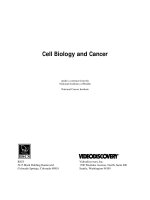
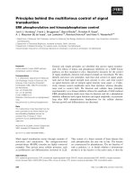
closely related to the apparent kM for sulfate of the transporters. Least square fittings (Figure 1) revealed that the
growing isotherms of the four complemented yeasts can
be properly described by single hyperbolic MichaelisMenten functions, with kG for sulfate in the micromolar
range, indicating that these proteins are high-affinity sulfate transporters; in particular the kG values were 5.46 ±
0.22 μM (BjSultr1;1), 1.74 ± 0.05 μM (BjSultr1;2a), 1.73 ±
0.07 μM (BjSultr1;2b), and 1.74 ± 0.05 μM (BjSultr1;2c).
Lancilli et al. BMC Plant Biology 2014, 14:132
/>
Page 6 of 15
Figure 1 Estimation of the growth constant (kG) dependent on sulfate of the yeasts expressing the Brassica juncea sulfate transporters.
The duplication times (dt) of the complemented yeast cells were calculated by fitting the equation A600(t) = A600(t0) ekt to the experimental data
reported in Additional file 5. kG was estimated by expressing the growth rates (dt−1) of complemented yeasts as a function of sulfate concentrations
in the media, and by fitting the Michaelis-Menten equation to the data. Data points and error bars are means and SE of two experiments run in
triplicate (n = 6).
Effect of Cd exposure and sulfate limitation on sulfate
uptake and sulfur allocation in Brassica juncea plants
All the data presented in this paragraph derived from experiments aimed at comparing environmental conditions
(Cd exposure and sulfate limitation) in which sulfate uptake induction should occur. For these purposes plants
were exposed to 10 and 25 μM Cd2+ for 2 days or grown
under sulfate limitation (50 and 10 μM SO4 2−) for a 10day period. Control plants were grown at 200 μM SO4 2−
in the absence of Cd.
Cadmium exposure neither significantly influenced
the growth of shoots and roots, nor produced any apparent symptom of stress; conversely, lowering sulfate
concentration in the growing solution significantly increased root growth without affecting shoots, as indicated by the values of the shoot/root ratio which
decreased from 3.82 to 2.20 (Table 1). The total amount
of Cd retained by roots increased as the metal concentration in the external medium did, whereas it reached
similar values in the shoots of plants grown at 10 and
25 μM Cd2+ (Table 1).
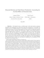
Cadmium exposure and sulfate limitation deeply affected the sulfate uptake capacity of the root, as indicated by the values of 35S-sulfate uptake, measured at
200 μM SO4 2− external concentration (Figure 2A, B). In
Cd exposed plants the rate of sulfate uptake increased
up to 0.9-fold with respect to the untreated control at
the highest Cd external concentration (25 μM). Similar
behaviors were observed in sulfur-starved plants, in which
the rate of sulfate uptake increased as the sulfate concentration in the external medium decreased, reaching
values 1.2-fold higher than in sulfur-sufficient control
(200 μM SO4 2−). These trends were closely associated
to changes in the transcript level of BjSultr1;1 and in
the cumulative amount of the three transcripts of the
BjSultr1;2 variants (BjSultr1;2 pool), which significantly
accumulated as the severity of the stresses increased
(Figure 2C, D).
Taken as a whole these preliminary results indicate that
48 h Cd exposure and 10-day sulfate limitation produced
similar induction of sulfate uptake. Since such effects
should presumably be related to changes in the sulfur
Lancilli et al. BMC Plant Biology 2014, 14:132
/>
Page 7 of 15
Table 1 Dry weight of roots and shoots and Cd accumulation
Experimental condition
Dry weight
Roots
Cd2+ content
Shoots/Roots
Shoots
Roots
Shoots
μmol g−1 DW
g
Control
0.063 ± 0.003 (a)
0.241 ± 0.010 (a)
3.82
ND
ND
10 μM Cd2+
0.069 ± 0.003 (a)
0.252 ± 0.011 (a)
3.65
25.81 ± 1.18 (a)
5.66 ± 0.25 (a)
25 μM Cd2+
0.066 ± 0.004 (a)
0.231 ± 0.012 (a)
3.50
97.33 ± 3.99 (b)
5.00 ± 0.22 (a)
50 μM SO4 2−
0.112 ± 0.005 (b)
0.251 ± 0.011 (a)
2.24
ND
ND
10 μM SO4
0.110 ± 0.005 (b)
0.243 ± 0.011 (a)
2.20
ND
ND
2−
Plants were exposed to different Cd concentrations (10 and 25 μM) for 48 h or grown under different sulfate concentrations (50 and 10 μM) for 10 days. Control
plants were grown under 200 μM SO4 2− and were not exposed to Cd. Cadmium content was measured by ICP-MS. Values are means ± SE of three experiments
run in triplicate (n = 9). Different letters indicate significant differences (P < 0.05). ND, not detectable.
nutritional status of the plants, we analyzed the levels of
NPTs, GSH and sulfate of both roots and shoots, assuming the pools of these intermediates as the main
diagnostic indicators of the sulfur nutritional status.
Cd exposure produced significant changes in the NPT
levels of the root, which progressively increased as Cd
concentration in the external medium did (Figure 3A),
whilst at the same time a decrease of the total GSH
pools was observed (about 30% with respect to the control in all analyzed conditions; Figure 3B). Such a trend
was probably related to PC biosynthesis and accumulation according to the progressive increase in Cd root
content (Table 1). The sulfate pools of the root were not
affected by Cd exposure (Figure 3C). Quite similar behaviors were observed in the shoots of Cd exposed
plants, since the NPT levels increased with Cd concentration in the external medium and the sulfate concentration was not affected by the presence of the metal;
however, a Cd-dependent increase in the GSH levels was
observed (Figure 3A, B, C).
As expected, a stepwise contraction in the levels of all
the diagnostic indicators was observed in the root of
plants grown for 10 days under sulfate limitation. Indeed, NPT, GSH and sulfate contents measured in the
Figure 2 Changes in sulfate uptake capacity of Brassica juncea roots. Plants were exposed to different Cd concentrations for 48 h (A, C) or
grown under different sulfate concentrations for 10 days (B, D). (A, B) Sulfate influxes were evaluated by measuring the rate of 35SO4 2− absorption
into roots of intact plants over a 15 min pulse. The incubation solutions contained 200 μM SO4 2−. Bars and error bars are means and SE of three
experiments run in triplicate (n = 9). Different letters indicate significant differences (P < 0.05). (C, D) Semi-quantitative RT-PCR analysis of
BjSultr1;1 and BjSultr1;2 gene expression. PCRs were carried out for 24 cycles where cDNAs were exponentially amplified. For BjSultr1;2 pool,
primers were designed on conserved sequences of the three BjSultr1;2 variants, and gave overlapping amplification products of 1046 bp. PCR
products were separated in agarose gel and stained with SYBR Green I. Signals were detected using a laser scanner with 532 nm laser and
526 nm filter. BjTub, tubulin. A representative set of data from three independent experiments is given.
Lancilli et al. BMC Plant Biology 2014, 14:132
/>
Page 8 of 15
Figure 3 Effects of Cd exposure and sulfate limitation on the sulfur nutritional status of Brassica juncea plants. Plants were exposed to
different Cd concentrations for 48 h (A, B, C) or grown under different sulfate concentrations for 10 days (D, E, F). (A, D) NPT contents of roots
(black bars) and shoots (grey bars) are expressed as GSH equivalents. (B, E) Total GSH contents of roots (black bars) and shoots (grey bars).
(C, F) Sulfate contents of roots (black bars) and shoots (grey bars). Bars and error bars are means and SE of three experiments run in triplicate
(n = 9). Different letters indicate significant differences between treatments (P < 0.05). ND, not detectable.
root tissues dramatically decreased as sulfate availability
in the external medium did (Figure 3D, E, F). Following
sulfate limitation, sulfate content of the shoot steadily
decreased, reaching the minimal value at 10 μM SO4 2−
external concentration; differently, NPT and GSH levels
did not significantly change when we lowered sulfate external concentration from 200 to 50 μM, whilst a sharp
decrease in the level of these compounds was observed
by moving toward the lowest (10 μM) sulfate concentration analyzed (Figure 3D, E, F).
We also analyzed the dynamic of root-to-shoot sulfate
translocation by measuring the concentration of the anion
in the xylem sap of Cd-exposed or sulfur-starved plants.
In these experiments, sulfate translocation was estimated
as the amount of sulfate ions loaded and transported in
the xylem sap for 1.5 h. Results indicate that the amount
of sulfate ions transported in the xylem sap progressively
increased following Cd exposure (Figure 4A); differently,
sulfate translocation increased when shifting sulfate external concentration from 200 to 50 μM, and sharply decreased when moving toward the lowest (10 μM) sulfate
concentration analyzed (Figure 4B).
Quantitative analysis of the expression of the three
BjSultr1;2 variants
Since the three BjSultr1;2 forms are not polymorphic
enough to be distinguished by means of a simple PCR
(Additional file 8), we developed a suitable method to study
changes in their expression by coupling semi-quantitative
RT-PCR analysis with the use of an opportune restriction
enzyme.
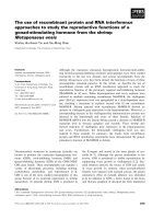
Sequence analysis revealed that the three BjSultr1;2
cDNAs have restriction site polymorphisms for the ClaI
endonuclease, which enabled us to discriminate the
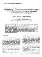
three variants after digestion. As detailed in Figure 5A:
i) BjSultr1;2a (1968 bp) is not cut by ClaI; ii) BjSultr1;2b
(1959 bp) is cut by ClaI 1752 bp downstream of the start
codon; iii) BjSultr1;2c (1959 bp) is cut twice by ClaI,
1098 and 1752 bp downstream of the start codon. As a
consequence the digestion of the cDNA clones with ClaI
produces characteristic restriction patterns with some
diagnostic bands useful to discriminate the three forms
(Figure 5B). The characteristic undigested 1968 bp band
is a diagnostic marker of BjSultr1;2a presence, the 1752 bp
fragment only results from the digestion of BjSultr1;2b,
Lancilli et al. BMC Plant Biology 2014, 14:132
/>
Page 9 of 15
to 25 μM Cd2+ or 10-day sulfate limitation (10 μM SO4 2−)
on the expression of the three BjSultr1;2 variants. A restriction analysis using the ClaI endonuclease followed the amplification reactions.
Results show that the cumulative amount of the
BjSultr1;2 transcripts in the roots was significantly
higher in Cd exposed plants than in the untreated control
ones (+217%, Figure 6A) as already shown in Figure 2C.
Such a behavior resulted from changes in the expression
of BjSultr1;2b and BjSultr1;2c only (Figure 6A). In fact,
the densitometric analysis of each diagnostic band indicated that BjSultr1;2b and BjSultr1;2c transcript levels significantly increased by 585% and 301%, respectively, whilst
the BjSultr1;2a expression was not affected by Cd exposure (Figure 6B, Additional file 9). Similar behaviors were
observed by analyzing changes in the expression pattern
of the three BjSultr1;2 forms in plants exposed for 48 h to
a lower (10 μM) Cd concentration (Additional file 10). In
fact, also in this condition the response to Cd, though to a
lesser extent, was only ascribable to specific increases in
the relative amount of BjSultr1;2b (+150%) and BjSultr1;2c
(+75%) transcript.
By contrast, in the case of sulfate limitation, the increase
in the cumulative amount of the BjSultr1;2 transcripts
(+455% with respect to the sulfur-sufficient control) was
ascribable to changes in the transcript levels of all the
three forms (Figure 6C). In particular, the BjSultr1,2a,
BjSultr1;2b, and BjSultr1;2c transcript levels significantly increased by 371%, 483%, and 618%, respectively
(Figure 6D, Additional file 9).
Figure 4 Effects of Cd exposure and sulfate limitation on sulfate
translocation in Brassica juncea plants. Plants were exposed to
different Cd concentrations for 48 h (A) or grown under different sulfate
concentrations for 10 days (B). At the end of the experimental periods,
shoots were separated from roots and the xylem sap exuded from the
cut (root side) surface was collected to be analyzed for sulfate content.
Bars and error bars are means and SE of three experiments run in
triplicate (n = 9). Different letters indicate significant differences (P < 0.05).
whilst both the 1098 and 654 bp bands are specifically
produced following the digestion of BjSultr1;2c. Finally
the 207 bp band is a digestion product shared among
BjSultr1;2b and BjSultr1;2c, and therefore does not give
any result useful for our purposes.
Starting from this rationale, we designed a couple of
primers amplifying at the same time the entire open reading frames of the three clones with the same efficiency (data
not shown), and we used these oligos for the semiquantitative RT-PCR analysis of the effects of 48 h exposure
Discussion
Brassica juncea (L.) Czern & Coss (AABB, n = 18) is
believed to have originated from the interspecific hybridization of two base “diploid” genomes provided by
Brassica rapa L. (AA; n = 10) and Brassica nigra L. (BB;
n = 8) [24,40]. Both the diploid parents are thought to
be ancient polyploids since they still exhibit highly replicated genomes, each containing three paralogous subgenomes closely related to that of Arabidopsis thaliana
[21-23]. In spite of the whole-genome triplication event –
thought to have occurred between 13 and 17 million years
ago – most comparative studies have shown that the number of each ancestral gene retained in the genome of the
modern diploid Brassica is variable, since paralogous
regions exhibit interspersed gene losses and insertions.
Interestingly, in the recently sequenced B. rapa genome
the extent of gene loss among triplicated genome segments varies, with one of the three copies consistently
retaining a disproportionately large fraction of the genes
expected to have been present in its ancestor [23]. Such
evolutionary events are thought to be the biological basis
of the immense plasticity of Brassica species and may have
led to a diversification of the genes retained in more than
Lancilli et al. BMC Plant Biology 2014, 14:132
/>
Page 10 of 15
Figure 5 Restriction analysis of the three BjSultr1;2 cDNAs. (A) The three BjSultr1;2 variants have restriction site polymorphisms for the Cla I
endonuclease. Black arrows indicate the relative position of Cla I restriction sites in each open reading frame. The expected lengths of the restriction
fragments obtained after digestion with ClaI are indicated. (B) Characteristic restriction patterns obtained from the digestion of each cDNA with ClaI.
Single cDNAs were obtained by PCR using a recombinant plasmid, containing a unique BjSultr1;2 clone, as template. u, undigested; d, digested.
one copy, in terms of function and/or expression. Searching for orthologs of the Arabidopsis high-affinity sulfate
transporter genes involved in sulfate uptake and retained
in the genome of B. rapa – one of the two parents of
B. juncea – revealed the existence of three distinct loci,
annotated as Bra022623, Bra015641 and Bra008340. The
first locus encodes for a putative high-affinity sulfate transporter closely related to Arabidopsis AtSultr1;1, whilst the
other two loci encode for two different forms of a highaffinity sulfate transporter functional related to Arabidopsis
AtSultr1;2, indicating these gene loci as paralogs. As
expected, a much more complex picture was found in
the allopolyploid B. juncea in which we were able to
identify an orthologous form of AtSultr1;1 (BjSultr1;1)
and three orthologous forms of AtSultr1;2 (BjSultr1;2a/b/c).
From the results obtained by the sequence analysis, and
in the absence of any other information so far available
about the B. nigra genome, we can reasonably suppose:
i) BjSultr1;2a as the ortholog of Bra015641 on the genome A or B of B. juncea; ii) BjSultr1;2b/c as allelic variants
orthologous of Bra008340 on the A or B genome of B.
juncea or a homeologous gene pair related to Bra008340
on the A and B genomes of B. juncea. Moreover, since
the progeny derived from self-fertilization inherited all
the three BjSultr1;2 variants (data not shown), it seems
likely to exclude that BjSultr1;2b and BjSultr1;2c would
be allelic, making plausible the hypothesis they are instead
present at different homeologous gene loci on A and B genomes, each in homozygous configuration; otherwise, a
simple mendelian segregation would be observed. In any
case, since the three BjSultr1;2 forms would share a common ancestor gene, they may either have retained their
original functions and expressions, or – as it is often the
case – have accumulated deleterious mutations or have
evolved novel gene interactions through the processes of
sub-functionalization and/or neo-functionalization [25,26].
Results of complementation tests in the yeast mutant
strain CP154-7A, defective in its two sulfate transporters
and thus unable to grow on media containing low concentrations of sulfate as the sole sulfur source [31], proved
the capacity of BjSultr1;1 and BjSultr1;2a/b/c to transport
sulfate ions across the plasma-membrane (Additional
file 4). Moreover, kinetic analysis of the growth (G) isotherms of complemented yeasts, revealed that BjSultr1;1
and BjSultr1;2a/b/c have high affinities for sulfate, as revealed by the kG values similar to the apparent kM values
of other plant high-affinity sulfate transporters [9,41-44]
indicating that all the B. juncea clones have retained
their functions. It is also worthy of note that the sulfate
transporter BjSultr1;1 has an apparent affinity for sulfate
Lancilli et al. BMC Plant Biology 2014, 14:132
/>
Page 11 of 15
Figure 6 RT-PCR analyses of the three BjSultr1;2 forms in the roots of Brassica juncea grown under Cd exposure or sulfate limitation.
Plants were exposed to 25 μM Cd2+ for 48 h (A, B) or grown under 10 μM SO4 2− for 10 days (C, D). (A, C) The entire ORFs of the three BjSultr1;2
forms were amplified and PCR products, digested (d) or not digested (u) with ClaI endonuclease, were electrophoresed on agarose gel and
stained with SYBR Green I. cDNA loading was normalized using BjTub as an internal control. Signals were detected using a laser scanner with
532 nm laser and 526 nm filter. (B, D) Densitometric analysis. Arrows indicate the relative position of each electrophoretic band obtained after
digestion of PCR products with ClaI. A representative set of data from three independent experiments is given. For statistical analysis, see
Additional file 9.
(kG = 5.46 μM) three times lower than those of the three
BjSultr1;2 forms (Figure 1). Finally, phylogenetic analyses indicate these transporters as functionally related
to AtSultr1;1 and AtSultr1;2 and then we can infer their
probable function in mediating root sulfate uptake from
the soil solution [14,43,44].
Physiological analysis reveals that Cd exposure as well
as sulfate limitation induces sulfate uptake in B. juncea
roots. Such behaviors seem to be related to the induction of high-affinity sulfate transporters belonging to the
group I, as indicated by the increase in the transcript
levels of BjSultr1;1 and BjSultr1;2 pool (Figure 2). Thus –
at the physiological level – B. juncea also retains the typical responses of sulfate uptake to Cd or sulfur shortage
[9,15]. The apparent quantitative discrepancy between the
changes in the transcript levels of BjSultr1;1 and BjSultr1;2
pool and the resulting increases in sulfate uptake may be
due to additional regulatory mechanisms working in parallel with the transcriptional control of the high-affinity sulfate transporter genes [45].
Considering the current model of demand-driven regulation of sulfate uptake, such inductions should be related
to the sulfur nutritional status reached by plants in
the two growing conditions. Changes in the amounts
of sulfur-containing compounds that we assume as the
main diagnostic indicators of the sulfur nutritional status
of root and shoot clearly indicate that Cd exposure and
sulfate limitation influence sulfur allocation throughout
the whole plant, generating deeply different local nutritional states which make it difficult to individuate an unequivocal and common nutritional signal related to the
expression of sulfate transporters (Figure 3). Indeed, as
expected, lowering sulfate concentration in the external
medium necessarily results in a significant contraction
of all the analyzed sulfur pools of the roots, whilst Cd
stress produces a typical increase in the level of NPTs,
probably due to the activation of GSH-dependent PC
biosynthesis, without affecting the sulfate content of
the roots.
Root responses to Cd exposure seem to be due to
homeostatic mechanisms driven by increases in the sulfur need of the plants, since, as previously reported [9],
the effect of Cd on sulfate uptake capacity is closely related to the NTP levels of both root and shoot and then
to the strength of the Cd-induced additional sink for thiols
(Additional file 11). Under Cd exposure the NPT levels in
plant tissues significantly increase, reaching values 8.5
(root) and 1.3 (shoot) fold higher than in the control at
the highest concentration analyzed. Since the sulfate pools
of both root and shoot seem not to be affected by Cd
Lancilli et al. BMC Plant Biology 2014, 14:132
/>
exposure, it appears clear that the additional sulfur required to sustain thiol biosynthesis necessarily derives from
the activation of sulfate uptake. On the other hand, root responses to sulfate shortage appear likely to be dependent
on the need for allocating the limiting nutrient in the best
and most efficient way. In fact, the stepwise decrease in the
sulfate content of the roots seems to be related not only to
a decrease in sulfate availability, but also to a transient activation of sulfate translocation making shoots less sensitive
to sulfate deficiency (Figure 4B) [46]. Noticeably, Cd exposure neither decreases root sulfate content (Figure 3C) nor
inhibits root sulfate uptake (Figure 2A) and sulfate transporter gene expression (Figure 2C), but rather significantly
enhances sulfate translocation (Figure 4A) and its metabolism as shown by the significant increases in the GSH levels
of the shoot (Figure 3B). Such an effect should be related
to the activation of both PC biosynthesis and mechanisms
involved in controlling oxidative damage due to the accumulation of free Cd ions in the leaves [47,48]. Moreover,
from these results we can also speculate that the overaccumulation of GSH in the shoot could help roots in detoxifying Cd through reallocating mechanisms involving
phloem translocation, as previously reported in other species [49,50]. In this way, the excess of sulfate taken up by
roots would partly bypass root assimilation to be directly
metabolized in organs less affected by Cd stress, without
however affecting the root sulfate pool (Figure 3C).
Taken as a whole our data clearly show that dissimilar
nutritional and metabolic states may result in quite similar
responses in sulfate uptake, suggesting that multi-signalling
pathways may control the expression of the high-affinity
sulfate transporters of the roots. Moreover, the fact that the
negative relationships between the levels of nutritional signals (sulfate and GSH) and sulfate uptake capacity of the
roots, existing in sulfur-starved plants, are not found in the
Cd exposed ones, seems to further support this conclusion.
Although it is difficult to indicate unambiguous nutritional signals, we can make some educated guesses on
the general structure of the hypothetical signaling pathways involved in the modulation of sulfate uptake by
analyzing the expression pattern of the three BjSultr1;2
forms under Cd exposure and sulfate limitation. Since
BjSultr1;2a seems to have lost its capacity to respond to
Cd stress, but, at the same time, retains its response to
sulfate shortage, we can speculate about the existence of
at least two distinct signal transduction pathways. The
first one modulates root sulfate uptake, ensuring adequate nutrient supply when plants experience lowering
in the sulfate concentration of the soil solution, probably
through a cis-acting sulfur responsive element as previously suggested [19], whilst the second one we postulate
is likely to be involved in meeting sulfate uptake with
the plant metabolic sulfur demand, which may increase
following heavy-metal stress. All the three BjSultr1;2 forms
Page 12 of 15
are controlled by the first pathway as indicated by the analysis of their respective contribution to the increase in the
cumulative amount of the BjSultr1;2 transcripts under sulfur starvation, but only two forms (BjSultr1;2b/c) seem to
have retained the ancient characteristic to be controlled by
the second regulatory pathway. In this context the differential transcriptional behaviors of the three BjSultr1;2 forms
could be explained by hypothesizing the presence of both
“cadmium-” and “sulfur-sensitive” regions in the promoter
of an ancient Brassica Sultr1;2 form, which – following
polyploidization – may have evolved in sub-functionalized
forms, whose combined actions result in molecular and
physiological responses to Cd exposure and sulfate limitation similar to those known in species with non-redundant
genome [8,9,15]. If this were not the case an interference
of the metal with the signal transduction pathways involved in the regulation of sulfate uptake should be postulated, as suggested in the recent paper of Shahbaz and
co-workers [51], in which they extensively discuss the
effect of copper accumulation on sulfur metabolism-related
gene expression. However, copper stress in Brassica seems
to produce significant increases in both sulfate uptake and
tissue sulfate content without substantially altering plant
sulfur demand [52], differently from Cd exposure which
produces increases in sulfate uptake closely related to
the strength of the additional sink for thiols it induces
(Additional file 11). Moreover, if any sort of Cd interference occurs we should also suppose the existence of at
least two signal transduction pathways controlling the
expression of BjSultr1;2a/b/c under Cd exposure: the
first inhibited by Cd, and the second Cd insensitive. Finally, we cannot exclude that other molecular mechanisms
may be involved in the differential expression of the three
BjSultr1;2 forms under Cd stress, as for instance those
suggested for rice phosphate transporters [53]; it could be
interesting to investigate if the short 9 bp-insertion located at the 5′ end of the BjSultr1;2a coding sequence
(Additional file 8) can play a role in the regulation of its
expression.
Conclusions
Taken as a whole our data agree with the main molecular and physiological evidence obtained in Arabidopsis
which support the idea that the regulation of AtSultr1;1
and AtSultr1;2 – the two transporters mediating sulfate
uptake from the soil solution – must necessarily involve
independent signaling pathways, as extensively shown by
Rouached and co-workers [15,54]. Moreover, we can also
conclude that different sulfur nutritional and metabolic
conditions may be perceived by a single sulfate transporter gene. Such a finding reveals that the mechanisms
involved in sulfate uptake regulation may be more complex than previously thought, and partially accounts for
the lack of unambiguous nutritional signals, since the
Lancilli et al. BMC Plant Biology 2014, 14:132
/>
activity of each transporter may result from a complex
interplay among multiple regulatory pathways.
Additional files
Additional file 1: Dendrogram showing sulfate transporter family
of Arabidopsis thaliana and high-affinity sulfate transporters of
Brassica juncea. The dendrogram was constructed on the bases of
amino acid sequences using MEGA 5.05 software. Accession numbers for
A. thaliana (TAIR; are: AtSultr1;1, At4g08620;
AtSultr1;2, At1g78000; AtSultr1;3, At1g22150; AtSultr2;1, At5g10180;
AtSultr2;2, At1g77990; AtSultr3;1, At3g51895; AtSultr3;2, At4g02700;
AtSultr3;3, At1g23090; AtSultr3;4, At3g15990; AtSultr3;5, At5g19600;
AtSultr4;1, At5g13550; AtSultr4;2, At3g12520. Accession numbers for B.
juncea (GenBank; are: BjSultr1;1,
JX896426; BjSultr1;2a, JX896427; BjSultr1;2b, JX896428; BjSultr1;2c,
JX896429.
Additional file 2: Nucleotide identity (%) between the coding
sequences of the three BjSultr1;2 variants and other Brassica
Sultr1;2 coding sequences.
Additional file 3: Dendrogram showing high affinity sulfate
transporters of Arabidopsis thaliana, Brassica juncea, Brassica napus,
and Brassica rapa. The dendrogram was constructed on the bases of
amino acid sequences using MEGA 5.05 software. Accession numbers for
A. thaliana (TAIR; are: AtSultr1;1, At4g08620;
AtSultr1;2, At1g78000. Accession numbers for B. juncea and B. napus
(GenBank; are: BjSultr1;1, JX896426;
BjSultr1;2a, JX896427; BjSultr1;2b, JX896428; BjSultr1;2c, JX896429; BnSultr1;1,
AJ416460; BnSultr1;2, AJ311388. Accession numbers for B. rapa (BRAD;
are: Bra022623; Bra015641; Bra008340.
Additional file 4: Phenotypic complementation of the yeast double
sulfate transporter mutant CP154-7A by the sulfate transporters of
Brassica juncea. Yeast mutant cells expressing BjSultr1;1, BjSultr1;2a,
BjSultr1;2b, and BjSultr1;2c under the control of the galactose-inducible
GAL10 promoter or harboring the empty pESC-TRP vector were grown at
28°C for 3 d on a minus-sulfur minimal medium (−S) or on minimal
media containing 100 μM sulfate (SO4 2−) or 100 μM DL-homocysteine
(HCys) as sole sulfur sources.
Additional file 5: Growth curves of complemented yeast cells. (A, B,
C, D) Complemented yeasts were incubated at 28°C for 25 h in liquid
media containing different sulfate concentrations (● 0 μM; ○ 1 μM; ▼
2.5 μM; Δ 5 μM; ■ 7.5 μM; □ 10 μM; ◆ 25 μM; ◇ 50 μM; ▲ 100 μM) or
100 μM HCys (▽) as sole sulfur source. Absorbance was measured at
600 nm (A600) along time. Data points and error bars are means and SE
of two experiments performed in triplicate (n = 6).
Additional file 6: Sulfate content in complemented yeast cells.
Complemented yeast cells expressing BjSultr1;1, BjSultr1;2a, BjSultr1;2b,
and BjSultr1;2c were incubated in liquid media containing different
sulfate concentrations as sole sulfur source. At the end of the incubation
period yeasts were harvested and processed to determine their sulfate
content. Values are means ± SE of two experiments run in triplicate (n = 6).
Additional file 7: Growth analysis of CP154-7A yeast mutant
expressing an high- or a low-affinity sulfate transporter of Zea mays
(ZmST1;1) or Arabidopsis thaliana (AtSultr2;1), respectively. ZmST1;1
and AtSultr2;1 coding sequences were amplified by RT-PCR from total
RNA isolated from maize and Arabidopsis roots, respectively, and cloned
in the pESC-TRP vector as described in Methods. (A) Complemented yeast
cells were incubated in liquid media containing two sulfate concentrations
(0.1 or 0.5 mM) as sole sulfur source. Yeast growth was monitored by
measuring the A600 nm at different times. (●) ZmST1;1 at 0.1 mM sulfate;
(○) ZmST1;1 at 0.5 mM sulfate; (▼) AtSultr2;1 at 0.1 mM sulfate; (▽)
AtSultr2;1 at 0.5 mM sulfate. (B) Growth curves of yeast cells expressing
ZmST1;1 (● 0 μM; ○ 1 μM; ▼ 2.5 μM; △ 5 μM; ■ 7.5 μM; □ 10 μM; ◆ 25 μM;
◇ 50 μM; ▲ 100 μM). (C) Growth curves of yeast cells expressing AtSultr2;1
(● 0 μM; ○ 0.05 mM; ▼ 0.1 mM; △ 0.15 mM; ■ 0.2 mM; □ 0.25 mM; ◆ 0.5
mM; ◇ 1 mM; ▲ 1.5 mM; ▽ 2 mM; 2.5 mM; 3 mM). (D, E) Estimation
of the growth constant (kG) for sulfate. The duplication times (dt) of the
Page 13 of 15
complemented yeast cells were calculated by fitting the equation A600(t) =
A600(t0) ekt to the experimental data reported in B and C. kG was determined
by expressing the growth rates (dt-1) of complemented yeasts as a function
of sulfate concentrations in the media, and by fitting the Michaelis-Menten
equation to the data. Results reveal that the kG values for sulfate were similar
to the kM values measured for each sulfate transporters by using conventional
methods (Nocito et al. Plant Physiol, 2006 141:1138-1148; Takahashi et al. Plant
J, 2000 23:171-182). Data points and error bars are means and SE of two
experiments performed in triplicate (n = 6).
Additional file 8: Alignment of nucleotide sequences of the three
BjSultr1;2 forms. Shared nucleotides are highlighted in grey.
Additional file 9: Changes in the transcript relative amount of the
three BjSultr1;2 forms in the roots of Brassica juncea grown under
Cd exposure or sulfate limitation. Plants were exposed to 25 μM Cd2+
for 48 h (+Cd) or grown under 10 μM SO4 2− for 10 days (−S). Control
plants were grown under 200 μM SO4 2− and were not exposed to
cadmium. The entire ORFs of the three BjSultr1;2 forms were amplified
and PCR products were digested with ClaI endonuclease, electrophoresed on
agarose gel, and finally stained with SYBR Green I. Signals were detected using
a laser scanner with 532 nm laser and 526 nm filter and densitometrically
analyzed using ImageJ 1.46 software. cDNA loading was normalized using
BjTub as an internal control. Bars and error bars are means and SE of three
independent experiments run in triplicate (n = 9). Asterisks indicate significant
differences between control and treated plants (P ≤ 0.001).
Additional file 10: RT-PCR analyses of the three BjSultr1;2 forms in
the roots of Brassica juncea exposed to 10 μM Cd. Plants were
exposed or not to 10 μM Cd2+ for 48 h. (A) The entire ORFs of the three
BjSultr1;2 forms were amplified and PCR products were digested with ClaI
endonuclease, electrophoresed on agarose gel, and finally stained with
SYBR Green I. cDNA loading was normalized using BjTub as an internal
control. Signals were detected using a laser scanner with 532 nm laser
and 526 nm filter. A representative set of data from three independent
experiments is given. (B) Densitometric analysis. Arrows indicate the
relative position of each electrophoretic band obtained after digestion of
PCR products with ClaI. (C) Statistical analysis. Bars and error bars are
means and SE of three independent experiments run in triplicate (n = 9).
Asterisks indicate significant differences between control and treated
plants (P ≤ 0.001).
Additional file 11: Relationship between NPT content and sulfate
uptake capacity in plant of Brassica juncea exposed to different Cd
concentrations. Plants were exposed for 48 h to differentCd2+
concentrations: 0 (white), 10 (grey), and 25 (black) μM. Circles, roots;
triangles, shoots. Data points and error bars are means and SE of three
experiments run in triplicate (n = 9).
Abbreviations
Cd: Cadmium; Cys: Cysteine; GSH: Glutathione; HCys: DL-homocysteine;
HEPES: 4-(2-HydroxyEthyl)-1-PiperazineEthaneSulfonic acid; ICP-MS: Inductively
coupled plasma-mass spectrometry; Met: Methionine; NPT: NonProtein thiol;
OAS: O-acetylserine; ORF: Open reading frame; PC: Phytochelatin;
PCR: Polymerase chain reaction; RT-PCR: Reverse transcription - PCR;
SE: Standard error of the mean.
Competing interests
The authors declare that they have no competing interests.
Authors’ contributions
CL and FFN conceived and designed the experiments and wrote the
manuscript. CL and BG carried out the physiological and molecular analyses.
BG, FFN, MC, and JCD conceived and performed the experiments with yeast.
GL performed ICP-MS analysis. GAS acquired the funds. CL, JCD, MC, GAS,
and FFN discussed and critical revised the manuscript. All authors read and
approved the final manuscript.
Acknowledgements
This work was supported by the Italian Ministry of Education, University, and
Research - PRIN 2009. We would like to thank Alessandro Ferri for its precious
support during the revision of the manuscript.
Lancilli et al. BMC Plant Biology 2014, 14:132
/>
Author details
1
Dipartimento di Scienze Agrarie e Ambientali – Produzione, Territorio,
Agroenergia, Università degli Studi di Milano, 20133 Milano, Italy. 2Biochimie
et Physiologie Moléculaire des Plantes, Unité mixte de recherche, Montpellier
SupAgro (Département Biologie et Ecologie), INRA, CNRS, Université de
Montpellier 2, 34060 Montpelliercedex 2, France.
Received: 1 February 2014 Accepted: 6 May 2014
Published: 16 May 2014
References
1. Leustek T, Martin MN, Bick JA, Davies JP: Pathways and regulation of sulfur
metabolism revealed through molecular and genetic studies. Annu Rev
Plant Phys 2000, 51:141–165.
2. Takahashi H, Kopriva S, Giordano M, Saito K, Hell R: Sulfur assimilation in
photosynthetic organisms: molecular functions and regulations of
transporters and assimilatory enzymes. Annu Rev Plant Biol 2011,
62:157–184.
3. Cram W, Rennenberg H, Brunold C, De Kok LJ, Stulen I: Uptake and
transport of sulfate. In Sulfur Nutrition and Sulfur Assimilation in Higher
Plants: Fundamental, Environmental and Agricultural Aspects. The Hague:
SPB Academic Publishing; 1990:3–11.
4. Clarkson D, Hawkesford M, Davidian J-C: Membrane and long-distance
transport of sulfate. In Sulfur Nutrition and Sulfur Assimilation in Higher
Plants: Fundamental, Environmental and Agricultural Aspects. Edited by De
Kok LJ, Stulen I, Rennenberg H, Brunold C, Rauser WE. The Hague: SPB
Academic Publishing; 1993:3–19.
5. Saito K: Sulfur assimilatory metabolism. The long and smelling road. Plant
Physiol 2004, 136:2443–2450.
6. Rausch T, Wachter A: Sulfur metabolism: a versatile platform for
launching defence operations. Trends Plant Sci 2005, 10:503–509.
7. Lee S, Leustek T: The effect of cadmium on sulfate assimilation enzymes
in Brassica juncea. Plant Sci 1999, 141:201–207.
8. Nocito FF, Pirovano L, Cocucci M, Sacchi GA: Cadmium-induced sulfate
uptake in maize roots. Plant Physiol 2002, 129:1872–1879.
9. Nocito FF, Lancilli C, Crema B, Fourcroy P, Davidian J-C, Sacchi GA: Heavy
metal stress and sulfate uptake in maize roots. Plant Physiol 2006,
141:1138–1148.
10. Nocito FF, Espen L, Crema B, Cocucci M, Sacchi GA: Cadmium induces
acidosis in maize root cells. New Phytol 2008, 179:700–711.
11. Noctor G, Mhamdi A, Chaouch S, Han Y, Neukermans J, Marquez-Garcia B,
Queval G, Foyer CH: Glutathione in plants: an integrated overview. Plant
Cell Environ 2012, 35:454–484.
12. Lappartient AG, Touraine B: Demand-driven control of root ATP sulfurylase
activity and SO2−
4 uptake in intact canola. The role of phloem-translocated
glutathione. Plant Physiol 1996, 111:147–157.
13. Lappartient AG, Vidmar JJ, Leustek T, Glass ADM, Touraine B: Inter-organ
signaling in plants: regulation of ATP sulfurylase and sulfate transporter
genes expression in roots mediated by phloem-translocated compound.
Plant J 1999, 18:89–95.
14. Davidian J-C, Kopriva S: Regulation of sulfate uptake and assimilation - the
same or not the same? Mol Plant 2010, 3:314–325.
15. Rouached H, Wirtz M, Alary R, Hell R, Arpat AB, Davidian J-C, Fourcroy P,
Berthomieu P: Differential regulation of the expression of two high-affinity
sulfate transporters, SULTR1.1 and SULTR1.2, in Arabidopsis. Plant Physiol
2008, 147:897–911.
16. Hawkesford MJ: Plant responses to sulphur deficiency and the genetic
manipulation of sulphate transporters to improve S-utilization efficiency.
J Exp Bot 2000, 51:131–138.
17. Jobe TO, Sung D-Y, Akmakjian G, Pham A, Komives EA, Mendoza-Cózatl DG,
Schroeder JI: Feedback inhibition by thiols outranks glutathione depletion: a
luciferase-based screen reveals glutathione-deficient γ-ECS and glutathione
synthetase mutants impaired in cadmium-induced sulfate assimilation.
Plant J 2012, 70:783–795.
18. Buchner P, Prosser IM, Hawkesford MJ: Phylogeny and expression of
paralogous and orthologous sulphate transporter genes in diploid and
hexaploid wheats. Genome 2004, 47:526–534.
19. Maruyama-Nakashita A, Nakamura Y, Watanabe-Takahashi A, Inoue E, Yamaya T,
Takahashi H: Identification of a novel cis-acting element conferring sulfur
deficiency response in Arabidopsis roots. Plant J 2005, 42:305–314.
Page 14 of 15
20. Maruyama-Nakashita A, Nakamura Y, Tohge T, Saito K, Takahashi H:
Arabidopsis SLIM1 is a central transcriptional regulator of plant sulfur
response and metabolism. Plant Cell 2006, 18:3235–3251.
21. Lysak MA, Koch MA, Pecinka A, Schubert I: Chromosome triplication found
across the tribe Brassiceae. Genome Res 2005, 15:516–525.
22. Lysak MA, Cheung K, Kitschke M, Bureš P: Ancestral chromosomal blocks
are triplicated in Brassiceae species with varying chromosome number
and genome size. Plant Physiol 2007, 145:402–410.
23. Wang X, Wang H, Wang J, Sun R, Wu J, Liu S, Bai Y, Mun JH, Bancroft I,
Cheng F, Huang S, Li X, Hua W, Wang J, Wang X, Freeling M, Pires JC,
Paterson AH, Chalhoub B, Wang B, Hayward A, Sharpe AG, Park B-S,
Weisshaar B, Liu B, Li B, Liu B, Tong C, Song C, Duran C, et al: The genome
of the mesopolyploid crop species Brassica rapa. Nature Genet 2011,
43:1035–1039.
24. U N: Genome analysis in Brassica with special reference to the
experimental formation of B. napus and peculiar mode of fertilization.
Jpn J Bot 1935, 7:389–452.
25. Lynch M, Force A: The probability of duplicate gene preservation by
subfunctionalization. Genetics 2000, 154:459–473.
26. He X, Zhang J: Rapid subfunctionalization accompanied by prolonged
and substantial neofunctionalization in duplicate gene evolution.
Genetics 2005, 169:1157–1164.
27. Rauser WE: Compartmental efflux analysis and removal of extracellular
cadmium from roots. Plant Physiol 1987, 85:62–65.
28. Tamura K, Peterson D, Peterson N, Stecher G, Nei M, Kumar S: MEGA5:
Molecular evolutionary genetics analysis using maximum likelihood,
evolutionary distance, and maximum parsimony methods. Mol Biol Evol
2011, 28:2731–2739.
29. Schneider CA, Rasband WS, Eliceiri KW: NIH Image to ImageJ: 25 years of
image analysis. Nat Methods 2012, 9:671–675.
30. Hamilton R, Watanabe CK, de Boer HA: Compilation and comparison of
the sequence context around the AUG startcodons in Saccharomyces
cerevisiae mRNAs. Nucleic Acids Res 1987, 15:3581–3593.
31. Cherest H, Davidian JC, Thomas D, Benes V, Ansorge W, Surdin-Kerjan Y:
Molecular characterization of two high affinity sulfate transporters in
Saccharomyces cerevisiae. Genetics 1997, 145:627–635.
32. Gietz D, Jean AS, Woods RA, Schiestl RH: Improved method for high
efficiency transformation of intact yeast cells. Nucleic Acids Res 1992,
20:1425.
33. Cherest H, Surdin-Kerjan Y: Genetic analysis of a new mutation conferring
cysteine auxotrophy in Saccharomyces cerevisiae: updating of the sulfur
metabolism pathway. Genetics 1992, 130:51–58.
34. Tabatabai MA, Bremner JM: A simple turbidimetric method of
determining total sulfur in plant material. Agron J 1970, 62:805–806.
35. Nocito FF, Lancilli C, Dendena B, Lucchini G, Sacchi GA: Cadmium retention
in rice roots is influenced by cadmium availability, chelation and
translocation. Plant Cell Environ 2011, 34:994–1008.
36. Griffith OW: Determination of glutathione and glutathione disulfide
using glutathione reductase and 2-vinylpyridine. Anal Biochem 1980,
106:207–212.
37. Hawkesford MJ: Transporter gene families in plants: the sulphate
transporter gene family - redundancy or specialization? Physiol Plantarum
2003, 117:155–163.
38. Hawkesford MJ, De Kok LJ: Managing sulphur metabolism in plants.
Plant Cell Environ 2006, 29:382–395.
39. Takahashi H, Buchner P, Yoshimoto N, Hawkesford MJ, Shiu S-H: Evolutionary
relationships and functional diversity of plant sulfate transporters. Front
Plant Sci 2012, 2:119.
40. Pradhan AK, Pental D: Genetics of Brassica juncea. In Genetics and
Genomics of the Brassicaceae. Edited by Bancroft I, Schmidt R. New York
Dordrecht Heidelberg London: Springer; 2011:323–345.
41. Smith FW, Ealing PM, Hawkesford MJ, Clarkson DT: Plant members of a
family of sulfate transporters reveal functional subtypes. P Natl Acad Sci
USA 1995, 92:9373–9377.
42. Smith FW, Hawkesford MJ, Ealing PM, Clarkson DT, Vanden Berg PJ, Belcher AR,
Warrilow AGS: Regulation of expression of a cDNA from barley roots
encoding a high affinity sulphate transporter. Plant J 1997, 12:875–884.
43. Takahashi H, Watanabe-Takahashi A, Smith FW, Blake-Kalff M, Hawkesford MJ,
Saito K: The roles of three functional sulphate transporters involved in
uptake and translocation of sulphate in Arabidopsis thaliana. Plant J 2000,
23:171–182.
Lancilli et al. BMC Plant Biology 2014, 14:132
/>
Page 15 of 15
44. Yoshimoto N, Takahashi H, Smith FW, Yamaya T, Saito K: Two distinct
high-affinity sulfate transporters with different inducibilities mediate
uptake of sulfate in Arabidopsis roots. Plant J 2002, 29:465–473.
45. Yoshimoto N, Inoue E, Watanabe-Takahashi A, Saito K, Takahashi H:
Posttranscriptional regulation of high-affinity sulfate transporters in
Arabidopsis by sulfur nutrition. Plant Physiol 2007, 145:378–388.
46. Kataoka T, Hayashi N, Yamaya T, Takahashi H: Root-to-shoot transport of
sulfate in Arabidopsis. Evidence for the role of SULTR3;5 as a component
of low-affinity sulfate transport system in the root vasculature. Plant
Physiol 2004, 136:4198–4204.
47. Nocito FF, Lancilli C, Giacomini B, Sacchi GA: Sulfur metabolism and
cadmium stress in higher plants. Plant Stress 2007, 1:142–156.
48. Jozefczak M, Remans T, Vangronsveld J, Cuypers A: Glutathione is a key
player in metal-induced oxidative stress defenses. Int J Mol Sci 2012,
13:3145–3175.
49. Mendoza-Cózatl DG, Jobe TO, Hauser F, Schroeder JI: Long-distance
transport, vacuolar sequestration, tolerance, and transcriptional
responses induced by cadmium and arsenic. Curr Opin Plant Biol 2011,
14:554–562.
50. Li Y, Dankher OP, Carreira L, Smith AP, Meaghe RB: The shoot-specific
expression of γ-glutamylcysteine synthetase directs the long-distance
transport of thiol-peptides to roots conferring tolerance to mercury and
arsenic. Plant Physiol 2006, 141:288–298.
51. Shahbaz M, Stuiver CEE, Posthumus FS, Parmar S, Hawkesford MJ, De Kok LJ:
Copper toxicity in Chinese cabbage is not influenced by plant sulphur
status, but affects sulphur metabolism-related gene expression and the
suggested regulatory metabolites. Plant Biology 2014, 16:68–78.
52. Shahbaz M, Tseng MH, Stuiver CEE, Koralewska A, Posthumus FS, Venema JH,
Saroj P, Schat H, Hawkesford MJ, De Kok LJ: Copper exposure interferes with
the regulation of the uptake, distribution and metabolism of sulfate in
Chinese cabbage. J Plant Physiol 2010, 167:438–446.
53. Secco D, Baumann A, Poirier Y: Characterization of the rice PHO1 gene
family reveals a key role for OsPHO1;2 in phosphate homeostasis and
the evolution of a distinct clade in dicotyledons. Plant Physiol 2010,
152:1693–1704.
54. Rouached H, Secco D, Bulak Arpat A: Getting the most sulfate from soil:
regulation of sulfate uptake transporters in Arabidopsis. J Plant Physiol
2009, 166:893–902.
doi:10.1186/1471-2229-14-132
Cite this article as: Lancilli et al.: Cadmium exposure and sulfate
limitation reveal differences in the transcriptional control of three
sulfate transporter (Sultr1;2) genes in Brassica juncea. BMC Plant Biology
2014 14:132.
Submit your next manuscript to BioMed Central
and take full advantage of:
• Convenient online submission
• Thorough peer review
• No space constraints or color figure charges
• Immediate publication on acceptance
• Inclusion in PubMed, CAS, Scopus and Google Scholar
• Research which is freely available for redistribution
Submit your manuscript at
www.biomedcentral.com/submit