A prospective study on the epidemiology of onychomycosis in tertiary care hospital
Bạn đang xem bản rút gọn của tài liệu. Xem và tải ngay bản đầy đủ của tài liệu tại đây (176.67 KB, 6 trang )
Int.J.Curr.Microbiol.App.Sci (2018) 7(8): 3765-3770
International Journal of Current Microbiology and Applied Sciences
ISSN: 2319-7706 Volume 7 Number 08 (2018)
Journal homepage:
Original Research Article
/>
A Prospective Study on the Epidemiology of Onychomycosis
in Tertiary Care Hospital
Vinay Hajare1*, G.P. Aaftab2 and Abdul Hadi Waseem3
Ram Mandir, Shahabazar, Gulbarga 585101, India
*Corresponding author
ABSTRACT
Keywords
Onychomycosis,
Dermatophytes,
Trichophyton.
Article Info
Accepted:
20 July 2018
Available Online:
10 August 2018
Fungal infection of nails or onychomycosis is non-life threatening disease commonly
caused by dermatophytes. The infection is also caused by non dermatophytes like yeasts
and non dermatophytic moulds. There are various factors which play an important role in
causation of onychomycosis. These predisposing factors are aging, fall in the immune
status, diabetes, immunosuppressive therapy for cancer and organ transplantation, HIV,
long term antibiotics, occlusive footwear, immune deficiency diseases and occupations
involving continuous contact with water, for instance swimmers, fishermen, clothes and
dish washers. Climatic conditions also play an important role in the causation of
onychomycosis. The present study was carried out in a tertiary care hospital for a period of
8 months. The aim of the study was to determine various predisposing factors and
causative agents of onychomycosis. The sample was placed in a sterile petridish and
transported to microbiology laboratory. The sample was then divided into two parts, one
for direct microscopy under high power objective using 20-25% KOH and the other part
for culture on Sabouraud’s dextrose agar (SDA) with cyclohexamide. The cultures were
kept at 25°C and 37°C for up to six weeks. Confirmation of the organism was done based
on morphology of fungus in LPCB (Lactose phenol cotton blue) mount, culture of fungus
on SDA and slide culture. Among the 68 patients selected based on clinical presentation,
26 yielded fungal pathogens in culture. A total of 15 (57.6%) isolates were dermatophytes
and 11 (42.3%) were non dermatophytes. Among the dermatophytes, 7 (26.9%) cases
yielded Trichophyton which was the most commonly isolated fungus followed by
Microsporum 5 (19.2%), Epidermophyton 3 (11.5%). Among the non dermatophytes,
candida was isolated from 3 (11.5%) cases, Aspergillus was isolated from 2 (7.6%),
Pyrenochaeta from 2 (7.6%) cases, Curvularia from 2 (7.6%) cases and only 1 (3.8%)
case yielded Fusarium. It was seen that males were more prone to onychomycosis
compared to females. Incidence of toe nail onychomycosis was higher compared to finger
nail onychomycosis. This study suggests that the isolation of the organism with culture is
very important as it will aid the clinician to rule out bacterial causes and choose
appropriate antifungal therapy.
Introduction
Fungal infection of nails or onychomycosis is
non-life threatening disease commonly caused
by dermatophytes. The infection is also caused
by non dermatophytes like yeasts and non
dermatophytic moulds. There are various
factors which play an important role in
3765
Int.J.Curr.Microbiol.App.Sci (2018) 7(8): 3765-3770
causation
of
onychomycosis.
These
predisposing factors are, aging, fall in the
immune status, diabetes, immunosuppressive
therapy for cancer and organ transplantation,
HIV, long term antibiotics, wearing of
occlusive footwear, immune deficiency
diseases and occupations involving continuous
contact with water, for instance swimmers,
fishermen, clothes and dish washers Kaur et
al., (2007).Climatic conditions also favour
onychomycosis. It was concluded that the
prevalence of onychomycosis was low in
tropical countries (3.8%) than in subtropical
and temperate zones (18%) (Bramono et al.,
2001).
Although onychomycosis is merely a cosmetic
problem, it can cause a more serious health
problem
in
HIV
infected
patients.
Onychomycosis in non immunocompromised
patients can cause negative effects like social
and
emotional
embarrassment,
nonwillingness to let their hands and feet to be
seen and patients may fear that they might
transmit the infection to their family members,
relatives
and
co-workers.
Differential
diagnosis to onychomycosis infection includes
psoriasis, lichen planus, onychogryphosis and
nail trauma. Onychomycosis represents upto
20% of nail disorders (Charif et al., 1997;
Bronson et al., 1983). The prolonged therapy
with its adverse effects may discourage the
patients.
The dermatophyte Trichophyton rubrum is the
major cause of onychomycosis (Charif et al.,
1997). The second most commonly isolated
fungal pathogen from onychomycosis patients
is the dermatophyte Trichophyton tonsurans
(Bronsonet et al., 1983). Other dermatophytes
causing onychomycosis are Trichophyton
mentagrophytes,
Trichophyton
megninii,
Trichophyton schoenleinii, Microsporum
gypseum and Epidermophyton floccosum. Non
dermatophytic fungi like Fusarium oxysporum
(Zaias
et
al.,
1972),
Scytalidium,
Scopulariopsis,
Candida,
Acremonium,
Fusarium solani, Aspergillus, Arachnomyces,
Pyrenochaeta unguis hominis have also been
isolated from cases of onychomycosis.
Classification of onychomycosis
According to the clinical presentation and the
route of invasion, onychomycosis can be
classified into four types.
1)
Distal
lateral
subungual
onychomycosis
(DLSO):
This
is
characterised by invasion of the nail bed and
the underside of the nail plate, beginning at
the
hyponychium
and
leading
to
hyperkeratosis
or
onycholysis
with
thickening of the subungual region. The nail
may appear yellowish brown in colour
(Cohen et al., 1992).
2)
Proximal subungual onychomycosis
(PSO): also known as proximal white
subungual onychomycosis is a condition
where the organism invades the nail from the
proximal nail fold through the cuticle area. It
may present with hyperkeratosis, proximal
onycholysis, leukonychia and destruction of
the proximal nail plate, involving all the
layers of the nail (Dompmartin et al., 1990).
3)
White superficial onychomycosis
(WSO): which occurs when the fungi
invades the superficial layer of the nail plate
leading to formation of opaque white patches
on the external nail plate which coalesce and
spreads as the disease progresses finally
causing the nail to become rough, soft and
crumbly (Cohen et al., 1992).
4)
Candida infection of the nail: In this
condition the organism invades the entire
nail plate causing onycholysis and
paronychia. Candida infection is more
commonly seen in women than in men
(Andre et al., 1987) and over the middle
3766
Int.J.Curr.Microbiol.App.Sci (2018) 7(8): 3765-3770
finger of women which frequently comes in
contact with the organism residing in the
vagina or intestine (Zaias et al., 1996).
The present study was carried out in a tertiary
care hospital for a period of 8 months. The
aim of the study was to determine various
predisposing factors and causative agents of
onychomycosis.
et al., 2018). Urease test and India Ink staining
was performed to differentiate candida from
Cryptococcus as Cryptococcus shows positive
reaction for urease test and it is a capsulated
organism unlike candida which is noncapsulated and shows negative reaction for
urease test. The capsule can be demonstrated
by negative staining with India ink or Nigrosin
(Jagdish Chander, 2017).
Materials and Methods
Results and Discussion
Inclusion criteria: Patients presenting with
distal subungual onychomycosis, proximal
subungual onychomycosis, white superficial
onychomycosis, paronychia, onycholysis,
hyperkeratosis, yellowish brown discoloration
and dystrophy were selected for the study.
Based on the clinical presentation 68 patients
were selected among which fungus was
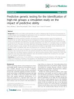
isolated from 28 (38.2%) cases. Male patients
were more prone to onychomycosis18 (69.2%)
compared to female patients 8 (30.7%) (Chart
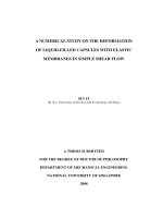
1). It was seen that 16 (61.5%) isolates were
from the toe nails, 7 (26.9%) isolates were
from finger nails and only 3 isolates (11.5%)
were from both toe and finger nails(Chart
2).Out of the 26 isolates, 13 (50%) isolates
were
from
Proximal
subungual
onychomycosis, 8 (30%) were from distal
lateral subungual onychomycosis, 2 (7.6%)
from white subungual onychomycosis and 3
(11.5%) cases were from candida infection
(Table 1). A total of 15 (57.6%) isolates were
dermatophytes and 11 (42.3%) were other
than dermatophytes (Table 2). Among the
dermatophytes, Trichophyton was most
commonly isolated 7 (26.9%), followed by
Microsporum 5 (19.2%), Epidermophyton 3
(11.5%). Among the non dermatophytes,
Candida was isolated from 3 (11.5%) cases,
Aspergillus was isolated from 2 (7.6%),
Pyrenochaeta from 2 (7.6%) cases,
Curvularia from 2 (7.6%) cases and only 1
(3.8%) case yielded Fusarium. Comparison of
various
predisposing
factors
for
Onychomycosis in males and females is
depicted in Table 3. Onychomycosis is a
cosmetic problem and a chronic disease which
has a long duration of treatment (Fig. 1 and 2).
Collection and transport of Sample: The
nails of the selected patients were cleansed
with 80% ethanol to remove contaminating
bacteria from the site. The sample was then
obtained by vigorous scraping on nail bed,
underside of nail plate and hyponychium. The
sample was placed in a sterile petridish and
transported to microbiology laboratory (Kaur
et al., 2007).
Processing of the sample: The sample was
then divided into two parts, one for direct
microscopy under high power objective using
20% KOH and the other part for culture on
Sabouraud’s dextrose agar (SDA) with
cyclohexamide, as it prevents the growth of
non dermatophytic fungi. SDA without
cyclohexamide and with 5% chloramphenicol
was used to grow non dermatophytic fungi.
The cultures were kept at 25°C and 37°C for
up to six weeks. No growth in the media after
six weeks was reported as negative (Boni et
al., 1998). Confirmation of the organism was
done based on morphology of fungus in LPCB
(Lactose phenol cotton blue) mount done from
the material obtained from the culture of
fungus on SDA and slide culture (Ramudamu
3767
Int.J.Curr.Microbiol.App.Sci (2018) 7(8): 3765-3770
Table.1 Table depicting distribution of various types of onychomycosis based on clinical
presentation
Clinical presentation
Proximal subungual onychomycosis
Distal lateral subungual onychomycosis
White subungual onychomycosis
Candidal
Isolates (n=26)
13 (50%)
8 (30%)
2 (7.6%)
3 (11.5%)
Table.2 Various fungal pathogens isolated from 26 onychomycosis cases
Dermatophytes
Trichophyton
Microsporum
Epidermophyton
Total
Non dermatophytes
Candida
Aspergillus
Pyrenochaeta
Fusarium
Penicilium
Curvularia
Total
7 (26.9%)
5 (19.2%)
3 (11.5%)
15 (57.6%)
3 (11.5%)
2 (7.6%)
2 (7.6%)
1 (3.8%)
1 (3.8%)
2 (7.6%)
11 (42.3%)
Fig.1 Gender wise distribution of Onychomycosis
3768
Int.J.Curr.Microbiol.App.Sci (2018) 7(8): 3765-3770
Table.3 Comparison of various predisposing factors among Onychomycosis cases (n=26)
Risk factors
Trauma
Immunocompromised
Diabetes
Occupations not involving trauma
Males
8 (30.76%)
3 (11.53%)
1 (3.84%)
5 (19.23%)
Females
4 (15.38%)
3 (11.53%)
2 (7.69%)
Fig.2 Fungal isolation from different sites
Our study showed an isolation rate of 38.2%
which was low when compared to Heikkila et
al., (1995), who isolated fungus from 91
(56.17%) clinical samples among the 162
patients selected based on clinical presentation.
In the present study it was seen that males were
very prone to onychomycosis compared to
females which correlates with the study
conducted by Sigurgeirsson et al., (2014). In our
study, fungus was more commonly isolated
from cases presenting with proximal subungual
onychomycosis which was in contrary to study
by Adekhand et al., (2015) who isolated fungus
more commonly from distal lateral subungual
onychomycosis. In comparison to Aditya et al.,
(2000), our study also showed a higher
incidence of toe nail onychomycosis.
Dermatophytes were the most common
organisms isolated. Our results were almost
similar to the findings of Gupta et al., (2000)
who also showed a higher incidence of
onychomycosis by dermatophytes. Among the
dermatophytes, Trichophyton was most
commonly isolated. Our study had similar
results with Mugge et al., (2006). Verylittle is
known
about
the
risk
factors
for
onychomycosis. Trauma is the major cause of
onychomycosis accounting for 8 (30.76%) in
males and 4 (15.38%) in females, followed by
occupations not involving trauma such as fisher
men, clothes and utensil washers, swimmers
etc. Even in this group men are predominantly
infected. The incidence of onychomycosis in
diabetes and immunocompromised patients was
less.
In conclusion, onychomycosis is a growing
public health concern. Dermatophytes are the
primary cause of onychomycosis when
compared
with
non-dermatophytes.
Onychomycosis occurs more commonly in men
compared to women. The cause may be related
to the occupations where the incidence of
trauma is more like carpentry, agriculture, wood
cutting, iron smith and in some instances it may
3769
Int.J.Curr.Microbiol.App.Sci (2018) 7(8): 3765-3770
be non-occupational like using occlusive
footwear and many other such factors. Diabetes
and immune compromised conditions promote
onychomycosis. Isolation of the organism with
culture is very important as it will aid the
clinician to rule out bacterial causes and choose
appropriate antifungal therapy.
References
Adekhandi S, Pal S, Sharma N, Juyal D, Sharma
M, Dimri D. Incidence and epidemiology
of onychomycosis in patients visiting
tertiary care hospital in India. Cutis, 2015;
95(1): E20-5.
Aditya K. Gupta, Hem C. Jain, Charles W. Lynde,
Paul Mac Donald, Elizabeth A. Cooper,
and Richard C. Summerbell. Prevalence
and epidemiology of onychomycosis in
patients visiting physicians offices: A
multicentre Canadian Survey of 15000
patients. J Am Acad Dermatol Vol 4,
2000 Aug; 43(2 Pt 1):244-8.
Andre J, Achten G. Onychomycosis. Int J
Dermatol., 1987; 26: 481-490.
Boni e, Elewski, Onychomycosis: Pathogenesis,
Diagnosis, and Management, Clinical,
Microbiol, Rev. July 1998: 11(3):415429.
Bramono. The Asian Achilles survey, Presented in
the 6th Asian Dermatological Congress,
Bangkok: November 2001
Bronson D M, D R Desai, S Barskey and S
McMillen Foley. An epidemic of
infection with Trichophyton tonsurans
revealed in a 20 year survey of fungal
infections in Chicago. J Am Acad
Dermatol., 1983; 8: 322-330
Charif M A, Elewski B E. A Historical
perspective on Onychomycosis. Dermatol
Ther., 1997: 3: 43-45
Cohen J L, Scher R K, Papper A S. The nail and
fungal infections. Cutaneous fungal
infections, New York, NY: Igaku-Shoin
Inc; 1992. Pp. 106-122
Dompmartin D, Dompmartin A, Deluol A M,
Grosshans
E,
Coulaud
J
P.
Onychomycosis and AIDS: Clinical and
laboratory findings in 62 patients. Int J
Dermatol 1990; 29: 337-339.
H. Heikkila, S. Stubb, The prevalence of
onychomycosis in Finland, vol 133, issue
5, November 1995, page 699-703.
Jagdish Chander, Text Book of Medical
Mycology, 4th Edition (2107).
Kaur R, Kasyap B, Bhalla P. A five year survey of
onychomycosis in New Delhi, India:
Epidemiological and Laboratory aspects.
Indian J Dermatol 2007;52:39-42
Mugge, Haustein UF, Nenoff P. Causative agents
of onychomycosis- A retrospective study.
J
Dtsch
Dermatol
Ges.2006
Mar;4(3):218-28
Ramudamu, Mandira, W Lyngdoh, Valarie;
Prasad, Abhijit; Rajbongshi, Jyotismita;
Durairaj, Elantamilan. (2018). A Study on
the
Mycological
Profile
of
Onychomycosis in a Tertiary Care
Hospital in Northeast India. Indian
Journal of Applied research. 8. 306-9.
Sigurgeirsson B, Baran R. Prevalence of
onychomycosis in the global populationA literature Study. Journal of European
Academy
of
Dermatology
and
Venereology2014 Nov; 28(11): 1480-91.
Zaias N, Tosti A, Rebell G, Morelli R, Bardazzi F,
Bieley H, Zaiac N, Glick B, Paley B,
Allevato M, Baran R. Autosomal
dominant pattern of Distal subungual
onychomycosis caused by Trichophyton
rubrum. J Am Acad Dermatol 1996; 34:
302-304.
Zaias N. Onychomycosis. Arch Dermatol 1972;
105: 263-274
How to cite this article:
Vinay Hajare, G.P. Aaftab and Abdul Hadi Waseem. 2018. A Prospective Study on the Epidemiology
of Onychomycosis in Tertiary Care Hospital. Int.J.Curr.Microbiol.App.Sci. 7(08): 3765-3770.
doi: />
3770