Characterization of in vivo metabolites in rat urine following an oral dose of masitinib by liquid chromatography tandem mass spectrometry
Bạn đang xem bản rút gọn của tài liệu. Xem và tải ngay bản đầy đủ của tài liệu tại đây (1.55 MB, 18 trang )
Kadi et al. Chemistry Central Journal (2018) 12:61
/>
Open Access
RESEARCH ARTICLE
Characterization of in vivo metabolites
in rat urine following an oral dose of masitinib
by liquid chromatography tandem mass
spectrometry
Adnan A. Kadi1, Sawsan M. Amer2, Hany W. Darwish1,2 and Mohamed W. Attwa1,2*
Abstract
Masitinib (MST) is an orally administered drug that targets mast cells and macrophages, important cells for immunity,
by inhibiting a limited number of tyrosine kinases. It is currently registered in Europe and USA for the treatment of
mast cell tumors in dogs. AB Science announced that the European Medicines Agency has accepted a conditional
marketing authorization application for MST to treat amyotrophic lateral sclerosis. In our work, we focused on studying in vivo metabolism of MST in Sprague–Dawley rats. Single oral dose of MST (33 mg kg−1) was given to Sprague–
Dawley rats (kept in metabolic cages) using oral gavage. Urine was collected and filtered at 0, 6, 12, 18, 24, 48, 72 and
96 h from MST dosing. An equal amount of ACN was added to urine samples. Both organic and aqueous layers were
injected into liquid chromatography-tandem mass spectrometry (LC–MS/MS) to detect in vivo phase I and phase
II MST metabolites. The current work reports the identification and characterization of twenty in vivo phase I and
four in vivo phase II metabolites of MST by LC–MS/MS. Phase I metabolic pathways were reduction, demethylation,
hydroxylation, oxidative deamination, oxidation and N-oxide formation. Phase II metabolic pathways were the direct
conjugation of MST, N-demethyl metabolites and oxidative metabolites with glucuronic acid. Part of MST dose was
excreted unchanged in urine. The literature review showed no previous articles have been made on in vivo metabolism of MST or detailed structural identification of the formed in vivo phase I and phase II metabolites.
Keywords: Masitinib, In vivo metabolism, Sprague–Dawley rats, Phase II glucuronide conjugates
Introduction
Cancer became a major reason of death [1]. More than
four millions new cancer cases reported in developed
countries [2, 3]. Molecular targeting strategies were used
to treat distributed cancer depending on identifying the
tumor suppressors and oncogenes involved in the progress of human cancers [4]. Tyrosine kinase inhibitors
(TKIs) (e.g. masitinib) are compounds that target tyrosine kinases enzymes, which are responsible for the activation of numerous proteins in a number of cell signaling
pathways. They initiate or stop many functions inside
*Correspondence: ;
1
Department of Pharmaceutical Chemistry, College of Pharmacy, King
Saud University, P.O. Box 2457, Riyadh 11451, Saudi Arabia
Full list of author information is available at the end of the article
living cells [5]. Blocking the selected activation of these
proteins has been shown to have therapeutic benefits in
cancer diseases and central nervous system disorders
mast cells and macrophages [6, 7]. Tyrosine kinase inhibitors (TKIs) are considered a very important class of targeted therapy [8].
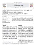
MST (Fig. 1) is new orally administered TKIs. It is
already registered in Europe and USA for the treatment of mast cell tumors in dogs [9]. MST is approved
under the trade name masivet in Europe and Kinavet in
the USA at a dose of 12.5 mg kg−1 per day [10]. Toxicity
profile of MST is lower than other TKIs [11]. MST selectively inhibits c-kit tyrosine kinase blocking stem cell factor induced proliferation. It exhibits more activity and
selectivity against KIT than imatinib in in vitro studies
[11]. In 3 October 2016, AB Science announced that the
© The Author(s) 2018. This article is distributed under the terms of the Creative Commons Attribution 4.0 International License
(http://creativecommons.org/licenses/by/4.0/), which permits unrestricted use, distribution, and reproduction in any medium,
provided you give appropriate credit to the original author(s) and the source, provide a link to the Creative Commons license,
and indicate if changes were made. The Creative Commons Public Domain Dedication waiver (http://creativecommons.org/
publicdomain/zero/1.0/) applies to the data made available in this article, unless otherwise stated.
Kadi et al. Chemistry Central Journal (2018) 12:61
Page 2 of 18
Table 1 List of materials and chemicals
Fig. 1 Chemical structure of MST
EMA has accepted a conditional marketing authorization
application for MST to treat ALS in human. MST found
to be effective for the treatment of severely symptomatic
indolent or smouldering systemic mastocytosis [12].
Drug metabolism research is an integral part of the
drug discovery process and is very often the factor that
determines the success of a given drug to be marketed
and clinically used [13]. Drug metabolism research is
generally conducted using in vitro and/or in vivo techniques. In vitro techniques involve the incubation of
drugs with different types of in vitro preparations (e.g.
liver microsomes, hepatocytes) isolated from rats and
subsequent sample processing and analysis using spectroscopic techniques [14, 15]. In vivo techniques involve
the administration of a single dose of the drug to rat, and
the subsequent collection of urine that contain the drugs
and their potential metabolites. In this work, we focused
in the in vivo phase I metabolites and in vivo phase II
MST metabolites identification using LC–MS/MS [16].
All measurements were done using Agilent LC–MS/MS
system that consisted of LC (Agilent HPLC 1200) coupled to MS/MS detector (6410 QqQ MS) through an
electrospray ionization source (Agilent Technologies,
USA) [17].
MST chemical structure contains cyclic tertiary amine.
Phase I metabolism of cyclic tertiary amines produces
metabolites of oxidative products including N-dealkylation, ring hydroxylation, α-carbonyl formation, N-oxygenation, and ring opening metabolites that can be
formed through iminium ion intermediates [18, 19].
Chemicals and methods
Chemicals
All chemicals are listed in Table 1.
In vivo metabolism of MST in Sprague–Dawley Rats
Rat dosing protocol
Male Sprague–Dawley rats (n = 6, average: 340 g, 4 weeks
of age) were housed individually in special purpose
metabolism cages. Cages are placed in the animal care
facility in a 12 h light/dark cycle (7:00–19:00) and were
allowed free access to standard animal feed and water
Namea
Source
Masitinib
LC Labs (USA)
Tween 80
Eurostar Scientific Ltd. (UK)
Ammonium formate, HPLC grade
acetonitrile (ACN), Dimethyl
Sulfoxide (DMSO), Polyethylene
glycol 300 (PEG 300) and formic
acid
Sigma-Aldrich (USA).
Water (HPLC grade)
Milli-Q plus purification system
(USA)
Sprague–Dawley rats
Animal Care Center, College of
Pharmacy, King Saud University
(Saudi Arabia)
a
All solvent are HPLC grade and reference powders are of AR grade
that were placed in the special food and water compartments attached to the metabolism cages. Rats were acclimated in metabolism cages for 72 h prior to the start of
the study. MST was formulated in (4% DMSO, 30% PEG
300, 5% Tween 80, HPLC H2O) for oral dosing of rats.
Doses were individually calculated for each rat such that
everyone receives a specific dose. The average dose of
MST (Kinavet-CA1) in dogs was 10 mg kg−1. By using the
following equations [20–22]:
mg
kg
Rat
mg
kg
= Dog
Rat
mg
kg
= 10 ∗ 20/6
Rat
mg
kg
= 200/6
Rat
mg
kg
= 33.3
∗ Km ratio
mg
kg
So the dose for each rat was 33.3 mg/kg. All rats except
one were given a single dose of MST. All MST doses were
administered by oral gavage. Urine draining into the special urine compartments fitted to the metabolism cages
were collected prior to drug dosing as blank control reference and at 6, 12, 18, 24, 48, 72 and 96 h following MST
dosing. Urine samples taken from all metabolism cages
were pooled together, labeled, and stored at (− 20 °C).
Sample preparation
Urine samples were thawed to room temperature and
filtered over 0.45 µm syringe filters. Liquid liquid extraction (LLC) was used to extract MST and its related
metabolites. Equal volume of ice cold acetonitrile (ACN)
was added to each sample then vigorously shaken by
vortexing for 1 min. Phase separation [23, 24] between
Kadi et al. Chemistry Central Journal (2018) 12:61
Page 3 of 18
an aqueous sample and a water-miscible solvent (ACN)
into two layers achieved by using ice cold ACN that was
added to urine and the mixture was stored at 4 °C overnight [25]. Low temperature leads to phase separation
of ACN/urine mixture. The pH of urine and the nature
of urine matrix which contains high concentration of
salt participated in phase separation [26]. As we did not
want to miss any MST-related metabolites, both layers
were removed and evaporated to dryness under stream of
nitrogen. The dried extracts were reconstituted in 1 mL
of mobile phase and transferred to 1.5 mL HPLC vials
for LC–MS/MS analysis. Control urine samples obtained
from rats prior to drug dosing were prepared in the exact
way described for each method of sample purification.
LC–MS/MS conditions
The LC–MS/MS parameters optimized for chromatographic separation and identification of rat urine extract
components are listed in Table 2.
Identification of in vivo MST metabolites
MST-related metabolites were concentrated in the ACN
layer while endogenous urine components and polar
metabolites (e.g. glucuronide conjugates) were found in
the aqueous layer. Extracted ion chromatograms for the
expected metabolites were used to find metabolites in
the total ion chromatogram of both organic and aqueous layers. PI studies were for the suspected compounds
and results were interpreted and compared with the
PI of MST. Mass scan and PI scan modes of the triple
quadrupole mass analyzer were used for detection of
in vivo phase I and phase II MST metabolites. PI mass
spectra were used to propose the metabolite chemical
structure by reconstructing the marker daughter ions.
Results and discussion
Identification of in vivo phase I metabolic pathways of MST
The in vivo metabolites of MST underwent fragmentations similar to that of the parent ion that allowed us to
identify and determine changes in the metabolite structures. The product ion mass spectra of some metabolites exhibited particular fragmentation pathways that
provided more structural information as shown below.
Comparison of PI mass spectra between urine extracts
with control samples in addition to the comparison
of PI of MST and its anticipated metabolites (Table 3)
resulted in the detection of twenty in vivo phase I and
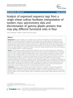
four phase II metabolites (Fig. 2). Ten in vivo phase I
metabolites are reported in the case of in vitro metabolism [27]. We concentrated on the structural identification of the new ten in vivo phase I and the other four
in vivo phase II MST metabolites. Metabolic pathways
for in vivo phase I metabolites were supposed to be
N-demethylation, N-oxide formation, oxidation, oxidative deamination, reduction, oxidative cleavage, benzyl
oxidation and hydroxylation while for phase II metabolites were N-conjugation of MST and the N-demethyl
metabolite with glucuronic acid and oxidative metabolites glucuronidation.
Table 2 Adjusted parameters of the supposed LC–MS/MS methodology
Parameters of LC
Parameters of MS/MS
HPLC
Agilent 1200
Mass spectrometer Agilent 6410 QQQ
Gradient mobile phase
A: H2O (10 mM Ammonium formate,
pH:4.1)
Ionization source
Positive ESI
Drying gas: N2 gas
Flow rate (12 L/min)
Pressure (55 psi)
B: ACN
Flow rate: 0.2 mL/min
Run time: 45 min
Injection volume: 20 µL
Agilent eclipse plus C18 column
Gradient system
Length
50 mm
ESI temperature: 350 °C
Internal diameter
2.1 mm
Capillary voltage: 4000 V
Particle size
1.8 μm
Collision gas
High purity N2
Temperature:
24 °C
Modes
Mass scan and product ion (PI)
Time
%B
Analyte
0
5
MST and its related in vivo phase I and phase II
metabolites
Mass parameters
Fragmentor voltage: 130 V
40
40
43
40
45
5
Post time (15 min)
5
Collision energy of 20 eV
Kadi et al. Chemistry Central Journal (2018) 12:61
Page 4 of 18
MST excretion of in rat urine
M2, M3 and M4 in vivo phase I metabolite
Part of the MST oral dose was excreted unmetabolized
in rat urine. MST parent ion was detected at m/z 499 in
full mass scan spectrum. MST of and its major in vivo
metabolites (M1 and MO6) excretion in urine was
observed after 6 h of dosing. Comparative concentrations of MST, M1 and MO6 were high after 6 h and then
began to decline by time until almost vanished after 96 h
from dosing as shown in the overlayed PI chromatograms
(Check Additional file 1). Peak area ratios of MST and its
major metabolite (M1 and MO6) in urine were plotted
against time. Peak area ratio of each MST, M1 and MO6
were measured at different collection time considering
the biggest peak is 100% (Fig. 3) [28].
Fragmentation of MST (Fig. 4) was explained in
Scheme 1. Comparison of PI of MST with suspected
peaks allowed the identification of metabolic changes in
the supposed in vivo metabolites.
M2, M3 and M4 were detected at m/z 501 at different
retention times in mass scan spectrum of organic urine
extract. PI scan for the three metabolites gave different
daughter ions. In the case of M2, parent ion at m/z 501
was fragmented to one ion at m/z 401. The daughter
ion at m/z 401 supposed that there is no change in the
methyl piperazine group. The metabolic pathway for M2
metabolite was supposed to be the reduction of the carbonyl group.
In the case of M3, parent ion at m/z 501 was fragmented to ions at 400.2 and 367.2 (Fig. 5). Metabolic
pathways for M3 were supposed to be hydroxylation of
pyridine ring and N-demethylation (Scheme 2).
In the case of M4, parent ion at m/z 501 was fragmented to two daughter ions at m/z 483 and at m/z 399
(Fig. 6). The daughter ion at m/z 399 supposed that there
all metabolic changes occured in the methyl piperazine group. Metabolic pathways for M4 metabolite were
hydroxylation and N-demethylation of N-methyl piperazine (Scheme 3).
M1 in vivo phase I metabolite
The major metabolic pathway for MST is N-demethyalation. M1 was detected at m/z 485 in mass scan spectrum.
Table 3 In vivo phase I MST metabolites
[M + H]+
PI
RT (min)
MST
499
399
24.9
M1
485
399
27.9
N-demethylation
M2
501
401
26.6
Carbonyl group reduction
M3
501
400.2, 367.3
24.4
N-demethylation and Hydroxylation of pyridine ring
M4
501
482.9, 399.3
26.5
N-demethylation and Hydroxylation of N-methyl piperazine
M5
529
511, 429
25.1
Benzyl oxidation to carboxylic acid
M6
529
486, 400
26.9
Pyridine ring hydroxylation and N-methyl piperazine oxidation
M7
529
511,482 399, 247
29.6
Oxidation and Hydroxylation of N-methyl piperazine
MO1
515
497.2, 415, 396.8
21.7
N-oxide formation
MO2
515
497.2, 396.9
22.2
Benzylic hydroxylation
MO3
515
497.0, 400.1
23.0
Pyridine ring hydroxylation
MO4
515
497, 399, 415, 217
23.1
Pyridine ring N-oxidation
MO5
515
497, 399, 415, 217
24.0
N-oxidation
MO6
515
428, 415, 400, 381.3, 98.1,
28.0
Piperazine ring N-oxidation
M8
531
488, 402, 123
26.7
Pyridine ring hydroxylation and piperazine ring hydroxylation
M9
531
415, 381, 123
27.3
Piperazine ring hydroxylation and benzyl hydroxylation
M10
531
501, 401
29.3
Oxidative cleavage of N-methyl piperazine ring to carboxylic acid
M11
547
511
30.7
N-oxide formation of pyridine and piperazine ring and Benzylic hydroxylation [27]
MA1
431
255
10.2
Oxidative deamination
MA2
447
271
13.2
Phenyl hydroxylation and oxidative deamination
MA3
447
285, 271, 164, 111
14.5
Benzyl hydroxylation and oxidative deamination
In vivo phase I metabolic reaction
Kadi et al. Chemistry Central Journal (2018) 12:61
Fig. 2 PI chromatograms: a (MST), b (M1), c (M2–M4), d (M5–M7), e (M8–M10) and f (MO1–MO6)
Page 5 of 18
Kadi et al. Chemistry Central Journal (2018) 12:61
Page 6 of 18
Fig. 3 MST, M1 and MO6 excretion rate
Fig. 5 PI mass spectrum of parent ion (M3) at m/z 502
MO1 to MO6 in vivo phase I metabolite
Oxidized MST metabolite (M + O) was detected at m/z
515 in mass scan spectrum at different retention times.
Fragmentation of parent ions at m/z 515 gave different
daughter ions as shown in the Table 3. The structure of
each metabolite was supposed The metabolic pathway for
MO metabolites was supposed to be either by hydroxylation or N-oxidation of MST [27].
M5, M6 and M7 in vivo phase I metabolite
M5, M6 and M7 metabolites were detected at m/z 529
in full mass scan spectrum at different retention times.
PI scan for parent ions at m/z 529 gave different daughter ions. In the case of M5, parent ion at m/z 529 was
Fig. 4 PI of MST parent ion at m/z 499
O
N
S
N
H
NH
Masitinib
m/z: 499
Scheme 1 Supposed PI of MST
N
N
N
H
PI
O
S
N
H
N
m/z: 399
N
N
H
Kadi et al. Chemistry Central Journal (2018) 12:61
Page 7 of 18
O
HN
S
NH
OH
N
N
H
N
H
N
M3
m/z: 515
PI
O
S
N
H
N
OH
N
N
H2
S
O
N
N
H
N
m/z: 400
N
H
m/z:367
Scheme 2 Supposed PIs of M3
Fig. 6 PI mass spectrum of parent ion (M4) at m/z 501
Fig. 7 PI mass spectrum of parent ion (M5) at m/z 529
OH
O
HN
S
N
H
NH
N
N
N
H
M4
m/z: 501
PI
O
S
N
H
N
m/z: 399
Scheme 3 Supposed PIs of M4
O
N
N
H
N
NH
S
N
H
m/z: 483
N
N
N
H
Kadi et al. Chemistry Central Journal (2018) 12:61
Page 8 of 18
COOH
S
O
N
N
H
NH
N
N
H
M5
N
m/z: 529
PI
O
O
N
N
S
N
H
N
H
N
N
H
COOH
S
O
N
N
H
N
N
m/z: 429
m/z: 511
Scheme 4 Supposed PIs of M5
fragmented to ions at m/z 511 and at m/z 429 (Fig. 7).
The metabolic pathway for M5 was supposed to be benzyl oxidation to carboxylic acid (Scheme 4).
In the case of M6, parent ion at m/z 529 was fragmented to ions at 486 and 400 (Fig. 8). The metabolic
pathway for M6 was supposed to be hydroxylation and
oxidation of methyl piperazine ring (Scheme 5).
In the case of M7, parent ion at m/z 529 was fragmented to ions at 511, 399 and 98 (Fig. 9). Metabolic
pathways for M7 were supposed to be hydroxylation and
oxidation of methyl piperazine ring (Scheme 6).
M8, M9 and M10 in vivo phase I metabolite
M8, M9 and M10 metabolites were detected at m/z 531
in full mass scan spectrum at different retention times. PI
Fig. 8 PI mass spectrum of parent ion (M6) at m/z 529
O
O
S
N
H
N
NH
N
H
N
N
OH
M6
m/z: 529
PI
O
O
N
NH
N
H
m/z: 486
Scheme 5 Supposed PIs of M6
O
S
N
H
N
S
N
H
N
H2
m/z: 400
N
N
OH
Kadi et al. Chemistry Central Journal (2018) 12:61
Page 9 of 18
Fig. 9 PI mass spectrum of parent ion (M7) at m/z 529
Fig. 10 PI mass spectrum of parent ion (M8) at m/z 531
In the case of M9, parent ion at m/z 531 was fragmented to ions at 513, 415, 381 and 123 (Fig. 11). Metabolic pathways for M9 were supposed to be benzyl
hydroxylation and hydroxylation of methyl piperazine
ring (Scheme 8).
scan for parent ions at m/z 531 gave different daughter
ions. In the case of M8, parent ion at m/z 531 was fragmented to ions at 488, 402 and 123 (Fig. 10). Metabolic
pathways for M8 were supposed to be hydroxylation of
pyridine and hydroxylation of methyl piperazine ring
(Scheme 7).
O
O
N
H
N
NH
HO
S
N
H
N
N
M7
m/z: 529
PI
O
O
N
S
N
H
N
O
N
N
N
H
OH
N
N
H
NH
O
m/z: 399
Scheme 6 Supposed PIs of M7
O
S
N
H
N
H
m/z: 499
m/z: 511
N
H
N
N
O
N
HO
N
S
N
m/z: 247
N
Kadi et al. Chemistry Central Journal (2018) 12:61
Page 10 of 18
OH
O
S
N
H
N
NH
N
H
N
N
OH
M8
m/z: 531
PI
OH
N
NH
O
O
S
N
H
N
H
m/z: 488
N
S
N
H
N
H2
N
N
OH
m/z: 402
Scheme 7 Supposed PIs of M8
In the case of M10, parent ion at m/z 531 was fragmented to ions at 501 and 401 (Fig. 12). Metabolic pathways for M10 were supposed to be oxidative cleavage of
N-methyl piperazine ring to carboxylic acid (Scheme 9).
M11 in vivo phase I metabolite
Fig. 11 PI mass spectrum of parent ion (M9) at m/z 531
M11 was detected at m/z 547 in mass scan spectrum
of the urine organic extract. PI chromatogram of urine
organic extract at m/z 547 showed one peak at 30.72 min.
PI scan for M11 at m/z 547 gave daughter ions at m/z 511.
Metabolic reactions for M11 metabolite were supposed
to be hydroxylation of benzylic carbon, oxidation of pyridine nitrogen and oxidation of piperazine nitrogen.
Kadi et al. Chemistry Central Journal (2018) 12:61
Page 11 of 18
OH
O
OH
N
S
N
H
NH
N
H
N
N
M9
m/z: 531
PI
O
OH
N
O
S
N
NH
N
H
N
HN
S
N
NH
N
N
H
N
N
m/z:481
m/z:513
OH
O
S
N
H
N
H
N
N
m/z:415
Scheme 8 Supposed PIs of M9
In vivo phase I oxidative deamination metabolic pathway
(MA1, MA2 and MA3)
Fig. 12 PI mass spectrum of parent ion (M10) at m/z 531
The loss of the piperazine moiety by oxidative deamination and rapid further oxidation of the intermediate aldehyde to a carboxylic acid metabolite were observed for
MA1, MA2 and MA3 in the aqueous layer of the urine/
ACN mixture. Fragmentation of parent ions at m/z 431
and at m/z 447 gave different daughter ions. The structure of each metabolite was supposed.
MA1 was detected at m/z 431 in mass scan spectrum
of the aqueous layer urine extract. PI chromatogram
of urine aqueous extract at m/z 431 showed one peak
at 10.2 min. PI scan for MA1 at m/z 431 gave daughter
ions at m/z 255 (Fig. 13). The daughter ion at m/z 255
Kadi et al. Chemistry Central Journal (2018) 12:61
Page 12 of 18
O
OH
N
H
HO
NH
N
H
S
N
N
N
H
M10
m/z: 531
PI
OH
O
S
N
H
HO
NH
N
H
OH
N
S
N
H
N
N
N
N
H
m/z: 401
m/z: 501
Scheme 9 Supposed PIs of M10
supposed the loss of the piperazine moiety by oxidative
deamination and rapid further oxidation of the intermediate aldehyde to a carboxylic acid (Scheme 10).
MA2 and MA3 were detected at m/z 447 in mass scan
spectrum of the aqueous layer urine extract. PI chromatogram of urine aqueous extract at m/z 447 showed two
peaks at 18.6 and 19.5 min. PI scan for MA2 and MA3
at m/z 447 gave different daughter ions at two different
retention times (Figs. 14 and 15).
In the case of MA2, the daughter ion at m/z 271 supposed the loss of the piperazine moiety by oxidative
deamination and rapid further oxidation of the intermediate aldehyde to a carboxylic acid in addition to phenyl
hydroxylation (Scheme 11).
Fig. 13 PI mass spectrum of parent ion (MA1) at m/z 431
O
S
N
H
O
OH
N
MA1
m/z: 431.3
Scheme 10 Supposed PIs of MA1
NH
N
H
O
PI
N
H2
O
OH
m/z: 255
Kadi et al. Chemistry Central Journal (2018) 12:61
Page 13 of 18
In the case of MA3, the daughter ion at m/z 271 supposed the loss of the piperazine moiety by oxidative
deamination and rapid further oxidation of the intermediate aldehyde to a carboxylic acid. The other daughter ion at m/z 285 supposed benzyl hydroxylation
(Scheme 12).
Identification of in vivo phase II metabolic pathways
of MST
Phase II metabolic pathways were supposed to be
N-conjugation of MST and the N-demethyl metabolite
with glucuronic acid, and glucuronidation of oxidative
metabolites (Table 4). Phase II metabolites were found in
the aqueous layer of the rat urine extract in a very small
concentration compared to in vivo phase I metabolites.
Excretion of all in vivo phase II metabolites in urine was
observed after 12 h of rat dosing and disappeared rapidly
after 48 h of rat dosing.
Fig. 14 PI mass spectrum of parent ion (MA2) at m/z 447
MG1 in vivo phase II metabolite
MG1 was detected at m/z 675 in mass scan spectrum
of the aqueous layer urine extract. PI chromatogram of
urine aqueous extract at m/z 675 showed one peak at
18.9 min. PI scan for MG1 at m/z 675 gave daughter ions
at m/z 499 and 399 (Fig. 16). The daughter ion at m/z 399
supposed that direct N-conjugation of MST with glucuronic. The other daughter ion at 499 refers to the aglycone (MST) formed in the triple quadrupole by the loss
of anhydroglucuronic acid (Scheme 13).
Fig. 15 PI mass spectrum of parent ion (MA3) at m/z 447
HO
O
N
H
O
OH
Scheme 11 Supposed PIs of MA2
S
N
MA2
m/z: 447
HO
O
NH
N
H
PI
N
H2
O
OH
m/z: 271
Kadi et al. Chemistry Central Journal (2018) 12:61
Page 14 of 18
OH
O
S
N
H
O
N
NH
N
H
MA3
OH
m/z: 447
PI
OH
O
O
N
H2
O
OH
N
H
O
m/z: 271
O
OH
S
N
H2
NH
N
H
m/z: 164
m/z: 285
Scheme 12 Supposed PIs of MA3
Table 4 In vivo phase II MST metabolites
Mass scan
Daughter ions
Retention time (min)
Phase II metabolic pathway
MG1
675
499, 399
18.93
Direct N-conjugation with glucuronic acid
MG2
661
485
18.77
N-demethylation and direct N-conjugation with glucuronic acid
MG3
691
514.8
18.7
Glucuronidation of hydroxy MST at N-methyl piperazine ring
MG4
691
515.3, 414.9
19.46
Glucuronidation of hydroxy MST at benzyl carbon
MG2 in vivo phase II metabolite
MG2 was detected at m/z 661 in mass scan spectrum
of the aqueous layer urine extract. PI chromatogram of
urine aqueous extract at m/z 661 showed one peak at
18.7 min. PI scan for MG2 at m/z 661 gave daughter ions
at m/z 485 (Fig. 17). The daughter ion at 485 refers to the
aglycone (N-demethyl MST) formed in the triple quadrupole by the loss of anhydroglucuronic acid (Scheme 14).
MG3 and MG4 in vivo Phase II metabolites
Fig. 16 PI mass spectrum of parent ion (MG1) at m/z 675
MG3 and MG4 were detected at m/z 691 in mass scan
spectrum of the aqueous layer urine extract. PI chromatogram of urine aqueous extract at m/z 691 showed two
peaks at 18.6 and 19.5 min. PI scan for MG3 and MG4
at m/z 691 gave different daughter ions at two different
retention times (Figs. 18, 19).
Kadi et al. Chemistry Central Journal (2018) 12:61
Page 15 of 18
OHHO
H
O
H
OH
H
OH
H
O
H
O
N
S
N
H
N
N
N
N
H
m/z: 675
MG1
PI
O
N
S
N
H
NH
N
N
H
N
O
S
N
H
N
N
N
H
m/z: 399
m/z: 499
Scheme 13 Supposed PIs of MG1
Fig. 17 PI mass spectrum of parent ion (MG2) at m/z 661
HO H
OH
H
H H
HO
N
HO
O
H
H
O
O
S
N
H
N
Fig. 18 PI mass spectrum of parent ion (MG3) at m/z 691
N
N
N
H
MG2
m/z: 661
PI
O
HN
S
N
H
NH
N
N
N
H
m/z: 485
Scheme 14 Supposed PIs of MG2
Fig. 19 PI mass spectrum of parent ion (MG4) at m/z 691
Kadi et al. Chemistry Central Journal (2018) 12:61
HO
H
HO
HO
HHO
O
H
N
O
H
O
N
H
N
MG3
H
m/z: 691
PI
O
N
HO
N
S
N
H
N
H
O
Page 16 of 18
S
N
H
NH
N
N
N
H
In the case of MG3, the daughter ion at m/z 515 supposed that direct O-glucuronidation of hydroxy MST.
The daughter ion at 515 refers to the aglycone (hydroxy
MST) formed in the triple quadrupole by the loss of
anhydroglucuronic acid. (Scheme 15). Hydroxylation was
supposed to be in the N-methyl piperazine ring. In the
case of MG4, the daughter ion at m/z 515 supposed that
direct O-glucuronidation of hydroxy MST. The daughter
ion at 515 refers to the aglycone (hydroxy MST) formed
in the triple quadrupole by the loss of anhydroglucuronic
acid (Scheme 16). The other daughter at m/z 415 supposed that the hydroxylation of benzyl carbon.
m/z: 515
Scheme 15 Supposed PIs of MG3
O
OH
HO H
H
O
H
OH
O
H
N
N
S
N
H
N
OH
H
OH
N
H
N
MG4
m/z: 691
PI
OH
OH
O
N
NH
N
H
m/z: 515
Scheme 16 Supposed PIs of MG4
N
S
N
N
H
O
N
S
N
H
N
m/z: 415
N
H
Kadi et al. Chemistry Central Journal (2018) 12:61
Page 17 of 18
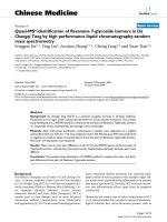
Fig. 20 Chemical structure of MST and identified metabolic pathways in Rat. The main metabolic pathway was the N-demethylation
Conclusions
MST was excreted partially unchanged in rat urine.
Twenty in vivo phase I metabolites were formed by oral
dosing of MST to Sprague–Dawley rats through six
metabolic pathways: N-demethylation, N-oxidation, oxidation, reduction, hydroxylation and oxidative deamination. Four in vivo phase II glucuronide conjugates were
found in the aqueous layer of rat urine extract (Fig. 20).
Ethics approval and consent to participate
Animal Care Center guidelines of Pharmacy College at King Saud Univesity
were applied for Rats’ maintenance. The Local Animal Care and Use Committee at KSU approved these guidelines.
Publisher’s Note
Springer Nature remains neutral with regard to jurisdictional claims in published maps and institutional affiliations.
Received: 21 August 2017 Accepted: 4 May 2018
Additional file
Additional file 1. Additional figures.
Authors’ contributions
AK, SA, HD and MA established the experiment design. Practical work was
performed by MA. Data were analyzed by AK, HD, SA and MA. HD and MA
wrote the first draft of the manuscript. AK and SA contributed in editing the
manuscript. AK, SA and HD supervised the research work. All authors read and
approved the final manuscript.
Author details
1
Department of Pharmaceutical Chemistry, College of Pharmacy, King Saud
University, P.O. Box 2457, Riyadh 11451, Saudi Arabia. 2 Analytical Chemistry
Department, Faculty of Pharmacy, Cairo University, Kasr El‑Aini St, Cairo 11562,
Egypt.
Acknowledgements
The authors would like to extend their sincere appreciation to the Deanship of
Scientific Research at the King Saud University for funding this work through
the Research Group Project No. RGP-322.
Competing interests
The authors declare that they have no competing interests.
Data availability
All data supporting the results in this article can be found in the manuscript or
the Additional file.
References
1. Jemal A, Siegel R, Ward E, Hao Y, Xu J, Murray T et al (2008) Cancer statistics, 2008. CA Cancer J Clin 58(2):71–96
2. Sinha R, El-Bayoumy K (2004) Apoptosis is a critical cellular event in cancer chemoprevention and chemotherapy by selenium compounds. Curr
Cancer Drug Targets 4(1):13–28
3. Cozzi P, Mongelli N, Suarato A (2004) Recent anticancer cytotoxic agents.
Curr Med Chem Anti Cancer Agents 4(2):93–121
4. Barinaga M (1997) From bench top to bedside. Science
278(5340):1036–1039
5. Schlessinger J (2000) Cell signaling by receptor tyrosine kinases. Cell
103(2):211–225
6. Özvegy-Laczka C, Cserepes J, Elkind NB, Sarkadi B (2005) Tyrosine kinase
inhibitor resistance in cancer: role of ABC multidrug transporters. Drug
Resist Updates 8(1):15–26
7. Steeghs N, Nortier JW, Gelderblom H (2007) Small molecule tyrosine
kinase inhibitors in the treatment of solid tumors: an update of recent
developments. Ann Surg Oncol 14(2):942–953
8. Natoli C, Perrucci B, Perrotti F, Falchi L, Iacobelli S (2010) Tyrosine kinase
inhibitors. Curr Cancer Drug Targets 10(5):462–483
9. Daly M, Sheppard S, Cohen N, Nabity M, Moussy A, Hermine O et al
(2011) Safety of masitinib mesylate in healthy cats. J Vet Intern Med
25(2):297–302
10. Marech I, Patruno R, Zizzo N, Gadaleta C, Introna M, Zito AF et al (2014)
Masitinib (AB1010), from canine tumor model to human clinical development: where we are? Crit Rev Oncol Hematol 91(1):98–111
11. Dubreuil P, Letard S, Ciufolini M, Gros L, Humbert M, Castéran N et al
(2009) Masitinib (AB1010), a potent and selective tyrosine kinase inhibitor
targeting KIT. PLoS ONE 4(9):e7258
Kadi et al. Chemistry Central Journal (2018) 12:61
12. Lortholary O, Chandesris MO, Livideanu CB, Paul C, Guillet G, Jassem E
et al (2017) Masitinib for treatment of severely symptomatic indolent
systemic mastocytosis: a randomised, placebo-controlled, phase 3 study.
Lancet 389(10069):612–620
13. Kumar GN, Surapaneni S (2001) Role of drug metabolism in drug discovery and development. Med Res Rev 21(5):397–411
14. Kadi AA, Al-Shakliah NS, Yin W, Rahman AM (2017) In vitro investigation of
metabolic profiling of newly developed topoisomerase inhibitors (ethyl
fluorescein hydrazones, EtFLHs) in RLMs by LC–MS/MS. J Chromatogr B
1054:93–104
15. Rahman AM, Al-Shakliah NS, Yin W, Kadi AA (2017) In vitro Investigation
of Metabolic Profiling of a Potent Topoisomerase Inhibitors Fluorescein
Hydrazones (FLHs) in RLMs by LC-MS/MS. J Chromatogr B 1054:27–35
16. Attwa MW, Kadi AA, Darwish HW, Alrabiah H (2018) LC-MS/MS reveals
the formation of reactive ortho-quinone and iminium intermediates
in saracatinib metabolism: phase I metabolic profiling. Clin Chim Acta
482:84–94
17. Attwa MW, Kadi AA, Darwish HW, Amer SM, Alrabiah H (2018) A reliable
and stable method for the determination of foretinib in human plasma
by LC-MS/MS: application to metabolic stability investigation and
excretion rate. Eur J Mass Spectrom 1469066718768327. https://doi.
org/10.1177/1469066718768327
18. Masic LP (2011) Role of cyclic tertiary amine bioactivation to reactive iminium species: structure toxicity relationship. Curr Drug Metab
12(1):35–50
19. Kadi AA, Attwa M, Darwish HW (2018) LC-ESI-MS/MS reveals the formation of reactive intermediates in brigatinib metabolism: elucidation of
bioactivation pathways. RSC Adv 8(3):1182–1190
Page 18 of 18
20. Shin J-W, Seol I-C, Son C-G (2010) Interpretation of animal dose and
human equivalent dose for drug development. J Korean Med 31(3):1–7
21. Nair AB, Jacob S (2016) A simple practice guide for dose conversion
between animals and human. J Basic Clin Pharm 7(2):27–31
22. Reagan-Shaw S, Nihal M, Ahmad N (2008) Dose translation from animal
to human studies revisited. FASEB J Off Publ Fed Am Soc Exp Biol
22(3):659–661 (Epub 2007/10/19)
23. Varaprath S, Salyers KL, Plotzke KP, Nanavati S (1999) Identification of
metabolites of octamethylcyclotetrasiloxane (D(4)) in rat urine. Drug
Metab Dispos Biol Fate Chem 27(11):1267–1273
24. Bylda C, Thiele R, Kobold U, Volmer DA (2014) Recent advances in sample
preparation techniques to overcome difficulties encountered during
quantitative analysis of small molecules from biofluids using LC–MS/MS.
Analyst 139(10):2265–2276
25. Yoshida M, Akane A (1999) Subzero-temperature liquid − liquid extraction of benzodiazepines for high-performance liquid chromatography.
Anal Chem 71(9):1918–1921
26. Valente IM, Gonçalves LM, Rodrigues JA (2013) Another glimpse over
the salting-out assisted liquid–liquid extraction in acetonitrile/water
mixtures. J Chromatogr A 1308:58–62
27. Amer S, Kadi AA, Darwish HW, Attwa MW (2017) Identification and
characterization of in vitro phase I and reactive metabolites of masitinib
using a LC-MS/MS method: bioactivation pathway elucidation. RSC Adv
7(8):4479–4491
28. Amer SM, Kadi AA, Darwish HW, Attwa MW (2017) LC–MS/MS method for
the quantification of masitinib in RLMs matrix and rat urine: application
to metabolic stability and excretion rate. Chem Cent J 11(1):136