In silico design and synthesis of targeted rutin derivatives as xanthine oxidase inhibitors
Bạn đang xem bản rút gọn của tài liệu. Xem và tải ngay bản đầy đủ của tài liệu tại đây (1.93 MB, 13 trang )
(2019) 13:71
Malik et al. BMC Chemistry
/>
RESEARCH ARTICLE
BMC Chemistry
Open Access
In silico design and synthesis of targeted
rutin derivatives as xanthine oxidase inhibitors
Neelam Malik1, Priyanka Dhiman1 and Anurag Khatkar2*
Abstract
Background: Xanthine oxidase is an important enzyme of purine catabolism pathway and has been associated
directly in pathogenesis of gout and indirectly in many pathological conditions like cancer, diabetes and metabolic
syndrome. In this research rutin, a bioactive flavonoid was explored to determine the capability of itself and its derivatives to inhibit xanthine oxidase.
Objective: To develop new xanthine oxidase inhibitors from natural constituents along with antioxidant potential.
Method: In this report, we designed and synthesized rutin derivatives hybridized with hydrazines to form hydrazides
and natural acids to form ester linkage with the help of molecular docking. The synthesized compounds were evaluated for their antioxidant and xanthine oxidase inhibitory potential.
Results: The enzyme kinetic studies performed on rutin derivatives showed a potential inhibitory effect on XO ability in competitive manner with IC50 value ranging from 04.708 to 19.377 µM and RU3a3 was revealed as most active
derivative. Molecular simulation revealed that new rutin derivatives interacted with the amino acid residues PHE798,
GLN1194, ARG912, GLN 767, ALA1078 and MET1038 positioned inside the binding site of XO. Results of antioxidant
activity revealed that all the derivatives showed very good antioxidant potential.
Conclusion: Taking advantage of molecular docking, this hybridization of two natural constituent could lead to
desirable xanthine oxidase inhibitors with improved activity.
Keywords: Rutin, Xanthine oxidase, Molecular docking, Antioxidant
Introduction
Xanthine oxidase (XO) having molecular weight of
around 300 kDa is oxidoreductase enzyme represented in
the form of a homodimer. Both the monomers of XO are
almost identical and each of them contains three domains
namely (a) molybdopterin (Mo-pt) domain at the C-terminal having 4 redox centers where oxidation takes place
(b) a flavin adenine dinucleotide (FAD) domain at the
centre generally considered as binding site domain and
(c) 2[Fe–S]/iron sulfur domain at the N-terminal [1–3].
The catalytic oxidation of XO is two substrates reaction
*Correspondence: ;
2
Laboratory for Preservation Technology and Enzyme Inhibition Studies,
Department of Pharmaceutical Sciences, M.D. University, Rohtak, Haryana,
India
Full list of author information is available at the end of the article
on the xanthine and oxygen at the enzymatic centre.
While xanthine undergoes oxidation reaction near to the
Mo-pt center/substrate binding domain of XO, simultaneously substrate oxygen undergoes reduction at FAD
center and electron transfer takes place leading to formation of superoxide anion (O2−) or hydrogen peroxide
(H2O2) free radicals. [4–8]. This catalytic reaction results
in formation uric acid as a final product and oxygen reactive species in form of free radicals. The excessive generation of uric acid leads to a condition like hyperuricemia
which is a key factor in development of gout [1, 9], and
uncontrolled amounts of reactive oxygen species causes
many pathological conditions like cardiovascular disorders, inflammatory diseases and hypertensive disorders.
Xanthine oxidase (XO; EC 1.17.3.2) has been considered as significantly potent drug target for the cure and
management of pathological conditions prevailing due
to high levels of uric acid in the blood stream. [10–17].
© The Author(s) 2019. This article is distributed under the terms of the Creative Commons Attribution 4.0 International License
(http://creativecommons.org/licenses/by/4.0/), which permits unrestricted use, distribution, and reproduction in any medium,
provided you give appropriate credit to the original author(s) and the source, provide a link to the Creative Commons license,
and indicate if changes were made. The Creative Commons Public Domain Dedication waiver (http://creativecommons.org/
publicdomain/zero/1.0/) applies to the data made available in this article, unless otherwise stated.
Malik et al. BMC Chemistry
(2019) 13:71
Page 2 of 13
Considering the above fact, by inhibiting XO selectively
could be better treatment plan for disorders caused by
XO directly or indirectly including gout, inflammatory
disease, oxidative damage and cancer [3, 18, 19]. Generally, XO inhibitors have been categorized into purine
and non-purines inhibitors differentiated on the basis
of their chemically derived skeleton structure. The first
purine derived XO inhibitor discovered and approved by
US FDA was Allopurinol as marketed drug for gout and
hyperuricemia [20, 21]. Considering the life threatening
side effects like Stevens–Johnsons syndrome caused by
allopurinol use, scientists turned their interest into nonpurine XO inhibitors and an immense accomplishment
has been received in this direction with development of
new drug Febuxostat [22–25]. This non-purine candidate produced minor and non-life threatening adverse
effects in comparison to Allopurinol [26–29]. Extending
our previous successful effort to achieve new xanthine
oxidase inhibitors from natural sources, in this report we
investigated and developed some new rutin derived xanthine oxidase inhibitor [30].
Rutin is a well characterized bioactive plant flavonoid
having great therapeutic importance for the treatment of
many disease like conditions including cytotoxicity, antioxidant activity, antibacterial property and anti-inflammatory action [31–34]. Due to these pharmacological
activities rutin is explored widely and great success have
been achieved in order to get drug like candidates.
OH
HO
OH
O
OH O
HO
O
HO
O
OH
O
OH
O
OH
CH3
OH
Rutin
Taking advantage of molecular docking techniques new
compounds with potential drugability for the targeted
enzyme might be achieved with a precise knowledge
of mechanism of action. With the combined approach
of molecular docking and synthetic chemistry, in this
research we developed some new potential compounds
against xanthine oxidase (Fig. 1).
evaluation of the human xanthine oxidase inhibitory
activity was performed by measuring hydrogen peroxide
(H2O2) production from oxidation of xanthine oxidase
by the substrate xanthine, utilizing the human xanthine
oxidase assay kit (Sigma USA). The progress of reaction
was observed through thin layer chromatography (TLC)
on 0.25 mm precoated silica gel plates purchased from
Merck, reaction spots were envisaged in iodine compartment and UV. Melting points were measured using a
Sonar melting point apparatus and uncorrected. 1H NMR
and 13C NMR spectra were documented in DMSO and
deuterated CDCl3 respectively on Bruker Avance II 400
NMR spectrometer at the frequency of 400 MHz using
tetramethylsilane standard (downfield) moreover chemical shifts were expressed in ppm (δ) using the residual
solvent line as internal standard. Infrared (IR) spectra
were recorded on Perkin Elmer FTIR spectrophotometer
by utilizing KBr pellets system.
Molecular docking
In silico docking studies was done with integrated Schrodinger software using Glide module for enzyme ligand
docking [35].
Protocol followed for docking procedures
Preparation of protein The 3D crystal structure of
human xanthine oxidase co-crystalised with salicylic acid
was retrieved from Protein Data Bank (PDB ID. 2E1Q).
The targeted protein structure was further refined in the
Protein Preparation Wizard to obtain the optimized and
chemically accurate protein configuration. For that, the
co-crystalised enzyme (XO) was retrieved directly from
Protein data bank in maestro panel followed by removal of
water molecules, addition of H atoms, addition of missing
side chains and finally minimization was done to obtain
the optimized structure.
Preparation of ligand The 3D-structures of rutin derived
compounds to be docked against XO were built in maestro building window. Ligand preparation was performed
in Ligprep module.
Chemicals and instrumentation
Active site prediction To predict the binding site/active
site Site Map application of glide was utilized. Out of top
three active site, the one having larger radius was selected.
Validation of binding site was done by redocking the salicylic acid and RMSD value was observed. RMSD value of
less than 0.2 validated the docking procedure and active
site was defined for docking of new rutin analogs.
For this research, the analytical grade chemicals necessary for synthesis and antioxidant activity were purchased from Hi-media Laboratories. The in vitro
Glide docking To carry out docking, Firstly the receptor grid generation tool was utilized to around the active/
Experimental
Malik et al. BMC Chemistry
(2019) 13:71
Page 3 of 13
OH
OH
HO
OH
OH
O
HO
OH
HO
OH
O
HO
O
OH
O
O
N
Cl
HO
O
NH2
RU4b1
NO2
H
N
CH3
OH
OH
N
HN HO
OH
O
OH
O
O
OH
OH
CH3
HO
OH
OH
O
HO
O
O
OH
RU3a3
N
HN HO
S
HN
RU3a2
NO2
OH
O
OH
OH
OH
HO
OH
O
HO
S
Phenyl thiosemicarbazide
S
H2N
NH2
N
H
Thiosemicarbazide
H2N
O
OH
N
HN HO
S
H2N
OH
O
HO
O
NO2
O
OH O
4-Nitrobenzenamine
HO
HO
OH
O
OH
OH
HO
O
OH
HO
OH
OH
O
N
Nicotinic acid
OH
OH
HO
N
HO
HO
HO
OH
O
Cinnamic Acid
O
Salicylic acid
OCH3
OCH3
H3CO
O
O
O
OCH3O
RU7c1
N
H3CO
O
O
OCH3O
RU7c2
O
O
OH
O
OH
O
CH3
OH
NO2
RU4b2
OCH3
OCH3
OCH3
OCH3
H3CO
CH3
O
O
OH
HO
O
RU3a1
CH3
O
OH
OH
Rutin
O
CH3
O
OH
H2N
HN
NO2
NH-NH2
O
OH
OH
O
O
OCH3O
O
OH
RU7c3
Fig. 1 Design strategy for the development of rutin derivatives
binding site of xanthine oxidase and glide docking with
extra precision was used to visualize the interaction of
protein and ligand. The top active ligand was selected for
wet lab synthesis and evaluation of pharmacological activity.
Synthetic procedures
Procedures for synthesis of rutin derivatives (Scheme 1)
(A)General procedure for synthesis of hydrazine derivatives RU3a(1–4)
0.001 mol of rutin was taken in round bottom flask
and dissolved in 50 ml of ethanol. Different hydra-
zines (0.001 mol) were added to the flask and reaction mixture was refluxed for 5–6 h at 40 °C. Completion of reaction was monitored by TLC. The
product thus obtained was filtered and filtrate was
concentrated to obtain the final product. The final
product was recrystallised to obtain the pure compound.
(B) General procedure for synthesis of anilline derivatives RU4b(1–2)
0.001 mol of the intermediate obtained above
was taken in round bottom flask and dissolved in
50 ml of ethanol. Different anillines (0.001 mol)
were added to the flask and reaction mixture was
refluxed for 8–10 h at 40 °C. Completion of reaction
Malik et al. BMC Chemistry
(2019) 13:71
Page 4 of 13
OH
OH
HO
OH
O
HO
O
OH
N
HN HO
OH
HO
O
OH
O
N
Cl
HO
O
RU4b1
NO2
H2N
HN
NH2
OH
O
NO2
H
N
O
HO
CH3
OH
H2N
OH
NO2
O
HO
OH
NO2
O
OH
H3CO
OH
RU3a2
O
OH
O
H2N
CH3
N
H
OH
NH2
HO
HO
O
Reflux
8-10 hrs
OH
OH
O
OH
N
HN HO
S
H2N
O
O
HO
O
N
NICOTINIC ACID
O
O
OCH3O
OCH3
OCH3
H3CO
O
OH
Reflux 5hr
OCH3O
RUI
HO
OH
O
CH3
OH
RU3a1
OCH3
OCH3
O
CINNAMIC ACID
Reflux 5hr
N
RU7c1
HO
O
OH
b) HCL,95% ethanol
reflux,2h;
OCH3
OCH3
CH3
Reflux
8-10 hrs
Rutin
a)CH3I K2CO3
DMF, RT,2d
RU4b2
O
OH
HO
O
O
S
OH
O
OH
OH
NH-NH2
S
Reflux
8-10 hrs
OH
O
HO
O
OH
HO
N
HO
N
HN HO
S
HN
Reflux
8-10 hrs
NO2
OH
O
OH
O
O
OH
OH
O
OH
HO
Reflux
8-10 hrs
OH
HO
OH
OH
CH3
OH
OH
CH3
RU3a3
OH
O
O
OH
OH
O
O
OH
OH
OH
HO
O
H3CO
O
O
OCH3O
O
HO
Reflux 5hr
OCH3
OCH3
O
H3CO
O
O
OCH3O
RU7c3
O
OH
RU7c2
Scheme 1 Synthesis of rutin derivatives
was monitored by TLC. The product thus obtained
was filtered and filtrate was concentrated to obtain
the final product. The final product was recrystallised to obtain the pure compound.
(C)General procedure for synthesis of methylated rutin
derivatives RU7c(1–3)
Rutin was methylated by methyl sulphate in presence of potassium carbonate and dimethyl formamide by stirring along with reflux at 40 °C for 48 h
to generate tetramethylated rutin. Acidolysis of
above was done to obtain the intermediate compound (RUI) by refluxing it with HCl and 95% ethanol for 4 h. The intermediate compound (RUI) was
then refluxed with different phenolic acid to obtain
their ester derivatives.
Spectral data RU3a1 yield 69.6%
Rf 0.6 [Mobile
Phase for TLC—Methanol:Glacial acetic acid:Formic
acid:Water (3:2.9:0.8:0.5)] M.pt. (231–232) IR (KBR pellets) cm−1 1) 3222 (O–H str., Ar), 1609 (C=N str.), 1501
(C=C str.), 1206 (O–CH3), 1128 (C=S Str.) 1H NMR
(400 MHz, DMSO-d6) δ 7.81 (dd, J = 7.5, 1.5 Hz, 1H),
7.59 (d, J = 1.5 Hz, 1H), 6.82 (d, J = 7.5 Hz, 1H), 6.48
(dd, J = 15.0, 1.5 Hz, 2H), 6.28 (t, J = 7.0 Hz, 1H), 4.13
(t, J = 7.0 Hz, 1H), 3.89–3.81 (m, 3H), 3.71 (dd, J = 12.4,
6.9 Hz, 1H), 3.67–3.54 (m, 3H), 2.32 (dt, J = 12.4, 7.0 Hz,
1H), 2.28–2.16 (m, 2H), 2.06–2.04 (m, 1H), 1.97–1.92
(m, 2H), 1.74–1.66 (m, 2H). 13C NMR (100 MHz, Chloroform-d) δ 180.16, 163.73, 155.81, 154.70, 152.34, 148.70,
145.50, 133.79, 133.45, 120.73, 120.41, 115.79, 115.09,
102.38, 99.59, 99.00, 91.11, 80.48, 73.58, 73.26, 72.40,
71.83 (d, J = 10.5 Hz), 66.02, 40.22, 37.43, 28.26, 26.90.
Malik et al. BMC Chemistry
(2019) 13:71
m/z found for C
28H33N3O15S: 683 (M+) 687 (M + 1)+.
Anal calcd for C28H33N3O15S: C, 52.91; H, 5.23; N, 6.61;
O, 35.20; S, 5.04 Found: C, 52.93; H, 5.21; N, 6.60; O,
35.19; S, 5.06.
RU3a2 yield 72.5%
Rf 0.7 [Mobile Phase for TLC—
Methanol:Glacial
acetic
acid:Formic
acid:Water
(3:2.9:0.8:0.5)] M.pt. (255–257) IR (KBR pellets) cm−1)
3468 (O–H str., Ar), 1639 (C=N str.), 1596 (C=C str.),
1218 (O–CH3), 1150 (C=S Str.) 1H NMR (400 MHz,
DMSO-d6) δ 7.78–7.60 (m, 3H), 7.49 (d, J = 1.5 Hz, 1H),
7.39–7.29 (m, 2H), 7.10–7.01 (m, 1H), 6.86 (d, J = 7.5 Hz,
1H), 6.52 (dd, J = 15.0, 1.5 Hz, 2H), 6.24 (t, J = 7.0 Hz,
1H), 4.04 (t, J = 7.0 Hz, 1H), 3.98–3.88 (m, 3H), 3.78 (dd,
J = 12.4, 6.9 Hz, 1H), 3.68–3.64 (m, 3H), 2.28 (dt, J = 12.4,
7.0 Hz, 1H), 2.14–2.11 (m, 2H), 2.09–2.06 (m, 1H), 1.87–
1.84 (m, 2H), 1.74–1.71 (m, 2H). 13C NMR (100 MHz,
Chloroform-d) δ 174.93, 164.50, 160.96, 155.78, 150.30,
148.16, 145.55, 139.23, 130.44, 128.67, 124.46, 123.85,
123.09, 122.39, 121.81, 116.06, 115.83, 103.40, 99.09,
97.71, 95.05, 82.37, 73.06 (d, J =
19.1 Hz), 72.87 (d,
J = 12.2 Hz), 72.47, 72.35, 71.92, 65.19, 41.10, 38.86,
29.40, 27.86. m/z found for C
34H37N3O15S: 759 (M+) 760
+
(M + 1) . Anal calcd for C34H37N3O15S: C, 53.75; H, 4.91;
N, 5.53; O, 31.59; S, 4.22. Found: C, C, 53.77; H, 4.93; N,
5.56; O, 31.59; S, 4.24.
RUT3a3 yield 61% R
f 0.6 [Mobile Phase for TLC—
Methanol:Glacial
acetic
acid:Formic
acid:Water
(3:2.9:0.8:0.5)] M.pt. (235–237) IR (KBR pellets) cm−1)
3475 (O–H str., Ar), 1641 (C=N str.), 1580 (C=C str.),
1220 (O–CH3), 1155 (C=S Str.) 1H NMR (400 MHz,
DMSO-d6) δ 7.70 (dd, J = 7.5, 1.5 Hz, 1H), 7.56 (d,
J = 1.5 Hz, 1H), 7.46–7.38 (m, 2H), 7.32–7.23 (m, 2H),
7.07–6.98 (m, 1H), 6.89 (d, J = 7.5 Hz, 1H), 6.35 (dd,
J = 15.0, 1.5 Hz, 2H), 6.19 (t, J = 7.0 Hz, 1H), 4.09 (t,
J = 7.0 Hz, 1H), 4.02–3.88 (m, 3H), 3.68 (dd, J = 12.4,
6.9 Hz, 1H), 3.66–3.54 (m, 3H), 2.33 (dt, J = 12.4, 7.0 Hz,
1H), 2.21–2.19 (m, 2H), 1.96–1.88 (m, 2H), 1.87–1.85 (m,
2H) (Additional file 1). 13C NMR (100 MHz, Chloroformd) δ 164.50, 160.96, 155.78, 150.30, 148.16, 145.55, 143.60,
132.14, 129.50, 124.46, 122.39, 121.81, 121.19, 118.32,
116.06, 115.83, 104.75, 94.15, 93.97, 91.01, 83.98, 79.41
(d, J = 19.1 Hz), 78.77 (d, J = 12.2 Hz), 77.09, 73.82, 68.48,
42.85, 37.51, 23.82, 23.17. m/z found for C33H36N2O15:
700 (M+) 701 (M + 1)+. Anal calcd for C33H36N2O15: C,
56.57; H, 5.18; N, 4.00; O, 34.25. Found: C, 56.58; H, 5.20;
N, 4.00; O, 34.27.
RU4b1 yield 74.3%
Rf 0.6 [Mobile Phase for TLC—
Methanol:Glacial
acetic
acid:Formic
acid:Water
(3:2.9:0.8:0.5)] M.pt. (259–260) IR (KBR pellets) c m−1 1)
1725 (C=O str.), 1631 (C=N str.), 1603 (C=C str.), 1234
(O–CH3), 1268 (C–O str., ester) 1H NMR (400 MHz,
DMSO-d6) δ 8.38 (d, J = 1.5 Hz, 1H), 8.15 (dd, J = 7.5,
1.5 Hz, 1H), 7.69 (dd, J =
7.5, 1.5 Hz, 1H), 7.2 (d,
Page 5 of 13
J = 1.5 Hz, 1H), 7.40 (d, J = 7.5 Hz, 1H), 6.81 (d, J = 7.5 Hz,
1H), 6.47 (dd, J = 10.8, 1.5 Hz, 2H), 6.22 (t, J = 7.0 Hz,
1H), 4.11 (t, J = 7.0 Hz, 1H), 3.98–3.90 (m, 3H), 3.79 (dd,
J = 12.4, 6.9 Hz, 1H), 3.71–3.61 (m, 3H), 2.42 (dt, J = 12.4,
7.0 Hz, 1H), 2.39– 2.31 (m, 2H), 2.29–2.28 (m, 1H),
1.87–1.77 (m, 2H). 13C NMR (100 MHz, Chloroform-d)
δ 169.14, 168.95, 168.11, 166.86, 150.94, 144.52, 144.24,
142.37, 140.47, 131.18, 128.56, 125.41, 123.81, 122.54 (d,
J = 14.8 Hz), 121.81, 113.64, 113.17, 106.71, 97.09, 96.89,
93.98, 82.37, 75.79 (d, J = 19.1 Hz), 73.17 (d, J = 12.2 Hz),
73.06, 72.69, 71.01, 65.19, 41.10, 38.86, 28.85, 27.44. m/z
found for H33ClN2O17: 764 (M+) 766 (M + 2)+. Anal calcd
for C33H33ClN2O17: C, 51.81; H, 4.35; Cl, 4.63; N, 3.66; O,
35.55. Found: C, 51.83; H, 4.36; Cl, 4.65; N, 3.64; O, 35.53.
RU4b2 yield 83.5%
Rf 0.8 [Mobile Phase for TLC—
Methanol:Glacial
acetic
acid:Formic
acid:Water
(3:2.9:0.8:0.5)] M.pt. (253–254) IR (KBR pellets) c m−1 1)
1785 (C=O str.), 1637 (C=N str.), 1561 (C=C str.), 1258
(O–CH3), 1234 (C–O str., ester) 1H NMR (400 MHz,
DMSO-d6) δ 8.21–8.14 (m, 2H), 7.79 (dd, J = 7.5, 1.5 Hz,
1H), 7.59 (d, J = 1.5 Hz, 1H), 7.32–7.25 (m, 2H), 6.75 (d,
J = 7.5 Hz, 1H), 6.44 (dd, J = 14.1, 1.5 Hz, 2H), 6.27 (t,
J = 7.0 Hz, 1H), 4.15 (t, J = 7.0 Hz, 1H), 3.98–3.95 (m,
3H), 3.88 (dd, J = 12.4, 6.9 Hz, 1H), 3.67–3.55 (m, 3H),
2.22 (dt, J = 12.4, 7.0 Hz, 1H), 2.14–2.11 (m, 2H), 2.09–
2.06 (m, 1H), 1.76–1.73 (m, 2H), 1.67–1.55 (m, 2H).
13
C NMR (100 MHz, Chloroform-d) δ 173.89, 164.58,
163.50, 158.34, 152.36, 151.92, 148.16, 146.53, 145.55,
128.56, 125.27, 124.36, 122.39, 121.81, 116.06, 115.83,
108.81, 93.06, 97.81, 90.53, 82.19, 73.80 (d, J = 19.1 Hz),
72.67 (d, J = 12.2 Hz), 72.36, 72.12, 71.08, 64.86, 42.81,
36.15, 28.55, 26.98. m/z found for C
33H34N2O17:730 (M+)
+
731 (M + 1) . Anal calcd for C33H34N2O17: C, 54.25; H,
4.69; N, 3.83; O, 37.23. Found: C, 54.27; H, 4.70; N, 3.85;
O, 37.25.
RU7C1 yield 83.5% R
f 0.8 [Mobile Phase for TLC—
Methanol:Glacial
acetic
acid:Formic
acid:Water
(3:2.9:0.8:0.5)] M.pt. (189–190) IR (KBR pellets) c m−1 1)
1715 (C=O str.), 1627 (C=N str.), 1607 (C=C str.), 1234
(O–CH3), 11,944 (C–O str., ester) 1H NMR (400 MHz,
DMSO-d6) δ 9.11 (d, J = 1.5 Hz, 1H), 8.77–8.70 (m, 1H),
8.14 (dt, J = 7.5, 1.5 Hz, 1H), 7.92 (dd, J = 7.5, 1.5 Hz,
1H), 7.68 (d, J = 1.5 Hz, 1H), 7.51 (t, J = 7.5 Hz, 1H),
6.93–6.83 (m, 2H), 6.23 (d, J = 1.5 Hz, 1H), 3.92 (s, 3H),
3.83 (d, J = 0.9 Hz, 6H), 3.76 (s, 3H). 13C NMR (100 MHz,
Chloroform-d) δ 174.99, 164.48, 164.18, 160.33, 157.96,
156.60, 153.53, 151.74, 150.80, 149.32, 138.25, 128.95,
123.72, 123.22, 122.87, 122.65, 113.70, 112.82, 107.81,
95.68, 93.25, 56.20, 55.88 (d, J = 2.6 Hz), 55.62. m/z found
for C25H21NO8:463 (M+) 464 (M + 1)+. Anal calcd for
C25H21NO8: C, 64.79; H, 4.57; N, 3.02; O, 27.62. Found: C,
64.80; H, 4.58; N, 3.00; O, 27.60.
Malik et al. BMC Chemistry
(2019) 13:71
RU7C2 yield 62.5% R
f 0.6 [Mobile Phase for TLC—
Methanol:Glacial
acetic
acid:Formic
acid:Water
(3:2.9:0.8:0.5)] M.pt. (186–188) IR (KBR pellets) c m−1 1)
1764 (C=O str.), 1619 (C=N str.), 1595 (C=C str.), 1277
(O–CH3), 1214 (C–O str., ester) 1H NMR (400 MHz,
DMSO-d6) δ 7.91 (ddd, J = 7.5, 6.5, 1.5 Hz, 2H), 7.67
(d, J = 1.5 Hz, 1H), 7.47 (td, J = 7.5, 1.5 Hz, 1H), 7.09
(td, J = 7.5, 1.5 Hz, 1H), 6.97–6.88 (m, 2H), 6.86 (d,
J = 1.5 Hz, 1H), 6.28 (d, J = 1.5 Hz, 1H), 3.97 (s, 3H), 3.80
(d, J = 0.7 Hz, 6H), 3.67 (s, 3H). 13C NMR (100 MHz,
Chloroform-d) δ 171.85, 168.95, 167.67, 165.22, 158.95,
157.67, 148.53, 146.92, 133.72, 131.16, 128.84, 124.78,
124.78, 123.22, 122.87, 116.52, 113.70, 108.53, 104.92,
92.81, 90.38, 53.06, 52.81, 52.76 (d, J = 2.6 Hz), 51.65. m/z
found for C
26H22O9:478 (M+) 479 (M + 1)+. Anal calcd
for C26H22O9: C, 65.27; H, 4.63; O, 30.10. Found: C, 65.27;
H, 4.63; O, 30.10.
RU7C3 yield 71%
Rf 0.7 [Mobile Phase for TLC—
Methanol:Glacial
acetic
acid:Formic
acid:Water
(3:2.9:0.8:0.5)] M.pt. (165–166) IR (KBR pellets) c m−1 1)
1710 (C=O str.), 1637 (C=N str.), 1596 (C=C str.), 1258
(O–CH3), 1194 (C–O str., ester) 1H NMR (400 MHz,
DMSO-d6) δ 7.98 (dd, J = 7.5, 1.5 Hz, 1H), 7.76 (d,
J = 1.5 Hz, 1H), 7.30–7.20 (m, 5H), 6.91–6.86 (m, 2H),
6.23 (d, J = 1.5 Hz, 1H), 3.93 (s, 3H), 3.88 (d, J = 0.9 Hz,
6H), 3.69 (s, 3H), 2.93–2.84 (m, 2H), 2.73 (td, J = 7.0,
0.8 Hz, 2H). 13C NMR (100 MHz, Chloroform-d) δ
175.20, 170.26, 164.48, 160.33, 157.96, 156.95, 150.80,
149.32, 139.89, 128.47–128.31 (m), 126.14, 123.22,
122.87, 113.70, 112.82, 107.81, 99.41, 98.77, 53.17, 53.06
(d, J = 2.6 Hz), 52.69, 51.86, 34.56, 30.26. m/z found
for C28H24O8:488 (M+) 489 (M + 1)+. Anal calcd for
C28H24O8: C, 68.85; H, 4.95; O, 26.20. Found: C, 68.87; H,
4.90; O, 26.20.
Evaluation of biological activity
In vitro evaluation of xanthine oxidase inhibitory activity
The method opted to evaluate the inhibitory potential
of rutin derivatives was a modified protocol of Sigma,
done by UV-spectrophotometric method by using xanthine oxidase activity assay kit purchased from sigma
(MAK078, sigma-aldrich.co, USA). The colorimetric
product obtained in the form of hydrogen peroxide generated during the oxidation of XO was determined by a
coupled enzyme technique, measured at 570 nm in a
96-well plate, using the plate reader EPOCH™ “MICROPLATE READER (BIOTEK).one unit of XO is defined
as the amount of enzyme that catalyzes the oxidation
of xanthine substrate, yielding 1.0 µmol of uric acid and
hydrogen peroxide per minute at 25 °C. Reagents used
were 44 µL of xanthine oxidase assay buffer, 2 µl xanthine
substrate solution and 2 µl of Xanthine Oxidase enzyme
solution. All the solutions mentioned above were mixed
Page 6 of 13
to prepare reaction mixture. The different concentrations
of synthesized derivatives having final volume 50 µl were
prepared in dimethyl sulfoxide (DMSO) and added to 96
well plate. To each well 50 µl of reaction mix was added
and mixed well. After 2–3 min initial measurement was
taken. The plates were incubated at 25 °C taking measurements at every 5 min. Allopurinol served as positive
control. Absorbance at different time intervals was noted
for further statistical analysis.
In vitro evaluation of antioxidant activity by DPPH method
The antioxidant potential of rutin derivatives was performed by DPPH method evaluated in the form of
IC50 estimated using the ELISA plate reader EPOCH™
“MICROPLATE READER (BIOTEK). This method opted
for evaluation of free radical scavenging activity of DPPH
was based on modified procedure described by Dhiman
et al. [36]. The tested compounds were prepared in methanolic solution and reacted with methanolic solution of
DPPH at 37 °C. The reaction mixture was prepared in
96-well plate by adding 50 µL of sample, 50 µl of methanol and 50 µl of DPPH solution prepared in 0.1 mM
methanol. The mechanism of action of DPPH assay was
based on the fact that DPPH radical get reduced during
its reaction with an antioxidant compound and results in
changes of color (from deep violet to light yellow). The
absorbance was read at 517 nm for 30 min at an interval of 5 min of using ELISA microplate reader. The mixture of methanol (5.0 ml) and tested compounds (0.2 ml)
serve as blank. Ascorbic acid served as positive control.
Hydrogen peroxide scavenging (H2O2) assay
To compare and best evaluate the antioxidant potential
of newly synthesized rutin derivatives, hydrogen peroxide assay was performed by the method described by
Patel et al. [37] with some modifications. The solution
of H2O2 (100 mM) was prepared via adding up different concentrations of synthesized derivatives ranging
from 5 to 80 μg/ml to H
2O2 solution (2 ml), prepared in
20 mM phosphate buffer of pH 7.4. Finally, the absorbance of H
2O2 was measured at 230 nm after incubating
for 10 min next to a blank reading of phosphate buffer
without H2O2. For every measurement, a fresh reading
of blank was taken to carry out background correction.
For control sample containing
H2O2 was scanned for
absorbance at 230 nm. Results calculated as percentage
of hydrogen peroxide inhibition was estimated by the
formula [(Ab–At)/A0] × 100, where A
b is the absorbance
of the control and At is the absorbance of compounds/
standard taken as l-ascorbic acid (5–80 μg/ml) are
shown in Table 5.
Malik et al. BMC Chemistry
(2019) 13:71
Page 7 of 13
Table 1 ADMET data of natural ligands calculated using Qik Prop simulation
Compound QPlogPo/w QPlogS QPlogHERG QPPCaco QPlogBB QPPMDCK QPlogKp QPlogKhsa Human oral Percent human
absorption oral absorption
RU3a1
RU3a2
− 1.084
0.866
RU3a3
0.444
RU4b1
− 0.044
RU4b2
0.407
RU7c1
3.322
RU7c2
4.878
RU7c3
− 0.334
Rutin
Allopurinol
− 0.28
− 1.365
− 3.257
− 5.488
511.672
− 4.593
− 7.183
605.947
− 3.745
− 6.548
563.916
− 5.496
758.912
− 4.15
− 6.511
941.594
− 5.717
− 6.59
2335.951
− 5.166
827.655
− 2.809
− 4.469
− 3.885
− 2.94
− 2.932
− 6.334
1460.431
− 6.168
743.251
− 0.839
569.551
− 2.173
625.905
− 1.139
853.322
− 2.192
641.237
− 1.381
793.01
− 2.757
730.468
− 0.63
1237.701
− 3.378
682.554
− 0.726
− 1.271
− 3.6
744.963
971.012
− 570.702
2
81
− 4.846
− 0.635
2
77
− 0.58
3
76
− 5.52
1
60
− 6.278
− 0.747
− 0.533
1
50
3
100
− 0.774
0.383
3
100
− 6.276
− 0.735
2
50
1
30
− 6.890
− 0.986
2
50
− 6.818
− 4.796
− 1.477
− 5.639
− 0.902
− 0.218
− 0.703
Descriptor standard range: QPlogPo/w, − 2.0 to 6.5; QPlogS, − 6.5 to 0.5; QPlogHERG, concern below –5; QPPCaco, < 25 poor, > 500 great; QPlogBB, − 3.0 to 1.2;
QPPMDCK, < 25 poor, > 500 great; QPlogKp, − 8.0 to − 1.0; QPlogKhsa, − 1.5 to 1.5; human oral absorption, 1, 2, or 3 for low, medium, or high; percent human oral
absorption, > 80% is high
ADMET studies
The pharmacokinetic and pharmacological parameters of
newly synthesized compounds were predicted with the
help of Schrodinger suite. In-silico ADMET-related properties were computed using Qikprop application of Schrodinger software (Table 1). QikProp program generates set
of physicochemically significant descriptors which further
evaluates ADMET properties. The whole ADME-compliance score-drug-likeness parameter is used to predict the
pharmacokinetic profiles of the ligands. This parameter
determines the number of property descriptors calculated
via QikProp which fall outside from the optimum range
of values for 95% of noted drugs. Initially, all compound
structures were neutralized before operated through Qikprop. The neutralizing step is crucial, as QikProp is unable
to neutralize ligands in normal mode. Qikprop predicts
both pharmacokinetically significant properties and physicochemically significant descriptors. It application run
in normal mode which predicted IC50 value for blockage
of HERG K + channels (log HERG), predicted apparent
Caco-2 cell permeability in nm/s (QPPCaco), brain/blood
partition coefficient (QPlogBB), predicted skin permeability (QPlogKp), prediction of binding to human serum
albumin (QPlogKhsa) and predicted apparent Madin–
Darby Canine Kidney (MDCK) cell permeability in nm/s
(QPPMDCK). Solubility of drug was predicted as octanol/
water partition coefficient (QPlogPo/w). Aqueous solubility of compound defined in terms of log S (S in mol dm−3)
is the concentration of the solute in a saturated solution
that is in equilibrium with the crystalline solid.
Result and discussion
Molecular docking
To rationalize the structure activity relationship observed
in this research and to foreknow the potential interaction
Table 2 Comparison of in vitro activity and molecular
docking studies
Compound
Docking score
Binding
energy [ΔG
(KJ/mol)]
IC50 (µM)
RU3a1
− 12.907
− 88.383
09.924 ± 0.01
− 13.244
− 91.242
04.870 ± 0.02
− 72.991
12.541 ± 0.45
− 61.268
17.428 ± 0.01
50.217
13.476 ± 0.25
− 45.549
20.867 ± 0.12
RU3a2
RU3a3
RU4b1
RU4b2
RU7c1
RU7c2
RU7c3
Rutin
Allopurinol
− 11.456
− 11.591
− 12.021
− 11.310
− 10.980
11.037
− 10.944
− 3.366
− 67.673
07.905 ± 0.15
− 60.323
15.037 ± 0.01
− 55.854
19.377 ± 0.38
− 17.231
10.410 ± 0.72
Italic values indicating standard drug
of the synthesized compounds with XO, molecular simulation studies were carried out using Schrödinger suite
(Schrödinger Release 2018-2, Schrödinger, LLC, New
York, NY, 2018).The crystal structure of xanthine oxidase
with PDB code 2E1Q was adopted for the docking calculations. Based on the docking score and binding energy
calculation, top ranking derivatives were established and
compared with the IC50 calculated from in vitro activity (Table 2). Important interactions were depicted as
hydrophobic regions, hydrogen bonding, polar interactions and pi–pi bonding visualized in the active pocket of
xanthine oxidase revealed through Site map application
of Schrodinger suite. The derivatives having better docking scores than rutin were kept for further synthetic procedures and the remaining were discarded. To observe
the binding interaction in detail, 3D poses of two most
Malik et al. BMC Chemistry
(2019) 13:71
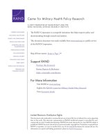
Fig. 2 3D pose of R
U3a3 inside the binding pocket
Page 8 of 13
Fig. 4 3D pose of R
U3a3 showing hydrogen bonding (yellow) with
GLN1194, ARG 912, GLY795, GLN 585 and π–π bonding (blue) with
PHE798
Fig. 5 3D pose of R
U3a1 inside the binding pocket
Fig. 3 2D pose of R
U3a3 inside the binding pocket
active compounds RU3a3 and RU3a1 were visualized and
compared with native rutin and standard drug Allopurinol. The residues of binding pocket involved in the interaction were reported as GLN 1194, ARG912, MET1038,
GLN1040, PHE798 and SER1080. Similar binding cavity
was observed by Li et al. during the docking analysis of
newly synthesized non-purine XO inhibitors [38].
Visual inspection of 3D poses of R
U3a3 displayed a
compact arrangement of polar and hydrophobic residues
around the ligand forming a narrow passage in XO binding cavity with a docking score/binding score of − 13.244
and binding energy − 91.242 kJ/mol. An interesting pi–pi
bonding was observed between benzene ring of phenyl
hydrazine and hydrophobic residue PHE 798 of active
site (Figs. 1, 2, 3). Along with this a strong hydrogen
bonding was observed between OH group of rutinoside
and polar residue GLN 1194 and negatively charged ARG
912 (Fig. 4). Similarly ARG 912 was found essential in the
study of Shen et al. during the comparison of curcumin
Malik et al. BMC Chemistry
(2019) 13:71
Page 9 of 13
Fig. 6 2D pose of R
U3a1 inside the binding pocket
Fig. 8 3D pose of rutin showing hydrogen bonding with GLN 1194
and MET1038
Fig. 7 3D pose of R
U3a1 showing hydrogen bonding with GLN 1194,
MET1038 and GLY 1039
derivatives with quercetin and leuteolin [39]. Another
hydrogen bonding was visualized between Chromene
moiety and the residues of active site namely GLY 795 ad
GLN585. Other hydrophobic amino acid residues closely
placed within the cavity were observed as PHE 798,
VAL1200, ALA1198, TYR 592, MET 1038 and ILE1229.
On the other hand, during the visualization of R
U3a1
the hydrogen bond was observed with OH group
of phenyl ring and hydrophobic residue MET 1038
(Figs. 5, 6). Another hydrogen bond was found similar to RU3a3 between OH group of rutinoside and polar
residue GLN1194 (Fig. 7). One more hydrogen bonding was observed between one of the OH group of
Fig. 9 3D pose of allopurinol showing hydrogen bonding with GLN
1194
dihydroxyphenyl ring and GLY1039. One more interaction was observed with the surrounding residue GLN 767
which forms a hydrogen bond with MOS 1328 (molybdenum metal ion) forming a closed channel to prevent
the entry of substrate in the binding site. Other residues
surrounding the ligand were observed as ARG 912, HIE
579, GLU 1261, ALA 1189 and ILE1198. When the 3D
poses of these two compounds were compared with the
native rutin structure, GLN 1194 forms 2 H-bonds, one
with the C=O group of rutin and another with OH group
Malik et al. BMC Chemistry
(2019) 13:71
Page 10 of 13
Table
3 In vitro xanthine oxidase inhibitory activity
of rutin derivatives
Compound
IC50 (µM) ± SEM
Compound
IC50 (µM) ± SEM
Rutin
20.867 ± 0.12
RU4b2
12.541 ± 0.45
RU3a1
09.924 ± 0.01
RU7c1
19.377 ± 0.38
RU3a2
07.905 ± 0.15
RU7c2
17.428 ± 0.01
RU3a3
04.870 ± 0.02
RU7c3
13.476 ± 0.25
RU4b1
15.037 ± 0.01
Allopurinol
10.410 ± 0.72
SEM, standard error of the mean
of rutinoside (Fig. 8). The amino acid residues GLU1261
and GLN 1194 were found to be interacted similarly
in the study of verbascoside by Wan et al. [40]. Beside
this one H-bond was formed between OH group of
chromene ring and MET1038. No pi–pi interaction was
in the native structure rutin. In case of Allopurinol, the
active site residues surrounding ligand were almost similar and placed near to MOS 1328. The hydrogen bond
was observed between purine ring of allopurinol and
GLN1194 (Fig. 9).
Fig. 11 Lineweaver–Burk plot for RU3a3 against different
concentrations
(RU7c1–RU7c3). All the compounds of hydrazine
series (RU3a1–RU3a3) were effective with IC50-values
ranging from 04.870 to 09.924 µM. Rutin hybridized
with phenyl hydrazine demonstrated highest activity
against xanthine oxidase. While thisemicarbazide and
phenylthiosemicarbazide derivatives of rutin showed
a slight decrease in activity indicating the role of sulfur group in diminishing the inhibition and NH–NH2
group in enhancing the activity of targeted enzyme.
Surprisingly, substitution of NH–NH2 with N
H2 group
leads to decrease of inhibitory activity. Ester derivatives of rutin synthesized after the hydrolysis of rutin
exhibited a weaker inhibition than the positive control
Allopurinol.
The results of in vitro activity showed 80% similarity
with the results of molecular docking with a few exceptions. In concordance with the screening and output of
In‑vitro xanthine oxidase inhibitory activity
In order to monitor the efficacy of different synthesized
rutin derivatives, xanthine oxidase inhibitory activity
was determined using xanthine oxidase activity assay
kit purchased from Sigma-aldrich Co. Allopurinol
(positive control) reported to inhibit xanthine oxidase
was also screened under identical conditions for comparison. The inhibition ratios revealed the xanthine
oxidase inhibitory activity of the synthesized rutin
derivatives and the results were summarized in Table 3.
As expected, these rutin derivatives exhibited remarkable activity comparable to the positive control. Based
on the in vitro activity; it was observed that hydrazine
(RU3a1–RU3a3) and anilline analogues ( RU4b1–RU4b2)
were considerably more effective than ester derivatives
OH
HO
Addition of thiosemicarbazide group
showed the XO inhibition moderately.
O
OH O
HO
O
HO
Rutin
Incorporation of hydrazide groups remarkably
increased the XO inhibitory action.
OH
O
OH
O
OH
O
OH
CH3
OH
Addition of phenylthiosemicarbazide group
significantly increased the XO inhibition.
Fig. 10 Structure activity relationship (SAR) of synthesized compounds
Presence of glycosidic 3-O-rutinoside linkage is
essential for the xanthine oxidase inhibitory potential, as
detachment of group diminished the XO inhibitory activity.
Malik et al. BMC Chemistry
(2019) 13:71
Page 11 of 13
Table 4 Km and Vmax values of xanthine
at different concentrations of RU3a3
S. no.
Fig. 12 Michaelis–Menten curve for RU3a3 at different
concentrations
molecular docking R
U3a3 comes out to be most active
rutin derivative showing very good interaction with xanthine oxidase at molecular level. Elimination of rutinoside from rutin to synthesize ester derivatives results in
a loss of potency with a threefold decrease of inhibitory
potential.
Structure activity relationship (SAR)
Few interesting notions about the relationship of activity and structures of synthesized compounds emerged
from the present research (Fig. 10): (A) Rutinoside
moiety seems to be important for the activity, as deletion of this leads to loss of activity could be seen from
xanthine oxidase inhibitory activity Table 3. Which
shows RU3a3 (Having rutinoside group) exhibited
highest activity with an IC50 value 04.870 µM among
all the compounds and RU7c1 showed lowest activity and fivefold decrease of activity with an IC50 value
19.377 µM. (B) Hydrazine derivatives were found to
be more effective than the aniline derivatives revealing the importance of NH–NH2 group. But substitution of sulfur group along with hydrazines decreases
the activity as in R
U3a3 and R
U3a2 and substitution of
phenyl group along with sulfur improves the activity
(RU3a1). (C) Substitution with ester group leads to a
decrease of inhibitory activity.
Enzyme kinetic analysis for XO‑inhibitory activity
To determine the XO-inhibitory mechanisms of newly
synthesized derivatives, we carried out kinetic studies
Conc. of RU3a3
(µM)
Km (µM)
oxidase
Vmax (µmol/min)
1.
0.0
27.21
119.6
2.
0.25
30.11
114.4
3.
0.5
32.90
108.2
4.
1.0
35.08
98.7
of most active compound R
U3a3 using Graph pad prism
software. Firstly Michaelis–Menten curve was plotted for
the enzyme activity at different concentrations of RU3a3
against different concentration of substrate (xanthine)
Fig. 11.
Then double reciprocal plot (Lineweaver–Burk) analysis was done in the presence (0.25, 0.5, and 1.0 µM)
and absence of R
U3a3 from in vitro data generated
during the oxidation of xanthine in presence of xanthine oxidase (Fig. 12). The x- and y axis intercepts of
the Lineweaver–Burk plot were utilized to calculate Km
and Vmax values of R
U3a3 at different concentrations
(Table 4).
A concentration-dependent decrease of
Vmax was
predicted in contrast to Km value which was found to
increasing when concentration of R
U3a3 was increased.
The intersection of linear straight lines drawn against
each concentration was located at same point, suggesting
that RU3a3 reacts in competitive manner during the inhibition of xanthine oxidase.
In‑vitro evaluation of antioxidant activity by DPPH
and H2O2 method
The antioxidant potential of newly synthesized compounds was evaluated by DPPH and Hydrogen peroxide
radical assay. The comparative analysis of
IC50 values
for both the assays was done and the results were found
to be impressive (Table 5). The results evinced a noteworthy inhibition of DPPH almost all the compounds
when compared with the positive control ascorbic acid.
In case of DPPH assay compound RU4b1 was demonstrated as most potent compound against oxidative stress
caused because of free radicals having an IC50 value of
02.647 ± 0.09 µM. Along with this compound RU3a1 also
showed very good antioxidant potential with an
IC50
value of 05.021 ± 0.10 µM. When the detailed structure
activity relationship was developed between these compounds, it was concluded that both the compounds having hydrazine linkage derived from phenyl hydrazine and
phenyl thiosemicarbazide. Similarly, during the analysis of hydrogen peroxide assay all the compounds with
hydrazines substitution showed very good antioxidant
Malik et al. BMC Chemistry
(2019) 13:71
Page 12 of 13
Table 5 Antioxidant activity of synthesized derivatives
by DPPH and H2O2 method
Compound
IC50 (µM) ± SEM
IC50 (µM) ± SEM
RU3a1
05.021 ± 0.10
09.134 ± 0.35
RU3a2
08.728 ± 0.02
04.146 ± 0.01
RU3a3
11.688 ± 0.01
06.561 ± 0.10
RU4b1
02.647 ± 0.09
09.863 ± 0.25
RU4b2
08.476 ± 0.25
04.378 ± 0.01
RU7c1
06.056 ± 0.13
14.731 ± 0.60
RU7c2
14.669 ± 0.01
12.126 ± 0.20
RU7c3
07.692 ± 0.42
17.884 ± 0.41
RU001
09.483 ± 0.08
18.623 ± 0.07
Ascorbic acid
22.195 ± 0.08
22.195 ± 0.08
SEM, standard error of the mean
potential having
IC50 in range of 04.146
± 0.01 to
09.134 ± 0.35 (Fig. 7). Compound R
U3a2 having phenyl
thiosemicarbazide substitution showed potential antioxidant activity among all the derivatives. Along with this
phenyl hydrazine substituted rutin derivative
(RU3a3)
also showed very good scavenging activity with an IC50
value of 06.561
±
0.10. When the detailed structure
activity relationship was developed between these compounds, it was concluded that both the compounds having hydrazine linkage derived from phenyl hydrazine and
phenyl thiosemicarbazide.
Conclusion
Starting from the structures of rutin as anti-XO hit previously identified, different series of novel analogues
were designed and synthesized to explore the structure–activity relationships associated with these xanthine oxidase inhibitors along with their antioxidant
potential. Different structural elements were identified
as essential for antioxidant and anti-XO properties, such
as the presence of rutinoside ( RU3a1, RU3a2 and R
U3a3)
comes out as important skeleton for the inhibitory
potential, presence of hydrazone linker along with phenyl group, while the associated xanthine oxidase inhibitory effect was found to follow a different trend for the
two series hydrazine
(RU3a1–3) and ester derivatives
(RU7c1–3). The newly synthesized derivatives with antioxidant and ani-XO IC50 values in the low micromolar
range and good selectivity indexes were identified. Contemporary synthetic efforts are focused towards the
insertion of the hydrazones and ester linkage by replacing the side linkage rutinoside of rutin with more stable groups while maintaining the overall length of new
derivatives. Molecular docking provide an improved
trail to design the new molecules with an avantgarde
stability and potency.
Additional file
Additional file 1. HNMR spectra of compound R
U3a3
Acknowledgements
The authors are highly thankful to the Head, Department of Pharmaceutical Sciences, M. D. University, Rohtak for providing essential facilities to
accomplish this research study. The authors are also thankful to Dr. Vinod
Devaraji Application Scientist Schrödinger LLC for his support to carry out the
computational work.
Authors’ contributions
Authors NM and AK have designed, synthesized and carried out the xanthine
oxidase inhibitory and antioxidant activity and the author PD, have carried
out the docking simulations with in silico ADMET studies. All authors read and
approved the final manuscript.
Funding
No funding received for this research work from outside sources.
Availability of data and materials
Not applicable.
Competing interests
The authors declare that they have no competing interests.
Author details
1
Faculty, Department of Pharmaceutical Sciences, M.D. University,
Rohtak 124001, India. 2 Laboratory for Preservation Technology and Enzyme
Inhibition Studies, Department of Pharmaceutical Sciences, M.D. University,
Rohtak, Haryana, India.
Received: 21 January 2019 Accepted: 2 May 2019
References
1. Berry CE, Hare JM (2004) Xanthine oxidoreductase and cardiovascular
disease: molecular mechanisms and pathophysiological implications. J
Physiol 555(3):589–606
2. Moriwaki Y, Yamamoto T, Higashino K (1997) Distribution and pathophysiologic role of molybdenum-containing enzymes. Histol Histopathol
12(2):513–524
3. Klinenberg JR, Goldfinger SE, Seegmiller JE (1965) The effectiveness of
the xanthine oxidase inhibitor allopurinol in the treatment of gout. Ann
Intern Med 62(4):639–647
4. Yu KH (2007) Febuxostat: a novel non-purine selective inhibitor of
xanthine oxidase for the treatment of hyperuricemia in gout. Recent Pat
Inflamm Allergy Drug Discov 1(1):69–75
5. Battelli MG, Bolognesi A, Polito L (2014) Pathophysiology of circulating xanthine oxidoreductase: new emerging roles for a multi-tasking
enzyme. Biochim Biophys Acta Mol Basis Dis 1842(9):1502–1517
6. Brass CA, Narciso J, Gollan JL (1991) Enhanced activity of the free radical
producing enzyme xanthine oxidase in hypoxic rat liver. Regulation and
pathophysiologic significance. J Clin Invest 87(2):424–431
7. Chambers DE, Parks DA, Patterson G, Roy R, McCord JM, Yoshida S, Parmley LF, Downey JM (1985) Xanthine oxidase as a source of free radical
damage in myocardial ischemia. J Mol Cell Cardiol 17(2):145–152
8. Desco MC, Asensi M, Márquez R, Martínez-Valls J, Vento M, Pallardó
FV, Sastre J, Viña J (2002) Xanthine oxidase is involved in free radical
production in type 1 diabetes: protection by allopurinol. Diabetes
51(4):1118–1124
9. Kuppusamy P, Zweier JL (1989) Characterization of free radical generation
by xanthine oxidase. Evidence for hydroxyl radical generation. J Biol
Chem 264(17):9880–9884
10. Dawson J, Walters M (2006) Uric acid and xanthine oxidase: future
therapeutic targets in the prevention of cardiovascular disease? Br J Clin
Pharmacol 62(6):633–644
Malik et al. BMC Chemistry
(2019) 13:71
11. Khosla UM, Zharikov S, Finch JL, Nakagawa T, Roncal C, Mu W, Krotova
K, Block ER, Prabhakar S, Johnson RJ (2005) Hyperuricemia induces
endothelial dysfunction. Kidney Int 67(5):1739–1742
12. Kaynar H, Meral M, Turhan H, Keles M, Celik G, Akcay F (2005) Glutathione
peroxidase, glutathione-S-transferase, catalase, xanthine oxidase, Cu–Zn
superoxide dismutase activities, total glutathione, nitric oxide, and
malondialdehyde levels in erythrocytes of patients with small cell and
non-small cell lung cancer. Cancer Lett 227(2):133–139
13. Griguer CE, Oliva CR, Kelley EE, Giles GI, Lancaster JR, Gillespie GY (2006)
Xanthine oxidase-dependent regulation of hypoxia-inducible factor in
cancer cells. Cancer Res 66(4):2257–2263
14. Kanellis J, Kang DH (2005) Uric acid as a mediator of endothelial dysfunction, inflammation, and vascular disease. Seminars in nephrology, vol 25.
New York, WB Saunders, pp 39–42
15. Miesel R, Zuber M (1993) Elevated levels of xanthine oxidase in serum of
patients with inflammatory and autoimmune rheumatic diseases. Inflammation 17(5):551–561
16. Wijermars LG, Bakker JA, de Vries DK, van Noorden CJ, Bierau J, Kostidis
S, Mayboroda OA, Tsikas D, Schaapherder AF, Lindeman JH (2016) The
hypoxanthine–xanthine oxidase axis is not involved in the initial phase of
clinical transplantation-related ischemia–reperfusion injury. Am J Physiol
Renal Physiol 312(3):F457–F464
17. Poles MZ, Bódi N, Bagyánszki M, Fekete É, Mészáros AT, Varga G, Szűcs S,
Nászai A, Kiss L, Kozlov AV, Boros M (2018) Reduction of nitrosative stress
by methane: neuroprotection through xanthine oxidoreductase inhibition in a rat model of mesenteric ischemia–reperfusion. Free Radic Biol
Med 120:160–169
18. Osada Y, Tsuchimoto M, Fukushima H, Takahashi K, Kondo S, Hasegawa
M, Komoriya K (1993) Hypouricemic effect of the novel xanthine oxidase
inhibitor, TEI-6720, in rodents. Eur J of Pharmacol 241(2–3):183–188
19. Krakoff IH, Meyer RL (1965) Prevention of hyperuricemia in leukemia
and lymphoma: use of allopurinol, a xanthine oxidase inhibitor. JAMA
193(1):1–6
20. Pacher PA, Nivorozhkin A, Szabó C (2006) Therapeutic effects of xanthine
oxidase inhibitors: renaissance half a century after the discovery of
allopurinol. Pharmacol Rev 58(1):87–114
21. Inkster ME, Cotter MA, Cameron NE (2007) Treatment with the xanthine
oxidase inhibitor, allopurinol, improves nerve and vascular function in
diabetic rats. Eur J Pharmacol 561(1–3):63–71
22. Sagor M, Taher A, Tabassum N, Potol M, Alam M (2015) Xanthine oxidase
inhibitor, allopurinol, prevented oxidative stress, fibrosis, and myocardial
damage in isoproterenol induced aged rats. Oxid Med Cell Longev. https
://doi.org/10.1155/2015/478039
23. Min HK, Lee B, Kwok SK, Ju JH, Kim WU, Park YM, Park SH (2015) Allopurinol hypersensitivity syndrome in patients with hematological malignancies: characteristics and clinical outcomes. Korean J Intern Med 30(4):521
24. Quach C, Galen BT (2018) HLA-B* 5801 testing to prevent allopurinol hypersensitivity syndrome: a teachable moment. JAMA Int Med
178(9):1260–1261
25. Takano Y, Hase-Aoki K, Horiuchi H, Zhao L, Kasahara Y, Kondo S, Becker MA
(2005) Selectivity of febuxostat, a novel non-purine inhibitor of xanthine
oxidase/xanthine dehydrogenase. Life Sci 76(16):1835–1847
26. Mayer MD, Khosravan R, Vernillet L, Wu JT, Joseph-Ridge N, Mulford DJ
(2005) Pharmacokinetics and pharmacodynamics of febuxostat, a new
non-purine selective inhibitor of xanthine oxidase in subjects with renal
impairment. Am J Ther 12(1):22–34
27. Nepali K, Singh G, Turan A, Agarwal A, Sapra S, Kumar R, Banerjee UC,
Verma PK, Satti NK, Gupta MK, Suri OP (2011) A rational approach for the
design and synthesis of 1-acetyl-3, 5-diaryl-4, 5-dihydro (1H) pyrazoles as
a new class of potential non-purine xanthine oxidase inhibitors. Bioorg
Med Chem 19(6):1950–1958
28. Becker MA, Kisicki J, Khosravan R, Wu J, Mulford D, Hunt B, MacDonald
P, Joseph-Ridge N (2004) Febuxostat (TMX-67), a novel, non-purine,
selective inhibitor of xanthine oxidase, is safe and decreases serum
urate in healthy volunteers. Nucleosides Nucleotides Nucleic Acids
23(8–9):1111–1116
Page 13 of 13
29. Khosravan R, Grabowski BA, Wu JT, Joseph-Ridge N, Vernillet L (2006)
Pharmacokinetics, pharmacodynamics and safety of febuxostat, a nonpurine selective inhibitor of xanthine oxidase, in a dose escalation study
in healthy subjects. Clin Pharmacokinet 45(8):821–841
30. Malik N, Dhiman P, Khatkar A (2017) In-silico design and ADMET studies of
natural compounds as inhibitors of xanthine oxidase (XO) enzyme. Curr
Drug Metab 18(6):577–593
31. Muhammad A, Arthur DE, Babangida S, Erukainure OL, Malami I, Sani
H, Abdulhamid AW, Ajiboye IO, Saka AA, Hamza NM, Asema S (2018)
Modulatory role of rutin on 2, 5-hexanedione-induced chromosomal and
DNA damage in rats: validation of computational predictions. Drug Chem
Toxicol 10:1–4
32. Roleira FM, Varela CL, Costa SC, Tavares-da-Silva EJ (2018) Phenolic
derivatives from medicinal herbs and plant extracts: anticancer effects
and synthetic approaches to modulate biological activity. Nat Prod Chem
57:115–156
33. Baldisserotto A, Vertuani S, Bino A, De Lucia D, Lampronti I, Milani R,
Gambari R, Manfredini S (2015) Design, synthesis and biological activity
of a novel Rutin analogue with improved lipid soluble properties. Bioorg
Med Chem 23(1):264–271
34. Gullon B, Lú-Chau TA, Moreira MT, Lema JM, Eibes G (2017) Rutin: a
review on extraction, identification and purification methods, biological
activities and approaches to enhance its bioavailability. Trends Food Sci
Technol 67:220–235
35. Friesner RA, Banks JL, Murphy RB, Halgren TA, Klicic JJ, Mainz DT, Repasky
MP, Knoll EH, Shelley M, Perry JK, Shaw DE (2004) Glide: a new approach
for rapid, accurate docking and scoring. 1. Method and assessment of
docking accuracy. J Med Chem 47(7):1739–1749
36. Dhiman P, Malik N, Verma PK, Khatkar A (2015) Synthesis and biological evaluation of thiazolo and imidazo N-(4-nitrophenyl)-7-methyl5-aryl-pyrimidine-6 carboxamide derivatives. Res Chem Intermed
41(11):8699–8711
37. Patel A, Patel A, Patel A, Patel NM (2010) Determination of polyphenols
and free radical scavenging activity of Tephrosia purpurea linn leaves
(Leguminosae). Pharmacogn Res 2:152–154
38. Li P, Tian Y, Zhai H, Deng F, Xie M, Zhang X (2013) Study on the activity
of non-purine xanthine oxidase inhibitor by 3D-QSAR modeling and
molecular docking. J Mol Struct 5(1051):56–65
39. Shen L, Ji HF (2009) Insights into the inhibition of xanthine oxidase by
curcumin. Bioorg Med Chem Lett 19(21):5990–5993
40. Wan Y, Zou B, Zeng H, Zhang L, Chen M, Fu G (2016) Inhibitory effect
of verbascoside on xanthine oxidase activity. Int J Biol Macromol
1(93):609–614
Publisher’s Note
Springer Nature remains neutral with regard to jurisdictional claims in published maps and institutional affiliations.
Ready to submit your research ? Choose BMC and benefit from:
• fast, convenient online submission
• thorough peer review by experienced researchers in your field
• rapid publication on acceptance
• support for research data, including large and complex data types
• gold Open Access which fosters wider collaboration and increased citations
• maximum visibility for your research: over 100M website views per year
At BMC, research is always in progress.
Learn more biomedcentral.com/submissions