Determination of adapalene in gel formulation by conventional and derivative synchronous fluorimetric approaches. Application to stability studies and in vitro diffusion test
Bạn đang xem bản rút gọn của tài liệu. Xem và tải ngay bản đầy đủ của tài liệu tại đây (1.32 MB, 10 trang )
Tolba and El‑Gamal Chemistry Central Journal (2016) 10:33
DOI 10.1186/s13065-016-0181-0
RESEARCH ARTICLE
Open Access
Determination of adapalene in gel
formulation by conventional and derivative
synchronous fluorimetric approaches.
Application to stability studies and in vitro
diffusion test
M. M. Tolba* and R. M. El‑Gamal
Abstract
Background: Adapalene is a retinoid analogue with actions similar to those of tretinoin. It is used in topical treat‑
ment of mild to moderate acne. A survey of the literature reveals that no spectrofluorimetric method has been
reported yet for determination of ADP, so it was thought necessary to develop a highly sensitive stability indicating
spectrofluorimetric method.
Results: Two highly sensitive spectrofluorimetric approaches were conducted for the assay of adapalene (ADP) in
its gel. In the first approach, ADP exhibits an intense native fluorescence at 389 nm after excitation at 312 nm using
borate buffer (pH 7.0)/ethanol system. This approach was successfully applied for routine analysis of ADP in its gel
and ideally suited to the in vitro diffusion test. To elucidate the inherent stability of ADP, bulk sample was subjected to
different stress conditions as specified by ICH guidelines. The acidic and oxidative degradation products were resolved
from the intact drug using second and first derivative synchronous fluorimetry at 346 and 312.45 nm, respectively
(the second approach). The synchronous fluorescence was scanned at Δ λ of 80 nm in case of acidic degradation and
at Δ λ of 100 nm in case of oxidative degradation. Good linearity was obtained for ADP over the range 2.0–14.0 ng/mL
with good correlation coefficient 0.999 in each approach. The approaches were carefully examined in terms of linear‑
ity, accuracy and precision. They were suitable for routine quality control laboratory. Moreover, the stability-indicating
power of the second approach was ascertained via forced degradation studies.
Conclusions: The proposed approaches were validated and successfully applied for the quantitative assay of a small
concentration of ADP in its pharmaceutical gel. The conventional spectrofluorimetry was ideally suited for in vitro
diffusion test. Stability studies were also conducted using different forced degradation condition according to ICH
recommendation.
Background
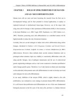
Chemically, adapalene (ADP) is 6-[3-(1-Adamantyl)4-methoxyphenyl]-2-naphthoic acid (Fig. 1). It is a
naphthoic acid derivative and retinoid analogue with
actions similar to those of tretinoin. It is used in
*Correspondence:
Department of Analytical Chemistry, Faculty of Pharmacy, University
of Mansoura, Mansoura 35516, Egypt
topical treatment of mild to moderate acne [1]. ADP
is a subject of monograph in European Pharmacopoeia
[2].
Only few analytical methods were reported for the
assay of ADP. These methods include high performance
liquid chromatography (HPLC) [3–8]. In addition, only
two derivative spectrophotometric methods were applied
for ADP determination in bulk drug and pharmaceutical
dosage form [9] or in liposomes [10].
© 2016 The Author(s). This article is distributed under the terms of the Creative Commons Attribution 4.0 International License
( which permits unrestricted use, distribution, and reproduction in any medium,
provided you give appropriate credit to the original author(s) and the source, provide a link to the Creative Commons license,
and indicate if changes were made. The Creative Commons Public Domain Dedication waiver ( />publicdomain/zero/1.0/) applies to the data made available in this article, unless otherwise stated.
Tolba and El‑Gamal Chemistry Central Journal (2016) 10:33
Page 2 of 10
fluorescence spectra of ADP and its degradation products were observed, therefore, we resorted to derivative synchronous fluorimetry (DSF). Where, ADP was
resolved from its acidic and oxidative degradation products by second (SDSF) and first (FDSF) derivative synchronous fluorimetry at 346 and 312.45 nm, respectively.
Experimental
Apparatus
Fig. 1 The structural formula for adapalene (ADP)
International Conference on Harmonization (ICH)
guideline Q1A on stability testing of new drug substances
and products requires that stress testing be carried out
to elucidate the inherent stability features of the active
substance which may be changed during storage and so,
ensure high quality, safety, and efficacy of the pharmaceutical product [11].
Moreover, the development of in vitro release study
serves as a good quality control tool to ensure batch to
batch uniformity and screen experimental formulation
during the product development. Determination of the
value of in vitro release helps to cross check the product
quality and product comparison [12].
A comprehensive literature survey revealed that no
spectrofluorimetric method has been reported yet for
the determination of ADP in its gel or in presence of its
degradation products. The reported methods concerned
with the stability of ADP are expensive, time consuming, sophisticated HPLC techniques [3–6]. Most of these
methods suffer from low sensitivity which restricted the
determination of ADP in low concentration in presence
of its degradation products. Moreover, some of these
methods showed narrow linearity range [5, 6] or failed to
separate the acidic and oxidative degradation products
from the parent drug [3, 6]. Regarding the pharmaceutical application, none of these methods are applicable to
in vitro dissolution test which is an important issue in
quality control laboratories.
Therefore, it was thought necessary to develop sensitive
stability indicating spectrofluorimetric method for determination of ADP and applicable to in vitro diffusion test.
In our study, two extremely sensitive spectrofluorimetric approaches were explored for the analysis of a very
small concentration of ADP down to 2.0 ng/mL. ADP
shows a strong native fluorescence at 389/312 nm (λem/
λex) in borate buffer (pH 7.0)/ethanol system. Depending on this fact, the first approach was conducted and
extended to study the inherent stability of ADP and the
in vitro diffusion test. Great overlapping between the
–– All fluorescence measurements were recorded with
a Perkin-Elmer UK model LS 45 luminescence spectrometer, equipped with a 150 W Xenon arc lamp,
grating excitation and emission monochromators and
a Perkin Elmer recorder. The slit widths were 10 nm for
both excitation and emission, and the photomultiplier
voltage was set to automatic option. Derivative spectra
were obtained using fluorescence data manager software, FL WINLAB, Version 4.00.02, Copyright 2001,
Perkin Elmer, Inc., UK.
–– A Consort P-901 pH-meter was used for pH measurements.
–– Thermostatically controlled shaking water bath (Grant
instrument Cambridge Ltd., Barrington Cambridge B2,
5002, England).
–– Modified Franz diffusion cell.
–– CAMAG UV-lamp, S/N 29000, dual wavelength
254/366 nm, 2 × 8 Watt (Switzerland) was used in the
UV-degradation study.
Materials and reagents
All the chemicals used were of analytical reagent grade,
and the solvents were of HPLC grade.
•• Adapalene was supplied by Glenmark (Cairo, Egypt)
with a certified purity of 99.70 % and was used as
received without further purification.
•• Adapalene® gel; batch # 011371, labeled to contain
0.1 % ADP (product of Borg Pharmaceutical Ind.,
Alexandria, Egypt). It was purchased from the local
pharmacy.
•• Ethanol (Fisher Scientific UK, Loughborough, Leics,
UK).
•• Acetonitrile, n-propanol and methanol were obtained
from Tedia (USA).
•• Boric acid, sodium acetate trihydrate, acetic acid
96 %, acetone, dimethyl formamide (DMF), methyl
cellulose (MC), tween-80, sodium hydroxide, hydrogen peroxide (30 %) and hydrochloric acid (32 %)
were all obtained from El-Nasr Pharmaceutical
Chemicals Company (ADWIC) (Abu Zaabal, Egypt).
•• Sodium dodecyl sulphate (SDS; 95 %), β-cyclodextrin
(β-CD), cetrimide (CTAB; 99 %) were purchased
from Winlab (UK).
Tolba and El‑Gamal Chemistry Central Journal (2016) 10:33
Standard solutions
Stock solution equivalent to 100.0 µg/mL of ADP was
prepared by dissolving 10.0 mg in 100.0 mL ethanol.
Other standard solution equivalent to 100.0 ng/mL was
prepared by appropriate dilution of the stock solution
with the same solvent. The solutions were found to be
stable for at least 7 days without alteration when kept in
the refrigerator.
Procedures
Construction of calibration graphs
Aliquots of ADP standard solution were transferred into
a series of 10 mL volumetric flasks so that the final concentration was in the range of 2.0–14.0 ng/mL. Then, 2 mL
borate buffer (0.2 M, pH 7.0) was added to each flask followed by completing the volume with ethanol and mixing
well. For the first approach, the fluorescence intensities of
the solutions were measured at 389 nm after excitation
at 312 nm. While, the second approach involved recording the synchronous fluorescence spectra of the solutions
by scanning at Δ λ = 100 nm and Δ λ = 80 nm in case of
acidic and oxidative degradation, respectively. The second
and first derivative synchronous fluorescence spectra were
derived. The peak amplitudes of the second (2D) or the
first (1D) derivative spectra were estimated at 346 nm and
312.45 nm for acidic and oxidative degradation, respectively. A blank experiment was performed simultaneously.
The relative fluorescence intensity (RFI) or the peak amplitude of the second (2D) or first (1D) derivative technique
was then plotted against the final drug concentration in
ng/mL to get the calibration graphs.
Analysis of ADP in semisolid pharmaceutical gel
An accurately weighed amount of the gel (0.1 g) was
transferred into a clean dry 100 mL beaker and about
80 mL of ethanol was added. The flasks were placed in a
water bath at 50 °C for 15 min followed by cooling. The
contents were quantitatively transferred into 100 mL
volumetric flask, completed to the mark with the same
solvent and filtered through cellulose acetate syringe filter. The subsequent dilutions were performed via diluting
an appropriate volume of this solution with ethanol and
the procedures described under “construction of calibration graphs” were then applied. The nominal content was
determined either from the previously plotted calibration
graph or using the corresponding regression equation.
Procedure for in vitro diffusion test
The release of ADP in 65 % hydroethanolic solution was
carried out using a modified diffusion cell according to
the method adopted by Deo et al. [12].
The donor half-cell consists simply of a glass tube
with an open end (3 cm in diameter) on which a
Page 3 of 10
semipermeable cellulose membrane was stretched and
fixed by rubber band to prevent leakage of water.
Five grams of Adapalene® gel were accurately weighed
and thoroughly spread on the membrane to occupy 3 cm
diameter circle. The donor cells were then immersed
upside-down in 250 mL beaker containing 50 mL hydroethanolic solution (65 %) (receptor compartment) which
was preheated and maintained at 37 ± 1 °C using thermostatically controlled water bath. The tubes height
was adjusted, so that the membrane was just below the
surface of the release medium. The whole assembly was
shaken at 25 strokes per minute during the entire time of
diffusion.
At the specified time interval, 2 mL was withdrawn
from the receiver compartment and replaced by equal
volume of fresh hydroethanolic solution and thus keeping a constant volume. Dilution of 0.1 mL was performed
with ethanol up to 10 mL in a volumetric flask. Appropriate volumes (0.1 mL) of this solution were then transferred to 10 mL volumetric flask, 2 mL of 0.2 M borate
buffer (pH 7.0) was added and the volume was completed
to the mark with ethanol. The % released amounts of
ADP were determined by conventional fluorimetry at
389 nm after excitation at 312 nm. Triplicate experiments
were carried out for each sample.
Preparation of the degradation products
For degradation studies, working solution equivalent
to 0.5 µg/mL was prepared by appropriate dilution of
the stock solution with ethanol. Aliquots of 5 mL of this
solution (equivalent to 2.5 µg) were then transferred
into series of small conical flasks for alkaline, acidic and
oxidative degradation. Then the following steps were
performed:
For alkaline and acidic degradation Aliquots of 5 mL of
2 M NaOH or different molarities of HCl (0.2–1 M) were
added to the flasks. The solutions were heated in a boiling water bath under reflux for different time intervals
(10–60 min). At the specified time, the contents of each
tube were cooled, neutralized to pH 7.0 and the solutions
were then transferred into a series of 25 mL volumetric
flasks. The volumes were completed with ethanol. Aliquots of these solutions (1.4 mL) were transferred into a
series of 10 mL volumetric flasks, followed by addition of
2.0 mL of 0.2 M borate buffer (pH 7.0) and completing the
volumes to the mark using ethanol. The procedure under
“construction of calibration graphs” for the first approach
was then conducted.
For oxidative degradation Five milliliters of 5–30 %
H2O2 were added to each flask. The solutions were then
heated in a thermostatically controlled water bath at 80 °C
Tolba and El‑Gamal Chemistry Central Journal (2016) 10:33
For day and UV light degradation Suitable aliquots of
the working solution (equivalent to 2.5 µg) were transferred into 25 mL volumetric flasks and completed to
volume with ethanol. The flasks were left in day light
or exposed to UV-light at 254 and 366 nm for 12 h in a
wooden cabinet, where the distance between the source
and the sample solution was kept at 15 cm. Aliquots of the
solution (1.4 mL) were transferred into a series of 10 mL
volumetric flasks and the procedure under ‘‘construction
of calibration graphs’’ was then applied.
Results and discussion
From scanning the fluorescence spectra of ADP, it was
found that ADP showed three different excitation peaks
at wavelengths of 230, 265 and 312 nm and only one
emission peak at 389 nm. In selection of excitation wavelength, we emphasize on the linearity and reproducibility
of the calibration graph even if the other excitation wavelengths showed higher sensitivity relative to the selected
one. Consequently, ADP fluorescence was measured at
389 nm after excitation at 312 nm (Fig. 2). Fortunately,
the approach was extremely sensitive and allowed the
determination of ADP in its pharmaceutical gel as alternative to the reported sophisticated HPLC methods. The
high precision and accuracy of the approach made it ideal
for in vitro diffusion test. Moreover, the inherent stability of ADP was investigated using the forced degradation
studies.
After preliminary studies, neither conventional nor synchronous fluorimetry was able to resolve ADP from the
degradation bands (Figs. 2, 3). Accordingly, we resorted
to derivative synchronous fluorimetry which efficiently
separated ADP from its acidic degradation product using
SDSF and allowed its quantitation at 346 nm (Fig. 4a).
Similarly, ADP was determined at 312.45 nm after application of FDSF to resolve its band from the oxidative degradation product (Fig. 4b). Thus, the stability-indicating
power of this approach was ascertained.
a
455.8
Synchronous fluorescence intensity
for different time intervals (10–60 min). At the specified
time intervals, the contents of each flask were cooled and
transferred to 25 mL volumetric flask. The volumes were
completed to the mark with ethanol. The procedure mentioned under “construction of calibration graphs” for the
first approach was then applied.
Page 4 of 10
(1)
400
350
300
(2)
250
200
150
100
50
0.8
200.0 220
240
260
280
300
320
340
360
380
400
420 440
nm
Synchronous fluorescence intensity
400.0
300
250
200
(2)
150
100
50
0.3
200.0
Fig. 2 Fluorescence spectra of: A, A′ ADP (14.0 ng/mL) in borate
buffer (pH 7.0)/ethanol system. B, B′ Blank (borate buffer (pH 7.0)/
ethanol system) where: (A, B) Excitation spectra. (A′, B′) Emission
spectra
b
(1)
350
220
240
260
280
300
320
nm
340
360
380
400
420
Fig. 3 Synchronous fluorescence spectra of: (a) (1) ADP (14.0 ng/mL)
(2) acidic degradation product; at Δ λ = 80 nm (b) (1) ADP (14.0 ng/
mL) (2) oxidative degradation product; at Δλ = 100 nm
Tolba and El‑Gamal Chemistry Central Journal (2016) 10:33
(1)
50
40
30
20
10
a
g
f
e
d
c
b
a
(2)
0
-10
D2
-20
-30
-40
-50
-60
600
Relative fluorescence intensity (RFI)
60.0
Page 5 of 10
400
200
0
3.0
-70
4.0
5.0
-80
pH
6.0
7.0
8.0
Fig. 5 Effect of pH on the native fluorescence intensity of ADP
(14.0 ng/mL)
-90
-105.0
294.0
320
340
360
380
400
nm
b
86.0
80
70
(2)
60
50
Table 1 Effect of diluting solvents on the relative fluorescence intensity of adapalene (14.0 ng/mL)
Diluting solvents
Relative fluorescence
intensity (RFI)
Ethanol
492
n-propanol
451
Methanol
450
c
Water
124
-50
d
Acetonitrile
165
-60
e
-70
f
Dimethyl formamide
40
30
20
10
D1 0
a
b
Acetone
380
1
nm 400
Fig. 4 Second and first derivative synchronous fluorescence spectra
of: (a) (1) (a–g) of ADP (2.0, 4.0, 6.0, 8.0, 10.0, 12.0, 14.0 ng/mL) at
346 nm (2) acidic degradation product (b) (1) (a–g) of ADP (2.0, 4.0,
6.0, 8.0, 10.0, 12.0, 14.0 ng/mL) at 312.45 nm (2) oxidative degradation
product
Various experimental parameters affecting the fluorescence intensities of ADP were investigated to achieve the
maximum sensitivity and the ultimate selectivity.
RFI
Optimization of experimental conditions
600
400
200
Effect of pH
o
su
rf
ac
ta
nt
0
N
To study the influence of pH on the fluorescence behavior of ADP, different types of buffers covering the whole
pH range were tested, such as 0.2 M acetate buffer (pH
3.6–6) and 0.2 M borate buffer (pH 6.5–10), in addition
to 0.1 M HCl and 0.1 M NaOH (Fig. 5). It was noticed
that increasing the pH resulted in a proportional increase
in the RFI of the drug up to 6.5, and then remained constant up to pH 8.0, after which a precipitation occurred at
pH 9 and 10. Therefore, borate buffer pH 7.0 was selected
as the optimum pH giving the highest sensitivity. Also,
trials were made by replacement of buffer by 0.1 M HCl
or NaOH. Indeed, utilizing 0.1 M NaOH resulted in a
ee
n
360
C
340
Tw
320
M
300
B
280
TA
g
260
51
C
-80
-90.0
235.0
(1)
D
-40
βC
-30
D
S
-20
S
-10
Fig. 6 Effect of surfactant on the native fluorescence intensity of
ADP (14.0 ng/mL)
high RFI but almost equal to the fluorescence intensity
achieved upon utilizing 0.2 M borate buffer with pH
7.0. And as a well-known fact that using buffer is more
favorable to resist changes in pH values, so borate buffer
Tolba and El‑Gamal Chemistry Central Journal (2016) 10:33
Page 6 of 10
was selected. On the contrary, using 0.1 M HCl showed
a marked quenching of the RFI of ADP which may be
attributed to its degradation.
Effect of diluting solvent
Various solvents including water, methanol, acetonitrile, ethanol, n-propanol, dimethyl formamide (DMF)
and acetone were tested to choose the most convenient
diluting solvent. It is noticeable from Table 1 that ethanol gave the highest fluorescence intensity so; it was the
solvent of choice. Also, n-propanol could be used. High
blank reading was observed in case of using methanol at
this wavelength. Water and acetonitrile showed a marked
decrease in the fluorescence intensity so they were not
selected. The initiated intersystem crossing process
caused by DMF made it unsuitable for ADP determination as it resulted in a marked decrease in the fluorescence intensity of ADP in addition to high blank reading
[13]. Complete quenching of the fluorescence intensity
was attained upon using acetone.
Effect of surfactant
Study of the impact of the surfactant was accomplished
using 0.5 % aqueous solutions of anionic surfactant (SDS),
cationic surfactant (CTAB), non-ionic surfactant (tween80) and different macromolecules such as methyl cellulose, and β-CD. As shown in Fig. 6, these surfactants
didn’t significantly affect the fluorescence intensity of
Table 2 Analytical performance data for the proposed approaches
Parameters
The first approach
at 389 nm
The second approach
SDSF at 346 nm
FDSF at 312.45 nm
Linearity range (ng/mL)
2.0–14.0
2.0–14.0
2.0–14.0
Intercept (a)
−4.714
−1.212
5.171
Slope (b)
35.357
3.975
3.729
Correlation coefficient (r)
0.9999
0.9998
0.9998
SD of residuals (Sy/x)
1.59
0.34
0.31
SD of intercept (Sa)
1.34
0.29
0.26
SD of slope (Sb)
0.15
0.03
0.03
Percentage relative standard deviation, % RSD
0.42
0.88
0.76
Percentage relative error, % error
0.16
0.33
0.29
Limit of detection, LOD (ng/mL)
0.13
0.24
0.23
Limit of quantitation, LOQ (ng/mL)
0.38
0.73
0.69
Table 3 Application of the proposed approaches for the determination of adapalene in pure form
Parameter Amount
First approach at 389 nm
taken (ng/mL)
Amount
% found
found (ng/mL)
ADP
Second approach
Amount found (ng/mL)
SDSF
at 346 nm
Comparison method [3]
% found
FDSF
SDSF
at 312.45 nm at 346 nm
FDSF
at 312.45 nm
Amount
% found
found (µg/mL)
2.0
2.00
100.00
1.991
1.992
99.54
99.62
20.0
99.22
4.0
4.008
100.20
4.001
3.977
100.02
99.43
30.0
99.88
40.0
101.15
6.0
5.988
99.80
5.976
5.962
99.59
99.36
8.0
8.053
100.66
8.134
8.000
101.68
100.00
10.0
9.948
99.48
9.915
10.146
99.15
101.46
12.0
11.956
99.63
11.918
12.023
99.31
100.19
14.0
14.049
100.35
14.066
13.900
100.47
99.29
Mean
100.02
99.97
99.91
SD
0.42
0.88
0.76
t
1.06 (2.31)*
0.73 (2.31)
0.93 (2.31)
F
2.88 (5.14)*
1.53 (19.33)
1.16 (19.33)
N.B. Each result is the average of three separate determinations
* The values between parentheses are the tabulated t and F values at P = 0.05 [15]
100.08
0.98
Tolba and El‑Gamal Chemistry Central Journal (2016) 10:33
ADP. Consequently, they were not incorporated in the
procedure.
Selection of optimum Δ λ in the second approach
Selection of optimum Δ λ is a very essential criterion
which should be considered during scanning of the
synchronous fluorimetry as it may significantly affect
sensitivity, resolution and symmetry of the bands. Consequently, a wide range of Δ λ (20–140 nm) was investigated. Good band shapes and adequate sensitivity were
obtained upon using Δ λ of 80 and 100 nm in case of
acidic and oxidative degradation, respectively. Lower and
higher values of Δ λ than the optimum ones showed low
fluorescence intensity for ADP and its degradation products. However, very low and very high Δ λ values caused
irregularities in the spectral shape.
Validation of the approaches
Linearity
It was investigated via replicate analysis of seven standard
concentrations of ADP; 2.0, 4.0, 6.0, 8.0, 10.0, 12.0, 14.0 ng/
mL. Calibration graphs of ADP were constructed by plotting either the RFI or the peak amplitude of (2D) or (1D)
against the drug concentration in ng/mL. The results of
the regression equations and correlation coefficients were
abridged in Table 2. In the approaches, good linearity for
ADP was achieved in the range of 2.0–14.0 ng/mL as indicated by higher value of correlation coefficients (>0.999).
Limit of quantitation (LOQ) and limit of detection (LOD)
These analytical parameters were computed by the equations specified by ICH Q2R1 recommendations [14] and
were presented in Table 2:
LOQ = 10 Sa /b and
LOD = 3.3 Sa /b
where Sa = standard deviation of the intercept of the calibration curve and b = slope of the calibration curve.
Accuracy and precision
The results of the present approaches were statistically
compared with those of the comparison method [3] to
ascertain these analytical features. No significant difference was observed using Student’s t test and variance
ratio F test [15]. The excellent recovery values demonstrated that the approaches were sufficiently accurate
over the specified range (Table 3).
The comparison HPLC method [3] was carried out on
C8 column using a blend of methanol: ammonium acetate buffer pH 4.0 (80:20, v/v) as a mobile phase and UV
detection at 270 nm.
The repeatability and intermediate precision of the applied
approaches were determined using three concentrations
Page 7 of 10
and three replicates of each concentration within the same
day or 3 different days. Small values of the relative standard deviations gave a good indication for the high precision
which characterizes these approaches (Table 4).
Selectivity
The proposed approaches were found to be selective for
ADP in its gel, where, satisfactory results were obtained
and no interference was observed (Table 5). Moreover,
the derivative synchronous fluorimetry was found to be
selective for ADP in presence of its acidic and oxidative
degradation products.
Applications
Adapalene® gel analysis
The present fluorimetric approaches were applied to the
analysis of ADP. Four samples were determined and three
replicate of each one. Satisfactory results were obtained
Table 4 Precision data for the determination of adapalene
applying the proposed approaches
Amount taken (ng/mL)
% found
% RSD
% error
6.0
100.11 ± 0.85
0.85
0.49
8.0
100.55 ± 0.37
0.37
0.21
10.0
99.88 ± 0.17
0.17
0.10
6.0
101.05 ± 1.15
1.14
0.66
8.0
99.93 ± 1.22
1.22
0.70
10.0
99.85 ± 0.55
0.55
0.32
6.0
99.67 ± 0.77
0.77
0.44
8.0
99.50 ± 0.25
0.25
0.14
10.0
100.02 ± 0.31
0.31
0.18
6.0
100.04 ± 0.81
0.81
0.47
8.0
99.02 ± 0.97
0.98
0.57
10.0
98.28 ± 1.13
1.15
0.66
6.0
99.20 ± 0.91
0.92
0.53
8.0
100.08 ± 0.77
0.77
0.44
10.0
99.31 ± 0.42
0.42
0.24
6.0
101.19 ± 1.37
1.35
0.78
8.0
99.74 ± 0.90
0.90
0.52
10.0
98.11 ± 1.09
1.11
0.65
The first approach at 389 nm
Intraday
Interday
The second approach
SDSF at 346 nm
Intraday
Interday
FDSF at 312.45 nm
Intraday
Interday
Tolba and El‑Gamal Chemistry Central Journal (2016) 10:33
Page 8 of 10
Table 5 Application of the proposed approaches for the determination of adapalene in its pharmaceutical Adapalene®
gel
Parameter
Amount
taken (ng/
mL)
Adapalene®
gela
6.0
(0.1 % ADP)
First approach at 389 nm Second approach
Amount
found
(ng/mL)
5.957
Comparisonmethod [3]
% found Amount found (ng/mL)
SDSF
at 346 nm
99.28
5.932
% found
FDSF
SDSF
at 312.45 nm at 346 nm
6.072
FDSF
at 312.45 nm
98.87
101.20
Amount
found
(µg/mL)
% found
20.0
100.07
8.0
8.082
101.03
7.922
8.026
99.03
100.33
30.0
98.80
Batch # 011371 10.0
10.007
100.07
10.022
9.905
100.22
99.05
40.0
101.03
12.0
11.900
99.17
11.972
11.986
99.77
99.88
99.89
99.47
100.12
99.97
S.D.
0.86
0.63
0.90
1.12
t
0.11
0.75
0.19
F
1.69
3.11
1.56
Mean
N.B. Each result is the average of three separate determinations
The tabulated t and F values are 2.57 and 9.55, respectively at P = 0.05 [15]
a
Product of Borg Pharmaceutical Ind., Alexandria, Egypt
In‑vitro diffusion test
The easy and excellent applicability of the first approach
for the determination of ADP in its pharmaceutical gel
encouraged us to conduct the in vitro diffusion test and
to study the percentage of its release. In-vitro release profile of ADP from its gel in hydroethanolic solution (65 %)
is shown in Fig. 7. It was found that increasing the time
resulted in a subsequent increase in the % release up to
3.5 h where it reach the maximum (30 %) after which no
more release was attained.
Stability studies
In forced degradation studies, the parent drug or the drug
product is subjecting to different stress conditions. These
studies play an essential role in establishing the intrinsic
stability of the drug and hence help in selecting the suitable pharmaceutical dosage forms, solving the problems
which may be appeared during the stages of storage and
packaging. Main degradation pathways involve acidic/
basic hydrolysis, oxidative, and photolytic-degradation.
40
30
% Release
for ADP in a good agreement with the label claims and
no interference was observed (Table 5).
Statistical analysis of the results obtained by the proposed approaches and the comparison [3] method
using Student’s t test and variance ratio F test at 95 %
confidence level [15] revealed no significant difference
between the performance of the approaches regarding
the accuracy and precision, respectively.
20
10
0
0
1
2
3
4
Time, hour
5
6
7
8
Fig. 7 The % release of ADP from adapalene® gel
According to ICH [11] guidelines, only small amount of
data concerned with methodology and basics for establishing a new forced degradation study was available.
Also, the required amount of the applied stress is not
sufficiently discussed. Stress conditions should be realistic and not excessive. So, our target was concentrated
on giving an appropriate and relevant degradation about
(10–30 %) and separating the produced degradation
products from the parent drug as possible.
Different forced degradation studies were tried to
study the inherent stability of ADP. ADP was not susceptible to alkaline degradation as evidenced by boiling
with 2 M NaOH for 2 h. On the other hand, ADP was
strongly affected by acidic condition as preliminary studies showed that almost all the drug was degraded upon
Tolba and El‑Gamal Chemistry Central Journal (2016) 10:33
Page 9 of 10
boiling with 1 M HCL for only 10 min. So, we directed
to use 0.3 M HCl instead where 28 % of the drug was
degraded after boiling for 10 min.
Similarly, ADP was susceptible to oxidative conditions
and the percentage of degradation was dependent on the
strength of the used hydrogen peroxide. After heating
ADP with 30 % H2O2 solution at 80 °C for 10 min., about
30 % of ADP was degraded.
The impact of day light on the stability of ADP was
checked after leaving its ethanolic solution on the bench
in day light for 12 h and no considerable degradation
was observed. Also, the solution of ADP in ethanol was
exposed to UV light at two different wave lengths; 254
and 366 nm for 12 h to manifest the effect of UV light
on its stability. About 25 % of ADP was degraded at the
mentioned wavelengths. Unfortunately, the photolytic
degradation product was not separated from the intact
drug in contrast to acidic and oxidative degradation
products.
Pathway of ADP degradation
The molecular rigidity is a key element in enhancing the
fluorescence behavior of many compounds as it prevents the internal conversion. Loss of rigidity of ADP
is proposed under acidic stress condition via breakage
of adamantine group and consequently fluorescence
diminishes. As ADP being a naphthalene derivative, its
photolytic irradiation may result in the degradation of
naphthalene moiety into the corresponding 2-formyl
cinnamaldehyde one [16, 17]. While, upon heating ADP
with 30 % H2O2 it undergoes oxidative degradation with
the formation of 1,4-naphthoquinone derivative. The
pathway of the degradation process was proposed and
postulated in Scheme 1.
Conclusions
The proposed approaches described highly sensitive and
simple fluorimetric methods for the quantitative assay of
a small concentration of ADP in its pharmaceutical gel
alternative to the reported sophisticated HPLC methods
[3–8] or the non-sensitive derivative spectrophotometric ones [9, 10]. The conventional spectrofluorimetry was
ideally suited for in vitro diffusion test. Stability studies
were also conducted using different forced degradation
condition according to ICH recommendation. Fortunately, second and first derivative synchronous fluorimetry were capable to resolve the band of ADP from its
acidic and oxidative degradation products at 346 and
312.45 nm after recording the synchronous fluorimetry
using Δ λ of 80 and 100 nm, respectively. Consequently,
the stability indicating power of this approach can be
assessed.
O
O
OH
H
O
UV light
H 3C
O
-CO2
H3C
H
O
H2 O2
O
HCl
OH
O
H 3C
O
OH
H3C
O
O
+
Scheme 1 The assumed pathways of acidic, oxidative and UV light degradation of ADP
O
Tolba and El‑Gamal Chemistry Central Journal (2016) 10:33
Abbreviations
ADP: adapalene; LOD: limit of detection; LOQ: limit of quantitation; ICH: Inter‑
national Conference on Harmonization.
Authors’ contributions
MMT proposed the subject, participated in the assay design, literature review,
conducted the validation of the assay, analysis of the samples, participated
in the results, discussion and participated in preparing the manuscript. RME
participated in the study design, assay design, conducted the validation of the
assay, analysis of the samples and participated in the results and discussion.
Both authors read and approved the final manuscript.
Competing interests
The authors declare that they have no competing interests.
Received: 26 February 2016 Accepted: 17 May 2016
References
1. Sweetman SC (ed) (2011) Martindale, the complete drug reference, 37th
edn. The Pharmaceutical Press, London
2. The European Pharmacopoeia (2010), 7th edn. vol 2, Council of Europe,
Strasbourg Cedex, pp 1324–1326
3. Patel RB, Patel MR, Chaudhari MD (2014) Stability indicating high perfor‑
mance liquid chromatographic method for estimation of adapalene in
tablet formulation. J Liq Chromatogr Relat Technol 37(3):379–390
4. Pujeri SS, Khader AMA, Seetharamappa J (2012) Development and valida‑
tion of a stability-indicating LC method for the assay of adapalene in bulk
drug and pharmaceutical formulations. J Anal Chem 67(6):585–590
5. Martins LA, Meneghini LZ, Junqueira CA, Ceni DC, Bergold AMA (2011)
Simple HPLC-dad method for determination of adapalene in topical gel
formulation. J Chromatogr Sci 49(10):796–800
Page 10 of 10
6. Mudasir M, Tabassum N, Ali J, Jan R (2008) Development and validation
of stability indicating HPLC method for adapalene and benzoic acid in
pharmaceutical gel formulations. Anal Chem 7(11):795–800
7. Zhang C, Zhao Y, Han C, Guo X (2008) Simultaneous determination
of adapalene, 2-phenoxyethanol and methyl-4-hydroxybenzoate in
adapalene gels using high performance liquid chromatography. Se pu
26(5):640–642
8. Ruhl R, Nau H (1997) Determination of adapalene (CD271/Differin) and
retinol in plasma and tissue by online solid-phase extraction and HPLC
analysis. Chromatographia 45:269–274
9. Adhikari L, Jagadev S, Sahu S, Moitra SK, Murthy PN (2012) Derivative
spectrophotometric determination of adapalene in its bulk and pharma‑
ceutical dosage form. Asian J Chem 24(3):1094–1096
10. An Y, Liu L (2010) Determination of adapalene content in liposomes
by first derivative UV spectrophotometry. Zhongguo Xinyao Zazhi
19(6):534–536
11. ICH Harmonized Tripartite Guidelines. Stability testing of new drug sub‑
stances and products, Q1A (R2) (2003). />MEDIA419.pdf. Accessed 25 Oct 2010
12. Deo SS, Inam F, Karmarkar NP (2013) Analytical method development
for determination of performance of adapalene in adapalene 0.1% gel
formulation using manual diffusion cell. Chem Sci Trans 2(1):251–257
13. Skoog DA, Holler FJ, Crouch SR (2007) Principles of instrumental analysis,
6th edn. Thomson, Belmont, p 406
14. ICH Harmonized Tripartite Guideline, validation of analytical procedures:
text and methodology, Q2(R1), Current Step 4 Version, Parent Guidelines
on Methodology 1996, Incorporated in 2005. />media/MEDIA417.pdf. Accessed 15 Feb 2008)
15. Miller JN, Miller JC (2005) Statistics and chemometrics for analytical
chemistry, 5th edn. Pearson Education Limited, Harlow, pp 39–73
16. Felix A, Andrew A, Mededode A (2014) Heterogeneous photocatalytic
degradation of naphthalene using periwinkle shell ash: effect of operating
variables, kinetic and isotherm study. South Afr J Chem Eng 19(1):31–45
17. Lair A, Ferronato C, Chovelon J, Herrmann J (2008) Naphthalene degrada‑
tion in water by heterogeneous photocatalysis: an investigation of the
influence of inorganic anions. J Photochem Photobiol 193(2–3):193–203