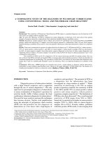
A comparative study of the metal binding behavior of alanine based bis-thiourea isomers
Bạn đang xem bản rút gọn của tài liệu. Xem và tải ngay bản đầy đủ của tài liệu tại đây (2.64 MB, 16 trang )
Fakhar et al. Chemistry Central Journal (2017) 11:76
DOI 10.1186/s13065-017-0304-2
Open Access
RESEARCH ARTICLE
A comparative study of the metal
binding behavior of alanine based
bis‑thiourea isomers
Imran Fakhar, Bohari M. Yamin and Siti Aishah Hasbullah*
Abstract
Two new symmetrical bis-thiourea, 2,2′-[{(terephthaloylbis(azanediyl)bis(carbonothioyl) bis(azanediyl)}dipropanoic
acid] (1A) and 3,3′-[{(terephthaloylbis(azanediyl)bis (carbonothioyl)bis(azanediyl)} dipropanoic acid] (1B) were synthesized by the reaction of terephthaloyl chloride with α- and β-alanine in good yields. Their binding properties were
investigated with various metal cations using UV–Vis titration experiments. Both isomers exhibited effective binding
with Ag+, Cu2+, Hg2+, Pb2+, Fe2+ and Fe3+ cations. However, in the presence of other cations, such as Na+, Ni2+, Co2+,
Cd2+, Zn2+, Mn2+, Mg2+, Ca2+, Sn2+, Al3+, and anions tetrabutylammonium Cl− and H2PO4−, no interaction occurred.
Both isomers displayed similar trends towards binding with metal cations.
Keywords: Bis-thiourea isomers, Binding study, α- and β-alanine, Metal cations
Introduction
Thiourea is an analogue of urea and was first synthesized
by Nencki [1]. Since then, thiourea compounds have
extensively been used as the building blocks of heterocyclic analogues [2]. Amongst this class of compounds,
benzoyl derivatives of thiourea have gained a great deal
of importance in the present day. Thiourea linkages have
contributed greatly to the observed enhancement in various activities [3], including antiviral [4], antibacterial [5,
6], antifungal [7], antitubercular [8, 9], herbicidal [10],
insecticidal [11], pharmacological properties [12], as
chelating agents [13, 14] and as anticancer compounds
[15]. In addition, benzoyl thiourea derivatives have often
been used in analytical and biological applications [16,
17].
Amino acids and their derivatives are significant constituents of chemical entities found within many natural
frameworks. The synthesis of biologically active amino
acid-coupled derivatives has recently become of major
interest [18–22].
*Correspondence:
School of Chemical Sciences and Food Technology, Faculty of Science
and Technology, Universiti Kebangsaan Malaysia, 43600 UKM Bangi,
Selangor, Malaysia
Thiourea and their amino acid derivatives coordinate
to several transition metal ions to form stable complexes.
Early useful suggestions of metal ions binding was provided by the old discipline of metal coordination chemistry by Werner [23]. Thioureas, along with its derivatives,
are versatile ligands, able to coordinate to metal centers as neutral ligands, monoanions, or dianions [24, 25].
According to Pearson’s hard and soft acid–base concept
thiourea, being a soft base, shows an affinity to bind with
soft acids like mercury, copper, silver, cadmium ions.
Conversely, amino acids, having carboxylic acid functionality, prefer interactions with hard acids like iron, lead,
aluminum ions [26]. The thiourea-based derivatives have
the ability to coordinate with several metal ions but have
not been much explored as receptors for the detection
of transition metal ions, this despite both urea and thiourea derivatives being frequently used as anion binding
receptors owing to their ability to act as hydrogen-bond
donors [27, 28]. However, recently some thiourea-based
derivatives and thiourea-based nanoparticles have been
used to detect metal ions [29, 30]. In view of these observations, the synthesis of two bis-thiourea isomers having
alanine linkers were planned followed by a comparative
study of their binding interactions against sixteen metal
cations (four soft, six mild and six hard ions) and two
© The Author(s) 2017. This article is distributed under the terms of the Creative Commons Attribution 4.0 International License
( which permits unrestricted use, distribution, and reproduction in any medium,
provided you give appropriate credit to the original author(s) and the source, provide a link to the Creative Commons license,
and indicate if changes were made. The Creative Commons Public Domain Dedication waiver ( />publicdomain/zero/1.0/) applies to the data made available in this article, unless otherwise stated.
Fakhar et al. Chemistry Central Journal (2017) 11:76
tetrabutyl ammonium anions. Both isomers were characterized by IR spectroscopy, 1H and 13C NMR spectroscopy, ESI–MS, and elemental analysis. Isomer 1B
was further confirmed by X-ray crystallography. Binding
studies of both isomers were studied by conducting titration experiments using UV–Vis spectroscopy.
Experimental
Materials and measurements
All the chemicals were obtained from ACROS Organics
(Geel, Belgium) and Sigma-Aldrich (Saint Louis, MO,
USA), and were utilized without further purification.
All solvents were distilled from CaH2 before use. Open
tube capillary method was used to determine the melting
points utilizing an Electrothermal 9100 (Electrothermal,
Southend, England) and were uncorrected. The micro
elemental investigation for CHNS were performed using
a Carlo Erba 1108 Elemental Analyzer (Milan, Italy).
The IR spectra of the isomers were obtained by KBr disc
method and were recorded on a Perkin Elmer Spectrum
GX spectrophotometer (Perkin Elmer, Waltham, MA,
USA) in the range of 400–4000 cm−1 with resolution
4 cm−1. UV–Vis estimations were performed on double
beam Varian UV 3.0 (Cary 100, Varian Australia Pty. Ltd.)
spectrophotometer with a quartz cell (1 cm path length)
in the scope of 200–800 nm with the highest resolution
of 1 nm. Nuclear Magnetic Resonance experiments (1H
and 13C NMR spectra) were done on a Bruker 400 MHz
instrument using DMSO-d6 as solvent. ESI–MS spectra
were recorded on a Micro Tof Q (Bruker, AXS Incorporation, and Madison, WI, USA). Single crystal X-ray
experiments were performed on a Bruker D-QUEST
diffractometer (Bruker, AXS Inc., Madison, WI, USA)
using graphite-monochromated Mo-Kα radiation
(λ = 0.71073 Å). Intensity data were measured at room
temperature by the ω-scan. Accurate cell parameters and
orientation matrix were determined by the full-matrix
least-squares fit of 25 reflections. Intensity data were
collected for Lorentz and polarization effects. Empirical
absorption correction was carried out using multi-scan.
The structure was solved by direct methods and leastsquares refinement of the structure was performed by
the SHELXL-2007 program [31]. All the non-hydrogen
atoms were refined anisotropically. The hydrogen atoms
were set in the calculated positions aside from the terminal N-atoms of thiourea moiety located from Fourier
maps and refined isotropically [32].
General procedure for the synthesis of isomers (1A and 1B)
Benzene-1,4-dicarbonyl chloride (terephthaloyl chloride) (0.609 g, 0.003 mol), was dissolved in dry acetone
(20 ml). A solution of ammonium thiocyanate (0.456 g,
Page 2 of 16
0.006 mol), antecedently dried (80 °C, 2 h) in dry acetone (15 ml) was prepared. Ammonium thiocyanate
was added slowly to the stirring solution of benzene-1,
4-dicarbonyl chloride, and the reaction mixture was
stirred at room temperature for 1 h. The white precipitate
of ammonium chloride were filtered off. α- or β-alanine
(0.534 g, 0.006 mol) in dry acetone (15 ml) was added
to the filtrate containing benzene-1,4-dicarbonyl isothiocyanate intermediate. The reaction mixture was then
refluxed for 24–30 h. The solution was allowed to cool to
RT and an excess of crushed ice added to the flask, bisthiourea analogues 1A and 1B were collected as precipitates which were then washed several times with water
and dried in a desiccator (using calcium sulfate as a drying agent). Both analogues were recrystallized from ethanol/DMSO to afford 1A and 1B in good yield (89.1 and
91.8%, respectively, Scheme 1).
Results and discussion
Characterization
2,2′‑[{(terephthaloylbis(azanediyl)bis(carbonothioyl)
bis(azanediyl)} dipropanoic acid] (1A) Using the general method outlined above, compound 1A was isolated
as a yellowish solid (0.760 g, 89.1%), mp: 214–215 °C,
[Found: C, 44.99; H, 4.19; N, 13.11; S, 15.01; O, 22.7%;
M+, 449.07. C16H18N4O6S2 requires C, 45.06; H, 4.25; N,
13.14; S, 15.04; O, 22.51%]; νmax (KBr/cm−1) 3358 (N–H),
3180 (C–Harom), 2929 (C–Haliph), 1728 (C=O), 1676
(COOH), 1545 (C–N), 1521 (Ar–C), 1012 (C=S); δH
(400 MHz, DMSO-d6, 1.50 (6H, d, J = 7.2 Hz, 2×CH3),
4.83 (2H, quint, J = 7.2 Hz, 2×CH), 8.00 (4H, s, Ar–H),
11.24 (2H, d, J = 6.8 Hz, 2×NH), 11.74 (2H, s, 2×NH).
δC (100 MHz, DMSO-d6) 17.5 (CH3), 53.5 (CH), 129.0
(CHarom), 136.3
(Carom), 168.2 (C=O), 173.3 (COOH),
180.1 (C=S); MS (EI): (m/z) = 449.07 [M + Na]+.
3,3′‑[{(terephthaloylbis(azanediyl)bis(carbonothioyl)
bis(azanediyl)} dipropanoic acid] (1B) Using the general method outlined above, compound 1B was isolated
as a white solid (0.784 g, 91.8%) as a white solid, mp:
203–204 °C, [Found: C, 45.09; H, 4.31; N, 13.01; S, 15.03;
O, 22.56%; M+, 449.47. C16H18N4O6S2 requires C, 45.06;
H, 4.25; N, 13.14; S, 15.04; O, 22.51%]; νmax (KBr/cm−1)
3330 (N–H), 3245 (C–Harom), 2950 (C–Haliph), 1711
(C=O), 1670 (COOH), 1554 (C–N), 1527 (Ar–C), 1025
(C=S); δH (400 MHz, DMSO-d6, 2.65 (4H, t, J = 6.0 Hz,
2×CH2), 3.82 (4H, d, J = 6.0 Hz, 2×CH2), 7.95 (4H,
s, Ar–H), 10.99 (2H, t, J = 5.6 Hz, 2×NH), 11.49 (2H,
s, 2×NH). δC (100 MHz, DMSO-d6) 32.6 (CH2), 41.0
(CH2), 127.8
(CHarom), 129.0
(Carom), 168.0 (C=O),
173.4 (C=OOH), 180.5 (C=S); MS (EI): (m/z) = 449.47
[M + Na]+.
Fakhar et al. Chemistry Central Journal (2017) 11:76
Page 3 of 16
Scheme 1 Synthesis of bis-thiourea alanine based isomers 1A and 1B
IR spectroscopy
IR spectra of both isomers were in accordance with the
vibrational frequencies of the functional groups as found in
the literature [3, 46]. The N–H stretching vibrations were
observed in the range 3330–3358 cm−1. The O–H stretching frequencies of the carboxylic groups were overlapped
by N–H stretching peak and hence could not be observed.
The C–H stretching vibrations for the sp2 carbon of the
aromatic ring of both isomers were observed in the range
3180–3245 cm−1 [33] whereas, the C–H stretching vibrations for the sp3 mode of the alkyl chain were observed
in the range 2930–2950 cm−1 [34]. The frequency for the
C=O and C=Ocarboxylic stretches were observed at 1728,
1676, 1711, and 1670 cm−1 for the isomers 1A and 1B,
respectively [35]. The ν (C–N) and ν (C=Caromatic) vibrational frequencies were observed at 1545, 1521 and 1554,
1527 cm−1 for isomers 1A and 1B, respectively. All of the
values mentioned were found in accordance with those
reported [3]. The ν (C=S) vibrational frequencies for both
isomers were observed at 1012 and 1025 cm−1. The lowering in the vibrational frequencies of (C=S) bonds were
due to mesomeric electron releasing effect of the nitrogen
bonded to the thiocarbonyl group (N–C=S). This lowering
of C=S stretching frequencies is due to an acquiring of a
partial polar character [36].
1
H NMR and 13C NMR spectroscopy
Bis-thiourea isomers were further characterized and confirmed by 1H, and 13C NMR. The proton chemical shifts
of the amide functionality appeared as a singlet at δ 11.74
and 11.49 ppm for isomers 1A and 1B, respectively. The
thioamide protons were observed as doublets at δ 11.24,
10.99 ppm for the isomers 1A and 1B, respectively. The
downfield signals of both amide and thioamide protons
are due to the formation of H-bonding between the
amino proton and the oxygen/sulfur atoms of carbonyl/
thiocarbonyl group, as well as the anisotropic effect [37].
All the aromatic protons for both isomers were identical
and found as singlets at δ 8.0 and 7.95 ppm for 1A and
1B, respectively. The chemical shift for the proton on the
chiral carbon of isomer 1A was observed at δ 4.83 ppm.
The signal was observed downfield due to the deshielding effect of the nearby electron withdrawing thioamide
group as well as the anisotropic effect of the carboxylic
carbonyl group. Isomer 1B contains no source of chirality and so two methylene groups are present. The methylene group proximal to the carboxylic acid were observed
downfield at δ 3.82 ppm as a doublet due to the anisotropic effect of the carbonyl group. Protons of the second
methylene group were observed as a triplet at δ 2.65 ppm
slightly downfield due to deshielding from the electron
withdrawing thioamide group. The methyl protons for
isomer 1A were observed as a doublet at δ 1.53 ppm.
The 13C NMR spectra for both isomers 1A and 1B
were in accordance with those that have been reported
previously [38]. The carbon chemical shifts of C=S,
C=Carboxylic and C=O were found at δ 180.1, 173.3 and
168.2 ppm for isomer 1A and at δ 180.5, 173.4 and 168.0
for isomer 1B, respectively. The aromatic carbons were
observed at δ129.0 and 136.3 ppm for isomer 1A and at
δ 127.8 and 129.0 ppm for isomer 1B, respectively. The
signal for the chiral carbon of isomer 1A was observed
at δ 53.5 ppm and that of the carbon bearing the methyl
group at δ 17.5 ppm. Whereas the chemical shifts of two
Fakhar et al. Chemistry Central Journal (2017) 11:76
methylene groups of isomer 1B were observed at δ 3.82
and 2.65 ppm, respectively.
Elemental analysis and ESI‑Mass spectroscopy
The CHNS analysis for both isomers were found to be in
close accordance with the theoretical values.
The ESI–MS spectra, for both isomers 1A and 1B,
showed sodium molecular ion peaks at m/z 449, which
is in accordance with the expected molecular ion peak
values.
Page 4 of 16
Table 1 Crystal data and structure refinement for isomer
1B
Identification code
boly370_0 m
Empirical formula
C16H18N4O6S2
Formula weight
426.46
Temperature
303(2) K
Wavelength
0.71073 Å
Crystal system
Monoclinic
Space group
C2/c
Unit cell dimensions
a = 26.9433(13) Å; α = 90°
b = 4.7668(2) Å; β = 100.926(2)°
X‑ray crystallography of isomer 1B
The isomer 1B crystallized in monoclinic system with space
group C2/c, a = 26.9433(13), b = 4.7668(2), c = 15.1750(7),
α = 90, β = 100.926(2), γ = 90, Z = 4 and V = 1913.65(15).
Crystallographic data for the structure determination has
been deposited with the Cambridge Crystallographic Data
number CCDC 1518921. The given crystal state and refinement parameters are given in Table 1.
The molecule 1B adopts a cis–trans configuration with
respect to the position of the propionic acid relative to
the S1 atom across the C(4)–N(1) bonds. Figure 1 shows
the conformational structure of the molecule with atoms
numbered.
The thiourea fragment, S(1)/N(1)/N(2)/O(3)/C(5) and
benzene ring are planar with maximum deviation of
0.073(2) Å for the N(1) atom from the least-squares plane
of the thiourea fragment. The thiourea moiety along with
benzene ring makes an angle of 90.0(3)° with the propionic acid fragment (Table 2). The bond lengths and
angles in isomer 1B is within normal ranges [39, 40].
In the molecule there are three intramolecular
H-bonds, N(1)…H(1)…O(3), C(3)…H(3B)…S(1) and
c = 15.1750(7) Å; γ = 90°
Volume
1913.65(15) Å3
Z
4
Density (calculated)
1.480 Mg m−3
Absorption coefficient
0.320 mm−1
F(000)
888
Crystal size
0.49 × 0.36 × 0.11 mm3
Theta range for data collection
2.87–28.31°
Index ranges
−35 <= h <= 35, −6 <= k <= 6,
−18 <= l <= 20
Reflections collected
29,791
Independent reflections
2376 [R(int) = 0.0372]
Completeness to theta = 28.31°
99.7%
Absorption correction
Semi-empirical from equivalents
Max. and min. transmission
0.9656 and 0.8588
Refinement method
Full-matrix least-squares on F2
Data/restraints/parameters
2
2376/0/128
Goodness-of-fit on F
1.064
Final R indices [I >2 sigma(I)]
R1 = 0.0538, wR2 = 0.1507
R indices (all data)
R1 = 0.0673, wR2 = 0.1618
Largest diff. peak and hole
0.335 and −0.357 e Å−3
Fig. 1 ORTEP diagram of the 3, 3′-[{(terephthaloylbis(azanediyl)bis(carbonothioyl)bis (azanediyl)}dipropanoicacid]. 1B was drawn at 50% probability
displacement ellipsoids. The dashed line indicates the intramolecular hydrogen bond
Fakhar et al. Chemistry Central Journal (2017) 11:76
Page 5 of 16
Table 2 Selected bond lengths (Å) and bond angles (°)
for isomer 1B
Table 3 Hydrogen bonds for isomer 1B [(Å) and (°)]
D–H…A
d(D–H)
d(H…A)
d(D…A)
<(DHA)
N(2)–H(2C)…O(1)
0.86
2.28
3.126(2)
168
N(1)–H(1)…O(3)
0.86
1.92
2.603(2)
134.9
126.52(18)
O(2)–H(2)…S(1)
0.82
2.26
3.072(2)
174
O(1)–C(1)–O(2)
122.1(2)
C(3)–H(3B)…S(1)
0.97
2.64
3.042(3)
105
1.214(3)
O(1)–C(1)–C(2)
124.6(2)
C(7)–H(7)…O(1)
0.93
2.20
3.123(3)
174
N(1)–C(4)
1.316(3)
O(2)–C(1)–C(2)
113.26(19)
C(8)–H(8)…O(3)
0.93
2.41
2.737(3)
100
N(1)–C(3)
1.464(3)
C(1)–C(2)–C(3)
112.6(2)
N(2)–C(5)
1.377(3)
N(1)–C(3)–C(2)
111.31(19)
Bond
Length (Å)
Bond
Angles (°)
S(1)–C(4)
1.672(2)
C(4)–N(1)–C(3)
123.7(2)
O(1)–C(1)
1.212(3)
C(5)–N(2)–C(4)
O(2)–C(1)
1.313(3)
O(3)–C(5)
N(2)–C(4)
1.399(2)
N(1)–C(4)–N(2)
116.77(18)
C(1)–C(2)
1.494(4)
N(1)–C(4)–S(1)
122.86(16)
C(2)–C(3)
1.514(3)
N(2)–C(4)–S(1)
120.35(16)
C(5)–C(6)
1.500(3)
O(3)–C(5)–N(2)
122.32(18)
C(6)–C(7)
1.371(3)
O(3)–C(5)–C(6)
120.38(19)
C(6)–C(8)
1.377(3)
N(2)–C(5)–C(6)
117.30(19)
C(7)–C(8)#1
1.382(3)
C(7)–C(6)–C(8)
118.52(19)
C(8)–C(7)#1
1.382(3)
C(7)–C(6)–C(5)
124.88(18)
C(8)–C(6)–C(5)
116.59(19)
Symmetry transformations used to generate equivalent atoms: #1 −x,−y,−z
Symmetry transformations used to generate equivalent atoms: #1 −x, −y, −z # 2
x, −y + 1, z − 1/2 #3 x, −y + 1, z+
C(8)…H(8)…O(3) (Table 3). In the crystal structure,
the molecules are linked by O(2)…H(2)…S(1), N(1)…
H(2C)…O(1) and C(7)…H(7)…O(1) intermolecular
H-bonds forming a 3-D network (Fig. 2).
Binding studies
UV–Vis spectra measurements
Firstly, stock solutions for both isomers (1A and 1B)
were prepared in DMSO (1 × 10⁻³ M) before making
Fig. 2 Molecular packing of 1B viewed down the b axis. Dashed lines denote C–H….O, O–H….S and N–H….O hydrogen bonds
Fakhar et al. Chemistry Central Journal (2017) 11:76
Page 6 of 16
stock solutions for both metal cations and tetrabutylammonium anions, also in DMSO (1 × 10⁻³ M). By
adding different volumes (0–600 µl) of metal ions and
terabutylammonium anions to a series volumetric flasks,
together with an equal volume (100 µl) of the isomers
1A and 1B, the work solutions were prepared. Each of
the work solutions were then diluted by adding DMSO
and shaken for several minutes. Readings were recorded
on UV–Vis spectrophotometer using quartz cuvettes
(1 cm path length) in the range of 200–800 nm with the
utmost resolution of 1 nm. The correlation coefficient
was computed using Pearson product-moment correlation strategy. By plotting a fit line curve using Sigma
Plot 12.0 (Systat Software Inc.), dissociation constant
(Kd) values were intended using a nonlinear regression
equation. Detection limit was figured by 3 σ/S, where ‘σ’
is the std. deviation and ‘S’ is the incline in the titration
curve. To demonstrate the veracity of information, more
than 20 arrangements of continuous data were gathered
in the UV–Vis titration tests until absorbance values
approached equilibrium.
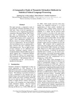
Theory and calculations
The correlation coefficient was utilized to quantify a linear association between the two factors (absorbance
vs concentration) amid the titration tests. The Pearson
product-moment correlation strategy was utilized as part
of this study to quantify the degree of linear dependence
between the two variables. The formula for correlation
coefficient ‘r’ can be accomplished by substituting assessments of the covariance and variance in the equation
below [47].
n
r = rxy =
n
xy −
x2 − (x)2
x
n
y
y2 −
y
2
where: r = correlation coefficient; x = concentration;
y = absorbance; n = no. of observations.
The detection limit was calculated by utilizing the
formula.
DL = 3σ S
where: σ = std. deviation of 5 blank values; S = slope of
the fit-line titration curve.
Fig. 3 Graphical representation of two-site binding
Clark’s theory of binding
Alfred Joseph Clark developed this concept in 1926, and
mathematically stated that for a bimolecular reaction [48]:
H + G⇆H − G
The equilibrium dissociation constant (Kd) or an equilibrium association constant (Ka), which are proportionally related, is demonstrated by the following:
Regardless of the mechanism, every reversible reaction achieves equilibrium conveyance of reactants and
products when the rates of both the forward and reverse
reactions reach equivalence. The general rate can be
communicated as:
d[H − G]
= kassn [H ][G] − kdiss [H − G].
dt
At the beginning of a reaction, the association rate
( kassn [H] [G]) would overwhelm. As more of the complex
is formed, the association rate would diminish and the
dissociation rate would increase. Eventually, the rates of
the opposing reactions would become equivalent, and be
described as:
d[H − G]
−d[H ]
−d[G]
=
=
dt
dt
dt
= kassn [H ][G] − kdiss [H − G] = 0.
Under these conditions:
[H ][G]
kdiss
=
= K d.
[H − G]
kassn
This expression demonstrates that the equilibrium concentration of reactants and products will have a constant
ratio (Kd) that is equivalent to the proportion of the forward and reverse rate constants. K
d is called the equilibrium dissociation constant.
In the present study the dissociation constant (also
termed as binding constant (Kd) was computed by the
Nonlinear Regression formula utilizing Sigma plot 12.0
(Systat Software Inc.).
For the two site mode of binding (Fig. 3), the nonlinear
regression equation is expressed as the following:
Fakhar et al. Chemistry Central Journal (2017) 11:76
y = Bmax1 ·
x
x
+ Bmax2 ·
Kd1 + x
Kd2 + x
Page 7 of 16
the most intense interactions with mild to soft Pearson
acidic ions.
where: Bmax = host–guest complex; y = absorbance;
x = [G]/[H].
Binding behavior and binding mechanism of bis‑thiourea
isomers
Comparison of binding behavior
Selectivity of bis‑thiourea isomers against cations
To inspect the coupling behavior of isomer 1A and 1B
against selected metal cations, titration experiments were
carried out. In the control experiment (isomers without metal cations), the absorption maxima of both isomers were seen at 265 nm, which can be allocated to an
intramolecular charge transfer (ICT) absorption band as
is the known case with thioureas [41]. Upon sequential
addition of cations to the test solutions, just Fe3+, Fe2+,
Cu2+, Pb2+, Hg2+, and A
g+ gave exceptional enhancement of emission intensity at 265 nm for both isomers
1A and 1B. The increase of emission absorbance intensity was credited to the conceivable formation of host–
guest complexes at two probable sites. The first and most
likely site of complexation is the carboxylate functionality of α/β-alanine [42], as shown by dissociation constant
Kd1 in Table 1. The second interaction would be from the
thiourea functionality via C=S and N–H [43] as shown
In the first place, the interaction properties of the isomers in DMSO were examined against sixteen metal
cations, four of which are soft metal ions such as A
g +,
2+
2+
2+
Cu , Co and Hg , six are mild metal ions such as
Fe2+, Ni2+, Pb2+, Mn2+ and Z n2+ and six are hard metal
ions such as N
a+, Ca2+, Mg2+, Fe3+, Cd2+, Sn2+ and A
l3+
according to the Pearson scale. The two tetrabutylammonium anions of C
l− and H2PO4− were also investigated.
Both isomers (1A and 1B) did not show any appreciable
interactions with both Cl− and H2PO4− ions. Whereas
both isomers showed reasonable interactions with six
metal ions, five which are soft to mild ( Ag+, Cu2+, Hg2+,
Fe2+, Pb2+) and one which is hard (Fe3+). The results of
interactions are shown in (Figs. 4, 5) for isomer 1A and
1B, respectively. On the Pearson scale thioureas are
considered soft bases and so would be expected to have
Fig. 4 Interactions of isomer 1A with various metal ions and tetrabutylammonium ions
Fakhar et al. Chemistry Central Journal (2017) 11:76
Page 8 of 16
Fig. 5 Interactions of isomer 1B with various metal ions and tetrabutylammonium ions
by dissociation constant Kd2 (Table 1). By looking at the
titration spectra of isomers 1A and 1B vs F
e3+, Ag+, and
2+
Cu (Figs. 6, 7, 10, 11, 16, 17), another band can be seen
to appear at 360–365 nm, which progressively expanded
on incremental addition of metal cations. This is due to
the deprotonation of the amino proton by counter anions. Fabrizzi et al. additionally reported a similar outcome for a urea based receptor [44]. The absorbance
maxima increased linearly with the concentration of all
the chosen cations in a given range (0–600 µl). Table 1
also shows the correlation coefficient values and detection limit values in the light of titration investigations.
Titration experiment curves and binding behaviors of
isomers 1A and 1B against metal ions are also shown
(Fig. 6 through to Fig. 17).
Binding mechanism
To explore the mechanism of complexation between isomers 1A and 1B and the chosen metal cations, continuous variation titration investigations were carried out. In
these tests, the concentration of cations was increased
incrementally, whereas the concentration of isomer 1A
and 1B were kept constant. In the light of these titration investigations, the stoichiometry of complexation
between isomer 1A/1B with metal cations were ascertained by a molar-ratio strategy [45], and the binding
constant (Kd) computed by nonlinear regression formula
[28]. The dissociation constant (Kd) values and stoichiometry of the complexation are shown in Table 4. The
graphical counts of the stoichiometry are also shown
(Inset: Figs. 6, 7, 8, 9, 10, 11, 12, 13, 14, 15, 16, 17).
Conclusions
Bis-thiourea isomers featuring amino acids (α and
β-alanine) have been successfully characterized using
spectroscopic methods, namely; IR, 1H NMR, 13C NMR,
ESI–MS, and elemental analysis (CHNS/O). Moreover, isomer 1B was further confirmed by X-ray crystallography, which revealed that the β-alanine side chain is
arranged in a cis–trans configuration. The spectroscopic
Fakhar et al. Chemistry Central Journal (2017) 11:76
Page 9 of 16
Table 4 Correlation coefficient, detection limit, stoichiometry of complexation and binding constants of both Isomers
with metal ions
Lig-metal ion
Isomer1A-Fe3+
Correlation coefficient
0.982
3+
Isomer1B-Fe
0.967
Isomer1A-Fe2+
0.998
Isomer1B-Fe2+
0.936
Isomer1A-Cu2+
0.998
Isomer1B-Cu2+
0.967
Isomer1A-Pb2+
0.982
Isomer1B-Pb2+
0.977
Isomer1A-Hg2+
0.967
Isomer1B-Hg2+
0.98
Isomer1A-Ag+
0.997
Isomer1B-Ag+
0.989
Detection limit
1.30 × 10⁻¹ M
−1
2.40 × 10
Complexation stoichi‑
ometry
Dissociation constant
Kd1
Kd2
1:4
5.45 × 10⁻¹⁷ M
6.760 M
1.42 × 10⁻¹⁸ M
6.835 M
6.04 × 10⁻¹⁸ M
6.149 M
2.84 × 10⁻¹⁷ M
1.269 M
9.57 × 10⁻¹⁷ M
5.201 M
6.87 × 10⁻¹⁸ M
4.557 M
M
1:4
1.50 × 10⁻¹ M
1:4
1.90 × 10⁻¹ M
1:4
1.14 × 10⁻¹ M
1:4
9.16 × 10⁻² M
1:4
2.02 × 10⁻¹ M
1:4
3.88 × 10⁻¹ M
1:4
3.16 × 10⁻¹ M
1:4
1.83 × 10⁻¹ M
1:4
2.10 × 10⁻¹ M
1:4
7.23 × 10⁻¹ M
1:4
3.81 × 10⁻¹⁷ M
4.539 M
1.15 × 10⁻¹⁷ M
7.380 M
5.92 × 10⁻¹⁷ M
9.852 M
5.69 × 10⁻¹⁷ M
5.310 M
5.56 × 10⁻¹⁸ M
7.916 M
1.64 × 10⁻¹⁷ M
1.717 M
Fig. 6 Titration of isomer 1A vs Fe3+ (Inset Binding behavior + stoichiometry)
results also revealed that both isomers exhibit a plane of
symmetry. The titration experiments confirmed the interaction of six metal ions; one ‘hard’ acid F
e3+, and five ‘soft’
2+
2+
2+
2+
+
acids Fe , Cu , Pb , Hg and Ag . All the remaining metal ions examined (Na+, Ca2+, Mg2+, Co2+, Ni2+,
Mn2+, Cd2+, Sn2+, Zn2+, and Al3+) showed no appreciable
interactions. In addition, no interaction was observed for
the tetrabutylammonium ions Cl− and H2PO4−. The stoichiometry of the complex (host–guest)formed for both
isomers was found to be 1:4. Binding constant K
d1 values
for both isomers were found to be very low due to complexation at carboxylate functionality of α and β-alanine.
Binding constant (Kd2) values were appreciably high as
compared to K
d1 values because of the complexation at
Fakhar et al. Chemistry Central Journal (2017) 11:76
Page 10 of 16
Fig. 7 Titration of isomer 1B vs Fe3+ (Inset Binding behavior + stoichiometry)
Fig. 8 Titration of isomer 1A vs Fe2+ (Inset Binding behavior + stoichiometry)
the thiourea functionality of isomers 1A and 1B. On comparing the binding constant (Kd2) for both isomers, values for isomer 1A were in the range 4.5–6.8 except for
(Pb2+) which was 1.2 and for isomer 1B binding constant
values were found in the range 4.5–9.8 except for (Ag+)
which was 1.7. The dissociation constant values for both
isomers with all metal ions were in relatively close proximity to each other. The next study will be focused on
Fakhar et al. Chemistry Central Journal (2017) 11:76
Page 11 of 16
Fig. 9 Titration of isomer 1B vs Fe2+ (Inset Binding behavior + stoichiometry)
Fig. 10 Titration of isomer 1A vs Cu2+ (Inset Binding behavior + stoichiometry)
the role of different side chain amino acids/secondary
amines towards binding behavior against various metals
and based on the data obtained in the present study, the
chemical sensor will be fabricated by using newly synthesized compounds for the detection of metal ions.
Fakhar et al. Chemistry Central Journal (2017) 11:76
Fig. 11 Titration of isomer 1B vs Cu2+ (Inset Binding behavior + stoichiometry)
Fig. 12 Titration of isomer 1A vs Pb2+ (Inset Binding behavior + stoichiometry)
Page 12 of 16
Fakhar et al. Chemistry Central Journal (2017) 11:76
Fig. 13 Titration of isomer 1B vs Pb2+ (Inset Binding behavior + stoichiometry)
Fig. 14 Titration of isomer 1A vs Hg2+ (Inset Binding behavior + stoichiometry)
Page 13 of 16
Fakhar et al. Chemistry Central Journal (2017) 11:76
Fig. 15 Titration of isomer 1B vs Hg2+ (Inset Binding behavior + stoichiometry)
Fig. 16 Titration of isomer 1A vs Ag+ (Inset Binding behavior + stoichiometry)
Page 14 of 16
Fakhar et al. Chemistry Central Journal (2017) 11:76
Page 15 of 16
Fig. 17 Titration of isomer 1B vs Ag+ (Inset Binding behavior + stoichiometry)
Authors’ contributions
IF, BMY and SAH initiated the study. All authors contributed to the synthesis
and characterization of new compounds, interpretation of the results and
preparation of the manuscript. The experiment and sample analysis were
conducted by IF with contributions from BMY and SAH. The binding studies
were conducted by IF and SAH with contributions from BMY. All authors read
and approved the final manuscript.
Acknowledgements
The authors wish to thank the School of Chemical Sciences and Food Technology, and the Universiti Kebangsaan Malaysia (DIP-2015-015) for providing
necessary facilities. We greatly appreciate the Ministry of Higher Education
for providing the funding of the project under Grants PRGS/2/2015/SG01/
UKM/02/1 and FRGS/1/2015/ST01/UKM/02/2 (Project leader Dr. Siti Aishah
Hasbullah). Mr. Imran Fakhar would also like to thank Mr. Kamran Fakhar for
providing financial assistance, his parents for providing moral support and
Mr. Hassanuddin for providing the technical assistance.
Competing interests
The authors declare that they have no competing interests.
Publisher’s Note
Springer Nature remains neutral with regard to jurisdictional claims in published maps and institutional affiliations.
Received: 24 December 2016 Accepted: 26 July 2017
References
1. Nencki M (1873) Zur Kenntniss des Sulfoharnstoffs. Ber Dtsch Chem Ges
6:598–600
2. Mitsuo K, Masato S, Keiko T, Tadashi A (2005) A convenient and efficient
method for the synthesis of mono- and N,N-disubstituted thioureas.
Tetrahedron Lett 46:5841–5843
3. Zullkiplee Wan S H W, Abd Ainaa N, Halim Zainab N, Maya A, Ariff M,
Hasnain H (2014) Bis-thiourea bearing aryl and amino acids side chains
and their antibacterial activities. Phosphorus Sulfur Silicon Relat Elem
189:832–838
4. Sun CW, Zhang XD (2007) Synthesis and crystal structure of S-(+)-N’-Tertbutylaminocarbonyl-N-[3-methyl-2-(4-chlorophenyl)butyryl] Thiourea.
Chin J Struct Chem 26:153–156
5. Aamer S, Khera Rasheed A, Naeem A, Muhammad L, Imran S, Ulrich F
(2010) Synthesis, characterization, crystal structures, and antibacterial
activity of some new 1-(3,4,5-trimethoxybenzoyl)-3-aryl thioureas. Turk J
Chem 34:335–345
6. Sohail S, Naghmana R, Muhammad A, Rizwan H (2010) Synthesis, characterization and antibacterial activity of nickel (II) and copper (II) complexes
of N-(alkyl(aryl)carbamothioyl)-4-nitrobenzamide. Eur J Chem 1:200–205
7. Eweis M, Elkholy SS, Elsabee MZ (2006) Antifungal efficacy of chitosan
and its thiourea derivatives upon the growth of some sugar-beet pathogens. Int J Biol Macromol 38:1–8
8. Dharmarajan S, Perumal Y, Murugesan D, Rathinasababathy T (2007) Antimycobacterial activity of novel 1-(5-cyclobutyl-1,3-oxazol-2-yl)-3-(sub)
phenyl/pyridylthiourea compounds endowed with high activity toward
multidrug-resistant Mycobacterium tuberculosis. J Antimicrob Chemother
59:1194–1196
9. Bedia KK, Sevim RKK-A (2003) In vivo metabolism of N-phenyl-N′-(3,5dimethylpyrazole-4-yl) thiourea in rats. Eur J Drug Metabol Pharmacokinet 28:273–278
10. Soung M-G, Park K-Y, Song J-H, Sung N-D (2008) Herbicidal activity and
molecular similarity of 1-(4-chloro-2-fluoro-5-propargyloxyphenyl)-3-thiourea derivatives. Soc Appl Biol Chem 51:219–222
11. Saeed A, Batool M (2007) Synthesis and bioactivity of some new 1-tolyl3-aryl-4-methylimidazole-2-thiones. Med Chem Res 16:143–154
12. Sohail S, Naghmana Muhammad A, Rizwan H, Peter GJ (2010) Synthesis,
spectroscopic characterization, crystal structure and pharmacological
properties of some novel thiophene-thiourea core derivatives. Eur J
Chem 1:221–227
13. Das DK (1984) N-α-(5-bromopyridyl)-N′-benzoyl thiourea (BrPBT) as
a new chelating agent for the spectrophotometric determination of
rhodium (III). Fresh J Anal Chem 318:612
Fakhar et al. Chemistry Central Journal (2017) 11:76
14. Shome SC, Mazumdar M, Haldar PK (1980) N-Alpha-pyridyl-N′-benzoyl
thiourea as a chelating agent for the determination of iridium. J Indian
Chem Soc 57:139–141
15. Ghora Mostafa M, Alsaid MS, Al-Dosary MS, Nissan YM, Attia SM (2016)
Design, synthesis and anticancer activity of some novel thioureido-benzenesulfonamides incorporated biologically active moieties. Chem Cent J
10:19. doi:10.1186/s13065-016-0161-4
16. Gün B, Gülten K, Nevzat K, Süheyla Ö, Ulrich F, Hakan A (2013) Synthesis
and characterization of novel thiourea derivatives and their nickel and
copper complexes. J Chem. doi:10.1155/2013/536562
17. Alkherraz Abdulfattah M, Lusta Zaineb I, Zubi Ahmed E (2014) Synthesis
and use of thiourea derivative (1-Phenyl-3-Benzoyl-2-Thiourea) for
extraction of cadmium ion. Int J Chem Nucl Metall Mater Eng 8:118–120
18. Walid F, Pavel P (2007) Synthesis and reactions of methyl 2-[3-(2-phenylquinazolin-4-yl)thioureido]alkanoates. ARKIVOC 1:236–243
19. Walid F, Ali Ibrahim A I (2007) Efficient synthesis of 1-substituted-4-phenyl-1, 4-dihydro-5H-tetrazole-5-thione and (1-phenyl-1H-tetrazol-5-yl)
thioacetyl derivatives. Heteroat Chem 18:637–643
20. Walid F, El Rayes SM, Ali AI (2007) Convenient synthesis of 1-substituted4-methyl-5-oxo[1,2,4]triazolo[4,3-a]quinazolines. ARKIVOC 16:173–186
21. El Rayes SM, Ali AI, Walid F (2008) A convenient synthesis of new amino
acid-coupled benzanilides. ARKIVOC 9:86–95
22. Al-M Najim A, Al-H Nahed, Faili NT, Pannecouque C (2010) Amino acid
derivatives. Part 5. Synthesis and anti-HIV activity of new sebacoyl precursor derived thioureido-amino acid and phthalimide derivatives. ARKIVOC
9:185–195
23. Werner Alfred (1893) Beitrag zur konstitution anorganischer verbindungen. Zeitschrift für Anorganische und Allgemeine Chemie 3:267–330
24. Rathod MS, Jadhao SZ (2012) Synthesis and structural investigation of
nickel metal–ligand (thiourea derivative) complexes. J Chem Pharm Res
4:1629
25. William H, Brian KN, Rickard CEF (2001) Platinum(II) complexes of
chelating and monodentate thiourea monoanions incorporating chiral,
fluorescent or chromophoric groups. Inorg Chim Acta 320:101–109
26. Pearson RG (1963) Hard and soft acids and bases. J Am Chem Soc
85:3533
27. Xuefang S, Zhenhua Y, Jiajia F, Peipei Z, Xiufang X (2015) The synthesis
and anion recognition property of symmetrical chemosensors involving
thiourea groups: theory and experiments. Sensors 15:28166–28176.
doi:10.3390/s151128166
28. Khansari ME, Wallace KD, Hossain MA (2014) Synthesis and anion recognition studies of a dipodal thiourea-based sensor for anions. Tetrahedron
Lett 55:438–440
29. Zhenyu Z, Shenzhou L, Chunming S, Dongmei X (2015) A single thioureaappended 1,8-naphthalimide chemosensor for three heavy metal ions:
Fe3+, Pb2+, and Hg2+. Sens Actuat B 208:258–266
30. Al-Kady AS, Gaber M, Hussein MM, El-Z Ebeid (2009) Fluorescence
enhancement of coumarin thiourea derivatives by Hg2+, Ag+, and silver
nanoparticles. J Phys Chem 113:9474
31. Sheldrick GM (1997) SHELXTL. Version 6.14 program for crystal structure
determination. University of Gottingen, Gottingen
Page 16 of 16
32. Hamza MA, Siti Aishah H, Yamin Bohari M (2015) Synthesis, characterization and X-ray structure of N-(4-bromobutanoyl-N’-(2-3- and 4-methylphenyl) thiourea. Chin J Struct Chem 34:379–385
33. Wen Y, Weiqun Z, Zhengjiang Z (2007) Structural and spectroscopic
study on N-2-fluorobenzoyl-N′-4-methoxyphenylthiourea. J Mol Struct
828:46–53
34. Hakan A, Ulrich F, Nevzat K, Gün B (2007) The molecular structure and
vibrational spectra of 2-chloro-N-(diethylcarbamothioyl)benzamide by
Hartree-Fock and density functional methods. Spectrochim Acta A Mol
Biomol Spectrosc 68:1347–1355
35. Sitti R, Bunbun B (2009) Kinetics of the oxidation of vitamin C. Prosiding
Seminar Kimia Bersama UKM-ITB VIII:9–11
36. Shankaranrayana ML, Patel CC (1965) Infrared absorption studies on
some derivatives of xanthic, dithiocarbamic and trithiocarbonic acids.
Spectrochim Acta A Mol Spectrosc 21:93–105
37. Abosadiyaa HM, Anouar EH, Hasbullah SA, Yamin BM (2015) Synthesis, X-ray, NMR, FT–IR, UV/Vis, DFT and TD–DFT studies of
N-(4-chlorobutanoyl)-N′-(2-, 3- and 4-methylphenyl)thiourea derivatives.
Spectrochim Acta A Mol Spectrosc 144:115–124
38. Freeman RA (1997) Handbook of Nuclear Magnetic Resonance, 2nd edn.
Longman, London
39. Frank HA, Olga K, David GW, Lee B, Guy Orpen A, Robin T (1987) Tables
of bond lengths determined by X-ray and neutron diffraction. Part 1
Bond lengths in organic compounds. J Chem Soc Perkin Trans 2:S1–S19.
doi:10.1039/P298700000S1
40. Hamza MA, El Salem A, Bohari MY (2009) synthesis and X-ray structure
study of cis–trans 3-(3-biphenyl carbonylthioureido)propanoicacid(i) and
n-(4-biphenyl carbonyl)-n’-(3-hydroxyphenyl)thiourea(II). Rasayan J Chem
2:572–576
41. Guangyan Q, Taolei S, Zhihong C, Xi Y, Xiaojun W, Yongbing H (2009)
‘Naked-eye’ enantioselective chemosensors for N-protected amino
acid anions bearing thiourea units. Chirality 21:363–373. doi:10.1002/
chir.20593
42. Abosadiya HA, Yamin BM (2007) Synthesis and molecular structure of
the complexes of trimethyl and tributyltin chloride with 3-(3-benzoylthiouredo)propionic acid. J Sains MIPA 13:73–76
43. Zhengqian L, Ying Z, Kai Y, Zhu Y, Yan L, Jun R (2014) A new fluorescence
“turn-on” type chemosensor for Fe3+ based on naphthalimide and coumarin. Dyes Pigments 105:711
44. Kalpana RD, Bryan M, Alamgir MA (2010) Rational design of a macrocyclebased chemosensor for anions. Tetrahedron Lett 51:1329–1332
45. Ukpebor EE, Okolo PO (2004) Stoichiometry of quinol/ammonium–nitrogen complex using spectrophotometry. J Chem Soc Pak 26:207–211
46. Kabbani AT, Ramadan H, Hammud HH, Ghannoum AM, Mouneimne
Y (2005) Synthesis of some metal complexes of N-[(benzoylamino)thioxomethyl]-aminoacid (HL). J Univ Chem Technol Metall 40:339–344
47. Asuero AG, Sayago A, González AG (2006) The correlation coefficient: an
overview. Crit Rev Anal Chem 36:41–59
48. Sauer RT. Review of chemical equilibrium. 1999. rsehero.
com/file/6535314/fa01lec07/. Accessed 11 July 2016