Influence of age on clinical presentation, diagnosis delay and outcome in pre-school children with acute appendicitis
Bạn đang xem bản rút gọn của tài liệu. Xem và tải ngay bản đầy đủ của tài liệu tại đây (780.57 KB, 9 trang )
Lounis et al. BMC Pediatrics
(2020) 20:151
/>
RESEARCH ARTICLE
Open Access
Influence of age on clinical presentation,
diagnosis delay and outcome in pre-school
children with acute appendicitis
Yasmine Lounis1, Julie Hugo2, Martine Demarche3 and Marie-Christine Seghaye1*
Abstract
Background: Unusual clinical presentation of acute appendicitis in preschool children leads to misdiagnosis and
complications.
We aimed to analyze the influence of age on clinical presentation, laboratory findings and complications in
preschool children with acute appendicitis.
Methods: From January 2012 until December 2017, 29 children younger than 6 years of age (median 50 months)
with acute appendicitis were enrolled in this retrospective study. Patients were grouped according to their age:
group 1: < 48 months (n = 13); group 2: > 48 months (n = 16), their clinical data, laboratory results and complications
were compared.
Results: In group 1, duration of nausea and vomiting was longer, alteration of general state was more frequent
and pain in the right fossa iliaca less frequent than in group 2 (p = 0.026, p = 0.000 and p = 0.029, respectively).
Heart rate was higher in group 1 than in group 2 (p = 0.012). Leucocyte and polynuclear neutrophil counts were
lower in group 1 than in group 2 (p = 0.028 and = 0.004, respectively) but C-reactive protein levels were not
different between groups. In the whole cohort however, C-reactive protein at admission value correlated negatively
with age (p = 0.025).
Abdominal ultrasound allowed diagnosis in 19/29 patients (65.5%), without any difference between groups.
Appendicular perforation was more frequent in group 1 than in group 2 (p = 0.003). Perforation was also related to
longer hospital stay (p = 0.018). Peritonitis occurred in 21/29 (72%), post-operative ileus in 5/29 (17%) and sepsis in
4/29 (14%) patients without any difference between groups. In the whole cohort, hospital stay correlated negatively
with age (p = 0.000). There was no mortality.
Conclusions: Among preschool children, those younger than 48 months present with longer duration of preadmission symptoms indicating longer infection course than in older children. Altered general state and higher
degree of tachycardia in the younger reflect higher systemic repercussions of the illness. Less specific abdominal
pain and dissociation of the inflammatory markers with lower leucocyte- and neutrophil counts and higher Creactive protein levels in the younger may contribute to further diagnosis delay and higher rate of perforation in
these patients.
Keywords: Acute appendicitis - children, Clinical presentation- diagnosis, Complications, Perforation
* Correspondence:
1
Department of Pediatrics, University Hospital Liège, Liège, Belgium
Full list of author information is available at the end of the article
© The Author(s). 2020 Open Access This article is licensed under a Creative Commons Attribution 4.0 International License,
which permits use, sharing, adaptation, distribution and reproduction in any medium or format, as long as you give
appropriate credit to the original author(s) and the source, provide a link to the Creative Commons licence, and indicate if
changes were made. The images or other third party material in this article are included in the article's Creative Commons
licence, unless indicated otherwise in a credit line to the material. If material is not included in the article's Creative Commons
licence and your intended use is not permitted by statutory regulation or exceeds the permitted use, you will need to obtain
permission directly from the copyright holder. To view a copy of this licence, visit />The Creative Commons Public Domain Dedication waiver ( applies to the
data made available in this article, unless otherwise stated in a credit line to the data.
Lounis et al. BMC Pediatrics
(2020) 20:151
Background
Acute appendicitis is rare condition in children under 6
years of age and is often diagnosed with delay in this age
group [1]. Indeed, an initial diagnostic error rate ranging
from 28 to 57% is reported in children 12 years old or
younger and can reach 100% in those 2 years of age or
younger [2]. A recent study showed a significant increase
of perforation in relation with age as follows: 100% < 1
year; 100% 1–2 years; 83,3% 2–3 years; 71,4% 3–4 years;
78,6% 4–5 years and 47,3% 5 years [3].
The diagnostic delay is partly due to unclear anamnesis and atypical clinical presentations found in twothirds of these young patients [4]. The most frequent
diagnosis in young children who are primary examined
in the context of abdominal pain with vomiting and
diarrhea and in whom acute appendicitis is finally diagnosed is acute gastro-enteritis [5].
This misdiagnosis is due to the fact that the classical
clinical symptoms and laboratory findings that are the
rule in older children and adolescents are missing in the
younger [6].
The banality of acute gastro-enteritis and the reinsurance of caregivers delay appropriate surgical treatment,
explaining higher rate of complications in younger children [7]. Besides diagnosis and treatment delay, appendicitis occurs on a particular terrain in children
characterized by the fragility of the appendicular wall
and by the relative immaturity of the large omentum.
This makes the condition more critical and more prone
to complications in a younger patient [8].
In the pediatric population, complicated intraabdominal infections are, in most of the cases, caused by
perforation of the appendix and may be one of the most
important causes of morbidity [9, 10]. Thus, in children
under 6 years of age two-third of appendicitis are complicated [11] with a perforation rate ranging from 57 to
100% in children younger than 4–5 years and 1 year of
age, respectively [12].
The aim of this retrospective study was to analyze
the incidence of primary symptoms, clinical- and laboratory parameters and complications in a cohort of
preschool children younger than 6 years of age in
whom acute appendicitis was diagnosed. The focus of
the study was set on the influence of age on the outcome variables.
Methods
The Ethics Committee of the University Hospital of
Liège approved this retrospective study.
Inclusion criteria: all children of both genders younger
than 6 years of age operated for acute appendicitis between January 2012 until December 2017 in our
department.
Page 2 of 9
Exclusion criteria: all children who did not fit the inclusion criteria or in whom the patient file was
incomplete.
Between January 2012 and December 2017, 369 children younger than 16 years of age were admitted in our
emergency department for acute appendicitis and underwent appendectomy. Thirty-four (8,9%) were preschool
children, younger than 6 years of age, 5 of them were excluded because of incomplete patient records. The
remaining 29 were eligible for the study. The number of
cases pro year was as follows: 2012: n = 4; 2013: n = 2;
2014: n = 5; 2015: n = 8; 2016: n = 6; 2017: n = 4.
Based on patient age distribution, patients younger
than 48 months (n = 13) were assigned to group 1 and
those older than 48 months (n = 16) to group 2.
Pediatricians and nurses of our institution had access
to an electronical patient record that precisely documents patient history including suspected diagnosis and
symptoms (type and duration of abdominal pain and its
localization, nausea and vomiting, diarrhea, anorexia),
demographic data (gender, age, weight, body mass index
(BMI)), rectal temperature, quality of the general state,
hemodynamic data (heart rate, blood pressure, capillary
refill time) and a complete examination of all organ
systems.
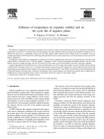
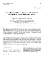
All patients were admitted for abdominal pain and
were managed according to an algorithm helping to
diagnose or exclude appendicitis (Fig. 1).
Pre-operative laboratory examination including determination of white blood cell (WBC)- and polynuclear
neutrophil (PN) count and C-reactive protein (CRP)
blood levels was performed in all patients. All patients
underwent pre-operative imaging by abdominal
ultrasound and abdominal computed tomography (CT)scanner if necessary. Direct signs of appendicitis on
ultrasound were thickening, hyperemia, and incompressibility of the appendix, layer dedifferentiation and
presence of an appendicolithis. Indirect signs were periappendicular fat infiltration, mesenteric adenomegaly
and reactive peritoneal effusion.
The Pediatric Appendicitis Score (PAS) [13] and the
score of Alvarado [14] were assessed in each case retrospectively, according to a previous study [15].
Urgent or scheduled appendectomy was performed
either by laparoscopy or by laparotomy, depending on
surgeon’s preference.
Complications such as appendix perforation, peritonitis (inflammation of the peritoneum with or without
purulent peritoneal liquid), or abscess formation were
diagnosed by ultrasound, at surgery and confirmed by
histological analysis.Children with perforated appendix,
peritonitis or persisting post-operative fever received
intravenous antibiotics (amoxicillin/clavulanic acid (100
mg/kg/day) with or without metronidazole (30 mg/kg/
Lounis et al. BMC Pediatrics
(2020) 20:151
Page 3 of 9
Fig. 1 Algorithm of the diagnostic procedure and treatment in preschool children with abdominal pain. WBC: white blood cell; PN: neutrophil;
CRP: C-reactive protein; US: ultrasound; CT: computed tomography. *: only in equivocal cases
day)) for up to 5 days. This was followed by oral antibiotherapy (amoxicillin/clavulanic acid (50 mg/kg/day)
or cefuroxime (50 mg/kg/) for 5 more days. In case of
sepsis associated with perforation or peritonitis, the
switch to broad spectrum intravenous antibiotics was
undertaken for 10–15 days (piperacillin 100 mg/kg/day/
tazobactam 12.5 mg/kg/day, or ceftazidime 50 mg/kg/
day). Oral relay was undertaken as described above or
with ciprofloxacin (20 mg/kg/day) for up to 10 days.
According to the clinical response to treatment, antibiotherapy was implemented by amikacin (15 mg/kg/
day), ampicillin (50 mg/kg/day), glazidim (50 mg/kg), or
vancomycin (60 mg/kg/day, 6H).
Children were discharged as soon as they were in good
general state, afebrile, painless and with feeding
autonomy.
Primary outcome variables were duration of preadmission symptoms that means the time period
Lounis et al. BMC Pediatrics
(2020) 20:151
Page 4 of 9
between presentation of the first symptoms of appendicitis and admission, clinical presentation and laboratory
findings. Secondary outcome variables were incidence of
operative complications and duration of hospital stay
and were analyzed by comparison of both patient groups
and with respect to the presence of appendicular
perforation.
Statistical analysis
Data were analyzed by the Statistical Package for The
Social Sciences SPSS 22,0, IBM corporation, Armonk,
USA.
Results are shown by the median and interquartile
range (IQR), according to the non-normal data distribution.Inter-group comparison of the median values was
performed by the non-parametrical Mann -Whithney U
test, distribution of categorical variables by the chisquare test and correlation analysis by calculating the
Spearman rank correlation coefficient.
P-values < 0.05 were considered significant, p-values
< 0.1 indicated a tendency toward significance.
Results
Demographic-, clinical patient data and laboratory results are summarized in Table 1.
Table 2 shows the incidence of outcome variables in
both patient groups.
In the whole cohort, age was 50 (25,5) months (group
1: 35 (10); group 2: 59 (16,25) months, respectively).
Median overall duration of symptoms before admission was 48 h (h) (72) in the whole group. It tended to
be longer in group 1 (72 h (72)) than in group 2 (24 h
(48)), p = 0.056).
Duration of nausea/vomiting before admission was significantly longer in group 1 than in group 2 (72 h (90)
versus 24 h (12,5), respectively) (p = 0.026), whereas duration of abdominal pain tended to be longer in group 1
than in group 2 (62 h (84) versus 19 h (46,5), respectively, p = 0.61). Duration of fever was not different between groups.
Upon apparition of the first symptoms and before admission in the emergency department, 17 patients (59%)
had had an ambulatory examination. Diagnosis of acute
appendicitis was made in only 5 of them. In the
Table 1 Demographic, Clinical and laboratory data in all patients, in group 1 and in group 2
All (N = 29)
Group 1 (N = 13)
Group 2 (N = 16)
P
50 (25.5)
35 (10)
59 (16.25)
0.000
Male (n)
14
7
7
Female (n)
Demographic data
Age (months)
Gender
0.43
15
6
9
Weight (kg)
16 (5.25)
14.5 (2.55)
18.8 (6.37)
0.000
2
15.4 (4.1)
14.5 (2.85)
16.1 (4.55)
0.23
Overall duration of symptoms (h)
48 (72)
72 (72)
24 (48)
0.056
Duration of abdominal pain (h)
36 (84)
62 (84)
19 (46.5)
0.061
Duration of nausea-vomiting (h)
31 (66)
72 (90)
24 (12.5)
0.026
48 (54)
67 (69)
30 (51)
0.19
Temperature at admission (°C)
37.5 (1.8)
37.8 (1.4)
37 (1.1)
0.062
Heart rate at admission (bpm)
130 (42.5)
153 (32)
120 (36.7)
0.012
Maximal temperature (°C)
38.8 (1)
38.9 (1.1)
38.6 (1.08)
0.33
17.2 (9.08)
12.2 (8.94)
17.9 (5.94)
0.028
BMI (kg/m )
Pre-admission symptoms
Duration of fever (h)
Clinical data
Laboratory data at admission
Leukocyte count (×109/L)
9
Neutrophil count (×10 /L)
12.9 (7.27)
9.6 (5.72)
14.9 (2.97)
0.004
CRP (mg%)
121.9 (145.2)
134 (140)
67.3 (146)
0.13
5 (2.5)
5 (2)
5 (3)
0.91
Scores
PAS score (/10)
Alvarado (/10)
Duration of hospital stay (d)
5 (3.5)
5 (2.5)
5.5 (4.5)
0.45
6 (4)
7 (5)
3 (3.75)
0.067
Data are shown by the median value and (interquartile range). Group 1: < 48 months; Group 2: > 48 months
Lounis et al. BMC Pediatrics
(2020) 20:151
Page 5 of 9
Table 2 Incidence of outcome variables in both patient groups
Pain right fossa iliaca
All patients
N = 29
Group 1
n = 13
Group 2
N = 16
P
11 (34.4%)
2 (15.4%)
9 (56.2%)
0.029
Alteration general state
16 (55.1%)
12 (92 .3%)
4 (25.0%)
0.000
Purulent peritoneal liquid
11 (34.4%)
8 (61.5%)
3 (18.7%)
0.023
Perforation
11 (34.4%)
9 (69.2%)
2 (12.5%)
0.003
Statistical analysis was performed by the χ2 test. Group 1: < 48 months; Group
2: > 48 months
remaining 12 patients, diagnosis was acute viral gastroenteritis (n = 4), urinary tract infection (n = 3), constipation (n = 2), viral infection (n = 2) and bronchitis (n = 1).
There was no difference between groups.
At admission, alteration of the general state was
present in 16 children and was more frequent in group 1
than in group 2 (p = 0.000).
All patients complained about abdominal pain that
was diffuse (n = 18) or located in the right fossa iliaca
(n = 11). This later was less frequent in group 1 than in
group 2 (p = 0.029).
The majority of the patients showed anorexia (n = 20),
fever (n = 18), nausea and/or vomiting (n = 16). Eleven
patients showed diarrhea, 11 constipation and 6 painful
urination, without any difference between groups.
Temperature at admission tended to be higher in
group 1 than in group 2 (37,8 °C (1,4) versus 37 °C (1,1),
respectively, (p = 0.062), whereas heart rate was significantly higher in group 1 than in group 2 (153 bpm (32)
versus 120 bpm (36,7), respectively, p = 0.012).
At admission 17 patients (59%) showed increased
WBC -, 23 patients (79%) increased PN count and 25
patients (86%) increased CRP. Fifteen children (52%) had
a combination of hyperleukocytosis and increased CRP.
Only one patient (3%) has no increased inflammatory
markers.
WBC- and PN count were significantly lower in
group 1 than in group 2 (WBC: 12.2 × 10⁹/L (8,94)
versus 17.9 × 10⁹/L (5,94), respectively, p = 0.028; PN:
9.6 × 10⁹/L (5,72) versus 14.9 × 10⁹/L (2,97), respectively, p = 0.004). CRP concentration was not different
between groups.PAS score was positive in 12 patients
(41%). Alvarado score was compatible with appendicitis in 9 children (31%), suggested probable or very
likely appendicitis in 7 children (27) % and 1 child
(3%), respectively.
PAS- and Alvarado scores were not different between
groups. Only 19 out of all patients (65.5%) displayed either direct or an association of direct and indirect signs
of appendicitis at this examination (group 1: n = 9; group
2: n = 10, p = 0.63). Abdominal CT-scan was performed
for diagnosis confirmation in 6 patients in whom second
ultrasound was not contributive (group 1: n = 1; group 2:
n = 5, p = 0.18).
Surgery took place either as immediate emergency
intervention or was scheduled at admission or not later
than in the early next morning if diagnosis was achieved
in late night in the majority of the cases (n = 25; 86%). In
4 cases (group 1: n = 1; group 2: n = 3), surgery was delayed until diagnosis confirmation or because of misdiagnosis and finally performed as emergency. Three out of
these patients had appendicular abscess and perforation.
Six patients had retro-caecal appendix (n = 3 each
group).
Eighteen (62%) patients underwent laparoscopy and 11
(38%) laparotomy, 4 of them after open conversion
(14%). Operation technique was not different between
groups.
At operation, perforation was reported in 9 patients of
group 1 and in 2 of group 2 (p = 0.003). Peritoneal liquid
was purulent in 8 patients of group 1 and in 3 of group
2 (p = 0.023). Peritonitis was the most frequent intraoperative finding in the whole cohort (72%). In 5 patients post-operative ileus occurred. Treatment consisted
of antalgic control, bowel rest, gastric liquid aspiration
and intravenous infusion of a crystalloid solution for hydration until bowel transit recovered after a median
delay of 2 days (IQR: 1.5 days). Alizapride chlorhydrate
was given as anti-emetic medication if necessary.
Twenty five children received intravenous combination
of amoxyciline/clavulanic acid that was associated with
metronidazole in 17 and followed by an oral relay, according to our protocol.
Four children developed sepsis and required a broad
spectrum antibiotherapy for a median duration of 16
days (IQR: 6.5 days): One patient received the association of intravenous amoxyciline/clavulanic acid and
metronidazole followed by oral amoxyciline/clavulanic
acid for a total of 10 days; One patient received intravenous amoxyciline/clavulanic acid and metronidazole
followed by oral ciprofloxacin for a total of 17 days; one
patient received intravenous amoxyciline/clavulanic acid
and metronidazole for 5 days that was switched to intravenous piperacillin/tazobactam for 5 days, followed by
oral ceftazidime for 5 more days (total 15 days). The last
patient did not respond to the initial intravenous amoxyciline/clavulanic acid and metronidazole association that
was enlarged with intravenous amikacin. The treatment
was switched to glazidim, ampicillin, vancomycin and
metronidazole 2 days later for a total of 18 days. Oral
treatment consisted of amoxyciline/clavulanic acid and
ciprofloxacin for 11 more days.
Two patients required a second surgery. There was no
difference in post-operative complications between
groups. Complications are summarized in Table 3.
Length of hospital stay was 6 days (4) and tended to be
longer in group 1 (7 (5)) than in group 2 (3 (3.75)) (p =
0.67). In the whole cohort, it correlated negatively with
Lounis et al. BMC Pediatrics
(2020) 20:151
Page 6 of 9
Table 3 Complications of acute appendicitis in all patients and in both patient groups
All patients (N = 29)
Group 1 (N = 13)
Group 2 (N = 16)
P
Peritonitis
21 (72%)
11 (85%)
10 (62.5%)
0.18
Appendicular abscess
12 (41%)
7 (54%)
5 (31%)
0.19
Perforation
11 (38%)
9 (69%)
2 (12.5%)
0.003
Post-operative ileus
5 (17%)
3 (23%)
2 (12.5%)
0.39
Sepsis
4 (14%)
1 (8%)
3 (18.7%)
0.38
Second surgery
2 (7%)
1 (8%)
1 (6%)
0.70
Statistical analysis was performed by the χ2-test. Group 1: < 48 months; Group 2: > 48 months
age (Spearman rank correlation coefficient − 0.668, p =
0.000) (Table 4). Patients with perforation had a longer
hospital stay than the others (7 days (4) versus 3.5 (3,75),
p = 0.018) (Table 5).
There was no mortality.
Discussion
Our study confirms that acute appendicitis in preschool children is rare, accounting for less than 10%
of all pediatric cases [3]. In our series, diagnosis of
appendicitis was made after a median period of 48 h
following the apparition of the first symptoms, the
majority of the patients having been assessed ambulatory and discharged with a diagnosis of a banal viral
infection, in particular gastro-enteritis or urinary tract
infection. This is in line with previous reports indicating that diarrhea is a frequent symptom of acute appendicitis explained by the effect of abdominal
infection on intestinal motility [7, 16, 17].
Indeed, diagnosis of appendicitis in preschool children
is challenging and burdened by a high rate of misdiagnosis resulting from atypical clinical signs and by
Table 4 Correlations between patient age and outcome
variables in the whole cohort (n = 29)
Spearman rank coefficient
P
* Overall duration of
pre-admission symptoms
−0.495
0.007
*Duration abdominal
pain before admission
−0.422
0.028
*Duration nausea-vomiting
before admission
−0.531
0.034
*Temperature at admission
−0.527
0.003
*Heart rate at admission
− 0.627
0.000
*White blood cell count at
admission
0,315
0,096
Age versus
*Neutrophil count at admission
0.442
0.016
*CRP-value at admission
−0.416
0.025
*Maximal CRP-value
−0.345
0.067
*Duration of hospital stay
−0.668
0.000
Statistical analysis was performed by the Spearman rank test
trivialization of abdominal pain in this age group [3]. On
the contrary to school children and adolescents, younger
children do not present the classical clinical picture with
initial anorexia and peri-umbilical pain that migrates in
the right fossa iliaca, vomiting and fever [1]. According
to that, in our series, the only constant symptom on admission was abdominal pain that was diffuse in the majority of cases. Fever, anorexia and transit alteration
were not observed in all patients.
Previous studies have shown that complications due to
appendicitis are more frequent and more severe in children
than in adults. Furthermore, patients with complicated appendicitis are more likely to be under 5 years of age and to
have had symptoms for a period exceeding 24 h, compared
to patients with uncomplicated appendicitis [18]. Our results are consistent with that. The non-specific clinical presentation of acute appendicitis in young children is thought
to be responsible for diagnosis delay and therefore for
higher rate of complications in this age group, as it has
been shown in patients younger than 5 years of age [3].
This fact is illustrated in our cohort in whom a high rate of
peritonitis (72%), appendicular abscess (41%), and appendicular perforation (38%) was observed.
This is in line with previous reports showing that
delayed diagnosis expressed by the duration of preadmission symptom is associated with appendix
perforation. Indeed, after 36 h of symptoms, the risk of
perforation increases by 5% each 12 h [19].
Diagnosis difficulties in children with appendicitis have
led to the attempt to use of scoring systems. In this
study, we applied retrospectively the Alvarado- and the
PAS scores but found the results not contributive, in accordance with previous report [20]. In a large previous
prospective study, both scoring systems were assessed
and compared but both scores gave a specificity lower
than 60% and none had a sufficient predictive value for
the diagnosis of acute appendicitis [6]. Since most items
entered for score calculation are clinical signs that have
a low incidence in pre-school children, patient age is
clearly expected to influence the predictive value of
Alvarado score and PAS. Hence, the controversial results
reported on appendicitis scores performance in children
may be explained by the important age variability in the
Lounis et al. BMC Pediatrics
(2020) 20:151
Page 7 of 9
Table 5 Demographic, Clinical and laboratory data in all patients, in patients with and without perforation
All (N = 29)
Perforated (N = 11)
Non-Perforated (N = 18)
P
50 (25.5)
35 (16)
58 (22,2)
0.002
Male (n)
14
6
8
Female (n)
Demographic data
Age (months)
Gender
0.33
15
5
10
Weight (kg)
16 (5.25)
15 (2.51)
18.8 (8,5)
0.002
2
15.4 (4.1)
14.5 (2.6)
16.1 (4.55)
0.025
BMI (kg/m )
Pre-admission symptoms
Overall duration of symptoms (h)
48 (72)
72 (52)
32 (53)
0.14
Duration of abdominal pain (h)
36 (84)
55 (96)
24 (51,5)
0.069
Duration of nausea-vomiting (h)
31 (66)
72 (90)
24 (32,5)
0.33
Duration of fever (h)
48 (54)
67 (96)
36 (51)
0.44
Temperature at admission (°C)
37.5 (1.8)
37.8 (1.2)
36,8 (3,4)
0.31
Heart rate at admission (bpm)
130 (42)
153 (25)
118,5 (41)
0.008
Maximal temperature (°C)
38.8 (1)
38.6 (1.0)
118,5 (41)
0.008
17.2 (9.08)
17.24 (11.93)
17.1 (8.74)
0.52
Clinical data
Laboratory data at admission
Leukocyte count (×109/L)
Neutrophil count (×109/L)
12.9 (7.27)
12.14 (7.34)
14.0 (7.65)
0.08
CRP (mg%)
121.9 (145.2)
134 (55.4)
61 (184.6)
0.41
PAS score (/10)
5 (2.5)
5 (2)
4.5 (4)
0.051
Alvarado (/10)
5 (3.5)
5 (3)
4.5 (4.2)
0.37
6 (4)
7 (4)
3.5 (3.75)
0.018
Scores
Duration of hospital stay (d)
Data are shown by the median value and (interquartile range)
different large series reported [6, 13, 15].Besides the lack
of specificity of clinical signs for diagnosing acute appendicitis in young children, laboratory examinations and
imaging are also imperfect diagnosis tools yet. Nevertheless, the literature admits that the elevation of biological
markers such as WBC count, PN count and CRP is often
observed in acute appendicitis, but it lacks of specificity,
especially when it is isolated [21, 22].
In our patients, first abdominal ultrasound was suggestive of appendicitis in only 65.5% of the cases. In most of
children in whom abdominal ultrasound was negative, appendix was either not- or incompletely visualized. A frequent cause for that is ectopic position of the appendix.
This explains the relative high rate of abdominal CTscanner that had to be performed in this cohort. Nevertheless, abdominal ultrasound should, due to the possibility to easily repeat examinations, remain the first choice
and the most frequently performed examination for the
diagnosis of appendicitis in the pediatric population [23,
24]. The role of magnetic resonance imaging for the diagnosis of acute appendicitis in young children remains to
be established [25].
An objective of this study was to analyze the influence
of age on outcome variables among the group of preschool children. Owing to the age distribution in our
series, we considered 2 patients groups younger or older
than 48 months of age. Our results show that, as expected,
the youngest had the longest duration of clinical symptoms before diagnosis and treatment. The youngest had
also more frequent alteration of the general state, indicating systemic involvement of the disease and less frequent
pain in the right fossa iliaca. This latter might be explained
by a preponderance of visceral abdominal pain in contrast
to parietal abdominal pain in younger children [26] and
not by appendix localization, in particular in retro-caecum
position that was equally present in both groups.
Children younger than 48 months of age tended to
have higher central temperature at admission, as a sign
of higher systemic repercussions of the abdominal infection in this group. They also showed significantly higher
heart rate that besides the fact that this decreases
physiologically with age may be explained by the combination of higher central
temperature and
hemodynamic adaptation to severe infection.
Lounis et al. BMC Pediatrics
(2020) 20:151
Interestingly, our results show that in younger children, there was dissociation in the inflammatory response with significantly lower WBC- and PN counts in
combination with higher CRP levels than in older children. Indeed, PN count at admission correlated positively with age and CRP negatively, and WBC- and PN
counts were significantly lower in children younger than
48 months than in the older ones.
This suggests age-related WBC and PN migration with
impaired recruitment from the bone marrow into the circulation in younger children in spite of an important inflammatory response to the bacterial infection reflected by
the induction of high levels of CRP in the liver [27]. This
observation points out the absolute necessity to measure
blood levels of CRP together with WBC count in order
not to misinterpret normal or low WBC counts that may
consolidate the presumption of banal viral infection, and
especially as a large literature review concluded that lower
WBC count decreased the likelihood of appendicitis in
children [28]. Low WBC- and PN counts in young children had certainly also contributed to the underscoring
and lack of specificity of both the Alvarado score and PAS
in our patient population.
In our series, and in accordance with previous reports
[2, 3, 12], younger children developed a higher rate of
complications such as perforation with purulent peritoneal liquid that is, as discussed above, the result of prolonged disease course. However, the rate of other
complications such as abscess, ileus or sepsis was not
different between groups.
Limitation section
This study has several limitations related to its retrospective
design and to the small patient cohort analyzed according
to the rarity of the disease in the elected age group.
Page 8 of 9
Abbreviations
bpm: beat per minute; CRP: C-reactive protein; CT: Computed tomography;
IQR: Interquartile range; PN: Polynuclear neutrophils; PAS: Pediatric
appendicitis score; WBC: White blood cells
Acknowledgements
none.
Authors’ contributions
All authors contributed to the study conception and design. Material
preparation, data collection and analysis were performed by YL, JH and MCS. The first draft of the manuscript was written by YL and MD and all authors commented on previous versions of the manuscript. All authors read
and approved the final manuscript.
Funding
none.
Availability of data and materials
The datasets used and/or analyzed during the current study are available
from the corresponding author on reasonable request.
Ethics approval and consent to participate
All procedures performed in studies involving human participants were in
accordance with the ethical standards of the institutional and/or national
research committee, and with the 1964 Helsinki declaration and its later
amendments or comparable ethical standards.
The agreement of the Ethics Committee of the University Hospital Liège was
obtained.
Since this is a retrospective anonymized study, the informed consent of the
children’s caregiver was not requested.
Administrative permission was given by the Department of Medical- and
Economical Information (SIME) of the institution to review retrospectively the
patient files.
Consent for publication
does not apply.
Competing interests
The authors declare that they have no competing.
Author details
1
Department of Pediatrics, University Hospital Liège, Liège, Belgium.
2
Department of Emergency Medicine, University Hospital Liège, Liège,
Belgium. 3Department of Pediatric Surgery, Regional Hospital Citadelle, Liège,
Belgium.
Received: 12 September 2019 Accepted: 26 March 2020
Conclusions
This study confirms that acute appendicitis in children
less than 6 years of age is a rare condition and is still related to a high risk of morbidity, especially appendix perforation, due to the diagnostic delay. This latter in turn
is the consequence of non-specific symptoms and the
non-pathognomonic clinical-and complementary examination results and increases with younger age.
Less specific, trivialized abdominal pain and dissociation of the inflammatory markers with lower leucocyteand neutrophil counts and higher C-reactive protein
levels in young children contribute to the diagnosis trap
of acute appendicitis in preschool children.The greatest
caution is therefore mandatory when evaluating a young
child with acute abdominal pain and the question of
whether it could be acute appendicitis systematically
addressed.
References
1. Almaramhy HH. Acute appendicitis in young children less than 5 years:
review article. Ital J Pediatr. 2017;43:15. />2. van den Bogaard VA, Euser SM, van der Ploeg T, de Korte N, Sanders DG, de
Winter D, et al. Diagnosing perforated appendicitis in pediatric patients: a
new model. J Pediatr Surg. 2016;51:444–8. />2015.10.054.
3. Pogorelic Z, Domjanovic J, Jukic M, Pericic TP. Acute appendicitis in children
younger than five years of age: diagnostic challenge for pediatric surgeons.
Surg Infect. 2019;21(3):239. />4. Bansal S, Banever GT, Karrer FM, Partrick DA. Appendicitis in children less
than 5 years old: influence of age on presentation and outcome. Am J Surg.
2012;204:1031–5. />5. Naiditch JA, Lautz TB, Daley S, Pierce MC, Reynolds M. The implications of
missed opportunities to diagnose appendicitis in children. Acad Emerg
Med. 2013;20:592–6. />6. Pogorelic Z, Rak S, Mrklic I, Juric I. Prospective validation of Alvarado score
and pediatric appendicitis score for the diagnosis of acute appendicitis in
children. Pediatr Emerg Care. 2015;31:164–8. />0000000000000375.
Lounis et al. BMC Pediatrics
7.
8.
9.
10.
11.
12.
13.
14.
15.
16.
17.
18.
19.
20.
21.
22.
23.
24.
25.
26.
27.
28.
(2020) 20:151
Horwitz JR, Gursoy M, Jaksic T, Lally KP. Importance of diarrhea as a
presenting symptom of appendicitis in very young children. Am J Surg.
1997;173:80–2. />Sakellaris G, Tilemis S, Charissis G. Acute appendicitis in preschool-age children.
Eur J Pediatr. 2005;164:80–3. />Pogorelic Z, Buljubasic M, Susnjar T, Jukic M, Pericic TP, Juric I. Comparison
of open and laparoscopic appendectomy in children: a 5-year single center
experience. Indian Pediatr. 2019;56:299–303.
Pogorelic Z, Silov N, Jukic M, Elezovic Baloevic S, Peričić TP, et al. Ertapenem
monotherapy versus gentamicin plus metronidazole for perforated
appendicitis in pediatric patients. Surg Infect. 2019;20:625–30. https://doi.
org/10.1089/sur.2019.025.
van den Boom AL, Gorter RR, van Haard PM, Doornebosch PG, Heij HA,
Dawson I. The impact of disease severity, age and surgical approach on the
outcome of acute appendicitis in children. Pediatr Surg Int. 2015;31:339–45.
/>Bonadio W, Peloquin P, Brazg J, Scheinbach I, Saunders J, Okpalaji C, et al.
Appendicitis in preschool aged children: regression analysis of factors
associated with perforation outcome. J Pediatr Surg. 2015;50:1569–73.
/>Samuel M. Pediatric appendicitis score. J Pediatr Surg. 2002;37:877–81.
/>Alvarado A. A practical score for the early diagnosis of acute appendicitis.
Ann Emerg Med. 1986;15:557–64. />Macco S, Vrouenraets BC, de Castro SM. Evaluation of scoring systems in
predicting acute appendicitis in children. Surgery. 2016;160:1599–604.
/>Marzuillo P, Germani C, Krauss BS, Barbi E. Appendicitis in children less than
five years old: a challenge for the general practitioner. World J Clin Pediatr.
2015;4:19–24. />Mallick MS. Appendicitis in pre-school children: a continuing clinical
challenge. A retrospective study. Int J Surg. 2008;6:371–3. />1016/j.ijsu.2008.06.003.
Pham XD, Sullins VF, Kim DY, Range B, Kaji AH, de Virgilio CM, et al. Factors
predictive of complicated appendicitis in children. J Surg Res. 2016;206:62–
6. />Bickell NA, Aufses AH Jr, Rojas M, Bodian C. How time affects the risk of
rupture in appendicitis. J Am Coll Surg. 2006;202:401–6. />1016/j.jamcollsurg.2005.11.016.
Ebell MH, Shinholser J. What are the most clinically useful cutoffs for the
Alvarado and pediatric appendicitis scores? A systematic review. Ann Emerg
Med. 2014;64:365–72.e2. />Shogilev DJ, Duus N, Odom SR, Shapiro NI. Diagnosing appendicitis:
evidence-based review of the diagnostic approach in 2014. West J Emerg
Med. 2014;15:859–71. />Anandalwar SP, Callahan MJ, Bachur RG, Feng C, Sidhwa F, Karki M, et al.
Use of white blood cell count and Polymorphonuclear leukocyte differential
to improve the predictive value of ultrasound for suspected appendicitis in
children. J Am Coll Surg. 2015;220:1010–7. />jamcollsurg.2015.01.039.
Löfvenberg F, Salö M. Ultrasound for appendicitis: performance and
integration with clinical parameters. Biomed Res Int. 2016;2016:5697692.
/>Rentea RM, Peter SDS, Snyder CL. Pediatric appendicitis: state of the art
review. Pediatr Surg Int. 2017;33:269–83. />Mittal MK. Appendicitis: role of MRI. Pediatr Emerg Care. 2019;35:63–6.
/>Kim JS. Acute abdominal pain in children. Pediatr Gastroenterol Hepatol
Nutr. 2013;16:219–24. />Markanday A. Acute phase reactants in infections: evidence-based review
and a guide for clinicians. Open Forum Infect Dis. 2015;2:ofv098. https://doi.
org/10.1093/ofid/ofv098.
Bundy DG, Byerley JS, Liles EA, Perrin EM, Katznelson J, Rice HE. Does this
child have appendicitis? Jama. 2007;298:438–51. />jama.298.4.438.
Publisher’s Note
Springer Nature remains neutral with regard to jurisdictional claims in
published maps and institutional affiliations.
Page 9 of 9