Prognostic nutrition index as a predictor of coronary artery aneurysm in Kawasaki Disease
Bạn đang xem bản rút gọn của tài liệu. Xem và tải ngay bản đầy đủ của tài liệu tại đây (1.12 MB, 8 trang )
Tai et al. BMC Pediatrics
(2020) 20:203
/>
RESEARCH ARTICLE
Open Access
Prognostic nutrition index as a predictor of
coronary artery aneurysm in Kawasaki
Disease
I-Hsin Tai1,2,3, Pei-Lin Wu1, Mindy Ming-Huey Guo1, Jessica Lee4, Chi-Hsiang Chu5, Kai-Sheng Hsieh1,6 and
Ho-Chang Kuo1*
Abstract
Background: Kawasaki Disease (KD) is considered a major acquired heart disease in children under the age of 5.
Coronary artery aneurysm (CAA) can occur in serious cases despite extreme therapy efforts. Previous studies have
reported low serum albumin level was associated with disease outcome, but no further investigation was
addressed yet.
Method: This retrospective (case-control) study randomly included children with KD who were admitted and
underwent laboratory tests before undergoing IVIG treatment in this institution, the largest tertiary medical center
in southern Taiwan from 2012 to 2016. Prognostic nutrition index (PNI), an albumin-based formula product, was
evaluated as a predictor of CAA the first time. The progression of CAA was monitored using serial
echocardiography for six months. We performed multivariable logistic regression analysis on the laboratory test and
PNI with the disease outcome of the KD patients.
Result: Of the 275 children, 149 had CAA, including transient dilatation, while the other 126 did not develop CAA
during the 6-month follow-up period. A multivariate logistic regression model revealed that PNI, gender, IVIG nonresponder, and platelet count are significant predictors of CAA with a 95% confidence interval estimator of 1.999,
3.058, 3.864 and 1.004, respectively. Using PNI to predict CAA presence gave an area under the receiver-operatingcharacteristics (ROC) curve of 0.596. For a cutoff of 0.5 in the logistic regression model and the PNI cut-off point is
taken as 55 together with IVIG non-responder, boy gender, and platelet count take into account, sensitivity and
specificity were 65.7 and 70.4%.
Conclusion: PNI could be a candidate of adjunctive predictor of coronary artery aneurysm in addition to IVIG nonresponder. Together with low PNI, IVIG non-responder, male gender and platelet count will give high odds to
predict coronary artery aneurysm within 6 months of illness.
Keywords: Prognostic nutrition index, Kawasaki Disease, Coronary artery aneurysm, Albumin, Lymphocyte
* Correspondence:
1
Kawasaki Disease Center and Pediatrics, Kaohsiung Chang Gung Memorial
Hospital, Taiwan, College of Medicine, Chang Gung University, #123 Da-Pei
Road, Niaosong District, Kaohsiung city 83301, Taiwan
Full list of author information is available at the end of the article
© The Author(s). 2020 Open Access This article is licensed under a Creative Commons Attribution 4.0 International License,
which permits use, sharing, adaptation, distribution and reproduction in any medium or format, as long as you give
appropriate credit to the original author(s) and the source, provide a link to the Creative Commons licence, and indicate if
changes were made. The images or other third party material in this article are included in the article's Creative Commons
licence, unless indicated otherwise in a credit line to the material. If material is not included in the article's Creative Commons
licence and your intended use is not permitted by statutory regulation or exceeds the permitted use, you will need to obtain
permission directly from the copyright holder. To view a copy of this licence, visit />The Creative Commons Public Domain Dedication waiver ( applies to the
data made available in this article, unless otherwise stated in a credit line to the data.
Tai et al. BMC Pediatrics
(2020) 20:203
Background
KD is the worldwide leading cause of acquired heart disease
in developed countries, and the most serious sequela is the
development of a CAA. Starting treatment with IVIG within
9 days of the onset of fever reduces the incidence of coronary
artery aneurysms from 25% to 3 ~ 5% [1] in absolute luminal
dimensions. The 2017 American Heart Association (AHA)
scientific statement defined different management protocols
for KD patients with and without regression of coronary artery aneurysm [2] 4–6 weeks after the onset of KD. This
protocol difference demonstrates a delayed regression of coronary dilation, which indicates a more severe coronary vasculitis and deservedly more aggressive therapy and
monitoring. Wu et al. showed that morbidity rates increased
in those patients whose CAA regression occurred more than
2 months later [3]. Therefore, early or late regression of coronary vasculitis is crucial for future prognosis stratification.
PNI has been used to predict and evaluate postoperative status in cancer patients for decades, ever since
it was first published in 1983 [4]. PNI has also been used
to predict mortality in patients with ST-segment elevation myocardial infarction (STEMI) [5]. PNI is currently
determined by albumin (ALB) and total lymphocyte
count (TLC), while its original formula used triceps skinfold thickness (TSF), serum transferrin concentration
(TFN), and delayed hypersensitivity reaction (DHC, no
reaction = 0, < 5 mm induration =1, and > 5 mm induration = 2) instead of the current TLC. Albumin has been
a consistent parameter in the PNI formula because various studies have shown its correlation with nutrition
and immune status. By definition, a higher albumin level
or lymphocyte count contributes to a greater PNI value,
which indicates a superior self-healing ability due to sufficient nutrition and improved immune capacity, which can
prevent opportunistic infectious pathogen invasion. In our
previous report, we found that the serum level of albumin
was associated with IVIG resistance in KD patients [6].
Although the definite cause of KD remains unknown,
evidence [7] has shown that KD is most likely caused by
an infectious agent(s) that produces a clinically apparent
disease in genetically predisposed individuals. Once a patient develops KD, the vasculopathy cause plasma leakage as well as serum albumin. That explained the palmar
and plantar erythema which usually accompanied by
swelling in acute KD children. Hypoalbuminemia is wide
known as risk factor for IVIG resistant KD, which correlated with CAA development. This study aimed to investigate whether there is causality between nutrition, an
albumin-based status, and CAA in KD.
Methods
Subjects’ enrollment & data collection
This study was approved by Chang Gung Memorial
Hospital’s institutional review board with IRB number
Page 2 of 8
102-3595C. We performed a retrospective case control
study with the clinical records of KD patients hospitalized at Kaohsiung Chang Gung Memorial Hospital between 2012 and 2016. We included patients diagnosed
with KD based on AHA guidelines [2]. and collected
data from KD with CAA (CAA present group) and agematched KD population without CAA formation as the
control group (CAA absent group). According to the latest AHA guideline, classic KD is diagnosed with the
presence of fever for at least 5 consecutive days with at
least 4 of the 5 principal clinical features (oral changes,
conjunctivitis, cervical lymphadenopathy, extremity
changes, and dysmorphism rash). If a patient has more
than four of the principal clinical features together with
limb induration, KD can be diagnosed with just 4 days of
fever. All of the participating patients underwent 2Dechocardiography of the coronary artery during admission, as well as at 2, 4, and 6–8 weeks and 3, 4, and 6
months from disease onset. Positive echocardiogram
findings of CAA were defined by a body surface area adjusted Z score of coronary segments exceeding 2.5 in accordance with AHA criteria [2]. All the patients were
diagnosed with KD and underwent IVIG treatment in
our hospital except three patients who were afebrile
spontaneously within 5 days of illness. Patients who received IVIG treatment elsewhere were excluded. The following laboratory data were collected prior to
administering IVIG: total white blood cell count (WBC),
the percentage of neutrophils and lymphocytes,
hemoglobin levels, platelet count (PLT), serum concentrations of C-reactive protein (CRP), aspartate aminotransferase (AST), alanine aminotransferase (ALT), and
serum albumin.
Afterwards, PNI was calculated according to the serum
level of albumin & total lymphocyte count [PNI = 10 x
albumin (mg/dl) + 0.005 x lymphocyte counts (109 L− 1)]
as previously reported [4].
Statistical analyses
All values are expressed as mean ± standard deviation
(SD), median (1st quantile, 3rd quantile), or number
(percentage), as appropriate. For all analytic results, a pvalue of 0.05 is considered statistically significant. We
adopted the independent t and Mann-Whitney U test to
identify the difference between the two groups for continuous variables according to the normality test. For independent variables, Pearson chi-square test was applied
to compare the proportion between both groups. We
used the ROC curve to analyze the optimal cut-off point
of a variable with Youden’s index criterion. To compare
the odds ratio of significant variables, we selected the
candidate variables using univariate logistic regression
with a p-value of 0.05 and the final model using multivariate logistic regression. All statistical analysis was
Tai et al. BMC Pediatrics
(2020) 20:203
performed using SPSS statistical software for Windows
version 13.0 (SPSS for Windows, version 13; SPSS, Chicago, IL).
Results
We enrolled 275 patients with KD from our search database in this study. We randomly and retrospectively included 149 KD patients with CAA formation and 126
age-matched KD patients without CAA formation as the
control group. The percentage of males was higher in
the CAA present group (76.5% vs. 54.0%, p < 0.001) than
the CAA absent group. We found no statistical differences in age for KD between the two groups (due to this
being an age-matched case control study). The median
age of these patients upon diagnosis of acute KD was
1.14 years and 1.31 years (p = 0.161), respectively
(Table 1).
The initial absolute values of the complete blood
count, differential count, and CRP, as well as the liver
function and albumin concentrations, in each group are
provided in Table 2. We found WBC to be higher in the
CAA present group than in the CAA absent group (13.5
Table 1 Characteristics of participants, N=275
Page 3 of 8
vs. 12.7 × 10³/mm3, p = 0.050), the neutrophil count to
be higher in the CAA present group (7.88 vs. 7.05 × 10³/
mm3, p = 0.015) than in the CAA absent group, the
platelet count to be higher in the CAA present group
(360.0 vs. 314.5 × 10³/mm3), p = 0.001) than in the CAA
absent group, the CRP levels to have no significant difference between the CAA present group (69.9 vs. 63.2
mg/L, p = 0.314) and the CAA absent group, and albumin levels to be significantly lower in the CAA present
group (3.7 vs. 3.9 g/dL), p = 0.002) than the CAA absent
group.
Furthermore, we found that prior to IVIG therapy, the
CAA present group had a significantly higher segmentto-lymphocyte ratio (SLR) (3.50 vs. 2.32, P = 0.004) and
platelet-to-lymphocyte ratio (PLR) (127.66 vs. 91.09, P <
0.001), while a significantly lower PNI (56.23 vs. 59.63,
P = 0.038), than the CAA absent group, as shown in
Table 3.
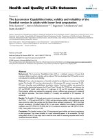
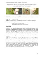
The ROC curve analysis (Fig. 1A) indicates that the
area under the ROC curve is 0.588, with a significance
0.023 for PNI. The PNI cut-off value is determined to be
55.24 with a sensitivity of 0.500 and a specificity of 0.678
Tai et al. BMC Pediatrics
(2020) 20:203
Page 4 of 8
Table 2 Baseline laboratory data of the CAA presence and absence group
by maximizing the Youden’s index. In the following
paragraph, we define the high-PNI group as PNI ≥ 55
and the low-PNI group as PNI < 55.
According to the multivariate analysis with logistic regression procedure (Table 4), the male gender, IVIG nonresponder, elevated platelet counts, and PNI-low group
positively correlated with the presence of CAA. The risk
of CAA formation was 3.058 greater in boys and 3.864
greater in the IVIG non-responder. As for PNI-low group,
the even earlier information acquired, the risk of CAA
was nearly twice as PNI-high group. The odds of IVIG-
resistant was 7.65 times greater for low-PNI patients than
for high-PNI patients (Fig. 1B) (p < 0.001).
Under multivariate analysis, male gender, higher platelet count and lower PNI value (< 55) before IVIG all had
significantly positive correlation to CAA presence in 6
months of KD-illness.
Discussion
PNI role in the history
Nutrition assessment results have previously been
proven to define the incidence of post-operative
Tai et al. BMC Pediatrics
(2020) 20:203
Page 5 of 8
Table 3 Blood cell ratio and PNI of CAA presence and absence group
complications, mortality, and morbidity in patients with heart
failure or malignant cancers [8–13]. While many nutritionists
suggest using the Controlling Nutritional Status (CONUT)
score to assess the nutrition status of acute heart failure, a
large retrospective cohort study demonstrated that PNI has
the same prognostic impact in patients with decompensated
heart failure [14, 15]. PNI was an independent predictor for
evaluating the correlation between nutritional status and malignancy or vital organ failure mortality by comparing subjects
of the high-PNI and low-PNI groups [12, 16, 17]. In addition
to being used with adult diseases, PNI can also predict the
clinical outcome of the pediatric population in the intensive
care unit after cardiac operation [18]. However, we found PNI
could predict CAA risk in acute KD patients in addition to
correlating with nutrition status.
Hypoalbuminemia in KD and CAL formation
KD is a form of chronic vasculitis that may last for
months to years in regard to pathophysiology. Therefore,
all KD patients with or without coronary ectasia are considered at high risk for accelerated atherosclerosis according to the epidemiological evidence and should
undergo nutrition counseling and diet education in an
effort to reduce their future cardiovascular burden [19].
Fig. 1 : PNI as predictor of CAL. A. The ROC curve analysis shows that the area under the ROC curve is 0.588 (0.513–0.663), with a significance of
0.023 for the prognostic nutritional index (PNI). The cut-off value of PNI is taken as 55.24, with a sensitivity of 0.500 and a specificity of 0.678 by
maximizing the Youden’s indexB. Low-PNI group has significant high odds (odds = 7.65) to be IVIG-resistant.
Tai et al. BMC Pediatrics
(2020) 20:203
Page 6 of 8
Table 4 Univariate/multivariate logistic regression model with CAA group
Research has identified that younger than 6 months of
age, male, incomplete KD, longer fever duration, higher
CRP levels (> 100 mg/l), and lower albumin levels (< 35
g/L) were all independent risk factors for CAA formation [20], thus indicating that both delayed initiation of
KD target therapy and hypoalbuminemia, which indicates a relatively poor nutritional status, result in higher
incidence rates of CAA complications in patients with
acute KD, despite the administration of IVIG therapy.
PNI predicts KD with CAA & IVIG non-responder
In the current study, we showed that PNI, an albumin
based long-term predictor of cancer, was also a
significant independent predictor of CAA in any coronary segment during the 6 months after the onset of illness (PNI < 55, estimator: 1.999, p = 0.030), as well as
gender, IVIG non-responder, and platelet count. However, the associations of pre-treatment platelet count
and CAA formation were relatively weak in this cohort,
with a 95% confidence interval of estimator between
1.002–1.007. To the best of our knowledge, this study is
the first to discuss the predictive value of PNI on CAA
formation in KD patients before they receive initial IVIG
therapy. Kobayashi et al. constructed a seven-variable
predictive model to identify IVIG-resistant KD using
pretreatment laboratory data. Although previous
Tai et al. BMC Pediatrics
(2020) 20:203
research has shown that most KD patients with CAA are
unresponsive to IVIG, the detailed mechanism between
IVIG non-responders and CAA formation has yet to be
explained. Our results are in line with Kuo et al.’s previously published studies demonstrating the significant relationship between hypoalbuminemia and IVIG-resistant
KD, which often indicates a higher incidence of CAA
[6]. Of particular interest is the discrepancy conclusion
from Japan [21] (Kobayashi et al., 2006) to Taiwan (Kuo
et al., 2010) regarding the correlation between IVIG
non-responder and hypoalbuminemia using multivariate
logistic regression models [6, 21]. Assuming that both
research methods were appropriately and strictly designed, we may presume that an unknown ongoing
process involved nutrition status, in addition to vascular
inflammation. However, early validation research on
Japan scoring models yield inconsistent result between
different races [2, 22–24]. It showed multiple ethnicityexclusive models are required. Our findings revealed that
a low pre-treatment PNI level (PNI < 55) correlated to a
high incidence of CAA complication in KD patients, as
well as IVIG non-responder.
PNI practice
Low-PNI alone before initial IVIG therapy have nearly
2-fold (estimator: 1.999, Table 4) risk to develop future
CAA. In the setting of low-PNI, IVIG non-responder,
male gender, and higher platelet count will give rise to
at least 8.8-fold higher risk to develop CAA. Therefore,
PNI in conjunction with IVIG response, gender, and
platelet will have better prediction of developing CAA
within 6 months of illness.
Conclusion
The utility of PNI as adjunctive predictor of coronary artery aneurysm in addition to IVIG non-responder, male
gender and platelet count will give high odds for predicting CAA formation in KD patients. The simply quick
formula allow physicians to identify patients that may
benefit from aggressive primary or advanced antiinflammatory therapies.
Abbreviations
KD: Kawasaki Disease; IVIG: intravenous immunoglobulin; CAA: coronary
artery aneurysm; PNI: Prognostic nutrition index
Acknowledgements
We would also like to show our gratitude to Ying-Hsien Huang MD, PhD,
Kawasaki Disease center & Kaohsiung Chang Gung Memorial Hospital for
sharing his pearls of wisdom with us during the course of this research.
Authors’ contributions
IHT analyzed and interpreted the patient data and was a major contributor
in writing the manuscript. IHT wrote the manuscript with support from PLW,
MMHG, and JL. CHC performed the calculations. KSH supervise the work.
HCK designed the experiment and analyzed the data. The authors read and
approved the final manuscript.
Page 7 of 8
Funding
This study was funded by the following grants: MOST: 108–2314-B-182-037MY3 from the Ministry of Science and Technology of Taiwan and
CMRPG8F1911, 1921, 1931, and 1941, and 8E0212 from Chang Gung
Memorial Hospital in Taiwan. Even though these institutes provided financial
support, they had no influence on the way we collected, analyzed, or
interpreted the data or prepared this manuscript.
Availability of data and materials
The datasets used and analyzed during the current study are available from
the corresponding author on reasonable request.
Ethics approval and consent to participate
This study was approved by Chang Gung Memorial Hospital’s institutional
review board with IRB number 102-3595C.
Consent for publication
Not applicable.
Competing interests
The authors declare that they have no competing interests.
Author details
1
Kawasaki Disease Center and Pediatrics, Kaohsiung Chang Gung Memorial
Hospital, Taiwan, College of Medicine, Chang Gung University, #123 Da-Pei
Road, Niaosong District, Kaohsiung city 83301, Taiwan. 2Department of
Pediatric Emergency China Medical University Children’s Hospital, China
Medical University, Taichung City, Taiwan. 3Department of Medicine, College
of Medicine, China Medical University, Taichung City, Taiwan. 4University of
Maryland Medical Center, Baltimore, MD, USA. 5Department of Statistics,
National Cheng Kung University, Tainan city, Taiwan. 6Department of
Pediatrics, Shuang Ho Hospital-Taiwan Medical University, New Taipei City,
Taiwan.
Received: 27 December 2019 Accepted: 29 April 2020
References
1. Newburger JW, Takahashi M, Gerber MA, Gewitz MH, Tani LY, Burns JC, et al.
Diagnosis, treatment, and long-term management of Kawasaki disease: a
statement for health professionals from the committee on rheumatic fever,
endocarditis and Kawasaki disease, council on cardiovascular disease in the
young. Am Heart Assoc Circulation. 2004;110(17):2747–71.
2. McCrindle BW, Rowley AH, Newburger JW, Burns JC, Bolger AF, Gewitz M,
et al. Diagnosis, treatment, and long-term Management of Kawasaki
Disease: a scientific statement for health professionals from the American
Heart Association. Circulation. 2017;135(17):e927–e99.
3. Chih WL, Wu PY, Sun LC, Lin MT, Wang JK, Wu MH. Progressive coronary
dilatation predicts worse outcome in Kawasaki disease. J Pediatr. 2016;171:
78–82 e1.
4. Dempsey DT, Buzby GP, Mullen JL. Nutritional assessment in the seriously ill
patient. J Am Coll Nutr. 1983;2(1):15–22.
5. Keskin M, Hayiroglu MI, Keskin T, Kaya A, Tatlisu MA, Altay S, et al. A novel
and useful predictive indicator of prognosis in ST-segment elevation
myocardial infarction, the prognostic nutritional index. Nutr Metab
Cardiovasc Dis. 2017;27(5):438–46.
6. Kuo HC, Liang CD, Wang CL, Yu HR, Hwang KP, Yang KD. Serum albumin
level predicts initial intravenous immunoglobulin treatment failure in
Kawasaki disease. Acta Paediatr. 2010;99(10):1578–83.
7. Burns JC, Glode MP. Kawasaki syndrome. Lancet. 2004;364(9433):533–44.
8. Zhang X, Li C, Wen T, Peng W, Yan L, Yang J. Postoperative prognostic
nutritional index predicts survival of patients with hepatocellular carcinoma
within Milan criteria and Hypersplenism. J Gastrointest Surg. 2017;21(10):
1626–34.
9. Zhou XW, Dong H, Yang Y, Luo JW, Wang X, Liu YH, et al. Significance of
the prognostic nutritional index in patients with glioblastoma: a
retrospective study. Clin Neurol Neurosurg. 2016;151:86–91.
10. Yang Y, Gao P, Chen X, Song Y, Shi J, Zhao J, et al. Prognostic significance
of preoperative prognostic nutritional index in colorectal cancer: results
from a retrospective cohort study and a meta-analysis. Oncotarget. 2016;
7(36):58543–52.
Tai et al. BMC Pediatrics
(2020) 20:203
11. Sheng J, Yang YP, Ma YX, Qin T, Hu ZH, Hong SD, et al. Low prognostic
nutritional index correlates with worse survival in patients with advanced
NSCLC following EGFR-TKIs. PLoS One. 2016;11(1):e0147226.
12. Goh BK, Kam JH, Lee SY, Chan CY, Allen JC, Jeyaraj P, et al. Significance of
neutrophil-to-lymphocyte ratio, platelet-to-lymphocyte ratio and prognostic
nutrition index as preoperative predictors of early mortality after liver
resection for huge (>/=10 cm) hepatocellular carcinoma. J Surg Oncol.
2016;113(6):621–7.
13. Filip B, Scarpa M, Cavallin F, Cagol M, Alfieri R, Saadeh L, et al. Postoperative
outcome after oesophagectomy for cancer: nutritional status is the missing
ring in the current prognostic scores. Eur J Surg Oncol. 2015;41(6):787–94.
14. Shirakabe A, Hata N, Kobayashi N, Okazaki H, Matsushita M, Shibata Y, et al.
The prognostic impact of malnutrition in patients with severely
decompensated acute heart failure, as assessed using the prognostic
nutritional index (PNI) and controlling nutritional status (CONUT) score.
Heart Vessel. 2018;33(2):134–44.
15. Cheng YL, Sung SH, Cheng HM, Hsu PF, Guo CY, Yu WC, et al. Prognostic
Nutritional Index and the Risk of Mortality in Patients With Acute Heart
Failure. J Am Heart Assoc. 2017;6(6):e004876.
16. Ke M, Xu T, Li N, Ren Y, Shi A, Lv Y, et al. Prognostic nutritional index
predicts short-term outcomes after liver resection for hepatocellular
carcinoma within the Milan criteria. Oncotarget. 2016;7(49):81611–20.
17. Jian-Hui C, Iskandar EA, Cai Sh I, Chen CQ, Wu H, Xu JB, et al. Significance of
Onodera's prognostic nutritional index in patients with colorectal cancer: a
large cohort study in a single Chinese institution. Tumour Biol. 2016;37(3):
3277–83.
18. Wakita M, Fukatsu A, Amagai T. Nutrition assessment as a predictor of
clinical outcomes for infants with cardiac surgery: using the prognostic
nutritional index. Nutr Clin Pract. 2011;26(2):192–8.
19. Kavey RE, Allada V, Daniels SR, Hayman LL, BW MC, Newburger JW, et al.
Cardiovascular risk reduction in high-risk pediatric patients: a scientific
statement from the American Heart Association Expert Panel on Population
and Prevention Science; the Councils on Cardiovascular Disease in the
Young, Epidemiology and Prevention, Nutrition, Physical Activity and
Metabolism, High Blood Pressure Research, Cardiovascular Nursing, and the
Kidney in Heart Disease; and the Interdisciplinary Working Group on Quality
of Care and Outcomes Research: endorsed by the American Academy of
Pediatrics. Circulation. 2006;114(24):2710–38.
20. Zhao CN, Du ZD, Gao LL. Corticosteroid therapy might be associated with
the development of coronary aneurysm in children with Kawasaki disease.
Chin Med J. 2016;129(8):922–8.
21. Kobayashi T, Inoue Y, Takeuchi K, Okada Y, Tamura K, Tomomasa T, et al.
Prediction of intravenous immunoglobulin unresponsiveness in patients
with Kawasaki disease. Circulation. 2006;113(22):2606–12.
22. Arane K, Mendelsohn K, Mimouni M, Mimouni F, Koren Y, Brik Simon D,
et al. Japanese scoring systems to predict resistance to intravenous
immunoglobulin in Kawasaki disease were unreliable for Caucasian Israeli
children. Acta Paediatr. 2018;107(12):2179–84.
23. Fabi M, Andreozzi L, Corinaldesi E, Bodnar T, Lami F, Cicero C, et al. Inability
of Asian risk scoring systems to predict intravenous immunoglobulin
resistance and coronary lesions in Kawasaki disease in an Italian cohort. Eur
J Pediatr. 2019;178(3):315–22.
24. Loomba RS, Raskin A, Gudausky TM, Kirkpatrick E. Role of the Egami score in
predicting intravenous immunoglobulin resistance in Kawasaki disease
among different ethnicities. Am J Ther. 2016;23(6):e1293–e9.
Publisher’s Note
Springer Nature remains neutral with regard to jurisdictional claims in
published maps and institutional affiliations.
Page 8 of 8