Radiosurgery and fractionated stereotactic body radiotherapy for patients with lung oligometastases
Bạn đang xem bản rút gọn của tài liệu. Xem và tải ngay bản đầy đủ của tài liệu tại đây (1.68 MB, 10 trang )
Kalinauskaite et al. BMC Cancer
(2020) 20:404
/>
RESEARCH ARTICLE
Open Access
Radiosurgery and fractionated stereotactic
body radiotherapy for patients with lung
oligometastases
Goda G. Kalinauskaite1,2* , Ingeborg I. Tinhofer1,3, Markus M. Kufeld2, Anne A. Kluge1,2, Arne A. Grün1,2,
Volker V. Budach1,2, Carolin C. Senger1,2† and Carmen C. Stromberger1,2†
Abstract
Background: Patients with oligometastatic disease can potentially be cured by using an ablative therapy for all
active lesions. Stereotactic body radiotherapy (SBRT) is a non-invasive treatment option that lately proved to be as
effective and safe as surgery in treating lung metastases (LM). However, it is not clear which patients benefit most
and what are the most suitable fractionation regimens. The aim of this study was to analyze treatment outcomes
after single fraction radiosurgery (SFRS) and fractionated SBRT (fSBRT) in patients with lung oligometastases and
identify prognostic clinical features for better survival outcomes.
Methods: Fifty-two patients with 94 LM treated with SFRS or fSBRT between 2010 and 2016 were analyzed. The
characteristics of primary tumor, LM, treatment, toxicity profiles and outcomes were assessed. Kaplan-Meier and Cox
regression analyses were used for estimation of local control (LC), overall survival (OS) and progression-free survival.
Results: Ninety-four LM in 52 patients were treated using SFRS/fSBRT with a median of 2 lesions per patient (range:
1–5). The median planning target volume (PTV)-encompassing dose for SFRS was 24 Gy (range: 17–26) compared to
45 Gy (range: 20–60) in 2–12 fractions with fSBRT. The median follow-up time was 21 months (range: 3–68). LC rates
at 1 and 2 years for SFSR vs. fSBRT were 89 and 83% vs. 75 and 59%, respectively (p = 0.026). LM treated with SFSR
were significantly smaller (p = 0.001). The 1 and 2-year OS rates for all patients were 84 and 71%, respectively. In
univariate analysis treatment with SFRS, an interval of ≥12 months between diagnosis of LM and treatment, noncolorectal cancer histology and BED < 100 Gy were significantly associated with better LC. However, none of these
parameters remained significant in the multivariate Cox regression model. OS was significantly better in patients
with negative lymph nodes (N0), Karnofsky performance status (KPS) > 70% and time to first metastasis ≥12 months.
There was no grade 3 acute or late toxicity.
Conclusions: Longer time to first metastasis, good KPS and N0 predicted better OS. Good LC and low toxicity rates
were achieved after short SBRT schedules.
Keywords: Oligometastases, SBRT, Radiosurgery, Lung metastases, CyberKnife
* Correspondence:
†
C. Senger and C. Stromberger contributed equally to this work.
1
Department of Radiation Oncology and Radiotherapy, Charité Universitätsmedizin Berlin, Augustenburger Platz 1, 13353 Berlin, Germany
2
Charité CyberKnife Center, Charité - Universitätsmedizin Berlin,
Augustenburger Platz 1, 13353 Berlin, Germany
Full list of author information is available at the end of the article
© The Author(s). 2020 Open Access This article is licensed under a Creative Commons Attribution 4.0 International License,
which permits use, sharing, adaptation, distribution and reproduction in any medium or format, as long as you give
appropriate credit to the original author(s) and the source, provide a link to the Creative Commons licence, and indicate if
changes were made. The images or other third party material in this article are included in the article's Creative Commons
licence, unless indicated otherwise in a credit line to the material. If material is not included in the article's Creative Commons
licence and your intended use is not permitted by statutory regulation or exceeds the permitted use, you will need to obtain
permission directly from the copyright holder. To view a copy of this licence, visit />The Creative Commons Public Domain Dedication waiver ( applies to the
data made available in this article, unless otherwise stated in a credit line to the data.
Kalinauskaite et al. BMC Cancer
(2020) 20:404
Background
Metastatic progression of cancer is linked to poor prognosis and is the leading cause of cancer-related deaths
[1]. Few decades ago, the diagnosis of metastatic disease
was related to lethal outcomes. This paradigm has changed after Hellman and Weichselbaum introduced the
concept of oligometastases: the intermediate state between non-metastatic cancer and highly palliative disseminated metastatic disease [2]. Patients with an
initially limited number of metastases or with progression of only few lesions after cytoreductive therapy
might be potentially cured or reach long-term survival
when treated with local ablation therapy for all lesions.
The search for prognostic biomarkers for discrimination
of potentially oligometastatic patients is still ongoing. In
some small prospective studies circulating tumor cells as
well as circulating tumor DNA in liquid biopsies were
able to predict treatment outcomes and response to ablative therapy [3]. However, until prognostic biomarkers
will be established for routine application, the selection
of patients that could benefit from local ablative therapy
rather than from palliation will be based on clinical
features.
The lungs are one of the most common metastatic
sites for various solid tumors [4, 5]. Stereotactic body
radiotherapy (SBRT) and surgical resection are frequently used treatment options for patients with a limited number of pulmonary lesions. Although SBRT
compared to surgery for lung metastases have not been
studied in a prospective randomized trial, retrospective
data suggest that both methods achieve equal results in
terms of local control and overall survival [6, 7]. Single
fraction radiosurgery (SFRS) is especially attractive as an
outpatient procedure in terms of patients’ compliance,
cost effectiveness and limited treatment time. However,
up to now there is no recommendation when to administer SFRS over fractionated SBRT (fSBRT). The aim of
this study was to analyze local control (LC) after SFRS
and fSBRT in patients with lung oligometastases and
identify prognostic clinical features for better survival
outcomes.
Methods
Study design
This retrospective study was approved by the institutional medical ethics committee of the Charité - Universitätsmedizin Berlin (EA1/214/16). We identified all
patients with lung metastases treated with curative
intended SFRS or fSBRT between January 2010 and
December 2016. Cases with an initially limited number
of lung metastases from various solid tumors or with
oligo-progression after systemic therapy were selected
for the study. Patients with disseminated disease or with
a second malignancy were excluded. The data on
Page 2 of 10
patients’ demographics, e.g. primary tumor and metastases, disease stage as determined by computed tomography (CT), magnetic resonance imaging or positron
emission tomography, treatment parameters, follow-up
and LC, overall survival (OS), progression-free survival
(PFS), distant metastases-free survival (DMFS) were calculated. Clinical follow-up was performed at 6 weeks after
SFRS/fSBRT and at 3, 6, 12, 18, and 24 months after treatment and annually thereafter. Acute and late adverse
events were scored using NCI Common Terminology Criteria for Adverse Events (CTCAE), version 4.0.
Treatment planning and delivery
SBRT was delivered using CyberKnife (CK) and Novalis
systems, both dedicated stereotactic linear accelerators.
For respiratory motion compensation, the CyberKnife
Synchrony® Respiratory Motion Tracking System was
used. In general, one gold fiducial (1.0 mm × 5.0 mm)
was placed centrally within the lung metastasis under
CT-guidance in local anesthesia. For lesions larger than
2 cm feasibility of X-sight lung tracking was evaluated. If
motion compensation was not possible (e.g. due to patients’ comorbidities or technical limitations) an internal
gross tumor volume (IGTV), defined as the gross tumor
volumes of all respiratory phases on a 4D CT was constructed. In these cases, patients were aligned on the
spine. High-resolution thin-slice native planning CT of
the chest with 1.0 to 2.0 mm slice thickness in supine
position was performed.
The gross tumor volume (GTV) was delineated on all
axial slices including spiculae in the lung window. The
clinical target volume (CTV) was equal to the GTV. The
planning target volume (PTV) was obtained by adding a
5–8 mm margin to the CTV.
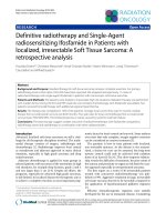
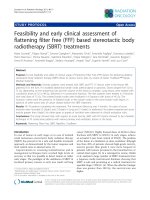
For CK treatments, doses were prescribed to the 70%
isodose covering the PTV and a total maximum of
100%. Novalis treatment was planned with less inhomogeneous dose distributions with the 80% isodose line of
the prescribed 100% dose encompassing the PTV and
allowing a maximum of up to 110% (Fig. 1).
The linear-quadratic model, assuming an alpha/beta
ratio of 10 Gy for tumor, was used to calculate the biologically equivalent dose (BED) and the equivalent dose
in 2 Gy fractions (EQD2) for PTV-encompassing total
dose. Dose constraints to organs at risk for single fraction treatment are shown in Table 1. Treatment planning for CK was performed in Multiplan® (Accuray)
using the Ray-Trace or Monte Carlo algorithm and for
Novalis in iPlan® (BrainLAB) using the Pencil Beam
algorithm.
Endpoints and statistical considerations
LC was defined as time from SFRS/fSBRT to tumor progression within the irradiation field or absence of
Kalinauskaite et al. BMC Cancer
(2020) 20:404
Page 3 of 10
Fig. 1 Treatment plan and dose distribution for (a) CyberKnife, (b) Novalis treatment system
progression at last available follow-up. LC was assessed
using routinely CT scans every 3 months. PET-CT and/
or biopsy of irradiated metastasis was performed in cases
of uncertain progression detected on CT images. OS was
calculated from the beginning of SFRS or fSBRT until
the death of any cause or the date of last follow-up. The
time to new metastases in the lung outside of the SFRS/
fSBRT field or in other organs was defined as DMFS and
was calculated from the start of SFRS/fSBRT. PFS was
defined as the time from the start of SFRS/fSBRT until
progression of the primary tumor, development of new
metastases or local failure.
LC was compared between lung metastases treated
with SFRS and fSBRT. The different fractionation regimens in the same patient were allowed, thus fractionation impact on OS, PFS and DMFS could not be
assessed.
OS, LC, DMFS and PFS after SFRS/fSBRT for lung
metastases were calculated using the Kaplan-Meier
method. Cox-regression analysis was used to obtain the
Hazard Ratio (HR) and 95% confidence intervals (CI) for
Table 1 Dose constrains for organs at risk of single fraction
radiosurgery
Organs at risk
Max critical volume
above threshold (cm3)
Threshold
dose (Gy)
Max point
dose (Gy)a
Spinal cord
<0.35
10.0
14.0
Esophagus
<5
11.9
15.4
Hearts/
pericardium
<15
16.0
22.0
Great vessels
<10
31.0
37.0
Trachea and
large bronchus
<4
10.5
20.2
Rib
<1
22.0
30.0
Ipsilateral Lung
(mean)
-
9.0
-
a
various covariates. Covariates with a p-value of ≤0.1
were included into the multivariate analyses carried out
with a Cox proportional hazards model with a threshold
of p < 0.05. The chi-squared test was performed in order
to compare variables between groups. A p-value of <
0.05 was considered as statistically significant. The data
processing and statistical analyses were accomplished
using FileMaker Pro 15 Advanced, Excel 2010 and IBM
SPSS Statistics 24 (SPSS Inc., Chicago, IL, USA).
Results
Patient and tumor characteristics
The clinical, treatment and follow-up data of 52 eligible
patients were assessed. Thirty-two patients were male
(61.5%) and 20 were female (38.5%) with a median age
of 66 years (range: 26–84) and a median Karnofsky performance status (KPS) of 80% (range: 60–100). The most
prevalent primary tumor was colorectal cancer (CRC) in
17 patients (32.7%). PET-CT staging before the SBRT
for lungs was performed in 7 (13.5%) patients. Twelve
patients (23.1%) had oligometastases at the time of
tumor diagnosis. The median time to first metastasis
was 19.5 months (range: 0–37.9). In 37 patients (71.2%)
metastases were limited to the lungs. Eight patients
(15.4%) had additional liver metastases and 3 patients
(5.8%) had brain metastasis. Forty-six patients (88.5%)
had systemic therapy prior to lung SBRT and 15 (28.8%)
after lung SBRT. Seventeen patients (32.7%) received immunotherapy at any time during the disease course. Patients’ and primary tumor characteristics are shown in
Table 2.
Treatment characteristics
Point defined as 0.035 cm3 or less
Overall, 94 lung metastases were treated using SFRS/
fSBRT with a median of 2 lesions per patient (range: 1–
5). Metastases and SFRS/fSBRT characteristics are
shown in Table 3 and Table 4. Forty-five metastases
Kalinauskaite et al. BMC Cancer
(2020) 20:404
Page 4 of 10
Table 2 Patient and primary tumor characteristics
Table 3 Metastases and treatment characteristics
Characteristics
LM and treatment characteristics
No. (%)
SFRS
(n=45)
fSBRT
(n=49)
p-value
Median
12.0
16.0
0.003
Range
5.0-35.0
5.0-70.0
Median
9.9
24.0
Range
2.4-90.8
5.8-164.5
peripheral
32
25
central
13
24
Age, years
Median
66
Range
26 - 84
Gender
Female
20 (38.5)
Male
32 (61.5)
KPS (%)
Median
80
Range
60 - 100
Primary tumor type
CRC
17 (32.7)
Sarcoma
8 (15.4)
Melanoma
7 (13.5)
HNC
6 (11.5)
RCC
5 (9.6)
NSCLC
3 (5.8)
Others
6 (11.5)
T-classification at initial diagnosis
T≤2
17 (32.7)
T>2
30 (57.7)
Unknown
5 (9.6)
N-classification at initial diagnosis
N0
18 (34.6)
N+
26 (50.0)
Unknown
8 (15.4)
M-classification at initial diagnosis
M0
36 (69.2)
M1
12 (23.1)
Unknown
4 (7.7)
Pre-SFRS/fSBRT systemic therapy
Yes
46 (88.5)
No
6 (11.5)
No. of LM treated with SFRS/fSBRT per patient
Metastasis diameter (mm)
Metastasis PTV (cm3)
<0.001
Metastasis location
0.092
Metastasis histology (CRC vs. non-CRC)
CRC
8
21
Non-CRC
37
28
0.009
PTV-encompassing prescription dose (Gy)
Median
24
45
Range
17-26
20-60
<0.001
Median
24
9.6
Range
17-26
4-16
Median
81.6
105.6
Range
45.9-93.6
42.6 – 151.2
PTV-encompassing single dose (Gy)
<0.001
Biological effective dose (Gy)
0.015
LM lung metastases, SFRS single fraction radiosurgery, fSBRT fractionated
stereotactic body radiotherapy, PTV planning target volume, CRC colorectal
cancer
central vs. 25 peripheral). Median diameter of metastases
was 14.5 mm (range: 5–70), with no significant difference between centrally and peripheral located lesions.
The median time from the diagnosis of lung metastases
to the start of SFRS/fSBRT was 4.5 months (range: 0–
Table 4 Fractionation regimens
Fractions and PTV- encompassing
single dose
No. of LM
(%)
BED
(Gy)
EQD2
(Gy)
1 x 22 Gy
2 (2.1)
70.4
58.7
Median
2
1 x 24 Gy
20 (21.3)
81.6
68.0
Range
1-5
1 x 25 Gy
12 (12.8)
87.5
72.9
1 x 26 Gy
5 (5.3)
93.6
78.0
No. of affected organs per patient
Median
1
3 x 12.5 Gy
3 (3.2)
84.4
70.3
Range
1-4
3 x 15 Gy
8 (8.5)
112.5
93.8
3 x 16 Gy
9 (9.6)
124.8
104.0
4 x 12 Gy
8 (8.5)
105.6
88.0
KPS Karnofsky performance status, CRC colorectal cancer, HNC head and neck
cancer, RCC renal cell carcinoma, NSCLC non-small cell cancer, SFRS single
fraction radiosurgery, fSBRT fractionated stereotactic body radiotherapy, LM
lung metastasis
(47.9%) were treated with SFRS of which only 12 were
located centrally. Metastases treated with fSBRT were almost equally distributed with respect to location (24
4 x 9.6 Gy
9 (9.6)
75.3
62.7
5 x 8 Gy
2 (2.1)
72.0
60.0
other regimens
16 (17.0)
LM lung metastases, PTV planning target volume, BED biologically effective
dose, EQD2 equivalent dose
Kalinauskaite et al. BMC Cancer
(2020) 20:404
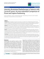
Fig. 2 Kaplan-Meier curves of (a) local control SFRS vs. fSBRT, (b) overall survival, (c) progression-free survival
Page 5 of 10
Kalinauskaite et al. BMC Cancer
(2020) 20:404
61). Before the therapy with CK a gold fiducial was implanted in 51 metastases, whereof 37 were treated with
SFRS and 14 with fSBRT using the Synchrony tracking
method. A total of 14 lung metastases were treated using
the X-sight lung tracking method. IGTV was used for all
29 metastases treated with Novalis. The median prescription dose for SFRS was 24 Gy (range: 17–26) compared to fSBRT with median 45 Gy (range: 20–60)
delivered in 2–12 fractions. The median diameter and
PTV were significantly smaller in metastases treated
with SFRS compared to fSBRT: 12 mm (range: 5–35)
and 9.9 cm3 (range: 2.4–90.8) vs. 16 mm (range: 5–70)
and 24.0 cm3 (range: 5.8–164.5), respectively.
Patient outcomes
The median follow-up time was 21 months (range: 3–
68). The 1-year and 2-year LC rates for SFSR vs. fSBRT
were 89 and 83% vs. 75 and 59%, respectively (p =
0.026). One and 2-year LC rates for metastases from
CRC vs. non-CRC were 59 and 46% vs. 90 and 80%, respectively (p = 0.001). In 5 out of 22 metastases with
local progression relapse was confirmed using PET-CT
and in 2 after histological examination. Eleven lesions
were repeatedly treated with local therapy: either with
repeated SBRT or with surgery. One and 2-year OS and
PFS rates were 84, 71 and 26%, 15%, respectively. At the
time of analysis 21 patients (41.4%) were dead. Disease
progression occurred in 42 patients (80.8%), of which 19
patients (36.5%) developed metastases in new organs.
The Kaplan-Meier LC, OS and PFS curves are shown in
Fig. 2.
Treatment with SFRS, an interval of < 12 months between diagnosis of metastases and the beginning of
SFRS/fSBRT as well as non-colorectal histology were significantly associated with better LC in univariate analysis
(Table 5). However, none of these parameters remained
significant in multivariate analysis. N0, KPS > 70% and
time to first metastasis ≥12 months were significantly associated with improved OS. PFS was significantly better
in patients with KPS > 70% and with maximum 3 metastases at the time of SBRT (Table 6). There was no difference regarding survival outcomes between patients with
oligorecurence and oligometastases.
Treatment related toxicity
The SFRS and fSBRT were safe and very well tolerated.
No treatment-related deaths and grade ≥ 3 toxicities occurred. Six patients (11.5%) developed asymptomatic
grade 1 pneumonitis (2 patients after SFRS and 4 patients after fSBRT) and one patient had grade 1 pulmonary fibrosis. Symptomatic and medical intervention
requiring grade 2 pneumonitis was diagnosed in one patient (1.9%) after SFRS with 25 Gy.
Page 6 of 10
Table 5 Univariate analysis of factors influencing local control
Covariate
HR (95% CI)
p-value
Time between diagnosis of LM and SBRT (months)
<12
1
≥12
2.5 (1.1-6.0)
0.027
Location of LM
central
1
peripheral
0.7 (0.2-1.7)
0.412
Histology
CRC
1
non-CRC
0.2 (0.1-0.6)
0.004
LM diameter (mm)
≤10
1
>10
2.2 (0.8-6.6)
0.150
3
PTV (cm )
≤10
1
>10
3.3 (0.9-11.3)
0.053
Fractionation regimens
SFRS
1
fSBRT
2.7 (1.0-7.0)
0.037
BED
<100Gy
1
≥100 Gy
2.7 (1.1-6.4)
0.021
HR Hazard ratio, CI confidence interval, LM. lung metastases, SBRT stereotactic
body radiotherapy, SFRS single fraction radiosurgery, fSBRT fractionated
stereotactic body radiotherapy, PTV Planning target volume, BED biologically
effective dose
Discussion
This analysis represents a single-center experience in
treating oligometastatic lung lesions with curative
intended SFRS and fSBRT. The 1-, 2-year LC and OS
rates for the entire cohort were 82, 70 and 84%, 71%, respectively. Our findings are comparable with the current
findings in the literature (Table 7) [8–16].
SBRT is an attractive non-invasive treatment option
providing good therapy outcomes with minimum toxicity. The BED ≥100 Gy, smaller tumor size, shorter
interval between diagnosis and treatment of metastases
are favorable prognostic factors influencing local control
of lung metastases after SBRT [9, 17–19]. The existing
data on fractionation schedules as well as dosage of
SBRT for lung metastases is limited by retrospective nature or non-randomized prospective study design.
Therefore, no standardized treatment regimens are yet
available. The primary results of TROG 13.01 SAFRON
II Phase II trial which compares SFRS to fSBRT for lung
metastases are expected soon [20].
According to our data, small lung metastases (median
PTV ≤ 9.9 cm3, median diameter 12 mm) might safely be
treated with SFRS applying 24–26 Gy (median Dmax of
Kalinauskaite et al. BMC Cancer
(2020) 20:404
Page 7 of 10
Table 6 Univariate and multivariate analysis of factors influencing overall and progression-free survival
Covariate
Overall survival
Progression-free survival
Univariate analysis
Multivariate analysis
HR (95% CI)
p-value
HR (95% CI)
p-value
Univariate analysis
0.81
NA
NA
HR (95% CI)
Multivariate analysis
p-value
HR (95% CI)
p-value
0.56
NA
NA
0.25
NA
NA
0.64
NA
NA
0.03
0.4 (0.2-0.7)
0.02
0.31
NA
NA
0.33
NA
NA
0.14
NA
NA
0.005
2.7 (1.4-5.4)
0.003
0.97
NA
NA
0.48
NA
NA
Age (years)
>70
1
≤70
1.1 (0.4-2.7)
1
0.8 (0.4-1.5)
Gender
Female
1
Male
1.6 (0.6-4.6)
1
0.31
NA
NA
0.29
NA
NA
1.2 (0.8-1.6)
Primary tumor
non-CRC
1
CRC
0.6 (0.2-1.4)
1
0.8 (0.5-1.6)
KPS
≤70%
1
>70%
0.4 (0.2-1.1)
1
0.09
0.3 (0.1-0.8)
0.03
0.08
1.5 (0.4-5.0)
0.48
0.5 (0.3-0.9)
T-classification
T≤2
1
T>2
2.4 (0.8-6.8)
1
1.4 (0.7-2.8)
N-classification
N0
1
N+
2.6 (0.9-7.3)
1
0.06
4.4 (1.2-15.6)
0.02
0.03
0.2 (0.1-0.7)
0.01
1.4 (0.7-2.7)
Time to first metastasis (months)
<12
1
≥12
0.3 (0.1-0.9)
1
0.6 (0.3-1.2)
No. of metastases before SBRT
<3
1
≥3
1.4 (0.6-3.3)
1
0.42
NA
NA
0.24
NA
NA
2.6 (1.3-5.1)
No. of affected organs
1
1
>1
1.6 (0.7-3.9)
1
1.1 (0.5-1.9)
Systemic therapy before SBRT
Yes
1
No
1.4 (0.3-6.3)
1
0.65
NA
NA
1.4 (0.5-4.1)
NA not assessed, HR Hazard ratio, CI confidence interval, CRC colorectal cancer, KPS Karnofsky performance status, SBRT stereotactic body radiotherapy
53 Gy and a median BEDmax of 81 Gy) with excellent 1and 2-year LC rates of 89 and 83%, implying that BED <
100 Gy using SFRS might be sufficient for durable control in small lung lesions. This observation, however,
contradicts the findings of other studies, where BED <
100 Gy was found to be a negative prognostic factor for
LC. Ricco et al. analyzed whether different lung metastases volumes and BED were associated with treatment
outcomes [17]. In this study, lesions after SBRT with
BED ≥100 Gy reached better LC rates. Moreover, in the
group with BED ≥100 Gy smaller metastases (volume <
11 cm3) were linked to improved LC and OS rates. The
median number of fractions employed was 3 (range: 1–
8), how many lesions were treated with SFRS remains
unclear. Other trials rarely report on the significance of
BED and fractionation regimens in terms of treatment
outcome for metastases according to their size [9, 12].
Nevertheless, the existing data on size-adapted SFRS for
lung metastases as well as primary lung tumors is promising with 1 year LC rates varying from 89.1–93.4% [15,
21–23]. However, diverse measurement units or target
volumes describing metastases size (e.g. diameter, GTV,
PTV) found in the literature make it difficult to
categorize lesions or to identify the optimal dose. Randomized, prospective studies are needed to determine
which fractionation schedule is the most suitable for
Kalinauskaite et al. BMC Cancer
(2020) 20:404
Page 8 of 10
Table 7 Overall survival and local control rates after SFRS/fSBRT or pulmonary metastasectomy according to various studies
Reference
Study design Year No.
Primary
Patients tumor
Nuyttens et al. [8] Phase 2
study
Rieber J et al. [9]
2015 30
Retrospective 2016 700
Various
No. of LM
1-5
Treatment
SFRS/fSBRT
Overall survival
Local control
1-year (%)
2-years (%)
1-year (%)
2-years (%)
-
63
79
-
Various
42% single
SFRS/fSBRT
75.1
54.4
-
81.2
Navarria et al. [10] Retrospective 2014 76
Various
1-5
fSBRT
84.1
73
95
89
Sharma A. et al.
[11, 12]
Retrospective 2018 206
Various
1-5
SFRS/fSBRT
-
63
-
85
Widder J et al.
[13]
Retrospective 2013 110
Various
3-5
fSBRT 42,
PME 68
SBRT: 87
PME: 98
SBRT: 86 PME:
74
SBRT: 94
PME: 93
SBRT:94 PME:
90
Sapir et al. [14]
Retrospective 2016 78
Sarcoma -
SBRT 26,
PME 127
-
SBRT: 57.9,
PME: 62.2
-
SBRT: 97.4
PME: 96.8
Filippi et al. [15]
Retrospective 2014 67
Various
1-5
SFRS
85.1
70.5
93
88.1
Agolli L [16]
Retrospective 2017 44
CRC
1 - 4 (61%
single)
SFRS/fSBRT
-
67.7
68.8
60.2
Present study
Retrospective 2019 52
Various
Median 2
SFRS/fSBRT
84
71
SFRS 89,
fSBRT 83
SFRS 83, fSBRT
59
LM lung metastases, SBRT stereotactic body radiotherapy, SFRS single fraction radiosurgery, fSBRT fractionated stereotactic radiotherapy
lung metastases according to the size in terms of therapy
outcomes, toxicity and patient’s compliance.
In the current study, 1- and 2-year LC rates for metastases from CRC compared with non-CRC were significantly worse. Recently, Jingu et al. investigated the
impact of primary tumor histology on LC rates after
SBRT for lung metastases in a metanalysis and systematic review. Analysis of 1920 patients (619 with CRC,
1301 non-CRC) showed that LC was significantly inferior in the CRC group (p < 0.00001). In addition, the dose
escalation (BED > 130 Gy) was associated with decreased
local recurrences [24]. Furthermore, Ahmed and colleagues concluded that lung metastases from rectal carcinoma are related with increased radio-resistance, and
therefore are more likely to relapse after SBRT. The authors recommend dose escalation with BED > 100 Gy for
radio-resistant tumors in order to improve treatment
outcomes [25]. In the present study, the median BED for
relapsed metastases from rectal cancer was 87.5 Gy
(range: 56–124.8), suggesting that an insufficient dose
for this histology may be responsible for lower LC rates
in patients with CRC. Therefore, SBRT with BED < 100
Gy should be used with caution in patients with lung oligometastases from rectal cancer.
We found time to the first metastasis ≥12 months,
KPS > 70% and N0 to be independent favorable prognostic factors for OS. Metachronous metastases with longer
metastasis free interval are associated with indolent
tumor histology and thus are frequently linked to better
outcomes, with the favoring time to metastasis diagnose
varying from ≥2 months to ≥75 months depending on
the primary tumor type [26–28]. Furthermore, in agreement with our results good performance score before
initiation of the SBRT was linked to better survival in
various studies [29, 30]. Absence of lymph node involvement was addressed as a prognostic factor mostly in
series on oligometastatic lung cancer [27, 31]. Unlike
our finding, no prognostic value of N classification was
reported in studies with cohorts of heterogenous primary tumor type, therefore this finding must be interpreted carefully. Despite the small sample size, we
identified two commonly reported prognostic factors
that might be useful for selecting oligometastatic patients for curative SBRT.
The major limitation of this study is its retrospective
design with inhomogeneous primary tumor types and
the limited number of patients. Therefore, neither a subgroup analysis based on metastasis histology nor an analysis of the effects of dose escalation was performed.
Treatment planning calculations with Ray-Tracing, Pencil Beam or Monte Carlo dose algorithms for lung might
produce differences in dose distribution for target and
organs at risk. However, there was no difference detected in the treatment outcomes in metastases planed
with different treatment algorithms. Since multiple metastases in the same patient were treated with different
fractionation, finding the prognostic value of SFRS vs.
fSBRT for survival outcomes was not feasible.
Conclusions
KPS > 70%, longer time to first metastasis and absence
of locoregional lymph node metastases were found to be
positive predictive factors for OS in patients with lung
oligometastases after SBRT. Long-term LC and low toxicity rates were achieved after short SBRT schedules.
Abbreviations
BED: Biologically effective dose; CRC: Colorectal cancer; CI: Confidence
interval; CT: Computed tomography; CTV: Clinical treatment volume;
Kalinauskaite et al. BMC Cancer
(2020) 20:404
CK: Cyberknife; DMFS: Distant metastases-free survival; EQD2: Equivalent dose
in 2 Gy fractions; fSBRT: Fractionated stereotactic body radiotherapy;
GTV: Gross tumor volume; HNC: Head and neck cancer; HI: Hazard ratio;
IGTV: Internal gross tumor volume; LC: Local control; non-CRC: Noncolorectal cancer; NSCLC: Non-small-cell lung cancer; OS: Overall survival;
PFS: Progression-free survival; PTV: Planning treatment volume; RCC: Renal
cell carcinoma; SFRS: Single fraction radiosurgery; SBRT: Stereotactic body
radiotherapy
Page 9 of 10
8.
9.
Acknowledgments
Not applicable.
10.
Availability of data and material
The datasets used and/or analyzed during the current study are available
from the corresponding author on reasonable request.
11.
12.
Authors` contributions
GK acquired, analyzed and interpreted the patient data, conducted the
statistical analysis, drafted the manuscript. CS2, IT and MK provided the idea
for the study. CS1, CS2 and IT contributed to data interpretation and
manuscript writing. AK provided technical support, preparation of figures
and critical review of the manuscript. GK, MK, AG, VB, CS1 and CS2 were
responsible for treatment, collection of patient data and follow-up. CS1 and
CS2 contributed equally. All authors read and approved the final version of
the manuscript.
Funding
This study was supported by scholarship for Goda Kalinauskaite from Berliner
Krebsgesellschaft, Ernst von Leyden-Stipendium.
Ethics approval and consent to participate
Analysis of patient data was approved by the institutional medical ethics
committee of the Charité - Universitätsmedizin Berlin (EA1/214/16). Because
of retrospective nature of this study we did not obtain written nor verbal
informed consents from the patients.
13.
14.
15.
16.
17.
18.
Consent for publication
Not applicable.
19.
Competing interests
The authors declare that they have no competing interests.
20.
Author details
1
Department of Radiation Oncology and Radiotherapy, Charité Universitätsmedizin Berlin, Augustenburger Platz 1, 13353 Berlin, Germany.
2
Charité CyberKnife Center, Charité - Universitätsmedizin Berlin,
Augustenburger Platz 1, 13353 Berlin, Germany. 3The Translational
Radiooncology and Radiobiology Research Laboratory, Charité Universitätsmedizin Berlin, Berlin, Germany.
Received: 17 July 2019 Accepted: 23 April 2020
References
1. Chaffer CL, Weinberg RA. A perspective on cancer cell metastasis. Science.
2011;331(6024):1559–64.
2. Hellman S, Weichselbaum RR. Oligometastases. J Clin Oncol. 1995;13:8–10.
3. Lindsay DP, Caster JM, Wang K, Myung JH, Chen RC, Chera B, et al.
Nanotechnology-based quantification of circulating tumor cells in
oligometastatic patients undergoing definitive radiation therapy. Int J Radiat
Oncol Biol Phys. 2017;99(2):S51.
4. Herold CJ, Bankier AA, Fleischmann D. Lung metastases. Eur Radiol. 1996;
6(5):596–606.
5. Budczies J, von Winterfeld M, Klauschen F, Bockmayr M, Lennerz JK, Denkert
C, et al. The landscape of metastatic progression patterns across major
human cancers. Oncotarget. 2015;6(1):570–83.
6. Lodeweges JE, Klinkenberg TJ, Ubbels JF, Groen HJM, Langendijk JA, Widder
J. Long-term outcome of surgery or stereotactic radiotherapy for lung
Oligometastases. J Thorac Oncol. 2017;12(9):1442–5.
7. Filippi AR, Guerrera F, Badellino S, Ceccarelli M, Castiglione A, Guarneri A,
et al. Exploratory analysis on overall survival after either surgery or
21.
22.
23.
24.
25.
26.
27.
stereotactic radiotherapy for lung Oligometastases from colorectal Cancer. J
Clin Oncol. 2016;28(8):505–12.
Nuyttens JJ, van der Voort van Zyp NC, Verhoef C, Maat A, van Klaveren RJ,
van der Holt B, et al. Stereotactic body radiation therapy for oligometastases
to the lung: a phase 2 study. Int J Radiat Oncol Biol Phys. 2015;91(2):337–43.
Rieber J, Streblow J, Uhlmann L, Flentje M, Duma M, Ernst I, et al.
Stereotactic body radiotherapy (SBRT) for medically inoperable lung
metastases-a pooled analysis of the German working group "stereotactic
radiotherapy". Lung Cancer. 2016;97:51–8.
Navarria P, Ascolese AM, Tomatis S, Cozzi L, De Rose F, Mancosu B, et al.
Stereotactic body radiotherapy (sbrt) in lung oligometastatic patients: role
of local treatments. Radiat Oncol. 2014;9(1):91.
Sharma A, Duijm M, Oomen-de Hoop E, Aerts JG, Verhoef C, Hoogeman M,
et al. Survival and prognostic factors of pulmonary oligometastases treated
with stereotactic body radiotherapy. Acta Oncol. 2019;58(1):74-80.
Sharma A, Duijm M, Oomen-de Hoop E, Aerts JG, Verhoef C, Hoogeman M,
et al. Factors affecting local control of pulmonary oligometastases treated
with stereotactic body radiotherapy. Acta Oncol. 2018;57(8):1031–7.
Widder J, Klinkenberg TJ, Ubbels JF, Wiegman EM, Groen HJ, Langendijk JA.
Pulmonary oligometastases: Metastasectomy or stereotactic ablative
radiotherapy? Radiother Oncol. 2013;107(3):409–13.
Sapir E, Tao Y, Lin T, Kollar L, Schipper M, Chugh R, et al. Surgical resection
or stereotactic body radiation therapy for sarcoma patients with pulmonary
metastases. Int J Radiat Oncol Biol Phys. 2016;96(2):S26.
Filippi AR, Badellino S, Guarneri A, Levis M, Botticella A, Mantovani C, et al.
Outcomes of single fraction stereotactic ablative radiotherapy for lung
metastases. Technol Cancer Res Treat. 2014;13(1):37–45.
Agolli L, Bracci S, Nicosia L, Valeriani M, De Sanctis V, Osti MF. Lung
metastases treated with stereotactic ablative radiation therapy in
Oligometastatic colorectal Cancer patients: outcomes and prognostic
factors after long-term follow-up. Clin Colorectal Cancer. 2017;16(1):58–64.
Ricco A, Davis J, Rate W, Yang J, Perry D, Pablo J, et al. Lung metastases
treated with stereotactic body radiotherapy: the RSSearch® patient Registry’s
experience. Radiat Oncol. 2017;12(1):35.
Oh Y, Taylor S, Bekele BN, Debnam JM, Allen PK, Suki D, et al. Number of
metastatic sites is a strong predictor of survival in patients with nonsmall
cell lung cancer with or without brain metastases. Cancer. 2009;115(13):
2930–8.
Wang Z, Kong Q-T, Li J, Wu X-H, Li B, Shen Z-T, et al. Clinical outcomes of
cyberknife stereotactic radiosurgery for lung metastases. J Thorac Dis. 2015;
7(3):407–12.
Siva S, Kron T, Bressel M, Haas M, Mai T, Vinod S, et al. A randomized phase
II study of stereotactic ablative body radiotherapy for metastases to the
lung (TROG 13.01 SAFRON II) (SAFRON II). BMC Cancer. 2016;16:183.
Ost MF, Carnevale A, Valeriani M, De Sanctis V, Minniti G, Cortesi E, et al.
Clinical outcomes of single dose stereotactic radiotherapy for lung
metastases. Clin Lung Cancer. 2013;14(6):699–703.
Trakul N, Chang CN, Harris J, Chapman C, Rao A, Shen J, et al. Tumor
volume-adapted dosing in stereotactic ablative radiotherapy of lung
tumors. Int J Radiat Oncol Biol Phys. 2012;84(1):231–7.
Videtic GM, Hu C, Singh AK, Chang JY, Parker W, Olivier KR, et al. A
randomized phase 2 study comparing 2 stereotactic body radiation therapy
schedules for medically inoperable patients with stage i peripheral nonsmall cell lung cancer: nrg oncology rtog 0915 (ncctg n0927). Int J Radiat
Oncol Biol Phys 2015;93(4):757–64.
Jingu K, Matsushita H, Yamamoto T, Umezawa R, Ishikawa Y, Takahashi N,
et al. Stereotactic radiotherapy for pulmonary oligometastases from
colorectal cancer: a systematic review and meta-analysis. Technol Cancer
Res Treat. 2018;17:1533033818794936.
Ahmed KA, Scott JG, Arrington JA, Naghavi AO, Grass GD, Perez BA, et al.
Radiosensitivity of lung metastases by primary histology and implications
for stereotactic body radiation therapy using the genomically adjusted
radiation dose. J Thorac Oncol. 2018;13(8):1121–7.
Hong JC, Ayala-Peacock DN, Lee J, Blackstock AW, Okunieff P, Sung MW,
et al. Classification for long-term survival in oligometastatic patients treated
with ablative radiotherapy: a multi-institutional pooled analysis. PLoS One.
2018;13(4):e0195149.
Ashworth AB, Senan S, Palma DA, Riquet M, Ahn YC, Ricardi U et al. An
individual patient data metaanalysis of outcomes and prognostic factors
after treatment of oligometastatic non-small-cell lung cancer. Clin Lung
Cancer 2014;15(5):346–55.
Kalinauskaite et al. BMC Cancer
(2020) 20:404
28. Inoue T, Katoh N, Aoyama H, Onimaru R, Taguchi H, Onodera S et al. Clinical
outcomes of stereotactic brain and/or body radiotherapy for patients with
oligometastatic lesions. Jpn J Clin Oncol. 2010;40(8):788–94.
29. Flannery TW, Suntharalingam M, Regine WF, Chin LS, Krasna MJ, Shehata
MK, et al. Long-term survival in patients with synchronous, solitary brain
metastasis from non-small-cell lung cancer treated with radiosurgery. Int J
Radiat Oncol Biol Phys. 2008;72(1):19–23.
30. Lancia A, Ingrosso G, Carosi A, Di Murro L, Giudice E, Cicchetti S, et al.
Oligometastatic cancer: stereotactic ablative radiotherapy for patients
affected by isolated body metastasis. Acta Oncol. 2017;56(11):1621–5.
31. Li S, Zhu R, Li D, Li N, Zhu X. Prognostic factors of oligometastatic non-small
cell lung cancer: a meta-analysis. J Thoracic Dis. 2018;10(6):3701–13.
Publisher’s Note
Springer Nature remains neutral with regard to jurisdictional claims in
published maps and institutional affiliations.
Page 10 of 10