CYP19A1 gene expression in the peripheral blood of Brazilian women with breast cancer relapse
Bạn đang xem bản rút gọn của tài liệu. Xem và tải ngay bản đầy đủ của tài liệu tại đây (598.27 KB, 8 trang )
Barros-Oliveira et al. BMC Cancer
(2020) 20:480
/>
RESEARCH ARTICLE
Open Access
CYP19A1 gene expression in the peripheral
blood of Brazilian women with breast
cancer relapse
Maria da Conceição Barros-Oliveira1,2, Danylo Rafhael Costa-Silva1,2, Larysse Cardoso Campos-Verdes1,2,
Renato de Oliveira Pereira3, Rozirene Araújo Silva3, Paulo de Tarso Moura-Borges3, Emerson Brandão Sousa3,
André Luiz Pinho-Sobral3, Pedro Vitor Lopes-Costa3, Alesse Ribeiro dos Santos3, Ione Maria Ribeiro Soares-Lopes3,
Jackeline Lopes Viana3, Mariella de Almeida Melo3, Fidelis Manes Neto3, Eid Gonçalves Coelho1,2,
Maria do Socorro Pires e Cruz1,2, Vladimir Costa-Silva1,2, Luiz Henrique Gebrim1,2,4 and
Benedito Borges Da Silva1,2,3*
Abstract
Background: The CYP19A1 gene, which encodes the enzyme responsible for androgen aromatization into
estrogens, may play an important role in breast cancer aggressiveness. However, no study has evaluated CYP19A1
gene expression in the peripheral blood of women with relapsed breast cancer.
Methods: In this cross-sectional study, CYP19A1 gene expression was quantified by RT-PCR in the peripheral blood
of 146 women with breast cancer who were first divided into two groups according to the expression of CYP19A1
(low and high); each group had 73 patients. Subsequently, women were divided into two groups: those without
recurrence (control, n = 85) and those with recurrence (study, n = 61). Statistical analysis of the data was performed
using ANOVA, the Mann-Whitney, Chi-square or Fisher’s exact test (p < 0.05).
Results: There were no significant differences between the relative expression of CYP19A1 mRNA in the low
expression group and the high expression group according to the variables studied. There were no significant
differences in CYP19A1 gene expression in the study and control groups (p = 0.8461). In the relapse group, CYP19A1
gene expression was significantly higher in the hybrid luminal subtype than in the triple-negative subtype (p =
0.0321), whereas it was significantly lower in HER2-negative cases than in HER2-positive cases (p < 0.0376). Women
with locoregional recurrence showed higher expression than women with distant recurrence (p < 0.0001).
Conclusions: The present study found no significant differences between women with high and low expression of
the CYP19A1 gene mRNA or between those in the study group and the control group. However, in women with
recurrence, there was increased expression of CYP19A1 mRNA in those who had the luminal hybrid subtype and
locoregional relapse and decreased expression in those negative for HER2.
Keywords: Breast cancer, CYP19A1, Gene expression, Relapse
* Correspondence:
1
Postgraduate Program of the Northeast Network of Biotechnology
(RENORBIO), Teresina, Northeast, Brazil
2
Federal University of Piaui, Teresina, Piaui, Brazil
Full list of author information is available at the end of the article
© The Author(s). 2020 Open Access This article is licensed under a Creative Commons Attribution 4.0 International License,
which permits use, sharing, adaptation, distribution and reproduction in any medium or format, as long as you give
appropriate credit to the original author(s) and the source, provide a link to the Creative Commons licence, and indicate if
changes were made. The images or other third party material in this article are included in the article's Creative Commons
licence, unless indicated otherwise in a credit line to the material. If material is not included in the article's Creative Commons
licence and your intended use is not permitted by statutory regulation or exceeds the permitted use, you will need to obtain
permission directly from the copyright holder. To view a copy of this licence, visit />The Creative Commons Public Domain Dedication waiver ( applies to the
data made available in this article, unless otherwise stated in a credit line to the data.
Barros-Oliveira et al. BMC Cancer
(2020) 20:480
Background
Breast cancer is the most common malignancy affecting
women worldwide, and in 2018, the global estimate of
impacted women was approximately 2,089,000, with a
mortality rate of nearly 627,000 [1, 2]. Its incidence is
higher in the most developed regions of the world compared to developing and underdeveloped regions [3].
In Brazil, which is a developing country, breast cancer
is the second most common malignancy in women after
non-melanoma skin cancer, with an estimated 59,700
new cases and 15,403 cases of death from the disease in
2018 [4]. Additionally, approximately 40% of patients
who develop disease recurrence die, especially in the first
2 to 3 years, when the risk of recurrence is higher [5–7].
Although physical examination and mammography
are important to ensure early diagnosis of the disease
and to reduce mortality, breast cancer is still frequently diagnosed in advanced stages in Brazil, resulting in high mortality rates, even with the current
therapeutic strategies [8].
It has been suggested that the most appropriate therapeutic and prognostic strategies for breast cancer may
be developed using genes that are associated with the
development, growth and aggressiveness of breast cancer
as biomarkers [9, 10]. This includes the CYP19A1 gene
that encodes the aromatase enzyme, which is involved in
estrogen biosynthesis, as it promotes androgen
aromatization in estrogens [11, 12]. The CYP19A1 gene
has been studied as a prognostic marker of breast cancer
due to its genetic control in estrogen biosynthesis [13,
14]. This gene has tissue-specific promoters, and principally, normal breast adipose tissue maintains low levels
of aromatase expression primarily via the I.4 distal promoter. However, in breast cancer, an exchange between
the I.4 and I.3 promoters and the I.7 and II promoters
occurs, leading to increased production of aromatase
and local estrogen [15, 16].
Some studies have examined CYP19A1 gene expression in breast cancer using quantitative reverse transcription polymerase chain reaction (RT-PCR), which is
considered a standard method for the quantitative measurement of gene expression; however, many of these
studies have shown controversial results [16, 17].
Miyoshi et al. found no significant association between
CYP19A1 expression levels and breast cancer [18]. On
the other hand, Friesenhengst et al. evaluated CYP19A1
expression in tumors of women with breast cancer, and
the results showed a significant association between high
CYP19A1 gene expression and estrogen receptor expression, menopausal status, metastasis-free survival, overall
survival, disease-free survival and local and distant recurrence [11]. Thus, the controversies surrounding the
gene expression of CYP19A1 in breast tumor studies
and, to the best of our knowledge, the absence of studies
Page 2 of 8
analyzing the peripheral blood of women with recurrent
breast cancer led to the design of this study.
Methods
Patients
This cross-sectional study involved 146 women from 34
to 80 years of age who had breast cancer and received
care at the Mastology Clinic of Perola Byington Hospital
(Sao Paulo, Brazil) between July and September 2018.
The Internal Review Board of the Federal University of
Piauı and Perola Byington Hospital approved the study
under number CAAE: 43447015.8.0000, and all the patients signed an informed consent form prior to admission. The women were first divided into two groups,
with low and high expression of CYP19A1 with 73 patients each. Subsequently, women were divided into two
groups, without recurrence (control, n = 85) and with recurrence (study, n = 61). Women who were over 18 years
of age, with and without breast cancer recurrence in the
operable stage, who were diagnosed and treated in the
past 10 years and had histologically confirmed diagnoses
(disease at diagnosis) were included in the study.
Women with a history of another neoplasm, a serious
concomitant disease or an initial diagnosis of metastatic
breast cancer were excluded from the study.
Blood sampling
Peripheral blood was collected by a specialized technician using a disposable syringe and needle after medical
consultation. The first 1 mL of peripheral blood was discarded to prevent contamination by epidermal cells. A 1
mL sample of total peripheral blood from each patient
was preserved in 3 mL TRIzol (Invitrogen; Thermo
Fisher Scientific, Inc.) and stored at − 80 °C until RNA
extraction.
Total RNA extraction and cDNA synthesis
RNA extraction was performed using TRIzol reagent
(Invitrogen; Thermo Fisher Scientific, Inc.) according to
the manufacturer’s instructions. RNA concentration, integrity and purity were analyzed using a NanoDrop 1000
spectrophotometer (Thermo Fisher Scientific, Inc.) and
agarose gel electrophoresis. Complementary DNA
(cDNA) was synthesized from 2000 ng RNA using
SuperScript III First-Strand Synthesis System (Invitrogen; Thermo Fisher Scientific, Inc.) with a total reaction
volume of 20 μL containing 50 μM Oligo (dT) 20, 10
mM DNTP, 1 mL 10X RT buffer, 0.1 M DTT, 40 U/μL
RNaseOUT and 200 U/μl SuperScript III RT. The incubation conditions for reverse transcription (RT) were
50 °C for 60 min and 70 °C for 15 min. The samples were
placed in long-term storage at 4 °C. The cDNA was kept
at − 20 °C and was diluted 10-fold prior to use in the
quantitative RT-PCR.
Barros-Oliveira et al. BMC Cancer
(2020) 20:480
Quantitative RT-PCR
CYP19A1 mRNA expression was determined by quantitative RT-PCR using Power SYBR Green PCR Master
Mix (Applied Biosystems; Thermo Fisher Scientific, Inc.)
and an ABI 7500 detection system equipped with SDS
v1.4 software. The following primers were used for
detection and quantitation of CYP19A1 mRNA: sense
primer, 5′-CACATCCTCAATACCAGGTCC-3′; antisense primer, 5′-CAGAGATCCAGACTCGCATG-3′.
BETA-ACTIN (ACTB) was used as an endogenous
normalization control. The following primers were used
for ACTB: sense primer, 5′-CACTGTGTTGGCGT
ACAGGT-3′ and antisense primer, 5′-AAATCTGGCA
CCACACCTTC-3′. Reactions were performed in a final
volume of 13 μL, containing 3 μL DNA sample, 6.4 μL
SYBR Green Master Mix (Applied Biosystems; Thermo
Fisher Scientific, Inc.), 0.4 μL primers (Custom TaqMan
Gene Expression Assays, Applied Biosystems; Thermo
Fisher Scientific, Inc.), and 2.9 μL of ultrapure sterile
water, in 96-well plates using the StepOne Real-Time
PCR System (Applied Biosystems, Foster City, CA,
USA). After initial denaturation for 10 min at 95 °C, the
samples were subjected to 40 amplification cycles, consisting of two steps: 15 s at 95 °C and 1 min at 60 °C.
Samples were evaluated in duplicate, and two negative
controls were added to each plate containing the same
reaction compounds, but the DNA sample was replaced
with water. Relative quantitation of CYP19A1 mRNA expression as a target was performed using the 2 ^ -ΔCT
method using the mean values obtained from the threshold cycle (CT) of 146 samples and the ACTB CT values
as an endogenous control.
Statistical analysis
To evaluate the associations between CYP19A1 expression and clinical and histopathologic variables, the
Mann-Whitney, Chi-square or Fisher’s exact test and
unidirectional ANOVA with multiple comparisons were
performed using the Bonferroni post-test method. The
values of p < 0.05 were interpreted as statistically significant. All statistical analyses were performed with GraphPad Prism software 6.0 (GraphPad Software, San Diego,
CA, USA).
Page 3 of 8
therapy (ET) based on 2nd international consensus
guidelines for advanced breast cancer, developed by
European School of Oncology and European Society of
Medical Oncology [19], responsive to ET, when relapses
occur after 2 years of adjuvant ET or resistant when a relapses occurs in the first 2 years of adjuvant ET. In the
current study, 44.27% of patients were considered responsive to endocrine therapy and 55.73% considered
unresponsive.
Association of relative expression of CYP19A1 mRNA with
the clinical and histopathologic features
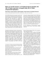
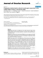
There were no significant differences in the relative expression of CYP19A1 mRNA in the study group compared to the control, p = 0.8461 (Fig. 1). In the study
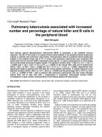
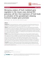
group, CYP19A1 mRNA expression was significantly
higher in patients with a hybrid luminal molecular subtype than in patients with a triple-negative subtype, p =
0.0321 (Fig. 2). There were no significant differences in
the relative expression of CYP19A1 mRNA in women
with locoregional recurrence in the pre or post menopause (p = 0.116). No other associations were observed
between the relative expression of CYP19A1 mRNA and
the other variables studied, such as age, use of tobacco,
menopausal status, grade, nodal status (N), tumor stage,
estrogen and progesterone receptor, HER2 and histological type.
Correlations between the median expression levels of
CYP19A1 mRNA and histopathologic features according to
recurrence
Using the median as the cut-off point, the median expression of CYP19A1 mRNA was classified as high or
low and then analyzed to determine its association with
clinical and histopathological features (Table 2). The
group of women with recurrence of breast cancer and
negative HER2 receptor expression showed reduced
CYP19A1 mRNA levels compared to those with positive
HER2 receptor expression (p < 0.0376). Further, the
mRNA expression of CYP19A1 was significantly higher
in women with locoregional recurrence than in women
with distant recurrence (p < 0.0001). There was no significant difference between the expression of CYP19A1
and the other variables studied.
Results
Correlations between CYP19A1 mRNA levels and
histopathologic features
Using the median as the cut-off point, the patients were
divided in low expression group and high expression
group of CYP19A1. There were no significant differences
between the relative expression of CYP19A1 mRNA in
the low expression group and the high expression group
according to the variables studied (Table 1). Patients
were classified according to their sensitivity to endocrino
Discussion
The detection of circulating CYP19A1 mRNA by RTPCR has been shown to be significantly increased in
breast cancer compared to normal controls, and quantitative RT-PCR is considered a sensitive and reliable
method for studying mRNA in a variety of sites, such as
bone marrow, lymph nodes, tissues and blood [20–23].
However, according to a survey of the literature, the
evaluation of CYP19A1 mRNA by quantitative RT-PCR
Barros-Oliveira et al. BMC Cancer
(2020) 20:480
Page 4 of 8
Table 1 Correlations between the levels of CYP19A1 mRNA and histopathologic features in women with breast cancer
Variables
Median age, 54 y
(range, 34–82 y)
N (%)
CYP19A1 Expression
Low (%)
p value
High (%)
Age
≤50 years
60 (41.1)
31 (21.2)
29 (19.9)
> 51 years
86 (58.9)
42 (28.8)
44 (30.1)
Yes
55 (37.7)
30 (20.5)
25 (17.1)
No
91 (62.3)
43 (29.5)
48 (32.9)
0.73
Tobacco use
0.39
Menopausal status
Premenopausal
63 (43.2)
31 (21.2)
32 (21.9)
Postmenopausal
83 (56.8)
42 (28.8)
41 (28.1)
0.86
G1
14 (9.6)
3 (2.1)
11 (7.5)
G2
107 (73.3)
57 (39.0)
50 (34.2)
G3
25 (17.1)
13 (8.9)
12 (8.2)
N0-N1
127 (87.0)
62 (42.5)
65 (44.5)
N2-N3
19 (13.0)
11 (7.5)
8 (5.5)
I
19 (13.0)
7 (4.8)
12 (8.2)
II
74 (50.7)
40 (27.4)
34 (23.3)
III
53 (36.3)
26 (17.8)
27 (18.5)
Luminal A
16 (11.0)
6 (4.1)
10 (6.8)
Luminal B
68 (45.2)
36 (24.7)
32 (21.9)
Her2 overexpression
23 (15.8)
12 (8.2)
11 (7.5)
Triple-negative
31 (21.2)
19 (13.0)
12 (8.2)
Hybrid Luminal
8 (6.8)
8 (5.5)
0 (0.0)
105 (71.9) 41 (28.1)
52 (35.6) 21 (14.4)
53 (36.3) 20 (13.7)
0.85
97 (66.4) 49 (33.6)
49 (33.6) 24 (16.4)
48 (32.9) 25 (17.1)
0.86
35 (24.0) 111 (76.0)
14 (9.6) 59 (40.4)
21 (14.4) 52 (35.6)
0.17
Ductal
104 (71.2)
50 (34.2)
54 (37.0)
0.73
Lobular
8 (5.5)
4 (2.7)
4 (2.7)
Other
34 (23.3)
19 (13.0)
15 (10.3)
Tumor Grade
0.07
N Classification
0.46
Tumor stage
0.40
Molecular subtype
0.06
Estrogen receptors
Positive Negative
Progesterone receptors
Positive Negative
HER2
Positive Negative
Histological type
Recurrence
Yes
61(41.8)
32 (21.9)
29 (19.9)
No
85 (58.2)
41(28.1)
44 (30.1)
0.61
Barros-Oliveira et al. BMC Cancer
(2020) 20:480
Fig. 1 Relative expression of CYP19A1 mRNA in the study group
compared to that in the control group
in the total blood of women with breast cancer recurrence was not reported, but some studies solely involving normal, peritumoral and tumoral tissues are
available [11, 18]. Peripheral blood has been used as a
clinical sample for gene expression analysis in breast cancer, since peripheral blood samples are readily available,
their acquisition is minimally invasive and they can be collected at low cost, thus making them an attractive alternative modality for diagnostic and prognostic purposes in
cancer research [24, 25]. According to some authors, there
is a correlation between CYP19A1 mRNA levels in peripheral blood leukocytes and target tissues [26, 27].
In the present study, comparison of peripheral blood
CYP19A1 gene expression levels by quantitative RT-PCR
between women with low and high expression and
women with nonrelapsed breast cancer (control) and
women with relapsed cancer (study) showed no statistically significant differences. In the group with relapsed
Fig. 2 Relative expression of CYP19A1 mRNA in the relapse group of
women who had the hybrid luminal molecular subtype compared
to that in those with the triple negative subtype
Page 5 of 8
cancer, CYP19A1 gene expression was significantly
higher in women with a hybrid luminal molecular subtype than in women with a triple-negative subtype. Regarding tumor characteristics, the group of women with
breast cancer recurrence showed a significant reduction
in CYP19A1 mRNA in women with HER2-negative tumors compared to those with HER2-positive tumors.
Additionally, CYP19A1 mRNA expression was significantly higher in women with locoregional recurrence
than in those with distant recurrence, and there was no
difference in relation to the other variables studied.
On evaluating the expression of the CYP19A1 gene
in breast cancer tissue, Friesenhengst et al. [11] detected an association between the high expression of
CYP19A1 in breast tumors and the incidence of
breast cancer recurrence. However, consistent with
our results, Girault et al. [28] and Licznerska et al.
[29] evaluated CYP19A1 gene expression in breast
cancer tissue and found no associations between
CYP19A1 mRNA levels and disease recurrence. Darlix
et al. [30] showed longer survival in women with hybrid luminal breast cancer than in those with triplenegative breast cancer, while other authors, such as
Friesenhengst et al. [11] and Brown et al. [31],
showed no association between CYP19A1 mRNA expression levels and molecular subtypes of breast cancer. However, these studies were performed in tumor
tissue and had a smaller sample size than the present
study.
Some authors have not shown an association between
CYP19A1 gene expression in women with breast cancer
and HER2 receptor status [11, 32, 33]. However, findings
similar to those in this study were found by Subbaramaiah et al. [34], who showed lower levels of aromatase
enzyme and CYP19A1 activity in HER2-negative tumors
than in HER2-positive tumors. Some authors have
shown that HER2 overexpression is the main determinant of increased expression of cyclooxygenase-2 and synthesis of prostaglandin E2 in breast tumor cells, which in
turn, leads to increased CYP19A1 gene expression and
aromatase activity [34–36].
Bollet et al. [32] showed a significant association between low expression of the CYP19A1 gene and an increased risk of locoregional recurrence. On the other
hand, other studies have shown an association between
the complete absence of CYP19A1 gene expression and
shorter relapse-free survival [29, 37]. Furthermore, consistent with our results, Friesenhengst et al. [11] and Salhab et al. [33] reported that high expression of the
CYP19A1 gene was associated with increased locoregional
recurrence. Estrogen synthesis in situ is believed to be primarily catalyzed by the enzyme aromatase, which is often
overexpressed in breast tumors, thus explaining the increased levels of CYP19A1 mRNA in patients with
Barros-Oliveira et al. BMC Cancer
(2020) 20:480
Page 6 of 8
Table 2 Correlations between the median expression levels of CYP19A1 mRNA and histopathologic features according to relapse
Variables
P values
Relapse (n = 61)
Nonrelapsed (n =85)
Low
High
Low
High
n (%)
n (%)
n (%)
n (%)
P values
Age
≤50 years
13 (21.3)
12 (19.7)
> 51 years
18 (29.5)
18 (29.5)
Yes
13 (21.3)
10 (16.4)
No
18 (29.5)
20 (32,8)
0.877
19 (22.4)
16 (18.8)
24 (28.2)
26 (30.6)
18 (21.2)
14 (16.5)
24 (28.2)
29 (34.1)
0.568
Tobacco use
0.488
0.327
Menopausal status
Premenopausal
12 (19.7)
14 (23.0)
Postmenopausal
19 (31.1)
16 (26.2)
0.529
20 (23.5)
17 (20.0)
23 (27.1)
25 (29.4)
3 (3.5)
7 (8.2)
22 (36.1)
30 (35.3)
29 (34.1)
4 (6.6)
9 (10.6)
7 (8.2)
36 (42.4)
40 (47.1)
6 (7.1)
3 (3.5)
0.574
G1
0 (0.0)
4 (6.6)
G2
26 (42.6)
G3
5 (8.2)
N0-N1
27 (44.3)
24 (39.3)
N2-N3
4 (6.6)
6 (9.8)
I
3 (4.9)
3 (4.9)
4 (4.7)
9 (10.6)
II
19 (31.1)
12 (19.7)
21 (24.7)
22 (25.9)
III
9 (14.8)
15 (24.6)
17 (20.0)
12 (14.1)
Luminal A
1 (1.6)
2 (3.3)
4 (4.7)
8 (9.4)
Luminal B
17 (27.9)
18 (29.5)
19 (22.4)
22 (25.9)
Her2 overexpression
4 (6.6)
6 (9.8)
8 (9.4)
5 (5.9)
Triple-negative
9 (14.8)
4 (6.6)
10 (11.8)
8 (9.4)
Positive
22 (36.1)
25 (41.0)
30 (35.3)
18 (21.2)
Negative
9 (14.8)
5 (8.2)
12 (14.1)
15 (17.6)
Negative
22 (36.1) 9 (14.8)
19 (31.1) 11 (18.0)
0.525
27 (31.8)
15 (17.6)
29 (34.1)
14 (16.5)
0.759
Negative
5 (8.2) 26 (42.6)
12 (19.7) 18 (29.5)
< 0.0376
9 (10.6)
33 (38.8)
9 (10.6)
34 (40.0)
0.955
Ductal
23 (37.7)
23 (37.7)
0.584
26 (30.6)
32 (37.6)
0.154
Lobular
3 (4.9)
1 (1.6)
1 (1.2)
3 (3.5)
Other
5 (8.2)
6 (9.8)
15 (17.6)
8 (9.4)
Locoregional
0 (0.0)
24 (39.3)
–
–
Distant metastasis
19 (31.1)
18 (29.5)
–
–
Tumor Grade
0.109
0.395
N Classification
0.454
0.273
Tumor stage
0.216
0.247
Molecular subtype
0.4455
0.182
Estrogen receptors
0.250
0.532
Progesterone receptors
Positive
HER2
Positive
Histological type
Recurrence type
< 0.0001
–
Barros-Oliveira et al. BMC Cancer
(2020) 20:480
locoregional breast recurrence compared with those in patients with recurrence in regions more distal to the tumor
such as liver, brain and bones [15, 38].
Friesenhengst et al. [11] showed that CYP19A1 mRNA
levels were significantly elevated in postmenopausal breast
cancer patients. Tüzüner et al. [39] showed that the expression levels of the CYP19A1 gene were significantly decreased in patients older than 50 years. However, in
agreement with our results, many studies have not shown
any association between CYP19A1 gene expression and
variables such as age, tobacco use, menopausal status,
grade, nodal status, tumor stage, estrogen receptor, progesterone and histological type [18, 28, 29, 32, 37].
Conclusion
The present study found no significant differences between women with high and low expression of the
CYP19A1 gene mRNA or between those in the study
and control groups. However, in women with recurrence, there was increased expression of CYP19A1
mRNA in those who had the luminal hybrid subtype and
locoregional relapse and decreased expression in those
negative for HER2; nevertheless, further studies should
be performed to consolidate the findings of the present
study.
Abbreviations
ACTB: Beta-actin; cDNA: Complementary DNA; CT: Threshold cycle; N: Nodal
status; RT: Reverse transcription; RT-PCR: Reverse transcription polymerase
chain reaction
Acknowledgements
The authors thank the patients who participated in the current study and
the Postgraduate Program of the Federal University of Piauí, Brazil.
Authors’ contributions
MCBO, DRCS, LCCV, ROP, RAS conceived the study idea, interpreted the data,
wrote and revised the manuscript. PTMB, EBS, ALPS and PVLC participated in
the design, coordination of the study, interpreted the data, and wrote and
revised the manuscript. ARS, IMRSL, JLV, MAM and FMN collected the
materials and conducted data extraction. EGC, MPC, VCS, LHG and BBS
analyzed and interpreted the data and revised the manuscript. All authors
have read and approved the manuscript.
Funding
The all authors declare no Funding.
Availability of data and materials
The datasets used and/or analysed during the current study are available
from the corresponding author on reasonable request.
Ethics approval and consent to participate
This study was approved by the Internal Review Board of the Federal
University of Piauí and Perola Byington Hospital of the Federal University of
Piauí. The written informed consent to participate in the study was be
obtained from all participants. No animal was involved in this study.
Consent for publication
Not applicable.
Competing interests
The authors declare that they have no competing interests.
Page 7 of 8
Author details
1
Postgraduate Program of the Northeast Network of Biotechnology
(RENORBIO), Teresina, Northeast, Brazil. 2Federal University of Piaui, Teresina,
Piaui, Brazil. 3Mastology Unit, Getulio Vargas Hospital, Teresina, Piaui, Brazil.
4
Perola Byington Hospital, São Paulo, São Paulo, Brazil.
Received: 27 November 2019 Accepted: 19 May 2020
References
1. Bray F, Ferlay J, Soerjomataram I, Siegel RL, Torre LA, Jemal A. Global cancer
statistics 2018: GLOBOCAN estimates of incidence and mortality worldwide
for 36 cancers in 185 countries. CA Cancer J Clin. 2018;68:394–424.
2. Ferlay J, Colombet M, Soerjomataram I, Mathers C, Parkin DM, Piñeros M,
et al. Estimating the global cancer incidence and mortality in 2018:
GLOBOCAN sources and methods. Int J Cancer. 2019;144:1941–53.
3. Ghoncheh M, Pournamdar Z, Salehiniya H. Incidence and mortality and
epidemiology of breast Cancer in the world. Asian Pac J Cancer Prev. 2016;
S3:43–6.
4. Quintanilha LF, Souza LN, Sanches D, Demarco RS, Fukutani KF. The impact
of cancer campaigns in Brazil: a Google trends analysis.
Ecancermedicalscience. 2019;13:963.
5. Jemal A, Thun MJ, Ries LA, Howe HL, Weir HK, Center MM, et al. Annual
report to the nation on the status of cancer, 1975-2005, featuring trends in
lung cancer, tobacco use, and tobacco control. J Natl Cancer Inst. 2008;100:
1672–94.
6. Gerber B, Freund M, Reimer T. Recurrent breast cancer: treatment strategies
for maintaining and prolonging good quality of life. Dtsch Arztebl Int. 2010;
107:85–91.
7. Voinea SC, Sandru A, Blidaru A. Management of Breast Cancer Locoregional
Recurrence. Chirurgia (Bucur). 2017;112:429–35.
8. Anderson KN, Schwab RB, Martinez ME. Reproductive risk factors and breast
cancer subtypes: a review of the literature. Breast Cancer Res Treat. 2014;
144:1–10.
9. Costa-Silva DR, da Conceição B-OM, Borges RS, Campos-Verdes LM, da SilvaSampaio JP, Escorcio-Dourado CS, et al. Insulin-like growth factor 1 gene
polymorphism in women with breast cancer. Med Oncol. 2017;34:59.
10. Campos-Verdes LM, da Silva-Sampaio JP, Costa-Silva DR, de Oliveira VA,
Junior AMC, Silva VC, et al. Genetic polymorphism of calcium-sensing
receptor in women with breast cancer. Med Oncol. 2018;35:23.
11. Friesenhengst A, Pribitzer-Winner T, Miedl H, Pröstling K, Schreiber M.
Elevated aromatase (CYP19A1) expression is associated with a poor survival
of patients with estrogen receptor positive breast Cancer. Horm Cancer.
2018;9:128–38.
12. Savolainen-Peltonen VV, Wang F, Turpeinen U, Hämäläinen E, Haanpää M,
et al. Estrogen biosynthesis in breast adipose tissue during menstrual cycle
in women with and without breast cancer. Gynecol Endocrinol. 2018;34:
1039–43.
13. Simpson E, Santen RJ. Celebrating 75 years of oestradiol. J Mol Endocrinol.
2015;55:T1–20.
14. Zhao H, Zhou L, Shangguan AJ, Bulun SE. Aromatase expression and
regulation in breast and endometrial cancer. J Mol Endocrinol. 2016;57:R19–33.
15. Bulun SE, Lin Z, Imir G, Amin S, Demura M, Yilmaz B, et al. Regulation of
aromatase expression in estrogen-responsive breast and uterine disease:
from bench to treatment. Pharmacol Rev. 2005;57:359–83.
16. Amatori S, Persico G, Fanelli M. Real-time quantitative PCR array to study
drug-induced changes of gene expression in tumor cell lines. J Cancer
Metastasis Treat. 2017;3:90–9.
17. E Hadi H, Abdellaoui-Maane I, Kottwitz D, E Amrani M, Bouchoutrouch N,
Qmichou Z, et al. Development and evaluation of a novel RT-qPCR based
test for the quantification of HER2 gene expression in breast cancer. Gene
2017; 605:114–122.
18. Miyoshi Y, Ando A, Hasegawa S, Ishitobi M, Taguchi T, Tamaki Y, et al. High
expression of steroid sulfatase mRNA predicts poor prognosis in patients with
estrogen receptor-positive breast cancer. Clin Cancer Res. 2003;9:2288–93.
19. Cardoso F, Costa A, Norton L, Senkus E, Aapro M, André F, et al. ESO-ESMO
2nd international consensus guidelines for advanced breast Cancer (ABC2).
Breast. 2014;23:489–502.
20. Bustin SA, Mueller R. Real-time reverse transcription PCR (qRT-PCR) and its
potential use in clinical diagnosis. Clin Sci (Lond). 2005;109:365–79.
Barros-Oliveira et al. BMC Cancer
(2020) 20:480
21. Wang S, Xu J, Zhang Q. Clinical significance of survivin and vascular
endothelial growth factor mRNA detection in the peripheral whole blood of
breast cancer patients. Neoplasma. 2016;63:133–40.
22. Gilbey AM, Burnett D, Coleman RE, Holen I. The detection of circulating
breast cancer cells in blood. J Clin Pathol. 2004;57:903–11.
23. Yie SM, Luo B, Ye NY, Xie K, Ye SR. Detection of Survivin-expressing
circulating cancer cells in the peripheral blood of breast cancer patients by
a RT-PCR ELISA. Clin Exp Metastasis. 2006;23:279–89.
24. Sharma P, Sahni NS, Tibshirani R, Skaane P, Urdal P, Berghagen H, et al. Early
detection of breast cancer based on gene-expression patterns in peripheral
blood cells. Breast Cancer Res. 2005;7:R634–44.
25. Aarøe J, Lindahl T, Dumeaux V, Saebø S, Tobin D, Hagen N, et al. Gene
expression profiling of peripheral blood cells for early detection of breast
cancer. Breast Cancer Res. 2010;12:R7.
26. Pignatti E, Casarini L, Scaltriti S, Wistuba J, Schlatt S, Rossi A, et al. Aromatase
expression in human peripheral blood leucocytes (PBLs) and in various
tissues in primates: studies in elderly humans and cynomolgus monkeys. J
Med Primatol. 2012;41:372–83.
27. Stratakis CA, Vottero A, Brodie A, Kirschner LS, DeAtkine D, Lu Q, et al. The
aromatase excess syndrome is associated with feminization of both sexes
and autosomal dominant transmission of aberrant P450 aromatase gene
transcription. J Clin Endocrinol Metab. 1998;83:1348–57.
28. Girault I, Lerebours F, Tozlu S, Spyratos F, Tubiana-Hulin M, Lidereau R, et al.
Real-time reverse transcription PCR assay of CYP19 expression: application
to a well-defined series of post-menopausal breast carcinomas. J Steroid
Biochem Mol Biol. 2002;82:323–32.
29. Licznerska BE, Wegman PP, Nordenskjold B, Wingren S. In situ levels of oestrogen
producing enzymes and its prognostic significance in postmenopausal breast
cancer patients. Breast Cancer Res Treat. 2008;112:15–23.
30. Darlix A, Griguolo G, Thezenas S, Kantelhardt E, Thomssen C, Dieci MV, et al.
Hormone receptors status: a strong determinant of the kinetics of brain
metastases occurrence compared with HER2 status in breast cancer. J
Neuro-Oncol. 2018;138:369–82.
31. Brown KA, Iyengar NM, Zhou XK, Gucalp A, Subbaramaiah K, Wang H, et al.
Menopause is a determinant of breast aromatase expression and its
associations with BMI, inflammation, and systemic markers. J Clin Endocrinol
Metab. 2017;102:1692–701.
32. Bollet MA, Savignoni A, De Koning L, Tran-Perennou C, Barbaroux C,
Degeorges A, et al. Tumor aromatase expression as a prognostic factor for
local control in young breast cancer patients after breast-conserving
treatment. Breast Cancer Res. 2009;11:R54.
33. Salhab M, Reed MJ, Al Sarakbi W, Jiang WG, Mokbel K. The role of
aromatase and 17-beta-hydroxysteroid dehydrogenase type 1 mRNA
expression in predicting the clinical outcome of human breast cancer.
Breast Cancer Res Treat. 2006;99:155–62.
34. Subbaramaiah K, Howe LR, Port ER, Brogi E, Fishman J, Liu CH, et al. HER-2/neu
status is a determinant of mammary aromatase activity in vivo: evidence for a
cyclooxygenase-2-dependent mechanism. Cancer Res. 2006;66:5504–11.
35. Harris RE, Robertson FM, Abou-Issa HM, Farrar WB, Brueggemeier R. Genetic
induction and upregulation of cyclooxygenase (COX) and aromatase
(CYP19): an extension of the dietary fat hypothesis of breast cancer. Med
Hypotheses. 1999;52:291–2.
36. Brueggemeier RW, Díaz-Cruz ES, Li PK, Sugimoto Y, Lin YC, Shapiro CL.
Translational studies on aromatase, cyclooxygenases, and enzyme inhibitors
in breast cancer. J Steroid Biochem Mol Biol. 2005;95:129–36.
37. Yoshimura N, Harada N, Bukholm I, Kåresen R, Børresen-Dale AL, Kristensen
VN. Intratumoural mRNA expression of genes from the oestradiol metabolic
pathway and clinical and histopathological parameters of breast cancer.
Breast Cancer Res. 2004;6:R46–55.
38. Zhang Z, Yamashita H, Toyama T, Omoto Y, Sugiura H, Hara Y, Wu X,
Kobayashi S, Iwase H. Quantitative determination, by real-time reverse
transcription polymerase chain reaction, of aromatase mRNA in invasive
ductal carcinoma of the breast. Breast Cancer Res. 2003;5:R250–6.
39. Tüzüner MB, Öztürk T, Eronat AP, Seyhan F, Kısakesen Hİ, Calay Z, et al.
Evaluation of local CYP17A1 and CYP19A1 expression levels as prognostic
factors in postmenopausal invasive ductal breast Cancer cases. Biochem
Genet. 2016;54:784–802.
Publisher’s Note
Springer Nature remains neutral with regard to jurisdictional claims in
published maps and institutional affiliations.
Page 8 of 8