Báo cáo khoa học: A functional polymorphism at the transcriptional initiation site in b2-glycoprotein I (apolipoprotein H) associated with reduced gene expression and lower plasma levels of b2-glycoprotein I docx
Bạn đang xem bản rút gọn của tài liệu. Xem và tải ngay bản đầy đủ của tài liệu tại đây (254.43 KB, 9 trang )
A functional polymorphism at the transcriptional initiation site
in b
2
-glycoprotein I (apolipoprotein H) associated with reduced
gene expression and lower plasma levels of b
2
-glycoprotein I
Haider Mehdi
1
, Susan Manzi
2
, Purnima Desai
1
, Qi Chen
1
, Cara Nestlerode
1
, Franklin Bontempo
2
,
Stephen C. Strom
3
, Reza Zarnegar
3
and M. Ilyas Kamboh
1
1
Department of Human Genetics, Graduate School of Public Health,
2
Department of Medicine and
3
Department of Pathology,
University of Pittsburgh, USA
Human b
2
-glycoprotein I (b
2
GPI), also known as apolipo-
protein H, has been implicated in haemostasis and the pro-
duction of anti-phospholipid antibodies. There is a wide
range of interindividual variation in b
2
GPI plasma levels
that is thought to be under genetic control, but its molecular
basis remains unknown. To understand the genetic basis of
b
2
GPI variation, we analyzed the 5¢ flanking region of the
b
2
GPI gene for mutation detection by DHPLC and identi-
fied a point mutation at the transcriptional initiation site
()1CfiA) with a carrier frequency of 12.1%. The mutation
was associated with significantly lower b
2
GPI plasma levels
(P < 0.0001) and low occurrence of anti-phospholipid
antibodies in lupus patients (4.8% antibody-positive group
vs. 16.6% in the antibody-negative group; P ¼ 0.019).
Northern blot analysis confirmed that the )1CfiAmutation
was associated with lower mRNA levels and it reduced the
reporter (luciferase) gene expression by twofold. Electro-
phoretic gel mobility shift assay (EMSA) revealed that the
)1CfiA mutation disrupts the binding for crude hepatic
nuclear extracts and purified TFIID. These results suggest
that the substitution of C with A at the b
2
GPI transcriptional
initiation site is a causative mutation that affects its gene
expression at the transcriptional level and ultimately b
2
GPI
plasma levels and the occurrence of anti-phospholipid anti-
bodies.
Keywords: b
2
-glycoprotein I; apolipoprotein H; anti-phos-
pholipid antibodies; polymorphism; lupus.
Human b
2
-glycoprotein I (b
2
GPI), also known as apolipo-
protein H, is a plasma glycoprotein of approximately
50 kDa [1], which is primarily expressed in liver and is
associated with very low-density lipoproteins, high-density
lipoproteins, and chylomicrons and it also exists in lipid-free
form in plasma [2,3]. The gene organization of b
2
GPI has
been characterized, which consists of eight exons, spanning
18 kb on chromosome 17q23–24 [4]. b
2
GPI is a single chain
polypeptide of 326 amino acids [5–8] that shows extensive
internal homology within its five consecutive homologous
segments of approximately 60 amino acid each. These
segments are referred to variously as GP-I domains [9], sushi
domains [10], short consensus repeats (SCR) or complement
control protein (CCP) repeats [5,11,12].
b
2
GPI has been implicated in a variety of physiological
pathways, including blood coagulation, haemostasis and
the production of anti-phospholipid antibodies (APA).
b
2
GPI inhibits the contact activation of the intrinsic
pathway by binding to and neutralizing negatively charged
macromolecules that might enter the blood stream and
therefore diminishes inappropriate activation of the blood
coagulation pathway [13–16]. In in vitro studies, b
2
GPI-
deficient plasma is unable to inhibit the contact activation
of blood coagulation [17] and therefore raises the possi-
bility that persons deficient in b
2
GPI may be more
susceptible to thrombosis. However, the role of b
2
GPI-
deficiency in thrombosis is controversial [18–20]. Recently,
b
2
GPI has become the subject of extensive study because
of its central role in the production of APA in sera of
patients with primary anti-phospholipid syndrome and
lupus. Originally, it was thought that APA in sera are
produced against simple anionic phospholipid molecules,
however, subsequent data showed that APA are produced
against a complex antigen consisting of both b
2
GPI and
anionic phospholipid [21–25].
There is a wide range of interindividual variation in b
2
GPI
plasma levels, ranging from immunologically undetectable to
as high as 35 mgÆdL
)1
, with a mean value of 20 mgÆdL
)1
in
whitepeopleand15 mgÆdL
)1
in black people [26–29]. Based
on family data, two autosomal codominant alleles b
2
GPI*N
(normal) and b
2
GPI*D (deficient), have been proposed to
control the expression of three quantitative phenotypes, NN
(normal), ND (intermediate) and DD (deficient). Homozy-
gous NN individuals have a b
2
GPI plasma concentration
between 16 and 35 mgÆdL
)1
, heterozygous ND individuals
between 6 and 15 mgÆdL
)1
, and individuals having values
less than 6 mgÆdL
)1
are classified as DD homozygotes.
Correspondence to H. Mehdi or M. I. Kamboh, Department of
Human Genetics, Graduate School of Public Health,
University of Pittsburgh, Pittsburgh, PA 15261, USA.
Fax: + 1 412 383 7844, Tel.: + 1 412 383 7193,
E-mail:
or ilyas.kamboh@mail. hgen.pitt.edu
Abbreviations: b
2
GPI, b
2
-glycoprotein I; APA, anti-phospholipid
antibodies; EMSA, electrophoretic gel mobility shift assay;
Luc, luciferase; DHPLC, denaturing high performance
liquid chromatography.
(Received 26 July 2002, accepted 20 November 2002)
Eur. J. Biochem. 270, 230–238 (2003) Ó FEBS 2003 doi:10.1046/j.1432-1033.2003.03379.x
The variation in b
2
GPI plasma levels is thought to be
under genetic control, but its molecular basis is
unknown. Therefore, it is critical to delineate the genetic
determinants of b
2
GPI variation. In our genetic associ-
ation studies of population-based [30] and lupus patient
[31] samples, we have shown that two polymorphisms in
the b
2
GPI gene, Cys306Gly and Trp316Ser, independ-
ently affect variation in b
2
GPI plasma levels. However,
our in vitro mutagenesis and expression studies revealed
that these mutations were not associated with altered
expression or secretion of recombinant b
2
GPI (rb
2
GPI)
[32]. We hypothesized that the Cys306Gly and
Trp316Ser mutations are in linkage disequilibrium with
two different functional mutations. There are three main
plausible regions in which mutations can directly affect
plasma protein levels: the promoter region, splice
junctions and the coding region. In our extensive survey
of the coding region and the intron–exon boundaries,
we have not found causative mutations, other than the
Cys306Gly and Trp316Ser. We therefore hypothesized
that the 5¢ flanking region of b
2
GPI harbors functional
mutations that determine the interindividual variation in
b
2
GPI plasma levels. Here we report the existence of a
mutation at position )1(CfiA) in the 5¢ flanking region
of b
2
GPI, which is in linkage disequilibrium with the
Trp316Ser mutation and is associated with significantly
lower b
2
GPI plasma and mRNA levels and the low
occurrence of APA. Furthermore, this mutation reduces
the expression of the luciferase (Luc) reporter gene by
twofold and disrupts the binding of nuclear trans-acting
factors.
Experimental procedures
Human subjects
For mutation detection, seven individuals with low b
2
GPI
plasma levels, ranging from 0.2 mgÆdL
)1
to 5.4 mgÆdL
)1
,
from our two previous studies [30,31], were subjected to
denaturing high performance liquid chromatography
(DHPLC) and DNA sequencing. For association with the
new mutation, we used 232 lupus women (mean age
45.11 ± 11.28 years) in which we have previously reported
b
2
GPI plasma levels, APA (anticardiolipin and lupus
anticoagulant), anti-b
2
GPI and b
2
GPI codons 306 and
codon 316 genotypes [31]. Normal human liver tissues
(n ¼ 50) were obtained from the NIH-funded program:
Liver Tissue Procurement and Distribution System
(LTPADS) at the University of Pittsburgh. The study was
approved by the Institutional Review Board.
Mutation detection by denaturing high performance
liquid chromatography (DHPLC) and DNA sequencing
Themutationdetectioninthe5¢ flanking region of the
b
2
GPI gene in seven selected subjects with lower b
2
GPI
plasma levels was performed by DHPLC. Briefly, a set of
PCR primers between nucleotides )132 and +74 (forward:
5¢-GAATGTGGGTCTCAGAGTTCC-3¢ and reverse:
5¢-GGCAGAGAAAACTCGAGAAC-3¢) were designed
to generate a 206 bp 5¢ flanking DNA fragment. For
DHPLC analysis, PCR was performed under oil-free
conditions using AmpliTaq Gold DNA polymerase
(Applied Biosystems, Foster City, CA, USA) mixed with
9 : 1 ratio of PfuTurbo DNA polymerase (Stratagene, La
Jolla, CA, USA) to eliminate the possibility of PCR-
induced mutations. The reactions were then denatured at
95 °C for 4 min and gradually reannealed by decreasing
the temperature to 25 °C over a period of 45 min, which
allowed the PCR-amplified DNA to form hetero- and
homo-duplexes. The PCR products were then analyzed
on the WAVE
TM
DNA Fragment Analysis System
(Transgenomic Inc., San Jose, CA, USA) using a linear
acetonitrile gradient. The melting temperature (T
m
)and
acetonitrile gradient were determined by the size and GC
content of the DNA fragments using
WAVEMAKER
4.0
software (Transgenomic Inc., San Jose, CA, USA). The
optimal mutation detection conditions were standardized
by analyzing the elution profiles of the PCR fragments at
temperatures, T
m
+2 and )2, and the eluted fragments
were detected by a UV detector. The PCR products
showing the DHPLC variant patterns were then sub-
cloned into a pCR II-TOPO cloning vector (Invitrogen,
Carlsbad, CA, USA) using the supplier’s standard
procedure. The positive clones with a full-length DNA
insert were subjected to DNA sequencing using Thermo
Sequenase Cy 5.5 Terminator Cycle Sequencing kit
(Amersham Pharmacia Biotech Inc., Piscataway, NJ,
USA). The sequencing reactions were then analyzed by
OpenGene Automatic DNA Sequencer System (Visible
Genetics, Suwanee, GA, USA) for mutation detection.
Genotyping for the -1CÔA mutation
The genomic DNA was isolated from buffy coats and
liver tissues using the QIAamp Blood kit (Qiagen,
Valencia, CA, USA). Genotyping for the )1CfiAmuta-
tion was performed by using a forward mismatch primer
starting at nucleotide )22 (5¢-GTCTTTTTAGCAGACG
AAA
GC-3¢; the mismatch base is underlined), which
creates the CviJ1 restriction site at nucleotide )1, in
combination with a reverse primer as described above to
PCR-amplify the genomic DNA. The amplified fragment
of 96 bp was digested with CviJ1 (Molecular Biology
Resources, Milwaukee, WI, USA) at 37 °Covernight
followed by electrophoresis on 6% (w/v) polyacrylamide
gel. The homozygous wild type (CC) yielded two
fragments of 22 bp and 74 bp, while the homozygous
mutant type (AA) remained uncut (96 bp).
Northern blotting
Total RNA was isolated from human liver tissues using
TRIZol solution (Gibco/BRL/Life Technologies, Inc.,
Rockville, MD, USA) according to the manufacturer’s
instructions. Total RNA concentrations were determined
by measuring the optical density at 260 nm. Northern blots
were prepared by separating 10 lgoftotalRNAon
formaldehyde containing 1% (w/v) agarose gels and
transferring them to Zeta probe membranes (Bio-Rad
Laboratories, Hercules, CA, USA). Blots were hybridized
with
32
P-labeled cDNA probes for b
2
GPI as described
elsewhere [6] or GAPDH using the manufacturer’s protocol
(Ambion, Inc., Austin, TX, USA).
Ó FEBS 2003 Functional polymorphism in b
2
GPI (Eur. J. Biochem. 270) 231
b
2
GPI quantification
Liver tissues were homogenized in radioimmunoprecipita-
tion buffer [20 m
M
Tris/HCl, pH 7.5, 0.15 m
M
NaCl, 2 m
M
EDTA, 1% (w/v) sodium deoxycholate, 1% (v/v) Triton
X-100, 0.1% (w/v) SDS] containing 1 m
M
phenyl-
methanesulfonyl fluoride followed by centrifugation at
1500 g for 15 min to remove the cellular debris. b
2
GPI levels
were measured by capture ELISA after diluting the lysates
(50, 100 and 200-fold) in NaCl/P
i
(0.137
M
NaCl, 2.7 m
M
KCl, 4.3 m
M
Na
2
HPO
4
.7H
2
O, 1.4 m
M
KH
2
PO
4
,pH7.3)
containing 1% (w/v) bovine serum albumin as described
elsewhere [30,31]. Total protein in liver lysates was measured
using the Bio-Rad Protein Assay Kit (Bio-Rad Laboratories,
Hercules, CA, USA) according to the manufacturer’s
instructions. Bovine serum albumin was used as standard
to determine the protein concentrations in the liver lysates.
Plasmid DNA constructs
The 5¢ flanking region of genomic DNA containing a
696-bp long DNA fragment (from nucleotide )622 to +74)
from heterozygous subjects (CA genotype) was PCR
amplified using a forward primer starting at nucleotide
)622 (5¢-CCAAGACATACTAAGAATGG-3¢)andthe
same reverse primer ending at nucleotide +74 (5¢-GGCA
GAGAAAACTCGAGAAC-3¢) as described above. The
696 bp long PCR amplified fragment ()1C or )1A allele)
was then ligated to the pCR II-TOPO cloning vector
(Invitrogen, Carlsbad, CA, USA) using the supplier’s
standard procedure. For PCR amplification, we used Pfu
DNA polymerase (Strategene, La Jolla, CA, USA) to reduce
the chances of PCR induced mutation followed by five
minute extension with AmpliTaq Gold DNA polymerase
(Applied Biosystems, Foster City, CA, USA) to facilitate TA
cloning into the pCR II-TOPO vector. The size of the DNA
insert and fidelity of DNA polymerase was confirmed by
restriction analysis and DNA sequencing. The wild ()1C)
and mutant ()1A) type fragments were then inserted
upstream of the firefly Luc reporter gene into the promoter-
less pGL3-basic vector (Promega, Madison, WI, USA) using
appropriate restriction enzymes followed by ligation with
T4DNA ligase (New England Biolabs, Inc., Beverly, MA,
USA), as described elsewhere [33]. The positive clones with
the full-length wild ()1C) and mutant ()1A) type fragments
were identified by restriction analysis and DNA sequencing.
Transient transfection and dual-luciferase assay
The wild ()1C) and mutant ()1A) type chimeric-firefly Luc
constructs (4 lg) were used to transiently cotransfect COS-1
cells along with Renila Luc control vector (pRL-CMV)
(1 lg) (Promega, Madison, WI, USA) using the DEAE-
dextran method as described earlier [34]. The transfection
control dish (mock transfected) received only DEAE-
dextran, but no-DNA and Luc-control dishes received only
pGL3-basic or pRL-CMV vector. After 48 h of transfection,
cells were washed twice with NaCl/P
i
and lysed in the lysis
buffer (Promega, Madison, WI, USA) followed by meas-
urement of Luc activity by TD-20/20 Luminometer (Turner
Design, Sunnyvale, CA, USA) using the dual luciferase
assay system (Promega, Madison, WI, USA), as described
elsewhere [33]. Actual Luc activity was calculated as the ratio
of firefly to Renila Luc activity for each experiment.
Preparation of nuclear extracts and electrophoretic gel
mobility shift assay (EMSA)
The nuclear extracts from mouse liver tissues were prepared
as reported earlier [35,36]. Purified TFIID was purchased
from Santa Cruz Biotechnology, Inc. (Santa Cruz, CA,
USA). The double-stranded wild ()1C) and mutant ()1A)
type oligonucleotides were labeled with [a
32
P] dCTP
(3000 CiÆmmol
)1
) (NEN, Boston, MA, USA) by filling in
and blunt ending with Klenow enzyme (Gibco BRL/Life
Technologies, Inc., Rockville, MD, USA). The labeled
probes were then gel purified and used in EMSA, as
described earlier [35,36]. Two micrograms of poly(dI-dC)
(Amersham Pharmacia Biotech, Piscataway, NJ, USA)
were used as the nonspecific competitor in the binding
reactions that were carried out at room temperature for
20 min before loading onto 5% nondenaturing polyacryl-
amide (19 : 1 acrylamide/bis acrylamide) gels. Gels were run
in 0.5· TBE buffer (45 m
M
Trisborate and 1 m
M
EDTA) at
a constant voltage of 190 V, then dried and autoradio-
graphed using intensifying screens. The concentration of
nuclear protein extracts used in each reaction was 2 lgand
that of the labeled probe was between 0.2 and 0.4 ng.
Statistical analysis
Allele frequencies were calculated by allele counting.
Hardy–Weinberg equilibrium was tested by a v
2
good-
ness-of-fit test. One-way
ANOVA
wasusedtotestformean
b
2
GPI levels among different genotype groups adjusted by
age. Pair-wise measure of linkage disequilibrium between
markers in the b
2
GPI gene was estimated by ID’I calcula-
tion [37]. Comparison of genotype and allele frequencies
between antibody positive and antibody negative groups
was made by 2 · 2 v
2
test and Z-test, respectively. The
b
2
GPI concentration in liver samples was calculated as
lg b
2
GPIÆmg
)1
total liver protein where average optical
density of the mixture of 11 samples is taken as standard for
b
2
GPI concentration (5 lg b
2
GPIÆmg
)1
total liver protein).
The luciferase activity of each construct was calculated as a
mean ± SD value of three experiments in triplicate after
adjusting the transfection efficiency by normalizing them
with the Rluc control value.
Results
Identification of the -1CÔA mutation
To enhance the likelihood of identifying functional muta-
tions associated with b
2
GPI-deficiency, DNA samples from
seven individuals previously identified with low b
2
GPI
plasma levels, ranging from 0.2 mgÆdL
)1
to 5.4 mgÆdL
)1
[30,31] were screened by DHPLC for mutation detection in
the 5¢ flanking region of the b
2
GPI gene between nucleo-
tides )132 and +74. A point mutation was identified at
position )1(CfiA) (Fig. 1A,B), which affects the consensus
sequence for transcriptional initiation site and TFIID
(Fig. 2A). Five of the seven samples with low b
2
GPI plasma
levels (3.4, 3.7, 4.3, 5.2 and 5.4 mgÆdL
)1
)hadthe)1CfiA
232 H. Mehdi et al.(Eur. J. Biochem. 270) Ó FEBS 2003
mutation (CA genotype, Fig. 1C) and they were also
heterozygous for the Trp316Ser mutation. Of the remaining
two subjects, one with b
2
GPI plasma levels 0.2 mgÆdL
)1
was
homozygous for the Cys306Gly mutation and the other
with b
2
GPI plasma levels 0.8 mgÆdL
)1
was wild type at all
three sites. We subsequently genotyped 232 subjects for the
)1CfiA mutation, and its distribution stratified by the
Cys306Gly and Trp316Ser polymorphisms is presented in
Table 1. The distribution of all polymorphisms was in the
Hardy–Weinberg equilibrium. The frequency of the )1A
allele was 0.0625 with a carrier frequency of 12.1%. The
carrier frequencies of the Gly306 and Ser316 alleles were
7.1% and 9.1%, respectively. There was a nearly complete
linkage disequilibrium between the )1CfiA and Trp316Ser
sites (P < 0.0001). Of the 21 individuals with the Trp316Ser
mutation, 20 had the )1CfiA mutation. On the other hand,
of the 16 individuals with the Cys306Gly mutation, only one
had the )1CfiA mutation, strongly indicating that these
two sites are in linkage equilibrium, as also reflected in the
pair-wise measure of linkage disequilibrium (Table 2).
Impact of the -1CÔA mutation on b
2
GPI
plasma levels
Table 3 presents the age-adjusted b
2
GPI plasma levels
among the )1CfiA genotype along with the Trp316Ser and
Cys306Gly genotypes. All three polymorphisms were signi-
ficantly associated with b
2
GPIplasmalevels(P < 0.0001).
In all cases the less common genotypes were associated with
lower levels as compared to the homozygous wild type
genotypes and the effect was gene-dosage related. When all
polymorphisms were included in the regression model, the
significant effect of the Trp316Ser polymorphism was lost
(P ¼ 0.29), but it remained significant for the )1CfiA
(P ¼ 0.035) and Cys306Gly (P < 0.001) polymorphisms.
This confirms our hypothesis that the effect of the
Trp316Ser polymorphism was due to its non-random
association with the )1CfiA mutation and that the effect
of the Cys306Gly is independent, which is perhaps mediated
by a yet to be discovered mutation.
Impact of the )1CfiA mutation on the occurrence
of anti-phospholipid antibodies (APA)
Previously we have reported the association of the
Trp316Ser polymorphism with the occurrence of APA
(anticardiolipin or lupus anticoagulant), but not with anti-
b
2
GPI in this sample [31]. As the Trp316Ser polymorphism
is tightly linked to the )1CfiA mutation, we predicted a
similar association with the )1CfiA mutation. As expected,
no association was observed between )1CfiAandanti-
b
2
GPI (data not shown). However, both )1CfiAand
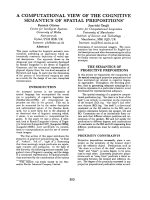
Fig. 1. Identification of the -1CÔA mutation.
(A) DHPLC profile of the )1CfiAmutation
where the heterozygous PCR fragments were
separated into two peaks (retention times
5.05 and 5.32) while the wild type PCR frag-
ment gave a single peak (retention time 5.30).
(B) Sequence differences between wild type
()1C) and mutant type ()1A) alleles. (C)
Genotyping for the )1CfiAmutationwhere
CviJI restriction pattern of homozygous wild
type (CC, 74 bp), homozygous mutant type
(AA, 96 bp), and heterozygous type (CA,
96 bp and 74 bp), were separated on 6%
polyacrylamide gel.
Ó FEBS 2003 Functional polymorphism in b
2
GPI (Eur. J. Biochem. 270) 233
Trp316Ser polymorphisms showed significant association
with APA (Table 4). The frequencies of the )1A (2.4% vs.
8.7%; P ¼ 0.0034) and Ser316 (1.6% vs. 6.9%;
P ¼ 0.0045) alleles were significantly lower in the APA-
positive group than the APA-negative group. For the
nucleotide )1 site, the age-adjusted odds ratio between
CA + AA and CC genotypes was 0.25 (95% CI ¼
0.07–0.86; P ¼ 0.028) and for the codon 316 site, the odds
ratio between Trp/Ser + Ser/Ser and Trp/Trp genotypes
was 0.22 (95% CI ¼ 0.05–0.94; P ¼ 0.042).
Fig. 2. Partial 5¢ flanking region of the b
2
GPI gene and EMSA probes.
(A) A partial 5¢ flanking sequence of human b
2
GPI (in lower case)
showing the transcriptional initiation site (nucleotide + 1; arrow) as
well as portion of exon 1 (upper case) including the 5¢ untranslated
region (UTR) upstream of the translation start site (ATG, nucleo-
tide +32) and a TFIID consensus sequence (underlined) [4]. A partial
5¢ flanking sequence of mouse b
2
GPI (italic) is aligned with the human
b
2
GPI sequence where the TFIID sequences at the transcriptional
initiation site are conserved between the two species, and are under-
lined and marked with the parallel bars. (B) Synthetic oligonucleotides
(wild and mutant types) corresponding to the 23 bp DNA fragment
from nucleotides )12 to +11 of the human b
2
GPI gene, which were
labeled with
32
P and used for EMSA. The mutant nucleotide at )1
position is indicated by lower case.
Table 1. Distribution of the b
2
GPI -1CÔA polymorphism in relation to
the Trp316Ser and Cys306Gly polymorphisms.
)1CfiA
CC CA AA Total
Trp316Trp
Trp/Trp 203 8 0 211
Trp/Ser 1 19 0 20
Ser/Ser 0 0 1 1
Total 204 27 1 232
Cys306Gly
Cys/Cys 183 26 1 210
Cys/Gly 14 1 0 15
Gly/Gly 1 0 0 1
Total 198 27 1 226
a
a
6 individuals with wild type genotypes at the )1CfiA and
Trp316Ser sites could not be genotyped for the Cys306Gly site due
to technical problems.
Table 2. Pair-wise measure of linkage disequilibrium between b
2
GPI
polymorphisms.
Pair-wise comparison P-value*
Nucleotide )1 vs. codon 316 < 0.0001
Nucleotide )1 vs. codon 306 0.294
codon 316 vs. codon 306 0.368
*P-values were obtained by v
2
–tests.
Table 3. Mean b
2
GPI plasma levels among b
2
GPI genotypes.
Polymorphic site/genotype b
2
GPI levels (mgÆdL
)1
) P-value
)1CfiA
CC (n ¼ 204) 18.45 ± 3.90
CA (n ¼ 27) 14.21 ± 4.22
AA (n ¼ 1) 9.40 < 0.0001
Trp316Ser
Trp/Trp (n ¼ 211) 18.39 ± 3.88
Trp/Ser (n ¼ 20) 13.37 ± 4.33
Ser/Ser (n ¼ 1) 9.40 < 0.0001
Cys306Gly
Cys/Cys (n ¼ 210) 18.41 ± 3.54
Cys/Gly (n ¼ 15) 10.49 ± 2.56
Gly/Gly (n ¼ 1) 0.20 < 0.0001
Table 4. Distribution of the b
2
GPI polymorphisms in APA-positive and
APA-negative groups.
Anti-phospholipid antibodies
Genotype Positive Negative
)1CfiA
CC 60 95.24% 120 83.33%
CA 3 4.76% 23 15.97%
AA 0 0.00% 1 0.69%
Total 63 144
A allele 0.024 0.087*
Trp316Ser
Trp/Trp 61 96.83% 125 86.81%
Trp/Ser 2 3.17% 18 12.50%
Ser/Ser 0 0.00% 1 0.69%
Total 63 144
Ser allele 0.016 0.069**
Cys306Gly
Cys/Cys 59 93.65% 133 92.36%
Cys/Gly 4 6.35% 10 6.94%
Gly/Gly 0 0.00% 1 0.69%
Total 63 144
Gly allele 0.032 0.042***
*P ¼ 0.0034, **P ¼ 0.0045, ***P ¼ 0.61 between APA-positive
and APA-negative groups.
234 H. Mehdi et al.(Eur. J. Biochem. 270) Ó FEBS 2003
Effect of the )1CfiA mutation on the
in vivo
level
of b
2
GPI transcripts
To examine if the )1CfiA mutation affects b
2
GPI
transcription that eventually determines low b
2
GPI plasma
levels, we screened 50 human liver tissues for the )1CfiA
mutation followed by determination of b
2
GPI plasma and
b
2
GPI mRNA levels in selected samples. We identified three
liver samples with low b
2
GPI levels (1.0, 1.3 and 2.1 lg
b
2
GPIÆmg
)1
total liver protein), but only two of them had
the )1CfiA mutation (CA genotype), while the third
sample was wild type (CC genotype). These results are
consistent with our findings in plasma samples, i.e. although
the )1CfiA mutation is associated with lower b
2
GPI levels,
not all b
2
GPI-deficient samples have this mutation. We then
performed Northern blot analysis on selected liver samples
with the mutant and wild type genotypes of the )1CfiA
mutation. Figure 3 shows the results of Northern blot for
one heterozygous (CA genotype) having a low b
2
GPI
protein level (1.0 lg b
2
GPIÆmg
)1
total liver protein) along
with three wild types (CC genotype) having normal b
2
GPI
protein levels (9.8, 8.2 and 9.1 lg b
2
GPIÆmg
)1
total liver
protein). While the level of GADPH mRNA (a house
keeping gene) was constant in each lane, b
2
GPI mRNA
level was significantly lower in the CA genotype (lane 3)
than the CC genotype (lanes 1, 2 and 4).
Effect of the )1CfiA mutation on reporter gene
expression
To further confirm that the )1CfiA mutation is associated
with low expression of the b
2
GPI gene, we performed
in vitro reporter gene expression assays. We constructed a
chimeric b
2
GPI-Luc vector to evaluate the promoter activity
of b
2
GPI within the 626 bp sequence in the 5¢ flanking
region and tested the effect of the )1CfiAmutationon
reporter (Luc) gene expression. Wild ()1C) and mutant
()1A) type constructs were subsequently tested for promo-
ter activity by cotransfecting COS-1 cells along with the
Renila Luc control vector (pRl-CMV) that was used to
adjust the transfection efficiency within different sets of
experiments. The promoter activity of each vector was
determined by the dual luciferase assay system that revealed
a twofold decrease in the promoter activity associated with
the mutant type allele ()1A) as compared to the wild type
allele ()1C) (Fig. 4). These results are similar to those seen
in the association studies (Table 3), in which the homozy-
gous AA mutant had almost one half of the b
2
GPI plasma
level (9.4 mgÆdL
)1
) than that observed in the CC wild type
homozygotes (18.45 mgÆdL
)1
). These results demonstrate
that the 626 bp 5¢ flanking region has some, if not all,
promoter activity and that the )1CfiA mutation down
regulates b
2
GPI gene expression.
Effect of the )1CfiA mutation on binding
of
trans
-acting factors
As the )1CfiA mutation disrupts the consensus sequence
for the b
2
GPI transcriptional initiation site and TFIID
(Fig. 2A), we designed double-stranded wild ()1C) and
mutant ()1A) type oligonucleotides as probes for EMSA to
evaluate the effect of this mutation on the binding of trans-
acting factors, using mouse liver nuclear extracts and
TFIID. We used mouse nuclear extracts because they were
easily available, and more importantly the consensus
sequence of the transcriptional initiation site is conserved
among human and mouse b
2
GPI (Fig. 2). EMSA of the
wild type ()1C) probe revealed two prominent and specific
bands of DNA-binding complexes (Fig. 5; lane 2), while the
mutant type ()1A) probe showed little or no binding to liver
nuclear proteins (Fig. 5; lane 3). We also found that the
purified TFIID bound weakly to the mutant type ()1A)
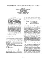
Fig. 3. Northern blot analysis to determine b
2
GPI mRNA levels and
capture ELISA to determine the b
2
GPI protein levels in human liver
samples carrying the wild (-1C) or mutant (-1A) type allele. Total RNA
was isolated from frozen human liver samples using TRIzol reagent
and 10 lg of total RNA was loaded in each lane for Northern blotting.
The corresponding liver samples were lysed in radioimmunoprecipi-
tation lysis buffer and b
2
GPI levels were determined by capture ELI-
SA. Total liver protein was estimated by Bio-Rad protein assay kit
where BSA was used as standard. (A) Northern autoradiograph dis-
playing levels of b
2
GPImRNAineachlane.(B)Northernautoradi-
ograph displaying levels of GAPDH mRNA in each lane. (C) b
2
GPI
levels (lgÆmg
)1
of total liver protein) for the corresponding liver
samples in each lane. The genotypes at nucleotide )1areindicated
beneath each lane.
Fig. 4. Effect of the -1CÔA mutation on reporter (Luc) gene expres-
sion. The effect of )1C (wild type) and )1A (mutant type) alleles was
measured as the mean of the firefly Luc levels, which were adjusted
with the Renila Luc levels which served as the reference for the
transfection efficiency. The results presented are from three inde-
pendent clones for each construct in triplicate, and each error bar
represents the standard error.
Ó FEBS 2003 Functional polymorphism in b
2
GPI (Eur. J. Biochem. 270) 235
probe (Fig. 5; lane 5) as compared to the wild type ()1C)
probe (Fig. 5; lane 4). These results demonstrate a sequence-
specific binding of liver nuclear extracts and TFIID to
b
2
GPI sequence at its transcriptional initiation site. These
results also confirm the location of the TFIID consensus
sequence at the b
2
GPI transcriptional initiation site, which is
disrupted by the )1CfiA mutation that would ultimately
affect the b
2
GPI gene expression.
Discussion
Human b
2
GPI, also known as apolipoprotein H, is a
plasma glycoprotein that has been implicated in a variety of
physiological pathways, including blood coagulation,
thrombosis, and the production of autoantibodies (APA).
b
2
GPI plasma levels vary significantly among individuals,
ranging from immunologically undetectable to as high as
35 mgÆdL
)1
, and family data indicate that this variation is
under genetic control [26–28,38]. We have recently deter-
mined the heritability of b
2
GPI plasma levels to be 66%
(Kamboh et al. unpublished data). In addition to the b
2
GPI
quantitative polymorphisms, we have originally described a
common protein polymorphism in the b
2
GPI gene [39] and
both polymorphisms were found to be tightly linked [38].
Thus, the family, heritability, and linkage data provide
strong support that b
2
GPI plasma variation is under genetic
control and that genetic variation in b
2
GPI is a major
determinant of this variation. In our attempt to deter-
mine the molecular basis of b
2
GPI plasma variation, we
conducted association studies and found that two of the
b
2
GPI mutations, Trp316Ser and Cys306Gly, were signifi-
cantly associated with b
2
GPI plasma levels and their effects
were independent of each other [30,31]. However, our
in vitro mutagenesis and expression study did not link these
mutations to an altered b
2
GPI gene expression [32].
Although, in vitro mutagenesis and expression study do
not rule out the possibility that these mutations might affect
the stability of b
2
GPI in vivo, we hypothesized that they are
in linkage disequilibrium with two different functional
mutations, as their effects on b
2
GPI plasma levels were
independent. To search for the functional mutations that
are associated with altered gene expression and b
2
GPI
plasma levels, we focused on a 626-bp fragment in the
5¢ flanking region of b
2
GPI that has been characterized
recently [4].
Here, we report a new point mutation ()1CfiA) at the
b
2
GPI transcriptional initiation site (Fig. 2A), which is
associated with low b
2
GPI plasma and mRNA levels as well
as a twofold reduced expression of the tagged-Luc gene. The
)1CfiA mutation was also in strong linkage disequilibrium
with the Trp316Ser mutation. In the univariate analysis,
both sites showed significant association with b
2
GPI plasma
levels. However, in multivariate analysis, the effect of
Trp316Ser was no longer significant, indicating that the
)1CfiA is the functional mutation. Of the 27 individuals in
the CA genotype group (Table 3), 18 had b
2
GPI plasma
levels between 4.3 mgÆdL
)1
and 15.9 mgÆdL
)1
, which would
fall in the heterozygous category (ND) based on the
quantitative polymorphism. The remaining nine individuals
fell in the normal (NN) category; seven in a narrow range
between 16.2 mgÆdL
)1
and 18.4 mgÆdL
)1
,andtwowith
20.5 mgÆdL
)1
and 20.6 mgÆdL
)1
. Although the bulk of
b
2
GPI plasma variation is under genetic control (66%
heritability) other nongenetic factors also influence this
variation [26,29] and this may explain higher than expected
b
2
GPI plasma levels observed in nine individuals with the
)1CfiA mutation. Alternatively, other genetic or non-
genetic factors modulate the effect of the )1CfiAmutation
on b
2
GPI plasma levels. We also found that the )1CfiA
mutation cannot explain the independent effect of
Cys306Gly on b
2
GPI plasma levels. This indicates that
another functional mutation is responsible for the lowering
effect of Cys306Gly on b
2
GPI plasma levels. Another
subject with only 0.8 mgÆdL
)1
b
2
GPI plasma levels was wild
type homozygous at nucleotide )1, codon 306 and 316 sites
and thus must be a carrier of a yet to be discovered
functional mutation. These data suggest that multiple
functional mutations in the b
2
GPI gene affect b
2
GPI plasma
levels.
In our earlier work, we found a protective effect of the
Trp316Ser polymorphism against the occurrence of APA
in the lupus sample [31]. In this study, the )1CfiA
mutation also showed a significant protective effect. The
carrier frequency of the )1A allele was almost fourfold
lower in the APA-positive group than the APA-negative
group (4.8% vs. 16.6%). As the Trp316Ser polymorphism
is in almost complete linkage disequilibrium with the
)1CfiA mutation, our data link the protective effect
directly to the )1CfiA mutation, as this is associated with
lower b
2
GPI plasma levels and consequently lower risk of
developing APA. Paradoxically, however, the Cys306Gly
Fig. 5. Effect of the -1CÔA mutation on the binding of crude nuclear
factors and purified TFIID using EMSA. The EMSA was performed
using
32
P-labeled 23 bp oligonucleotide ()12 to +11 nucleotides of the
b
2
GPI gene) carrying the wild (CC) or mutant (AA) type sequence at
nucleotide )1 followed by binding with crude nuclear extracts from
mouse liver or purified TFIID. The binding reactions were performed
at room temperature for 20 min in the presence of the nonspecific
competitor poly (dI–dC). Lanes 1 and 6 are wild and mutant type
probes without nuclear extracts or TFIID, respectively. Lanes 2 and 3
are wild and mutant type probes incubated with nuclear extracts,
respectively. Lanes 4 and 5 are wild and mutant type probes incubated
with TFIID, respectively.
236 H. Mehdi et al.(Eur. J. Biochem. 270) Ó FEBS 2003
mutation, which is also associated with lower b
2
GPI
plasma levels, was not associated with protection from the
presence of APA. Furthermore, although the )1CfiAand
Trp316Ser mutations were associated with protection
against APA, there were three (4.8%) individuals with
these two mutations, who were positive for APA but had
lower b
2
GPI plasma levels (3.7, 4.3 and 7.3 mgÆdL
)1
). This
indicates that the genetic basis of APA is complex and
other genetic and/or biological factors are involved in the
occurrence of APA.
The structural organization of the b
2
GPI gene, including
626 bp sequence in the 5¢ flanking region has been reported
together with the transcriptional initiation site 31 bp
upstream of the translation start codon [4], which com-
pletely agrees with the consensus for an initiator element,
PyPyA
+1
N(TA)PyPy known to sustain transcriptional
initiation [40]. The computer analysis for transcriptional
elements within this region did not reveal any TATA box or
CG rich region close to the transcriptional initiation site
(nucleotide +1) but a TFIID binding sequence was
identified between nucleotides )2 and +5 (CCACTTT)
that is disrupted by the )1CfiA mutation. Thus, we
predicted that lower b
2
GPI plasma levels associated with the
)1CfiA mutation might be due to its direct impact on
b
2
GPI transcription. Indeed, our Northern blot analysis on
liver samples containing the )1CfiA mutation confirmed
this prediction in which all samples containing the CA
genotype had lower mRNA levels (Fig. 3).
As Northern blot analysis revealed that the )1CfiA
mutation affects b
2
GPI transcription, we examined its effect
on b
2
GPI gene expression using tagged-Luc constructs
expressed in COS-1 cells. Although the promoter of b
2
GPI
is not yet characterized, we cloned the reported 5¢ flanking
region of the b
2
GPI gene (from nucleotide )622 to +74) in
front of the Luc gene for in vitro functional studies. The
reporter gene assay revealed that the 626 bp 5¢ flanking
region had some, if not all, the promoter activity and the
)1CfiA mutation is a functional substitution that suppres-
ses b
2
GPI gene expression by twofold. The twofold
difference observed between the )1A and )1C alleles in
the reporter gene assay is similar to that seen in the plasma
level difference between the AA (9.4 mgÆdL
)1
)andCC
(18.5 mgÆdL
)1
) genotypes (Table 3). As the effect of the
)1CfiAmutationonb
2
GPI gene expression was moderate,
this does not preclude the possibility that other sequence
variationinthe5¢ region of b
2
GPI might also have an effect
on the regulation of b
2
GPI expression. The functional
characterization of the b
2
GPI promoter would enable the
targeting of regulatory regions for mutation detection.
Further evidence that the )1CfiA mutation is functional
comes from our EMSA data that demonstrate an allele-
specific binding of nuclear factors and TFIID to the
mutation containing sequence; )1A has less affinity than
)1C. Our novel data demonstrate that the )1CfiA
mutation at the transcriptional initiation site is causative,
which regulates b
2
GPI gene expression at the transcriptional
level that ultimately affects b
2
GPI plasma levels.
In summary, we have identified a new polymorphism at
the transcriptional initiation site of the b
2
GPI gene that is
associated with less binding with a putative transcriptional
factor, lower gene expression, lower b
2
GPI plasma levels,
lower b
2
GPI mRNA levels and protection from the
occurrence of APA in lupus patients. Our data also indicate
that the molecular basis of plasma b
2
GPI deficiency is
heterogenous. The characterization of functional b
2
GPI
promoter and identification of sequence variation in these
regulatory elements may help to further delineate the
molecular basis of b
2
GPI deficiency.
Acknowledgements
This study was supported by a National Heart, Lung and Blood
Institute of Health grant HL 54900 and Central Research Development
Fund award by the University of Pittsburgh.
References
1. Schultz, H.E., Heide, K. & Haupt, H. (1961) Uber ein bisher
unbekanntes neidermolekul es b
2
Globulin des Humanserums.
Naturwissenschaften 48,719.
2. Polz, E. & Kostner, G.M. (1979) The binding of b
2
-glycoprotein I
to human serum lipoproteins: Distribution among density frac-
tions. FEBS Lett. 102, 183–186.
3. Polz, E. & Kostner, G.M. (1979) Binding of b
2
-glycoprotein I to
intralipid: Distribution of the dissociation constant. Biochem.
Biophys. Res. Commun. 90, 1305–1312.
4. Okkels, H., Rasmussen, T.E., Sanghera, D.K., Kamboh, M.I. &
Kristensen, T. (1999) Structure of the b
2
-glycoprotein I (apolipo-
protein H) gene. Eur. J. Biochem. 259, 435–440.
5. Lozier, J., Takahashi, N. & Putnam, F.W. (1984) Complete amino
acid sequence of human plasmid b
2
-glycoprotein I. Proc. Natl
Acad. Sci. USA 81, 3640–3364.
6. Mehdi,H.,Nunn,M.,Steel,D.M.,Whitehead,A.S.,Perez,M.,
Walker, L. & Peeples, M.E. (1991) Nucleotide sequence and
expression of the human gene encoding apolipoprotein H. Gene
108, 293–298.
7. Kristensen, T., Schousboe, I., Boel, E., Mulvihill, E.M., Hansen,
R.R., Moller, K.B., Moller, N.P.H. & Sottrup-Jensen, L. (1991)
Molecular cloning and mammalian expression of human b
2
-gly-
coprotein I cDNA. FEBS Lett. 289, 183–186.
8. Steinkasserer, A., Estaller, C., Weiss, E. & Sim, R.B. (1991)
Complete nucleotide and deduced amino acid sequence of human
b
2
-glycoprotein I. Biochem. J. 277, 387–391.
9. Davie, E.W., Ichinose, A. & Leytus, S.P. (1986) Structural features
of the proteins participating in blood coagulation and fibrinolysis.
Cold Spring Harbor Symposium on Quantitative Biology,LI,pp.
509–514. Cold Spring Harbor Laboratory, Cold Spring Harbor,
NY, USA.
10. Ichinose, A., Bottenus, R.E. & Davie, E.W. (1990) Structure of
transglutaminases. J. Biol. Chem. 265, 13411–13414.
11. Klickenstein, L.B., Wong, W.W., Smith, J.A., Weis, J.H., Wilson,
J.G. & Fearon, D.T. (1987) Human C3b/C4b receptor (CR1)
demonstration of long homologous repeating domains that are
composed of the short consensus repeats characteristic of C3/C4
binding proteins. J. Exp. Med. 165, 2095–1112.
12. Kristensen,T.,Deustachio,P.,Ogato,R.T.,Chung,L.P.,Reid,
K.B.M. & Tack, B.F. (1987) The superfamily of C3b/C4b-binding
proteins. Federation Proc. 46, 2463–2469.
13. Schousboe, I. (1985) b
2
-glycoprotein I: a plasma inhibitor of the
contact activation of the intrinsic blood coagulation pathway.
Blood 66, 1086–1091.
14. Nimpf, J., Bevers, E.M., Bomans, P.H.H., Till, U., Wurm, H.,
Kostner, G.M. & Zwaal, R.F.A. (1986) Prothrombinase activity
of human platelets is inhibited by b
2
-glycoprotein I. Biochim.
Biophys. Acta 884, 142–149.
15. Schousboe, I. (1988) In vitro activation of the contact activa-
tion system (Hageman factor system) in plasma by acidic
Ó FEBS 2003 Functional polymorphism in b
2
GPI (Eur. J. Biochem. 270) 237
phospholipids, and the inhibitory effect of b
2
-glycoprotein I on the
activation. Int. J. Biochem. 20, 309–315.
16. Schousboe, I. & Rasnussen, M.S. (1988) The effect of b
2
-glyco-
protein I on the dextran sulfate and sulfatide activation of the
contact system (Hageman factor system) in the blood coagulation.
Int. J. Biochem. 20, 787–792.
17. Halkier, T. & Magnussen, S. (1988) Contact activation of blood
coagulation is inhibited by plasma factor XIIIb-chain. Thromb.
Res. 51, 313–324.
18. Bancsi, L.F.J.M.M., van der Linden, I.K. & Bertina, R.M. (1992)
b
2
-glycoprotein I deficiency and the risk of thrombosis. Thromb.
Haemos. 67, 649–653.
19. Yasuda, S., Tsutsumi, A., Chiba, H., Yanai, H., Miyoshi, R.,
Horita,T.,Atsumi,T.,Ichikawa,K.,Matsuura,E.&Koike,T.
(2000) b
2
-glycoprotein I deficiency: prevalence, genetic back-
ground and effects on plasma lipoprotein metabolism and
hemostasis. Atherosclerosis 152, 337–346.
20. Sheng, Y., Reddel, S.W., Herzog, H., Wang, Y.X., Brighton, T.,
France, M.P., Robertson, S.A. & Krilis, S.A. (2001) Impaired
thrombin generation in b
2
-glycoprotein I null mice. J. Biol. Chem.
276, 13817–13821.
21. Galli, M., Confurius, P., Maassen, C., Hemker, H.C., DeBaets,
M.H., van Breda-Vriesman, P.J.C., Barbui, T., Zwaal, R.F.A. &
Beveres, E.M. (1990) Anticadiolipin antibodies (ACA) directed
not to cardiolipin but to a plasma protein cofactor. Lancet 335,
1544–1547.
22. McNeil, H.P., Simpson, R.J., Chesterman, C.N. & Krilis, S.A.
(1990) Anti-phospholipid antibodies are directed against a com-
plex antigen that includes a lipid binding inhibitor of coagulation:
b
2
-glycoprotein I (apolipoprotein H). Proc. Natl Acad. Sci. USA
87, 4120–4124.
23. Jones, J.V., James, H., Tan, M.H. & Mansouor, M. (1992) Anti-
phospholipid antibodies required b
2
-glycoprotein I (apolipopro-
tein H) as cofactor. J. Rhematol. 19, 1397–1402.
24. Roubey,R.A.,Pratt,C.W.,Buyon,J.P.&Winfield,J.B.(1992)
Lupus anticoagulant activity of autoimmune anti-phospholipid
antibodies is dependent upon b
2
-glycoprotein I. J. Clin. Invest. 90,
1100–1104.
25. Cabral, A.R., Cabiedes, J. & Alarcon-Segovia, D. (1995) Anti-
bodies to phospholipid-free b
2
-glycoprotein I in patients with
primary anti-phospholipid syndrome. J. Rheumatol. 22, 1894–1898.
26. Cleve, H. (1968) Genetic studies on the deficiency of b
2
-glyco-
protein I of human serum. Hum. Genet. 5, 294–304.
27. Koppe, A.L., Walter, H., Chopra, V.P. & Bajatzadeh, M. (1970)
Investigations on the genetics and population genetics of the
b
2
-glycoprotein I polymorphism. Hum. Genet. 9, 164–171.
28. Propert, D.N. (1978) The relationship of sex, smoking status, birth
rank and parental age to b
2
-glycoprotein I levels and phenotypes
in a sample of Australian Caucasian adults. Hum. Genet. 43, 281–
288.
29. Walter, H., Hilling, M., Brachtel, R. & Hitzeroth, H.W. (1979)
On the population genetics of b
2
-glycoprotein I. Hum. Hered. 29,
236–241.
30. Mehdi, H., Aston, C.E., Sanghera, D.K., Hamman, R.F. &
Kamboh, M.I. (1999) Genetic variation in the apolipoprotein H
(b
2
-glycoprotein I) gene affects plasma apolipoprotein H concen-
trations. Hum. Genet. 105, 63–71.
31. Kamboh, M.I., Manzi, S., Mehdi, H., Fitzgerald, S.,
Sanghera, D.K., Kuller, L.H. & Aston, C.E. (1999) Genetic
variation in the apolipoprotein H (b
2
-glycoprotein I) affects the
occurrence of anti-phospholipid antibodies and apolipoprotein H
concentrations in systemic lupus erythematosus. Lupus 8, 742–
750.
32. Mehdi, H., Naqvi, A. & Kamboh, M.I. (2000) A hydrophobic
sequence at position 313–316 (Leu-Ala-Phe-Trp) in the fifth
domain of apolipoprotein H (b
2
-glycoprotein I) is crucial for
cardiolipin binding. Eur. J. Biochem. 267, 1770–1776.
33. Luedecking, E.K., DeKosky, S.T., Mehdi, H., Ganguli, M. &
Kamboh, M.I. (2000) Analysis of genetic polymorphisms in the
transforming growth factor-b1geneandtheriskofAlzheimer’s
disease. Hum. Genet. 106, 565–569.
34. Mehdi, H., Ono, E. & Gupta, K.C. (1990) Initiation of translation
at CUG, GUG, and ACG codons in mammalian cells. Gene 91,
173–178.
35. Liu, Y., Beedle, A.B., Lin, L., Bell, A.W. & Zarnegar, R. (1994)
Identification of a cell-type-specific transcriptional repressor in the
promoter region of the mouse hepatocyte growth factor gene.
Mol. Cell. Biol. 14, 7046–7058.
36. Jiang, J.G., Bell, A., Liu, Y. & Zarnegar, R. (1997) Transcriptional
regulation of the HGF gene by COUP-TF and estrogen receptor.
J. Biol. Chem. 272, 3928–3934.
37. Lewontin, R.C. (1964) The interaction of selection and linkage. I.
General considerations; heterotic models. Genetics 49, 49–67.
38. Eiberg, H., Nielsen, L.S. & Mohr, J. (1989) Exclusion mapping of
apolipoprotein H (APOH) and relationship between electro-
phorative polymorphism. Cytogenet. Cell. Genet. 51, 994.
39. Kamboh, M.I., Ferrell, R.E. & Sephernia, B. (1988) Genetic stu-
dies of human apolipoproteins. IV. Structural heterogeneity of
apolipoprotein H (b
2
-glycoprotein I). Am.J.Hum.Genet.42, 452–
457.
40. Lo, K. & Smale, S.T. (1996) Generality of a functional initiator
consensus sequence. Gene 182, 13–22.
238 H. Mehdi et al.(Eur. J. Biochem. 270) Ó FEBS 2003