High tumor budding is a strong predictor of poor prognosis in the resected perihilar cholangiocarcinoma patients regardless of neoadjuvant therapy, showing survival similar to those without
Bạn đang xem bản rút gọn của tài liệu. Xem và tải ngay bản đầy đủ của tài liệu tại đây (2.56 MB, 11 trang )
Ito et al. BMC Cancer
(2020) 20:209
/>
RESEARCH ARTICLE
Open Access
High tumor budding is a strong predictor
of poor prognosis in the resected perihilar
cholangiocarcinoma patients regardless of
neoadjuvant therapy, showing survival
similar to those without resection
Takahiro Ito1, Naohisa Kuriyama1*, Yuji Kozuka2, Haruna Komatsubara2, Ken Ichikawa1, Daisuke Noguchi1,
Aoi Hayasaki1, Tekehiro Fujii1, Yusuke Iizawa1, Hiroyuki Kato1, Akihiro Tanemura1, Yasuhiro Murata1,
Masashi Kishiwada1, Shugo Mizuno1, Masanobu Usui1, Hiroyuki Sakurai1 and Shuji Isaji1
Abstract
Background: Tumor budding (TB) is used as an indicator of poor prognosis in various cancers. However, studies on
TB in perihilar cholangiocarcinoma are still limited. We examined the significance of TB in resected perihilar
cholangiocarcinoma with or without neoadjuvant therapy.
Methods: Seventy-eight patients who underwent surgical resection at our institution for perihilar
cholangiocarcinoma from 2004 to 2017, (36 with neoadjuvant therapy), were enrolled in this study. TB was defined
as an isolated cancer cell or clusters (< 5 cells) at the invasive front and the number of TB was counted using a 20
times objective lens. Patients were classified into two groups according to TB counts: low TB (TB < 5) and high TB
(TB ≥5).
Results: In this 78 patient cohort, high TB was significantly associated with advanced tumor status (pT4: 50.0% vs
22.2%, p = 0.007, pN1/2: 70.8% vs 39.6%, p = 0.011, M1: 20.8% vs 1.9%) and higher histological grade (G3: 25.0% vs
5.7%, p = 0.014). Disease specific survival (DSS) in high TB was significantly inferior compared to that in low TB
group (3-y DSS 14.5% vs 67.7%, p < 0.001). Interestingly, DSS in high TB showed similar to survival in unresected
patients. In addition, high TB was also associated with advanced tumor status and poor prognosis in patients with
neoadjuvant therapy. Multivariate analysis identified high TB as an independent poor prognostic factors for DSS (HR:
5.206, p = 0.001).
Conclusion: This study demonstrated that high TB was strongly associated with advanced tumor status and poor
prognosis in resected perihilar cholangiocarcinoma patients. High TB can be a novel poor prognostic factor in
resected perihilar cholangiocarcinoma regardless of neoadjuvant therapy.
Keywords: Perihilar cholangiocarcinoma, Tumor budding, Prognostic factor
* Correspondence:
1
Department of Hepatobiliary Pancreatic and Transplant Surgery, Mie
University Graduate School of Medicine, 2-174 Edobashi, Tsu, Mie 514-8507,
Japan
Full list of author information is available at the end of the article
© The Author(s). 2020 Open Access This article is licensed under a Creative Commons Attribution 4.0 International License,
which permits use, sharing, adaptation, distribution and reproduction in any medium or format, as long as you give
appropriate credit to the original author(s) and the source, provide a link to the Creative Commons licence, and indicate if
changes were made. The images or other third party material in this article are included in the article's Creative Commons
licence, unless indicated otherwise in a credit line to the material. If material is not included in the article's Creative Commons
licence and your intended use is not permitted by statutory regulation or exceeds the permitted use, you will need to obtain
permission directly from the copyright holder. To view a copy of this licence, visit />The Creative Commons Public Domain Dedication waiver ( applies to the
data made available in this article, unless otherwise stated in a credit line to the data.
Ito et al. BMC Cancer
(2020) 20:209
Background
Perihilar cholangiocarcinoma, which is an epithelial cell
malignancy localized to the area between the second degree bile ducts and the insertion of the cystic duct into
the common bile duct, represents the most common
form of cholangiocarcinoma [1, 2]. Although surgical resection remains the only curative treatment for perihilar
cholangiocarcinoma, resection is considered a significant
challenge for surgeons, and the prognosis of nonresected patients is very poor [3]. However, even if patients undergo curative resection, many patients have
cancer recurrence [4]. Predicting poor prognosis and
cancer recurrence is an important issue when planning
an adequate and effective therapeutic strategy.
Tumor budding (TB) is a histological phenomenon encountered in various cancers typically described as the
presence of single cells or clusters in the tumor stroma,
and was first described by Imai in Japanese literature [5].
TB is widely used in the field of colorectal carcinoma as
a prognostic factor and a correlated factor with advanced
stage [6]. In addition, it has been identified as a novel
prognostic factor in various types of cancer such as
esophageal [7], pancreatic [8], as well as, cancer of the
ampulla [9], and gall bladder [10]. With regards to cholangiocarcinoma, three reports on the impact of TB have
recently been published [11–13]. Ogino et al. [11] demonstrated that high TB grade was an independent adverse
prognostic factor in 195 perihilar cholangiocarcinoma patients by multivariate analysis. Okubo et al. [12] reported
the prognostic significance of TB in resected 299 patients
with biliary tree carcinoma (intrahepatic: n = 47 (16%), extrahepatic: n = 144 (48%), gallbladder: n = 50 (17%), ampulla: n = 58 (19%)). In addition, Tanaka et al. [13]
demonstrated that TB tumor budding was a significant
prognostic factor in 107 cases of intrahepatic cholangiocarcinoma and 54 cases of perihilar cholangiocarcinoma.
In recent years, neoadjuvant therapy has become a
critical treatment for improving the outcomes of various
cancers. Additionally, neoadjuvant therapy is becoming
the standard of treatment of locally advanced pancreatic
adenocarcinoma. However, there are few reports on the
impact of neoadjuvant therapy in the patients with cholangiocarcinoma, although the efficacy of neoadjuvant
therapy followed by surgery for “unresectable” locally advanced cholangiocarcinoma has been reported [14, 15].
Studies on the relationship of TB and neoadjuvant
therapy remain limited. In the field of rectal and esophageal carcinoma, where the use of neoadjuvant therapy is
common, there are several reports showing the prognostic significance of high TB in patients who underwent
neoajuvant therapy [7, 16, 17]. These studies have reported the association between high TB and poor prognosis in patients underwent neoadjuvant therapy for
esophageal carcinoma [7] and rectal carcinoma [16, 17].
Page 2 of 11
In contrast, the three previous studies on the significance of TB in cholangiocarcinoma have excluded patients with neoadjuvant therapy. Due to an expected
increase in the number of patients with perihilar cholangiocarcinima who will undergo neoadjuvant therapy
followed by curative-intend surgery, we considered
which prognostic factors should be analyzed, with additional focus on preoperative treatment. In the present
study, we aimed to elucidate the significance of TB in
resected perihilar cholangiocarcinoma. In addition, we
sought to examine the relationship of TB and neoadjuvant therapy.
Methods
Patients
Between January 2004 and December 2017, 81 patients
with perihilar cholangiocarcinoma underwent surgical
resection at our institution. Three patients with hospital
death were excluded, and the remaining 78 patients were
included in this study. In addition, 28 patients with locally advanced perihilar cholangiocarcinoma who did
not undergo surgical resection in the same period were
included in the survival analysis with comparison to
resected patients.
Preoperative management
Multidetector-row computed tomography (MDCT),
magnetic resonance cholangiopancreatography (MRCP),
endoscopic retrograde cholangiography (ERC), and
intraductal ultrasonography (IDUS) were used for preoperative diagnosis and tumor staging. Tumor and negative biopsies by ERC were used for confirming diagnosis
and definition of biliary cancer invasion. Endoscopic
retrograde biliary drainage (ERBD) tubes were inserted
into the future remnant liver in patients with obstructive
jaundice.
Neoadjuvant therapy
The use of neoadjuvant therapy was depended on clinical practice from 2004 to 2009. Chemotherapy or chemoradiotherapy (CRT) was used before and/or after
resection. As for chemotherapy, gemcitabine-based regimen in combination with S-1, cisplatin, and 5-FU were
used. Radiation therapy (RT) was used as local therapy
with a total dose of 36–45 Gy. From 2010, we had introduced a new protocol of preoperative chemotherapy.
After evaluation of tumor extension to the hepatic artery
(HA), portal vein (PV), and bile duct by preoperative imaging studies, two cycles of chemotherapy with gemcitabine (600 mg/m2 on days 7 and 21) plus S-1 (60 mg/m2
daily on days 1–21 every 4 weeks), followed by surgery,
was administrated in patients with locally advanced perihilar cholangiocarcinoma with (1) main, bilateral, or
contralateral PV and/or HA invasion with or without
Ito et al. BMC Cancer
(2020) 20:209
possible vascular reconstruction; or (2) invasion of the
right side of the umbilical portion and the left side of
the origin of the right posterior PV; or (3) regional
lymph node metastasis [18–20].
Page 3 of 11
Sub-analysis for comparison between groups with TB
counts 5–9 vs TB counts ≥5 was performed. The concordance rate was 94.5%. In disagreement cases, these
pathologists discussed the findings and reached a
consensus.
Surgical procedure
The type of hepatectomy was determined using the following factors: the indocyanine green retention rate at
15 min (ICGR15), the hepatic uptake ratio of 99mTcGSA scintigraphy at 15 min (LHL15), and the future
remnant liver volume using CT volumetry [21]. Right
hepatectomy with caudate lobectomy was applied to Bismuth type I, II, and IIIa tumors. Left hepatectomy with
caudate lobectomy was applied to Bismuth type IIIb tumors. If a tumor obviously extended over the second
order biliary radicles, such as Bismuth type IV tumors,
trisectionectomy or central bisectionectomy was selected. In several select patients, extrahepatic bile duct
resection without hepatectomywas performed due to
poor patient condition, such as older age and insufficient
remnant liver function. Portal vein embolization (PVE)
was indicated when the future remnant liver volume was
estimated as less than 40%. Tumor unresectability was
determined by preoperative or intraoperative evaluation
of tumor extension to hepatic parenchyma or major vessels, and by insufficient remnant liver function for
hepatectomy.
Histology and assessment of tumor budding
All resected specimens were fixed in 10% buffered formalin and paraffin-embedded (FFPE). FFPE blocks were
then sliced into 4-μm-thick sections and stained with
haematoxylin and eosin (H&E) for microscopic examination. Primary tumor staging (pT) and regional lymph
node metastasis (pN) were defined according to Union
for International Cancer Control (UICC) 8th edition. TB
was defined as an isolated cancer cell or clusters composed of fewer than 5 cancer cells at the site of tumor
invasive front according to previous studies [10–13].
The invasive front was defined as the peripheral to
whole primary tumor and in the most severe extended
area of tumor to the surrounding tissue according to
previous studies. The number of TB was counted in a
field measuring 0.785 mm2 using a 20 times objective
lens by microscopy. The independent two pathologists
were blinded to the clinical characteristics of the patients
and selected a single field for evaluation, so-called ‘hotspot’ that would include the maximal budding area for
determining the TB count. To find a hot-spot, whole invasive front of tumors were evaluated. The maximal
value of tumor budding was defined as TB counts for
each tumor. Based on previous studies [10, 12, 13], TB
counts were classified into two groups: low TB (TB
counts < 5) and high TB (TB counts≥5). In addition,
Statistical analysis
Continuous data are expressed as median and range.
Continuous and categorical variables were compared
using the Mann-Whitney U test and Chi-square test, respectively. Disease-specific survival time (DSS) was calculated from the date of initial treatment to the date of
cancer related death or the date of last follow-up if death
did not occur. Recurrence free survival time (RFS) was
calculated from the date of initial treatment to the date
of first recurrence of cancer or the date of last follow-up
without recurrence if recurrence did not occur in patients without R2 resection. Patients with R2 resection
were excluded from RFS analysis. Survival curves were
generated by the Kaplan–Meier method, and differences
in survival rates were analyzed using the Log rank test.
To identify predictors for disease specific survival, COX
hazard regression model with significant variables in
Table 1 Patient overview in all 78 patients
Factors
All patients (n = 78)
Age (y.o.)
69 (39–87)
Gender (Male / Female)
47 / 31
Neoadjuvant therapy
36
Chemotherapy
25
Chemoradiotherapy
11
PTPE
18
Type of liver resectiona
No liver resection (PD, Extra bile duct resection)
5
S1,5,6,7,8
27
S1,4,5,6,7,8
7
S1,2,3,4
26
S1,2,3,4,5,8
5
Others
8
Histological type: G1 / G2 / G3 / others
49 / 18 / 9 / 2
TNM (UICC 8th)
pT: T0 / T1 / T2 / T3 / T4
1 / 8 / 33 / 12 / 24
pN: N0 / N1 or N2
40 / 38
M1 (Intrahepatic / Extrahepatic)
6 (3 / 3)
Residual tumor status: R0 / R1 / R2
53 / 14 / 11
Postoperative hospital stay (median: days)
45.5 (13–325)
Complication ≥ C-D grade III
32
Postoperative chemotherapy
56
PTPE percutaneous transhepatic portal vein embolization, PD
pancreaticoduodenectomy, C-D Clavien-Dindo
a
Expressed as Couinaud’s hepatic segments resected
Ito et al. BMC Cancer
(2020) 20:209
univariate analysis was used for multivariate analysis. As
prognostic factors, age, gender, preoperative tumor
marker (CEA, CA19–9), TMN stage, status of tumor
lymphovascular (LV) and perinueral invasion, residual
tumor status (non-curative resection), tumor budding
status (High TB) were analyzed. All tests were twosided, and the significance level was p < 0.05, and the
confidence interval was determined at 95%. All analyses
were performed using IBM® SPSS® Statistics version 25
(IBM Corporation, Armonk, NY).
Results
Patient overview
Patient demographics of 78 patients are shown in
Table 1. Forty-seven were men and 31 were women,
with a median age of 69 years (range 39–87 years).
Thirty-six patients (46%) received neoadjuvant therapy
and 18 patients (23%) underwent PTPE prior to resection. The most common type of liver resection was right
hepatectomy with caudate lobectomy (n = 27, 35%),
followed by left hepatectomy with caudate lobectomy
(n = 26, 33%). Thirty-eight patients (49%) had tumor
with regional lymph node metastasis and 6 patients (8%)
had distant metastasis: intrahepatic metastasis (n = 3)
and extrahepatic metastasis (n = 3). Fifty-three patients
(68%) achieved R0 resection. Postoperative complication
with grade III or more (Clavien-Dindo classification) was
occurred in 32 patients (41%) and median (range)
Page 4 of 11
postoperative hospital days were 45.5 (13–325) days.
Median follow-up time was 2.4 years.
Distribution of TB counts and patients classification
according to TB status
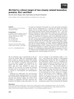
Fig. 1 shows typical histological picture with tumor invasive front with / without tumor budding and distribution
of TB counts. Fifty-four patients (69%) had TB counts
0–4, 12 patients (15%) had 5–9, and 12 patients (15%)
had 10 or more. Based on some previous studies [7, 8],
patients were classified into two groups according to TB;
low TB (TB counts < 5, n = 54) and high TB (TB counts
≥5, n = 24). Then we compared patient characteristics
and outcomes.
Tumor budding was associated with advanced
histological status and poor prognosis
As shown in Table 2, there were no significant differences
in preoperative patient characteristics and surgical information such as age, gender, preoperative treatment, tumor
markers, types of surgery. However, in high TB patients,
the rate of patients who underwent combined vascular resection (HA and/or PV) was tended to be lower than that
in low TB patients despite it was not statistically significant (67% vs. 43%, p = 0.050). In terms of histologically
factors, high TB patients had higher rates of tumor with
grade G3 (25% vs. 5.6%, p = 0.013), pT4 (50.0% vs. 22.2%,
p = 0.014), lymph node metastasis (70.8% vs. 38.9%, p =
Fig. 1 Representative histological findings at the invasive front of tumor and distribution of tumor budding (TB) counts. a High TB. b Low TB. c
Distribution of TB counts in the 78 patients. In the front of tumor with high TB, several single cells or clusters (white arrow) are detected (a). In
contrast, there are few TB in the front of tumor with low TB (b). Thirty-one percent of patients (24/78) had tumor with TB counts ≥5 (c)
Ito et al. BMC Cancer
(2020) 20:209
Page 5 of 11
Table 2 Patient characteristics in the low and high TB groups
p value
Variables
Low TB (n = 54)
High TB (n = 24)
Age (y.o.)
69 (40–87)
69 (39–78
0.630
Gender (Male/Female)
31 / 23
16 / 8
0.441
Neoadjuvant therapy
25 (46.3%)
11 (45.8%)
0.970
PTPE
12 (22.2%)
6 (25.0%)
0.788
CEA (ng/mL)
3.6 (0.6–38.4)
3.6 (0.7–28.0)
0.862
CA19–9 (U/mL)
76.6 (1.0–7898)
106.6 (1.0–9278)
0.681
CEA (ng/mL)
3.0 (0.7–32.2)
3.2 (0.5–32.3)
0.492
CA19–9 (U/mL)
56.9 (1.0–11,659)
89.7 (1.0–9278)
0.174
5 (9.3%)
0
Initial tumor marker
Preoperative tumor marker
Type of liver resection
No liver resection
0.109
S1,5,6,7,8
16 (29.6%)
10 (41.7%)
S1,4,5,6,7,8
19 (35.2%)
8 (33.3%)
S1,2,3,4
3 (5.6%)
2 (8.3%)
S1,2,3,4,5,8
3 (5.6%)
4 (16.7%)
Others
8 (14.8%)
0
HPD
3 (5.6%)
1 (4.2%)
Combined HA and/or PV resection
23 (42.6%)
16 (66.7%)
0.050
Operation time (min)
603 (383–1030)
622 (422–972)
0.482
Blood loss (ml)
2165 (166–9907)
2054 (587–6012)
0.987
51 / 3 (5.6%)
18 / 6 (25%)
0.013
42 / 12 (22.2%)
12 / 12 (50.0%)
0.014
0.797
Histological type
G1–2 / G3
TNM (UICC8th)
pT: T0–3 / T4
pN: N0 / N1–2
33 / 21 (38.9%)
7 / 17 (70.8%)
0.009
M: M0 / M1a
53 / 1 (1.9%)
19 / 5 (20.8%)
0.004
R0 / R1 / R2 (M1/ margin positive)
38 / 11 / 5 (1 / 4)
15 / 3 / 6 (5 / 1)
0.162
Residual tumor status
Postoperative hospital stay (days)
45.5 (13–61)
47.5 (17–325)
0.931
Postoperative complication ≥ grade IIIb
22 (40.7%)
10 (41.7%)
0.939
Postoperative chemotherapy
37 (68.5%)
19 (79.2%)
0.335
There were no significant differences in preoperative patient characteristics and surgical information. In High TB group, patients had higher rate of tumor with
grade G3 (25% vs 5.6%, p = 0.013), T4 (50.0% vs 22.2%, p = 0.014), Lymph node metastasis (70.8% vs 38.9%, p = 0.009), and distant metastasis (20.8% vs
1.9%, p = 0.004)
HPD hepatopancreaticoduodenectomy, HA hepatic artery, PV portal vein
a
M1 include 3 patients with intrahepatic metastasis. bClavien-Dindo classification
0.009), and distant metastasis (20.8% vs. 1.9%, p = 0.004).
There were no significant differences in postoperative factors such as length of postoperative hospital stay, complication and adjuvant therapy.
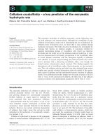
As for survival, high TB patients had significantly poor
as compared to low TB patients on both of DSS and RFS
(p < 0.001 in DSS, p = 0.001 in RFS, Fig. 2). From RFS
study, 11 patients with R2 resection (distant metastasis or
cancer positive surgical margin) were excluded. Median
survival time (MST) of DSS in high and low TB patients
was 19.2 and 56.4 months, respectively. MST of RFS in
high and low TB patients was 11.3 and 37.6 months, respectively. Interestingly, DSS after initial treatment in high
TB patients did not show statistical difference compared
to that in 28 unresected patients having locally advanced
tumor at our institution in the same period. When we
Ito et al. BMC Cancer
(2020) 20:209
Page 6 of 11
Fig. 2 Patient survival according to tumor budding. a Disease specific survival. b Recurrence free survival. On both disease specific survival (DSS)
and recurrence free survival (RFS), patients in high TB group had significantly poor survival compared to patients in low TB group (p < 0.001 in
DSS, p = 0.001 in RFS). Interestingly, DSS in high TB group did not show statistical difference compared to that in 28 unresected patients at our
institution in the same period (p = 0.103). *Eleven patients with R2 resection (distant metastasis or cancer positive surgical margin) were excluded
from RFS study
compared DSS and RFS between patients with TB5–9 and
those with TB 10 or more, there were no differences between two groups (Figure S).
Tumor budding was associated with advanced
histological features and poor survival in patients with
neoadjuvant therapy
To confirm the significance of TB in the patients who received neoadjuvant therapy (Table 3), we classified the 36
patients with neoadjuvant therapy into low TB (n = 25)
and high TB (n = 11), and classified the 42 patients without neoadjuvant therapy into low TB (n = 29) and high TB
(n = 13). Among the patients with neoadjuvant therapy,
high TB patients had a significantly higher rate of combined vascular resection (90.9% vs. 48.0%, p = 0.015) compared to low TB patients. In the patients without
neoadjuvant therapy, there were no significant differences
in pre- and intra-operative factors. In the patients with
neoadjuvant therapy, high TB patients, as compared to
low TB patients, had significantly higher rates of G3
(45.5% vs. 0%, p < 0.001), pT4 (63.6% vs. 24.0%, p = 0.023),
lymph node metastasis (72.7% vs. 32.0%, p = 0.023), and
distant metastasis (27.3% vs. 0%, p = 0.006). As for postoperative factors, there were no differences between the two
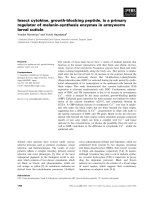
groups. Figure 3 shows patients survival according to TB
status in patients with or without neoadjuvant therapy. In
the patients with neoadjuvant therapy, high TB patients
had significantly poor survival as compared to low TB patients (p < 0.001 in DSS, p = 0.001 in RFS). In the patients
without neoadjuvant therapy, high TB patients had significantly poor DSS, as compared to Low TB patients, but
RFS had no significantly difference between two groups.
High tumor budding was an independent poor
prognostic factor by multivariate analysis for disease
specific survival
To identify predictors for DSS and to confirm the significance of TB, multivariate COX regression analysis
was performed. As shown in Table 4, pre-operative CEA
level (≥ 5 ng/ml), histological grade G3, T4, N1/2, M1,
LV invasion, non-curative resection, and High TB, were
identified as poor prognostic factors for DSS by univariate analysis. Multivariate analysis identified N1/2, LV invasion, non-curative resection, and High TB, as
independent significant poor prognostic factors for DSS.
In Fig. 4, we compared DSS according to independent
prognostic factors in the 78 patients. In all comparison
according to each factors (N1/2, LV invasion, noncurative resection, and high TB), patients with each
prognostic factors had significantly inferior survival
compared to those without it. Among four patient classifications, notably, DSS in only patients with high TB did
not show significantly difference compared to DSS in 28
unresected patients.
Discussion
In order to improve prognosis and implementation of
therapeutic strategies for patients with perihilar cholangiocarinoma, it is crucial to identify new significant
prognostic factors. In the present study, we first elucidated the prognostic significance of high TB (TB counts
≥5) at the tumor invasive front by analyzing our patient
database, including approximately half of patients having
received neoadjuvant therapy. In all patients, high TB
was significantly associated with advanced tumor status
including rates of pT4, pN1/2, M1, and histological
Ito et al. BMC Cancer
(2020) 20:209
Page 7 of 11
Table 3 Characteristics in the patients with or without neoadjuvant therapy
Patients with neoadjuvant therapy (n = 36)
Patients without neoadjuvant therapy (n = 42)
Factors
Low TB (n = 25)
High TB (n = 11)
p value
Low TB (n = 29)
High TB (n = 13)
p value
Age (y.o.)
70 (49–84)
69 (39–77)
0.520
67 (40–87)
69 (44–78)
> 0.999
Gender (Male / Female)
13 / 12
9/2
0.091
18 / 11
7/6
0.616
Chemotherapy
17 (68.0%)
8 (72.7%)
0.777
–
–
–
Chemoradiotherapy
8 (32.0%)
3 (27.3%)
4 (16.0%)
2 (18.2%)
0.871
8 (27.6%)
4 (30.8%)
0.833
CEA (ng/mL)
3.7 (1.3–14.6)
3.9 (0.9–16.3)
0.685
2.9 (0.6–38.4)
2.6 (0.7–28.0)
0.936
CA19–9 (U/mL)
61.5 (1.0–7898)
140.3 (1.0–1325)
0.435
87.5 (7.0–1115.7)
65.1 (1.0–9278)
0.872
CEA (ng/mL)
2.6 (0.9–9.6)
3.6 (1.1–24.4)
0.151
3.0 (0.7–32.2)
2.6 (0.5–32.3)
0.788
CA19–9 (U/mL)
33.2 (1.0–11,659)
151.3 (1.0–1158)
0.086
67.3 (13.7–977.2)
65.1 (1.0–9278)
Type of Neoadjuvant therapy
PTPE
Initial tumor marker
Preoperative tumor marker
Type of liver resectiona
0.936
0.114
No liver resection
0
0
5 (17.2%)
0
S1,5,6,7,8
9 (36.0%)
3 (27.3%)
10 (34.5%)
5 (38.5%)
S1,4,5,6,7,8
1 (4.0%)
1 (9.1%)
2 (6.9%)
3 (23.1%)
S1,2,3,4
9 (36.0%)
6 (54.5%)
7 (24.1%)
4 (30.8%)
S1,2,3,4,5,8
3 (12.0%)
1 (9.1%)
0
1 (7.7%)
Others
3 (12.0%)
0
5 (17.2%)
0
HPD
0
1 (9.1%)
0.126
3 (10.3%)
0
0.229
Combined HA and/or PV resection
12 (48.0%)
10 (90.9%)
0.015
11 (37.9%)
6 (46.2%)
0.616
Operation time (min)
625 (383–965)
672 (422–972)
0.261
597 (403–1030)
610 (435–746)
0.914
Blood loss (ml)
2212 (505–6916)
2170 (1459–6012)
0.612
1964 (166–9907)
1830 (587–3870)
0.727
25 / 0 (0%)
6 / 5 (45.5%)
< 0.001
26 / 3 (10.3%)
12 / 1 (7.7%)
0.787
19 / 6 (24.0%)
4 / 7 (63.6%)
0.023
23 / 6 (20.7%)
8 / 5 (38.5%)
0.226
Histological type
G1, 2 / G3
UICC 8th
pT: T0–3 / T4
pN: N0 / N1–2
17 / 8 (32.0%)
3 / 8 (72.7%)
0.023
16 /13 (44.8%)
4 / 9 (69.2%)
0.143
M: M0 / M1a
25 / 0 (0%)
8 / 3 (27.3%)
0.006
28 / 1 (3.4%)
11 / 2 (15.4%)
0.165
17 / 6 / 2 (0 / 2)
6 / 1 / 4 (3 / 1)
0.092
21 / 5 / 3 (1 / 2)
9 / 2 / 2 (2 / 0)
0.895
Postoperative hospital stay (days)
45 (13–161)
37 (17–136)
0.308
49 (25–117)
56 (18–325)
0.374
Postoperative complication ≥ grade IIIb
13 (52.0%)
6 (54.5%)
0.888
9 (31.0%)
4 (30.8%)
0.986
Postoperative chemotherapy
19 (76.0%)
9 (81.8%)
0.699
18 (62.1%)
10 (76.9%)
0.345
Residual tumor status
R0 / R1 / R2 (M1 / margin positive)
Patients in high TB groups had a significantly higher rate of combined vascular resection in patients received neoadjuvant therapy. In patients without
preoperative therapy, there were no significant differences in preoperative and operative factors. Among 36 patients with neoadjuvant therapy, the rates of G3,
T4, N1–2, and M1 in high TB group were significantly higher than those in low TB. As for postoperative factors, there were no differences between two groups in
both patients with and without neoadjuvant therapy
a
M1 include 3 patients with intrahepatic metastasis. b Clavien-Dindo classification
grade 3. Survival in patients with high TB was significantly inferior than that in patients with low TB. By
multivariate analysis, high TB was identified as one of
independent poor prognostic factors for DSS among 4
factors including regional lymph node metastasis, LV
invasion, and non-curative resection. Interestingly,
DSS in high TB group did not show statistical difference compared to that in unresected patients. In
addition, the impact of high TB in patients with neoadjuvant therapy showed similar results, withhigh TB
significantly associated with advanced tumor status
and poor prognosis.
Ito et al. BMC Cancer
(2020) 20:209
Page 8 of 11
Fig. 3 Patient survival according to tumor budding in patients with or without neoadjuvant therapy. a, b Disease specific survival (DSS) and
Recurrence free survival (RFS) in patients with neoadjuvant therapy. c, d DSS and RFS in patients without neoadjuvant therapy. In patients with
neoadjuvant therapy, patients in high TB group had significantly poor survival as compared to patients in low TB group (p < 0.001 in DSS, p =
0.001 in RFS). Similarly, in patients without neoadjuvant therapy, patients in high TB group had significantly poor DSS and had a tendency with
poor RFS, as compared to patients in low TB group (p = 0.004 in DSS, p = 0.127 in RFS). *6 patients with neoadjuvant therapy and 5 patients
without neoadjuvant therapy with R2 resection (distant metastasis or cancer positive surgical margin) were excluded from RFS study
Many studies have reported several prognostic factors,
such as presence of higher histological grade (G3), higher
T stage, lymph node metastasis, and positive surgical resection margin, associated with poor survival in resected
patients with cholangiocarcinoma [3, 22–24]. In the previous study on TB in extrahepatic cholangiocarcinoma,
Ogino, et al. [11] demonstrated high TB as an independent adverse prognostic factor in multivariate analysis,
along with higher T stage, lymph node metastasis, and
resected margin positive invasive carcinoma. The present
study similarly showed that high TB, N1/2, LV invasion,
and non-curative resection were identified independent
poor prognostic factors in all patients. Therefore, high TB
has potential to be a new pathological prognostic factor.
Evaluation of TB can easily provide useful feedback on
the patient’s clinical situation, which can then be easily
disseminated from pathologist to clinical physician,
because it can be examined in the H&E-stained specimens
as a part of conventional pathologic diagnosis. In the
present study, the number of TB was counted in a field
measuring 0.785 mm2 using a 20 times objective lens by
microscopy. The pathologist then decided on a “hot-spot”
location and calculated the TB counts, that were classified
into two groups by using 5 as a cut-off value. As for cutoff value, Okubo et al. classified patients according to ≥5
or < 5, whereas Ogino et al. [11] classified TB into three
grades: low-grade, 0–4 TB; intermediate-grade, 5–11 TB;
high-grade, TB. Meanwhile, in colorectal cancer and pancreatic ductal adenocarcinoma, other methods for evaluating evaluate TB has been reported [8, 25]. Several reports
used immunohistochemistry by cytokeratin to easily identify TB at stroma [8, 25]. Okubo et al. [12] demonstrated
the strong correlation between TB counts cytokeratinstained tissue and the H&E-stained tissue sections in
Ito et al. BMC Cancer
(2020) 20:209
Page 9 of 11
Table 4 Uni- and multivariate analysis for poor disease specific survival
Factors
Patient age (≥70 years)
Univariate analysis
Multivariate analysis
Hazard Ratio (95% CI)
p value
0.742 (0.385–1.430)
0.373
Hazard Ratio (95% CI)
p value
0.531 (0.241–1.319)
0.173
Gender (male)
0.779 (0.408–1.486)
0.779
Pre-operative CEA level (≥ 5 ng/ml)
2.071 (1.021–4.199)
0.044
Pre-operative CA 19–9 level (≥ 100 U/ml)
1.479 (0.767–2.852)
0.243
Histological grade: G3
3.350 (1.514–7.414)
0.003
1.145 (0.433–3.025)
0.785
T stage: T4
2.366 (1.258–4.452)
0.008
1.221 (0.598–2.492)
0.584
N stage: N1 or N2
2.111 (1.115–3.994)
0.022
2.354 (1.010–5.487)
0.047
M stage: M1
9.524 (3.434–26.411)
< 0.001
1.655 (0.481–5.689)
0.424
Lymphovascular invasion
7.654 (2.349–24.937)
0.001
5.307 (1.530–18.413)
0.009
Perinueral invasion
28.161 (0.546–1451.390)
0.097
Non-curative resection
2.792 (1.471–5.299)
0.002
2.456 (1.116–5.408)
0.026
High TB
4.493 (2.276–8.870)
< 0.001
5.206 (1.985–13.655)
0.001
Regional lymph node metastasis, lyphovascular invasion, non-curative resection, and High TB were identified as independent poor prognostic factors for DSS
cholangiocarcinoma. In colorectal cancer, evaluation of
TB in only H&E stained tissue is widely recognized and
performed [6]. Therefore, to easily evaluate TB, we considered using H&E stained tissue as a prognostic factor. In
the present study, there were no differences in DSS and
RFS between patients with TB5–9 and those with TB 10
or more, although further studies for cut-off value and
method of counting are warranted.
Many previous studies on various malignant tumors
have reported a correlation between high TB and advanced tumor. In cholangiocarcinoma, Ogino, et al. [11]
reported similar results to the current study: that the
high TB grade was associated with poor histological differentiation, higher pT factor, regional lymph node metastasis, and a higher rate of residual invasive tumor in
the resected margin. They considered that TB at the
tumor invasive front may cause cancer cell migration
through the extracellular matrix, invade lymphovascular
structures, and represent the first step towards cancer
metastasis. To progress to this point, cancer cells need
to change their own phenotype in a process known
as, epithelial-mesenchymal transition (EMT), which is
a multistep dynamic cellular phenomenon in which
epithelial cells lose their cell–cell adhesion and gain
migratory and invasive traits that are typical of mesenchymal cells [26]. In several reports, TB was found to
be associated with EMT [11, 27]. In addition, Ogino et al.
[11] have confirmed the correlation between TB and EMT
in cholangiocarinoma, demonstrating that TB counts are
significantly higher in EMT status in TB; the lowexpression of E-cadherin (epithelial marker) and highexpression of Vimentin (mesenchymal marker).
A noteworthy point of the present study, is the demonstration of the prognostic significance of TB in patients
with neoadjuvant therapy. There are several reports showing the significance of high TB in patients who underwent
neoajuvant therapy [7, 16, 17] for rectal and esophageal
carcinoma. Miyata et al. [7] showed that TB in the invasive
front of tumors was significantly correlated with prognosis
in 74 patients who received neoadjuvant chemotherapy
for advanced esophageal squamous cell carcinomas. In
their study, they discussed the mechanisms of TB formation. They speculated that TB in tumor received neoadjuvant chemotherapy for esophageal cancers may represent
cross-interaction between bone marrow-derived cells and
cancer cells in the invasive front. Several in vitro studies
demonstrated that bone marrow-derived cells, which are
recruited to the tumor invasion front through chemokine
signaling, promote tumor invasion and metastasis [28, 29].
In another study on the prognostic value of tumor budding in rectal cancer after neoadjuvant radiotherapy, Du
et al. [16] demonstrated that high grade TB was the major
factor affecting 5-year RFS. Meanwhile, Sannier et al. [17]
chose a more easily applicable technique for evaluation of
TB in patients who received neoadjuvant chemoradiotherapy for locally advanced rectal carcinoma without any cut
off. Consequently, the presence of TB, even in low numbers, is considered to have an adverse effect on outcome.
In our present study, there were no differences in TB
counts between patients with or without neoadjuvant
therapy. However, further studies are needed to clarify the
mechanism of TB formation in patients with neoadjuvant
therapy.
Interestingly, DSS in resected patients with high TB
did not show a significant difference compared to that in
unresected patients, suggesting that they may not have
better prognosis irrespective of whether they can achieve
R0 resection. For these patients, we should consider the
Ito et al. BMC Cancer
(2020) 20:209
Page 10 of 11
Fig. 4 Disease specific survival according to independent prognostic factors in the 78 patients. a N1/2 vs N0. b LV invasion positive (+) vs LV
invasion negative (−). c Non-curative resection vs curative resection d High TB vs Low TB. In all comparison according to each independent
prognostic factor, patients with each factor had significantly poor survival compared to those without it. DSS in patients with high TB did not
show significantly difference compared to DSS in unresected patients
necessity of additional peri-operative therapy. The TB
evaluation method employed in the present study, a
pathologist determined “hotspot,” is limited in that it
cannot evaluate TB before surgery. Therefore, there is
an urgent need to identify preoperative predictors of
high TB and establish new therapeutic strategies. This
should include improving surgical technique, as well as,
developing effective new preoperative and postoperative
adjunctive therapy.
Our present study included several limitations. This
study was retrospective study with a small number of
patients. In addition, the indications and types of neoadjuvant therapy in each patient were not uniform. However, it is noteworthy that TB was strongly associated
with poor prognosis even in small number cohort. Additional prospective studies are warranted.
Conclusion
Our present study demonstrated that high TB at the invasive front of tumors in resected perihilar
cholangiocarcinoma patients with or without neoadjuvant therapy, is strongly associated with advanced tumor
status and poor prognosis, including DSS/RFS. High TB
could be a novel prognostic factor in resected perihilar
cholangiocarcinoma even if patients received neoadjuvant therapy.
Supplementary information
Supplementary information accompanies this paper at />1186/s12885-020-6695-9.
Additional file 1: Figure S. Disease specific survival (DSS) and
recurrence free survival (RFS) according to TB counts. Both of DSS (A) and
RFS (B) did not show differences between patients with TB 5–9 and
those with TB 10 or more.
Abbreviations
CRT: Chemoradiotherapy; DSS: Disease-specific survival time; EMT: Epithelialmesenchymal transition; ERBD: Endoscopic retrograde biliary drainage;
ERC: Endoscopic retrograde cholangiography; HA: Hepatic artery;
IDUS: Intraductal ultrasonography; IRB: Institutional Review Board;
LV: Lymphovasclular; MDCT: Multidetector-row computed tomography;
MRCP: Magnetic resonance cholangiopancreatography; MST: Median survival
Ito et al. BMC Cancer
(2020) 20:209
time; PV: Portal vein; PVE: Portal vein embolization; RFS: Recurrence free
survival time; RT: Radiation therapy; TB: Tumor budding; UICC: Union for
International Cancer Control
Page 11 of 11
9.
10.
Acknowledgements
Not applicable.
11.
Authors’ contributions
NK had full access to all the data in the study and takes responsibility for the
integrity of the data and the accuracy of the data analysis. Concept and
design: TI, NK, YK, HK1, KI, DN, and SI. Acquisition, analysis, or interpretation
of data: TI, NK, YK, HK1, KI, and DN. Drafting of the manuscript. TI, NK, and SI.
Critical revision of the manuscript for important intellectual content: All
authors. Statistical analysis: TI, NK, YK, HK1, KI, DN, and SI. Administrative,
technical, or material support: YK, HK1, and AH. Supervision: AH, TF, YI, HK2,
AT, YM, MK, SM, MU, HS, and SI. 1. Haruna Komatsubara 2. Hiroyuki Kato. All
authors read and approved the final manuscript.
Funding
There was no funding to support this study.
12.
13.
14.
15.
16.
Availability of data and materials
The datasets used and/or analyzed during the current study are available
from the corresponding author on reasonable request.
Ethics approval and consent to participate
This study was reviewed and approved by the Mie University Institutional
Review Board (IRB#: H2018–064). Due to the retrospective nature without
identifiable patient information, the requirement for informed consent was
waived.
17.
18.
19.
Consent for publication
Not applicable.
20.
Competing interests
The authors declare that they have no competing interests.
Author details
Department of Hepatobiliary Pancreatic and Transplant Surgery, Mie
University Graduate School of Medicine, 2-174 Edobashi, Tsu, Mie 514-8507,
Japan. 2Pathology Division, Mie University Hospital, 2-174 Edobashi, Tsu, Mie
514-8507, Japan.
21.
1
Received: 15 January 2020 Accepted: 28 February 2020
References
1. DeOliveira ML, Cunningham SC, Cameron JL, et al. Cholangiocarcinoma:
thirty-one-year experience with 564 patients at a single institution. Ann
Surg. 2007;245(5):755–62.
2. Razumilava N, Gores GJ. Cholangiocarcinoma. Lancet. 2014;383(9935):2168–
79.
3. Nagino M, Ebata T, Yokoyama Y, et al. Evolution of surgical treatment for
perihilar cholangiocarcinoma: a single-center 34-year review of 574
consecutive resections. Ann Surg. 2013;258(1):129–40.
4. Groot Koerkamp B, Wiggers JK, Allen PJ, et al. Recurrence rate and pattern
of Perihilar Cholangiocarcinoma after curative intent resection. J Am Coll
Surg. 2015;221(6):1041–9.
5. Imai T. The growth of human carcinoma: a morphological analysis. Fukuoka
Igaku Zasshi. 1954;45:72–102.
6. Lugli A, Kirsch R, Ajioka Y, et al. Recommendations for reporting tumor
budding in colorectal cancer based on the international tumor budding
consensus conference (ITBCC) 2016. Mod Pathol. 2017;30(9):1299–311.
7. Miyata H, Yoshioka A, Yamasaki M, et al. Tumor budding in tumor invasive
front predicts prognosis and survival of patients with esophageal squamous
cell carcinomas receiving neoadjuvant chemotherapy. Cancer. 2009;115(14):
3324–34.
8. Karamitopoulou E, Zlobec I, Born D, et al. Tumour budding is a strong and
independent prognostic factor in pancreatic cancer. Eur J Cancer. 2013;
49(5):1032–9.
22.
23.
24.
25.
26.
27.
28.
29.
Ohike N, Coban I, Kim GE, et al. Tumor budding as a strong prognostic
indicator in invasive ampullary adenocarcinomas. Am J Surg Pathol. 2010;
34(10):1417–24.
Kai K, Kohya N, Kitahara K, et al. Tumor budding and dedifferentiation in
gallbladder carcinoma: potential for the prognostic factors in T2 lesions.
Virchows Arch. 2011;459(4):449–56.
Ogino M, Nakanishi Y, Mitsuhashi T, et al. Impact of tumour budding grade
in 310 patients who underwent surgical resection for Extrahepatic
Cholangiocarcinoma. Histopathology. 2019;74(6):861–72.
Okubo S, Mitsunaga S, Kato Y, et al. The prognostic impact of differentiation at
the invasive front of biliary tract cancer. J Surg Oncol. 2018;117(6):1278–87.
Tanaka M, Yamauchi N, Ushiku T, Shibahara J, Hayashi A, Misumi K,
Yasunaga Y, Morikawa T, Kokudo T, Arita J, Sakamoto Y, Hasegawa K,
Fukayama M. Tumor budding in intrahepatic Cholangiocarcinoma: a
predictor of Postsurgery outcomes. Am J Surg Pathol. 2019;43(9):1180–90.
Sumiyoshi T, Shima Y, Okabayashi T, et al. Chemoradiotherapy for initially
Unresectable locally advanced Cholangiocarcinoma. World J Surg. 2018;
42(9):2910–8.
Grendar J, Grendarova P, Sinha R, Dixon E. Neoadjuvant therapy for
downstaging of locally advanced hilar cholangiocarcinoma: a systematic
review. HPB (Oxford). 2014;16(4):297–303.
Du C, Xue W, Li J, Cai Y, Gu J. Morphology and prognostic value of tumor
budding in rectal cancer after neoadjuvant radiotherapy. Hum Pathol. 2012;
43(7):1061–7.
Sannier A, Lefèvre JH, Panis Y, Cazals-Hatem D, Bedossa P, Guedj N.
Pathological prognostic factors in locally advanced rectal carcinoma after
neoadjuvant radiochemotherapy: analysis of 113 cases. Histopathology.
2014;65(5):623–30.
Kuriyama N, Isaji S, Tanemura A, et al. Transhepatic Hilar approach for
Perihilar Cholangiocarcinoma: significance of early judgment of Resectability
and safe vascular reconstruction. J Gastrointest Surg. 2017;21(3):590–9.
Morizane C, Okusaka T, Mizusawa J, et al. Randomized phase II study of
gemcitabine plus S-1 versus S-1 in advanced biliary tract cancer: a Japan
clinical oncology group trial (JCOG 0805). Cancer Sci. 2013;104:1211–6.
Sasaki T, Isayama H, Nakai Y, et al. A randomized phase II study of
gemcitabine and S-1 combination therapy versus gemcitabine
monotherapy for advanced biliary tract cancer. Cancer Chemother
Pharmacol. 2013;71:973–9.
Ohkura Y, Mizuno S, Kishiwada M, et al. Benefit of technetium-99m
galactosyl human serum albumin scintigraphy instead of indocyanine green
test in patients scheduled for hepatectomy. Hepatol Res. 2014;44(10):E118–
28.
Kobayashi A, Miwa S, Nakata T, Miyagawa S. Disease recurrence patterns
after R0 resection of hilar cholangiocarcinoma. Br J Surg. 2010;97(1):56–64.
Sakamoto Y, Shimada K, Nara S, et al. Surgical management of infrahilar/
suprapancreatic cholangiocarcinoma: an analysis of the surgical procedures,
surgical margins, and survivals of 77 patients. J Gastrointest Surg. 2010;14(2):
335–43.
Komaya K, Ebata T, Yokoyama Y, et al. Recurrence after curative-intent
resection of perihilar cholangiocarcinoma: analysis of a large cohort with a
close postoperative follow-up approach. Surgery. 2018;163(4):732–8.
Karamitopoulou E, Zlobec I, Kölzer V, et al. Proposal for a 10-high-powerfields scoring method for the assessment of tumor budding in colorectal
cancer. Mod Pathol. 2013;26(2):295–301.
Kalluri R, Weinberg RA. The basics of epithelial-mesenchymal transition. J
Clin Invest. 2009;119(6):1420–8.
Masugi Y, Yamazaki K, Hibi T, Aiura K, Kitagawa Y, Sakamoto M. Solitary cell
infiltration is a novel indicator of poor prognosis and epithelialmesenchymal transition in pancreatic cancer. Hum Pathol. 2010;41:1061–8.
Karnoub AE, Dash AB, Vo AP, et al. Mesenchymal stem cells within tumour
stroma promote breast cancer metastasis. Nature. 2007;449(7162):557–63.
Kitamura T, Kometani K, Hashida H, et al. SMAD4-deficient intestinal tumors
recruit CCR1+ myeloid cells that promote invasion. Nat Genet. 2007;39(4):
467–75.
Publisher’s Note
Springer Nature remains neutral with regard to jurisdictional claims in
published maps and institutional affiliations.