Patterns of regional lymph node failure of locally advanced hypopharyngeal squamous cell carcinoma after first-line treatment with surgery and/or intensitymodulated radiotherapy
Bạn đang xem bản rút gọn của tài liệu. Xem và tải ngay bản đầy đủ của tài liệu tại đây (1.1 MB, 11 trang )
Wang et al. BMC Cancer
(2020) 20:283
/>
RESEARCH ARTICLE
Open Access
Patterns of regional lymph node failure of
locally advanced hypopharyngeal
squamous cell carcinoma after first-line
treatment with surgery and/or intensitymodulated radiotherapy
Dongqing Wang1,2, Shui Yu2, Limin Zhai2, Jin Xu2 and Baosheng Li1,2*
Abstract
Background: To identify the spatial patterns of regional lymph node failure of locally advanced hypopharyngeal
squamous cell carcinoma (SCC) after first-line treatment with surgery and/or intensity-modulated radiotherapy
(IMRT).
Methods: We retrospectively obtained the clinicopathological characters of 123 hypopharyngeal SCC patients, and
investigated the patterns of regional lymph node failure. Univariate and multivariate logistic regression were used
to determine the risk factors of regional lymph node failure.
Results: Forty patients (32.5% of total patients) were suffered regional lymph node failure. In these patients, the
ipsilateral neck level II nodal failure account for 55.0% (22/40) followed by level III 30.0% (12/40), level VIb 15.0% (6/
40), level VII 15.0% (6/40), and level IV 5.0% (2/40). In addition, 17.5% (7/40) patients suffered contralateral neck level
II nodal failure and 7.5% (3/40) patients suffered level III nodal failure. The common failure levels were the II (7/46,
15.2%), III (4/46, 8.7%), VIb (4/46, 8.7%), and VII (5/46, 10.9%) for treatment by surgery. The lymph node recurrence
and persistent disease at levels II (19/77, 24.7%) and III (10/77, 13.0%) remained the major cause of failure following
curative intent of IMRT. The postoperative radiation significantly decreased the risk of regional lymph node failure
(OR = 0.082, 95% CI: 0.007–1.000, P = 0.049); and the radiologic extranodal extension significantly increased the risk
of regional lymph node failure (OR = 11.07, 95% CI: 2.870–42.69, P < 0.001).
Conclusions: Whatever the treatment modality, the lymph node failure at level II and III was the most popular
pattern for hypopharyngeal SCC. Moreover, for patients who underwent surgery, the nodal failure at level VIb and
VII was frequent. Thus, postoperative radiation of level VIb and VII may give rise to benefit to locally advanced
hypopharyngeal SCC patients.
Keywords: Hypopharyngeal squamous cell carcinoma, Surgery, Radiotherapy, Chemotherapy, Failure pattern
* Correspondence:
1
Tianjin Medical University, Tianjin 300070, P.R. China
2
Department of Radiation Oncology, Shandong Cancer Hospital and Institute,
Shandong First Medical University and Shandong Academy of Medical
Sciences, Jinan 250117, P.R. China
© The Author(s). 2020 Open Access This article is licensed under a Creative Commons Attribution 4.0 International License,
which permits use, sharing, adaptation, distribution and reproduction in any medium or format, as long as you give
appropriate credit to the original author(s) and the source, provide a link to the Creative Commons licence, and indicate if
changes were made. The images or other third party material in this article are included in the article's Creative Commons
licence, unless indicated otherwise in a credit line to the material. If material is not included in the article's Creative Commons
licence and your intended use is not permitted by statutory regulation or exceeds the permitted use, you will need to obtain
permission directly from the copyright holder. To view a copy of this licence, visit />The Creative Commons Public Domain Dedication waiver ( applies to the
data made available in this article, unless otherwise stated in a credit line to the data.
Wang et al. BMC Cancer
(2020) 20:283
Background
Squamous cell carcinoma (SCC) of the hypopharynx is
relatively rare and accounts for 3 to 7% of all head and
neck cancers [1–3]. Notably, the hypopharyngeal SCC is
a lethal disease and the 10-year overall survival is only
13.8% [4, 5]. The poor prognosis of hypopharyngeal SCC
may result from the facts that early stage of hypopharyngeal SCC often fails to cause any signs or symptoms, and
this delays the diagnosis of hypopharyngeal carcinoma
[1–3]. Currently, the standard treatment for locally advanced hypopharyngeal SCC is multimodality treatment,
including induction chemotherapy, partial or total laryngopharyngectomy with lymph node dissection, and postoperative radiation or chemoradiation as dictated by
pathologic risk features, such as, positive margins or
extranodal extension (ENE) [6, 7]. As for advanced unresectable tumors, such as stage IVb diseases, and for patients requiring organ preservation, concurrent
radiotherapy (RT) and high-dose cisplatin is recommended treatment schedule in national comprehensive
cancer network (NCCN) guideline for cancer of hypopharynx [7].
The intensity-modulated radiotherapy (IMRT) plays
an important role as an adjunct to surgery or concurrent
with chemotherapy. Accurate target volume delineation
is critical to achieve favourable clinical outcomes. Recently, Biau et al. [8] updated the international consensus guidelines for the delineation of the neck node levels
of head and neck cancers. However, there is still no consensus on the extent to which prophylactic treatment regional nodal basin needs to be included in adjuvant
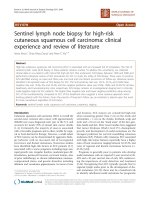
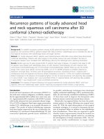
Fig. 1 The flow diagram for the inclusion
Page 2 of 11
IMRT. Moreover, the pattern of the lymph node failure
is still unclear in hypopharyngeal SCC patients.
In present study, we reported the follow-up results of
frequency and distribution of lymph node failure at each
nodal level for 123 patients with locally advanced hypopharyngeal SCC undergoing first-line treatment with
surgery and/or IMRT.
Methods
Population
Patients who were diagnosed as hypopharyngeal SCC
and confirmed by pathology at the Shandong Cancer
Hospital from January 2012 to November 2018 were
retrospectively reviewed. The inclusion criteria for the
present study were: (1) Clinical or pathological TNM
stage II–IVb according to AJCC 7th TNM classification
without distant metastasis, and (2) patients undergoing
radical surgery or IMRT. As indicated in Fig. 1, we excluded the patients (1) who presented with organ metastasis; (2) the radiation dose lower to 50Gy; and (3)
imaging studies unavailable for review at the time of initial treatment failure. The protocol of this study was
approved by the Institutional Review Board of the
Shandong Cancer Hospital.
Surgery treatment
Totally, 20 patients received total pharyngolaryngectomy
with unilateral neck dissection for 14 patients, bilateral
neck dissection for 6 patients. In addition, 20 patients
received partial pharyngolaryngectomy with unilateral
neck dissection for 15 patients, bilateral neck dissection
Wang et al. BMC Cancer
(2020) 20:283
for 5 patients. Notably, 6 patients were not treated by
laryngopharyngectomy after induction chemotherapy,
and only underwent the isolated unilateral neck dissection and postoperative RT. One patient could not be
treated by laryngopharyngectomy for his poor cardiopulmonary function. One patient with stage T1N2bM0 acquired disease progression for nodal disease, and
complete response for primary tumor after induction
chemotherapy. Four patients would like to preserve the
larynx and only removed the lymph node. The type of
performed neck dissection was selective dissection. Generally, neck dissection involved levels II, III, IV and V.
Level I and VIb were removed in partial patients according to tumor site, T stage, and lymph node metastasis
on preoperative imaging.
Radiotherapy treatment
A total 117 (95.1%) patients received IMRT during the
whole treatment procedure. Of these patients, 77 received chemoradiotherapy or definitive radiotherapy
alone, 40 received postoperative radiotherapy. Irradiation
is applied as a step and shoot IMRT technique using 6MV X ray in daily fractions of 1.8–2.2 Gy from Monday
to Friday. For definitive radiation therapy, the gross
tumor volume (GTV) encompass the primary tumor and
involved nodes. The clinical target volume (CTV) contains the GTV and areas of potential microscopic spread
as well as the lymph node areas for elective lymph node
irradiation. The planning target volume (PTV) accounts
for set-up variations by a margin of 0.3 cm. Generally,
we prescribe median dose 70Gy to gross tumor, 60Gy to
high-risk subclinical regions, 50Gy to low-risk subclinical regions. As for postoperative radiation therapy,
high-risk regions (CTVhigh) were given 60–66 Gy, and
the low-risk regions (CTVlow) 50 Gy in daily fractions of
1.8–2.0 Gy. The extension of the CTVs was defining by
radiation oncologists taking into account of clinical factors including TNM stage, number and distribution of
positive lymph nodes, size of metastatic lymph nodes,
extension of primary tumor beyond the midline, pathological resection status, and existence of extra-capsular
spread of nodal disease.
Page 3 of 11
days 1–4 infusion. Induction chemotherapy was administered in 1–3 cycles every 3 weeks.
Regional lymph node failure
The location of lymph nodes metastasis was divided into
several levels in the present study according to the
DAHANCA, EORTC, HKNPCSG, NCIC CTG, NCRI,
RTOG, TROG consensus guidelines for the delineation
of the neck node levels [9]: Ia, submental group; Ib, submandibular group; II, upper jugular group, and level II is
further subdivided into level IIa and level IIb by the posterior edge of the internal jugular vein; III, middle jugular group; IV, lower jugular group; V, posterior triangle
group, and level V is further subdivided into levels Va
(upper posterior triangle nodes) and Vb (lower posterior
triangle nodes) using the caudal edge of the cricoid cartilage as an anatomic landmark; VIa, anterior jugular
nodes; VIb, prelaryngeal, pretracheal, and paratracheal
nodes; VII, retropharyngeal nodes.
Local recurrence was defined as recurrence at the site of
the initial primary tumor, and regional failure was defined
as the development of recurrence in cervical lymph nodes.
Distant failure was defined as metastasis in an organ outside of the head and neck. The presence of failure was determined based on the information of a clinical evaluation,
systemic radiographic imaging and biopsy, and it was evaluated by Dongqing Wang and Shui Yu.
Lymphatic metastasis intensity (LMI) was used to describe as the ratio of the number of positive lymph
nodes to the number of examined lymph nodes. Lymphatic metastasis ratio (LMR) was defined as the ratio of
the number of patients with positive lymph node diagnosed by contrast-enhanced CT and/or magnetic resonance imaging divided by the number of the whole
population.
Follow-up
After completion of treatment, patients were followed by
every 3 months for the initial 3 years, and every 6 months
after 3 years. Progression-free survival (PFS) was considered as the time period from treatment completion to
the initial treatment failure.
Systemic therapy
Statistical analysis
Twenty-eight (22.8%) patients received concurrent chemoradiotherapy, of 25 patients received cisplatin 75 mg/
m2 as concurrent agents, 2 received nimotuzumab and 1
received cetuximab. Fifty-four patients (43.9%) received
induction chemotherapy. The induction chemotherapy
regimens were as follows: (1) cisplatin 75 mg/m2 plus 5fluorouracil (5-FU) 750–1000 mg/m2/d from days 1–4
infusion, (2) docetaxel 75 mg/m2 on day 1 plus cisplatin
75 mg/m2. Three patients received cisplatin 75 mg/m2
plus docetaxel 75 mg/m2 plus 5-FU 500 mg/m2/d from
Statistical analysis was performed using the SPSS statistical software, version 20.0 (IBM Corporation, Armonk,
NY, USA). LMI and LMR were presented as the frequencies and percentages. The mean PFS were determined by the Kaplan-Meier curve (log-rank test). The
lymph node recurrence rate at respective level in patient
treated with surgery, with or without postoperative RT
was analyzed by Chi-square test. The univariate and
multivariate logistic regression were used to determine
the risk factors of lymph nodal failure. The factors
Wang et al. BMC Cancer
(2020) 20:283
Page 4 of 11
Table 1 Patient, disease, and treatment characteristics
Characteristics
N (%)
Characteristics
Age, years
Median (range)
58 (41–82)
Female
Bilateral
N (%)
11 (8.9%)
Intensity-modulated radiotherapy
Gender
Male
Table 1 Patient, disease, and treatment characteristics
(Continued)
118 (95.9%)
Postoperative
40 (32.5%)
5 (4.1%)
Definitive
77 (62.6%)
Chemotherapy
Tumor site
Pyriform sinus
106 (86.2%)
Induction
54 (43.9%)
Posterior pharyngeal wall
9 (7.3%)
Concurrent
28 (22.8%)
Postcricoid area
8 (6.5%)
TNM stage
II
7 (5.7%)
III
25 (20.3%)
IVa
83 (67.5%)
IVb
8 (6.5%)
Clincal T stage
Results
Clinicopathological characters
T1
7 (5.7%)
T2
19 (15.4%)
T3
29 (23.6%)
T4
28 (22.8%)
Pathological T stage
T1
5 (4.1%)
T2
12 (9.8%)
T3
10 (8.1%)
T4
13 (10.6%)
Clincal N stage
N0
12 (9.8%)
N1
16 (13.0%)
N2a
2 (1.6%)
N2b
25 (20.3%)
N2c
19 (15.4%)
N3
3 (2.4%)
The clinicopathological characters were summarized in
Table 1. The median age was 58 years (range, 41 to 82
years) and majority was males (95.9%). Hypopharyngeal
subsite was piriform sinus in 106 cases (86.2%), posterior
hypopharyngeal wall in 9 (7.3%), and retrocricoid in 8
(6.5%). According to AJCC 7th criteria, clinical or pathological staging were 7 (5.7%) for stage II, 25 (20.3%) for
stage III and 91 (74.0%) for stage IV. One hundred and
seventeen (95.1%) patients underwent IMRT, 40 (32.5%)
postoperatively and 77 (62.6%) definitively. Forty patients (32.5%) received total or partial pharyngolaryngectomy with neck dissection, 6 received isolated
unilateral neck dissection. Twenty-eight (22.8%) patients
received concurrent chemoradiotherapy, and 54 (43.9%)
received induction chemotherapy. In addition, we summarized the treatment schedule based on the T and N
classification (Table 2).
Metastasis of lymph node
Pathological N stage
N0
6 (4.9%)
N1
7 (5.7%)
N2a
1 (0.8%)
N2b
26 (21.1%)
N2c
5 (4.1%)
N3
1 (0.8%)
Surgery
Total laryngopharyngectomy and neck dissection
20 (16.3%)
Partial laryngopharyngectomy and neck dissection
20 (16.3%)
Isolated neck dissection
6 (4.9%)
Node dissection
Ipsilateral
included age, TNM stage, adjuvant treatment with postoperative RT and chemotherapy, and radiologic extranodal extension (rENE). P values of < 0.05 indicated
significant difference.
35 (28.5%)
Forty-six neck dissections were performed: 35 ipsi- and
11 bi-lateral, 1148 lymph nodes were analyzed. A total
of 169 nodes in 40 (40/46, 86.9%) patients confirmed
lymphatic metastasis, the overall LMI was 14.7% (169/
1148). The LMI for ipsilateral neck was 16.4% (160/976),
whereas, only 5.2% (9/172) for contralateral neck. In
addition, we evaluated the LMI based on the level of
lymph node. We observed that the LMI was 20.0% (14/
70) for level II, 14.9% (7/47) for level III, 5.9% (3/51) for
level IV, 0% (0/27) for level V, 8.0% (2/25) for level VI,
and 0% (0/5) for level VII. Seventy-seven patients did
not receive resection of neck, and the LMR were 66.2%
(51/77) for level II, 48.1% (37/77) for level III, 13.0% (10/
77) for level IV, 5.2% (4/77) for level V, 13.0% (10/77)
for level VI, and 15.6% (12/77) for level VII.
Wang et al. BMC Cancer
(2020) 20:283
Page 5 of 11
Table 2 Treatment schedule by clinical/pathological T and N
classification
T
N
Cases
Treatment schedule (N, %)
T1–2
N0
7
S ± RT (3, 42.8%)
RT (2, 28.6%)
IC + RT (2, 28.6%)
T1–2
N1–3
36
IC + S + RT (2, 5.6%)
IC + RT/CRT (14, 38.9%)
S ± RT/CRT (14, 38.9%)
RT/CRT (6, 16.6%)
T3
N0–3
39
IC + S + RT (4, 10.3%)
IC + RT/CRT (16, 41.0%)
S + RT/CRT (9, 23.1%)
RT/CRT (10, 25.6%)
T4
N0–3
41
IC + S + RT (2, 4.9%)
IC + RT/CRT (14, 34.1%)
S ± RT/CRT (12, 29.3%)
RT/CRT (13, 31.7%)
Note: S Surgery, RT Radiotherapy, CRT Chemoradiotherapy, IC
Induction chemotherapy
Regional lymph node failure
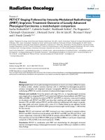
All patients were followed up for median time 12 months
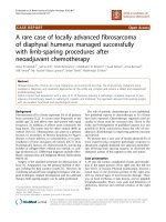
(3–84 months). The median PFS rates were 13 months
(95% CI 6.4–19.6 months) for surgery treatment, and 11
months (95% CI 9.1–12.9 months) for non-surgery treatment, no significant difference was observed (P = 0.732)
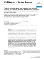
(Fig. 2). For all patients, local recurrence, cervical lymph
node failure, and distant metastasis accounted for 13.0%
(16/123), 32.5% (40/123), and 13.8% (17/123), respectively.
Of the cervical lymph node failure, 26 patients were isolated regional lymph node failure, 9 were both nodal failure and local recurrence, and 5 were both nodal failure
and distant metastasis (Fig. 3). The second primary cancers were found in 19 patients (15.4%), with esophagus
cancer 18 patients, and lung cancer one patient.
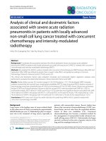
Of the 40 regional nodal failures, failures involved ipsilateral neck level II in 22 patients (55.0%), III in 12 patients (30.0%), IV in 2 patients (5%), VIb and VII both in
6 patients (15.0%). The nodal failures involved contralateral neck level II in 7 patients (17.5%), III in 3 patients
(7.5%). Furthermore, respective one patient was found
nodal failure at level Ib and Va in ipsilateral neck, and
level VIb and VII in contralateral neck (Fig. 4). Notably,
another one patient occurred axillary lymphatic failure
accompanied by bone metastasis. For patients undergoing surgery, the most commonly failure levels were the
II (7/46, 15.2%), III (4/46, 8.7%), VIb (4/46, 8.7%), and
VII (5/46, 10.9%). The detailed results of lymph node recurrence at respective level was reported in Table 3 for
patients undergoing surgery with or without postoperative RT. The rate of lymph node failure at levels II, III,
VIb, and VII was observed higher for patients who did
not receive postoperative irradiation (Fig. 5), however,
probably because of small sample size (N = 6), borderline
significant difference was observed at level VII (33.3% vs.
Fig. 2 Progression-free survival of hypopharyngeal carcinoma
following radical surgery and/or radiotherapy
7.5%, P = 0.058, OR = 0.162, 95% CI: 0.021–0.128), and
no significant difference at level III (Table 3). In contrast, for patients undergoing IMRT, the most commonly failure levels were the II (19/77, 24.7%), and III
(10/77, 13.0%), then followed by VIb (2/77, 2.6%), VII
(1/77, 1.3%), and IV (1/77, 1.3%).
Risk factors for lymph node failure
Table 4 showed the risk factors of lymph node failure
for patients treated by surgery. The postoperative
Fig. 3 Patterns of failure of hypopharyngeal carcinoma following
radical surgery and/or radiotherapy
Wang et al. BMC Cancer
(2020) 20:283
Page 6 of 11
Fig. 4 The spatial patterns of lymph node failure of hypopharyngeal carcinoma following radical surgery and/or radiotherapy
radiation strongly associated with lower risk nodal failure
(OR = 0.086, 95% CI: 0.009–0.814, P = 0.012), and pathologic N stage had a trend towards significance on univariate analysis (OR = 0.218, 95% CI: 0.042–1.142, P = 0.057).
In multivariate analysis, non postoperative radiation was
an independent risk factor (OR = 0.082, 95% CI: 0.007–
1.000, P = 0.049). Table 5 reported the radiologic extranodal extension (OR = 11.07, 95%: CI 2.870–42.69, P < 0.001)
was significantly increased the lymph node recurrence and
persistence for patients treated by IMRT.
Discussion
Our results demonstrate that 47.2% of the hypopharyngeal SCC patients were found local-regional failure and
distant metastasis with median time to the initial treatment failure was 13 months (95% CI 6.4–19.6 months)
for surgery, and 11 months (95% CI 9.1–12.9 months)
for IMRT. The most commonly failures in hypopharyngeal SCC are mainly attributed to cervical lymph node
failure, account for 32.5% of patients.
It is well know that hypopharyngeal carcinoma characterized by aggressive clinical behavior and high risk tendency to invade cervical lymph nodes. The lymph node
metastasis is an important prognostic factor, therefore,
control of regional metastasis is an essential part of
treatment for hypopharyngeal cancer. Presently, there is
no agreement on the best treatment approach for hypopharyngeal SCC. Definitive chemoradiation strategy
arose from the RTOG 91–11 trial [10, 11] which demonstrated improved loco-regional control and laryngeal
preservation rates has become an important approach
for locally advanced hypopharyngeal cancer. By means
of prophylactic neck irradiation (PNI), the incidence of
nodal failure can be reduced to 4% in head and neck
Table 3 The lymph node recurrence (N, %) at respective level in patient with hypopharyngeal carcinoma treated by surgery with or
without postoperative RT
Nodal level
Postoperative RT
N = 40
Non postoperative RT
N =6
Total
N = 46
iIb
0
1 (16.7%)
1 (2.2%)
P value
Odds ratio (95% CI)
iII
3 (7.5%)
2 (33.3%)
5 (10.9%)
0.058
0.162 (0.021–0.128)
iIII
1 (2.5%)
1 (16.7%)
2 (4.3%)
0.113
0.128 (0.007–2.387)
iIV
1 (2.5%)
0
1 (2.2%)
iVa
1 (2.5%)
0
1 (2.2%)
iVIb
2 (5.0%)
2 (33.3%)
4 (8.7%)
0.022
0.105 (0.011–0.964)
iVII
3 (7.5%)
2 (33.3%)
5 (10.9%)
0.058
0.162 (0.021–0.128)
cII
1 (2.5%)
3 (50.0%)
4 (8.7%)
< 0.001
0.026 (0.002–0.328)
cIII
2 (5.0%)
1 (16.7%)
3 (6.5%)
0.280
0.263 (0.020–3.456)
cIV
0
0
0
cV
0
0
0
cVIb
1 (2.5%)
0
1 (2.2%)
cVII
1 (2.5%)
0
1 (2.2%)
Note: i ipsilateral neck, c contralateral neck, RT Radiotherapy
Wang et al. BMC Cancer
(2020) 20:283
Page 7 of 11
Fig. 5 The lymph node recurrence rate at respective level for hypopharyngeal carcinoma undergoing surgery with or without postoperative
radiotherapy (RT)
cancers [12]. Therefore, PNI is an important IMRT component in the treatment of hypopharyngeal cancer.
In the present study, nodal involvement mainly concerned levels II (66.2%) and III (48.1%), then followed by
levels IV (13.0%), VI (13.0%), and VII (15.6%), while level
V showed involvement in 5.2% of patients. As comparing
with ipsilateral neck, the risk of metastasis for contralateral neck tend to be lower (LMI: 16.4% vs. 5.2%). These
results are in agreement with our previous study and the
literature [13]. However, few studies have reported the
outcomes of regional lymph node failure for locally advanced hypopharynx SCC after treatment with IMRT.
Sommat et al. [14] reported a retrospective analysis of
58 patients (III–IVb 95%) with hypopharyngeal cancer
treated with curative intent RT. In Sommat’s study, 88%
of patients managed to achieve complete response 3
Table 4 Univariate and multivariate analysis of lymph node failure in patient with hypopharyngeal carcinoma treated by surgery
(N = 46)
Variable
Ipsilateral nodal failure
N = 10
Contralateral/Bilateral
nodal failure
N =6
41–58
8
6
58–82
2
0
Age (years)
TNM stage
II
0
0
III-IV
10
6
T1–2
4
2
T3–4
6
4
Pathologic T stage
Pathologic N stage
N0–1
0
2
N2–3
10
4
Yes
8
3
No
2
3
Postoperative radiation
Chemotherapy
Yes
5
4
No
5
2
Univariate
Odds ratio (95% CI)
P value
Multivariate
Odds ratio (95% CI)
P value
2.852 (0.666–12.22)
0.149
1.727 (0.348–8.563)
0.503
1.654 (1.299–2.106)
0.170
1.045 (0.508–2.151)
0.905
0.862 (0.256–2.894)
0.809
1.126 (0.054–23.63)
0.939
0.218 (0.042–1.142)
0.057
0.201 (0.006–6.691)
0.370
0.086 (0.009–0.814)
0.012
0.082 (0.007–1.000)
0.049
1.008 (0.299–3.403)
0.989
1.559 (0.311–7.813)
0.589
Wang et al. BMC Cancer
(2020) 20:283
Page 8 of 11
Table 5 Univariate and multivariate analysis of lymph node failure in patient with hypopharyngeal carcinoma treated by intensitymodulated radiotherapy (N = 77)
Variable
Ipsilateral nodal failure
N = 21
Contralateral/Bilateral nodal failure
N=3
Age (years)
41–58
12
2
58–82
9
1
II
0
0
III
5
0
IV
16
3
T1
3
1
T2
5
0
T3
8
2
T4
5
0
TNM stage
Clinical T stage
Clinical N stage
N0
1
0
N1
5
0
N2
13
3
N3
2
0
Yes
11
1
No
10
2
rENE
Chemotherapy
Yes
17
3
No
4
0
Univariate
Odds ratio
(95% CI)
P value
Multivariate
Odds ratio
(95% CI)
P value
1.488 (0.555–3.992)
0.429
1.777 (0.545–5.797)
0.340
1.104 (1.012–1.204)
0.120
0.875 (0.141–5.420)
0.886
1.520 (0.548–4.215)
0.420
1.414 (0.749–2.670)
0.285
0.627 (0.222–1.772)
0.377
0.812 (0.225–2.937)
0.751
12.25 (3.353–44.75)
< 0.001
11.07 (2.870–42.69)
< 0.001
0.761 (0.200–2.894)
0.668
0.500 (0.106–2.359)
0.381
Note: rENE Radiologic extranodal extension
months after completion of treatment, loco-regional recurrence remained the major cause of failure following
curative intent RT. Most deaths occurred in patients
who succumbed to loco-regional rather than systemic
failure. However, only 50% of patients undergone IMRT
in Sommat’s study, half part of patients treated using a
2-dimensional technique. Daly et al. [15] recruited 42
patients with newly diagnosed SCC of hypopharynx (23
patients) and larynx (19 patients) underwent IMRT, 11
postoperatively and 31 definitively at Stanford University
Medical Center. Median follow-up was 30 months, 5 patients developed a loco-regional failure or had persistent
disease, with a median time to failure of 12.1 months.
Three local failures occurred within the high-dose region
and 3 occurred in regional nodes. No marginal misses
were observed. The author considered that loco-regional
relapses occurred in the high-dose volumes, suggesting
that target volume delineation was adequate but further
dose-escalation and more aggressive treatment may be
needed. Huang et al. [16] retrospectively reviewed 47 patients with locally advanced resectable SCC of
hypopharynx underwent primary surgery or definitive
IMRT with concurrent platinum-based chemotherapy
(CCRT). The 5-year survival rate, disease-free survival,
and loco-regional progression-free survival of surgery
and CCRT group was 33 and 56%, 25 and 41%, 15 and
53%, respectively. Loco-regional progression was the
main cause of failure in both groups. Eleven patients had
neck failure; 8 in the ipsilateral neck, 2 in the contralateral neck, and 1 in the tracheostoma site. All were infield failure in the PTV2 (60Gy). One retrospective study
[17] reported by Chun et al. included 54 patients receiving definitive radiotherapy with or without chemotherapy. Thirty patients received IMRT and 24 patients
received three dimensional conformal radiotherapy.
With median follow-up time 42.3 months, there were 20
loco-regional failures discovered. Estimated crude locoregional recurrence free survival at 3 years were 64.1%.
Of the 20 loco-regional failures, 14 were isolated local
failures, 4 were isolated regional nodal failures, and 2
were both. Of the 6 regional nodal failures, failures involved ipsilateral neck level II in 3 patients, ipsilateral
Wang et al. BMC Cancer
(2020) 20:283
neck level III in 1 patient, paraesophageal lymph node in
1 patient, and bilateral neck level II in 1 patient. Among
the loco-regional failures, 17 were observed in the PTV
high region, while 2 were in the PTV intermediate region and 1 patient had out-of-feld failure (paraesophageal lymph node), but was also accompanied by local
failure within the PTV High region. Pignon et al. [18]
found that IMRT failure in the low-neck supraclavicular
field was very uncommon.
Our center has employed IMRT for the definitive
treatment of head and neck cancers nearly for 10 years.
Our study results demonstrated the poor outcome expected in hypopharyngeal cancer with median PFS rates
were approximately 1 year after first-line treatment. The
regional cervical lymph node recurrence and persistent
disease remained the major cause of failure following
curative intent of IMRT. Approximately 70% of nodal
failures were observed in the PTV high or intermediate
regions. In our study, the most commonly failure levels
were the II (24.7%), and III (13.0%). However, the nodal
failures at level IV, VIb and VII was uncommon, the rate
of nodal failure only 1.3–2.6%. In our study, lymph node
failure was mostly involved in ipsilateral neck, only 2 patients developed isolated level II failure in contralateral
neck, and one patient developed level II failure in bilateral necks. Regarding our patients received IMRT enrolled in this study, more than half of patients have
severe lymph node involvement and were not suitable
candidates for selective lymph node dissection. Approximately 80% of them displayed lymph node metastasis
with liquefactive necrosis in lymph nodes. After completion of IMRT treatment, majority of them in our cohort
presented nodal residue. In our study, ENE with radiological evidence was observed significantly associated
with lymph node recurrence and persistent diseases. In
the recently released eighth edition of the AJCC TNM
staging, ENE has been added as a prognostic variable for
regional lymph node metastasis in addition to the number and size of metastatic lymph nodes [19]. Pitifully, because of extra capsular extension (for example vessels
and soft tissue invasion), or nodal failures accompanied
by local recurrence or distant metastasis, or severe late
treatment toxicities, ultimately only 2 patients received a
salvage node dissection within 6 months of follow-up
time. Aside from 5 patients with local-regional failure received salvage surgery after definitive radiotherapy, most
patients were received chemotherapy or combining with
targeted therapy. Chun et al. [17] suggest that salvage
surgery after definitive radiotherapy should be considered for patients who show residual disease after 6
months, because residual tumors show progression soon
after 6 months.
In patients undergoing surgical resection with or without postoperative adjuvant IMRT. Seventeen patients
Page 9 of 11
were observed regional lymph node failure, 10 of them
were isolated nodal failure, 4 patients accompanied by
local recurrence, and 3 patients accompanied by distant
metastasis (one patients occurred axillary lymphatic and
scapula metastasis). Of the 16 patients with nodal failure,
failures involved level II in 7 patients, levels III and VIb
both in 4 patients, level VII in 5 patients. Furthermore,
nodal failure involved in ipsilateral neck level IV and V
was both one patient.
Regarding 46 patients undergone lymph node dissection with 35 ips- and 11 bilateral neck dissection in this
study. Six of them observed contralateral neck failure,
with level II in 4 patients, level III in 3 patients, level
VIb and VII both in one patient. Among these 6 patients, 3 patients had received postoperative radiation
with radiation dose of 50–66Gy. Previously multi-center
randomized clinical trials have confirmed post-operative
radiation or chemoradiation improves loco-regional control and overall survival in the presence of extracapsular nodal extension [6, 7]. Although we fail to analyzed the correlation of pathologic ENE with node failure
after surgery in our study, we found that the most commonly failure levels were the II (15.2%), III (8.7%), VIb
(8.7%), and VII (10.9%). Comparing with patients receiving definitive radiotherapy, node failure rates at levels II
and III were lower for patients receiving surgery as firstline treatment (15.2% vs. 24.7%; 8.7% vs. 13.0%),
whereas, node failure at levels VIb and VII were exhibited higher (8.7% vs. 2.6%; 10.9% vs. 1.3%). The reason
probably because the selective neck dissection always included the nodes in level II and III, whereas, the nodes
in level VI and VII failed to remove from patients routinely in our study.
One retrospective study [13] include larynx (110 patients) and hypopharynx (26 patients) SCC undergoing
total laryngectomy or pharyngolaryngectomy with neck
dissection. Levels IIa and III were invaded in 28.7 and
25.7% of patients, respectively. Level VIb lymph-node involvement was 23.8% in patients who underwent level
VIb neck dissection. Lymph-node recurrence rate was
10.3% in levels II to IV, and 13.2% in VIb. The author
concluded that because high rate of involvement and recurrence of level VIb, systematic elective bilateral neck
dissection might be needed. Previous retrospective studies [20, 21] indicated that pyriform sinus apex or postcricoid invasion, or tumor diameter exceeding 3.5 cm
showed a trend in favor of paratracheal lymph node involvement. In our previous study, esophagus invasion
was also highly correlated with increased risk of developing level VIb metastasis. It is noteworthy that lymph
node at level VII (retropharyngeal lymph node) can not
be removed routinely by surgery, and hardly be detected
by imaging before surgery. Currently, there is no consensus regarding the delineation of lymphatic clinical target
Wang et al. BMC Cancer
(2020) 20:283
volume for post-operative radiation therapy for hypopharyngeal cancer. In present study, we found that not
receiving postoperative radiation therapy was strongly
associated with higher risk nodal failure. Five in 6 patients who failed to receiving postoperative radiation occurred nodal failure. Compared to the patients who
received postoperative RT, the lymph node recurrence
rate of level VII and VIb in ipsilateral neck was higher in
patients who did not recevive postoperative RT (33.3%
vs. 7.5%, P = 0.058; 33.3% vs. 5.0%, P = 0.022, Fig. 5). Furthermore, three patients (50.0%) occured nodal failure at
level II in contralateral or bilateral necks for patients not
receiving adjuvant radiation therapy, which was much
higher than patients who recevive postoperative RT
(50.0% vs. 2.5%, P < 0.001; OR = 0.026, 95%CI: 0.002–
0.328). Based on results found in our study, irradiation
of the level VIb and VII should be recommended, especially for the primary tumors originated from posterior
pharyngeal wall (PPW), PPW invasion, postcricoid invasion, and esophagus invasion [22, 23].
The limitations of our study include its retrospective
nature. The follow up time is relatively short. We did
not perform the dosimetric analysis of the patterns of
failure, and fail to confirm if CTV delineation is adequate. The prognosis associated factors, including the
evaluation of the surgical margins, perineural invasion
for hypopharyngeal cancer could not be taken into
account.
Conclusions
Based on our results, we concluded that whatever the
treatment modality, levels II and III in ipsilateral neck
were most commonly failure regions. The regional cervical
lymph node recurrence and persistent disease remained
the major cause of failure following curative intent of definitive IMRT. Because of high rate of node failure of level
VIb and VII after surgery, post-operative radiation field
should be include these territories, particularly in the setting of locally advanced disease. Our results provide a
clear rationale for efforts in the future aimed at improving
local-regional control, which including accurate target volume delineation, optimal prescribed radiation dose and
fraction, possibly identification areas of radio-resistance
within the tumour. Further clinical research is needed to
assess the utilization of IMRT combined with novel systemic agents in locally advanced hypopharyngeal SCC.
Abbreviations
SCC: Squamous cell carcinoma; LMR: Lymphatic metastasis ratio;
LMI: Lymphatic metastasis intensity
Acknowledgements
The authors thank Dr. Xianbin Zhang from Shandong Cancer Hospital and
Institute for language editing. The authors have obtained permission from
Dr. Zhang.
Page 10 of 11
Authors’ contributions
DQ W participate in imaging analysis and drafting the article. LM Z and BS L
participated in the design of the study. SY and JX participated in clinical
follow-up work. All authors have read and approved the manuscript, and
consent for publication.
Funding
Data collection and writing in this work was supported by the National
Natural Science Foundation of China (grant numbers 81530060 and
81874224).
Availability of data and materials
We declared that the materials and data of this study are available from the
first author on reasonable request.
Ethics approval and consent to participate
This study protocol was approved by Shandong Cancer Hospital ethics
committee. The written informed consent to participate was given.
Consent for publication
Not applicable.
Competing interests
The authors declare that they have no competing interest.
Received: 5 September 2019 Accepted: 26 March 2020
References
1. Global Burden of Disease Cancer Collaboration, Fitzmaurice C, Allen C,
Barber RM, Barregard L, Bhutta ZA, Brenner H, et al. Global, Regional, and
National Cancer Incidence, Mortality, Years of Life Lost, Years Lived With
Disability, and Disability-Adjusted Life-years for 32 Cancer Groups, 1990 to
2015: A Systematic Analysis for the Global Burden of Disease Study. JAMA
Oncol. 2017;3(4):524–48.
2. Parkin DM, Bray F, Ferlay J, Pisani P. Global cancer statistics, 2002. CA Cancer
J Clin. 2005;55(2):74–108.
3. Garneau JC, Bakst RL, Miles BA. Hypopharyngeal cancer: a state of the art
review. Oral Oncol. 2018;86:244–50.
4. Chung EJ, Jeong WJ, Jung YH, Kwon SK, Kwon TK, Ahn SH, Sung MW, Keam
B, Heo DS, Kim JH, Wu HG, Lee KW, Eom KY, Rho YS. Long-term oncological
and functional outcomes of induction chemotherapy followed by (chemo)
radiotherapy vs definitive chemoradiotherapy vs surgery-based therapy in
locally advanced stage III/IV hypopharyngeal cancer: multicenter review of
266 cases. Oral Oncol. 2019;89:84–94.
5. Lefebvre JL, Andry G, Chevalier D, Luboinski B, Collette L, Traissac L, de
Raucourt D, Langendijk JA. EORTC head and neck Cancer group. Laryngeal
preservation with induction chemotherapy for hypopharyngeal squamous
cell carcinoma: 10-year results of EORTC trial 24891. Ann Oncol. 2012;23(10):
2708–14.
6. Argiris A, Karamouzis MV, Raben D, Ferris RL. Head and neck cancer. Lancet.
2008;371(9625):1695–709.
7. Pfister DG, Spencer S, Adelstein D, et al.. NCCN Clinical Practice Guidelines
in Oncology (NCCN Guidelines) Head and Neck Cancer Version 1. 2018.
www.nccn.org. Accessed May 19, 2018.
8. Biau J, Lapeyre M, Troussier I, Budach W, Giralt J, Grau C, Kazmierska J,
Langendijk JA, Ozsahin M, O'Sullivan B, Bourhis J, Grégoire V. Selection of
lymph node target volumes for definitive head and neck radiation therapy:
a 2019 update. Radiother Oncol. 2019;134:1–9.
9. Grégoire V, Ang K, Budach W, Grau C, Hamoir M, Langendijk JA, Lee A, Le
QT, Maingon P, Nutting C, O'Sullivan B, Porceddu SV, Lengele B. Delineation
of the neck node levels for head and neck tumors: a 2013 update.
DAHANCA, EORTC, HKNPCSG, NCIC CTG, NCRI, RTOG, TROG consensus
guidelines. Radiother Oncol. 2014;110(1):172–81.
10. Forastiere AA, Zhang Q, Weber RS, Maor MH, Goepfert H, Pajak TF, et al.
Long-term results of RTOG 91-11: a comparison of three nonsurgical
treatment strategies to preserve the larynx in patients with locally advanced
larynx cancer. J Clin Oncol. 2013;31:845–52.
11. Forastiere AA, Goepfert H, Maor M, Pajak TF, Weber R, Morrison W, et al.
Concurrent chemotherapy and radiotherapy for organ preservation in
advanced laryngeal cancer. N Engl J Med. 2003;349:2091–8.
Wang et al. BMC Cancer
(2020) 20:283
12. Rabuzzi DD, Chung CT, Sagerman RH. Prophylactic neck irradiation. Arch
Otolaryngol. 1980;106(8):454–5.
13. Rivière D, Mancini J, Santini L, Giovanni A, Dessi P, Fakhry N. Lymph-node
metastasis following total laryngectomy and total pharyngolaryngectomy
for laryngeal and hypopharyngeal squamous cell carcinoma: frequency,
distribution and risk factors. Eur Ann Otorhinolaryngol Head Neck Dis. 2018;
135(3):163–6.
14. Sommat K, Yong SK, Fong KW, Tan TW, Wee JT, Soong YL. A 13-year single
institutional experience with definitive radiotherapy in Hypopharyngeal
Cancer. Ann Acad Med Singap. 2017;46(1):32–6.
15. Daly ME, Le QT, Jain AK, Maxim PG, Hsu A, Loo BW Jr, Kaplan MJ, Fischbein
NJ, Colevas AD, Pinto H, Chang DT. Intensity-modulated radiotherapy for
locally advanced cancers of the larynx and hypopharynx. Head Neck. 2011;
33(1):103–11.
16. Huang WY, Jen YM, Chen CM, Su YF, Lin CS, Lin YS, Chang YN, Chao HL, Lin
KT, Chang LP. Intensity modulated radiotherapy with concurrent
chemotherapy for larynx preservation of advanced resectable
hypopharyngeal cancer. Radiat Oncol. 2010;5:37.
17. Chun SJ, Keam B, Heo DS, Kim KH, Sung MW, Chung EJ, Kim JH, Jung KC,
Kim JH, Wu HG. Optimal timing for salvage surgery after definitive
radiotherapy in hypopharyngeal cancer. Radiat Oncol J. 2018;36(3):192–9.
18. Pignon JP, le Maitre A, Maillard E, Bourhis J, Group M-NC. Meta-analysis of
chemotherapy in head and neck cancer (MACH-NC): an update on 93
randomised trials and 17,346 patients. Radiother Oncol. 2009;92:4–14.
19. Lydiatt WM, Patel SG, O'Sullivan B, et al. Head and neck cancers-major
changes in the American joint committee on cancer eighth edition cancer
staging manual. CA Cancer J Clin. 2017;67(2):122–37.
20. Chung EJ, Kim GW, Cho BK, Park HS, Rho YS. Pattern of lymph node
metastasis in hypopharyngeal squamous cell carcinoma and indications for
level VI lymph node dissection. Head Neck. 2016;38(Suppl 1):E1969–73.
21. Dequanter D, Shahla M, Zouaoui Boudjeltia K, Paulus P, Lothaire P. Neck
and mediastinal node dissection in pharyngolaryngeal tumors. Eur Ann
Otorhinolaryngol Head Neck Dis. 2013;130:5–7.
22. Harada R, Isobe K, Watanabe M, Kobayashi H, Horikoshi T, Motoori K, et al.
The incidence and significance of retropharyngeal lymph node metastases
in hypopharyngeal cancer. Jpn J Clin Oncol. 2012;42:794–9.
23. Wu Z, Deng XY, Zeng RF, Su Y, Gu MF, Zhang Y, et al. Analysis of risk factors
for retropharyngeal lymph node metastasis in carcinoma of the
hypopharynx. Head Neck. 2013;35:1274–7.
Publisher’s Note
Springer Nature remains neutral with regard to jurisdictional claims in
published maps and institutional affiliations.
Page 11 of 11