Circulating T cell subsets are associated with clinical outcome of anti-VEGF-based 1st-line treatment of metastatic colorectal cancer patients: A prospective study with focus on primary
Bạn đang xem bản rút gọn của tài liệu. Xem và tải ngay bản đầy đủ của tài liệu tại đây (1.42 MB, 9 trang )
Bencsikova et al. BMC Cancer
(2019) 19:687
/>
RESEARCH ARTICLE
Open Access
Circulating T cell subsets are associated
with clinical outcome of anti-VEGF-based
1st-line treatment of metastatic colorectal
cancer patients: a prospective study with
focus on primary tumor sidedness
Beatrix Bencsikova1,2, Eva Budinska2, Iveta Selingerova2,3, Katerina Pilatova2,3, Lenka Fedorova3, Kristina Greplova2,3,
Rudolf Nenutil2,4, Dalibor Valik2,3, Radka Obermannova1,2, Michael A. Sheard2 and Lenka Zdrazilova-Dubska2,3*
Abstract
Background: In a prospective study with long-term follow-up, we analyzed circulating T cell subsets in patients
with metastatic colorectal cancer (mCRC) in the context of primary tumor sidedness, KRAS status, and clinical
outcome. Our primary goal was to investigate whether baseline levels of circulating T cell subsets serve as a
potential biomarker of clinical outcome of mCRC patients treated with an anti-VEGF-based regimen.
Methods: The study group consisted of 36 patients with colorectal adenocarcinoma who started first-line
chemotherapy with bevacizumab for metastatic disease. We quantified T cell subsets including Tregs and CD8+ T
cells in the peripheral blood prior to therapy initiation. Clinical outcome was evaluated as progression-free survival
(PFS), overall survival (OS), and objective response rate (ORR).
Results: 1) mCRC patients with KRAS wt tumors had higher proportions of circulating CD8+ cytotoxic T cells among
all T cells but also higher measures of T regulatory (Treg) cells such as absolute count and a higher proportion of
Tregs in the CD4+ subset. 2) A low proportion of circulating Tregs among CD4+ cells, and a high CD8:Treg ratio at
initiation of VEGF-targeting therapy, were associated with favorable clinical outcome. 3) In a subset of patients with
primarily right-sided mCRC, superior PFS and OS were observed when the CD8:Treg ratio was high.
Conclusions: The baseline level of circulating immune cells predicts clinical outcome of 1st-line treatment with the
anti-VEGF angio/immunomodulatory agent bevacizumab. Circulating immune biomarkers, namely the CD8:Treg
ratio, identified patients in the right-sided mCRC subgroup with favorable outcome following treatment with 1stline anti-VEGF treatment.
Keywords: Metastatic colorectal cancer, T cell subsets, Regulatory T cells, Antitumor immune response, Anti-VEGF,
Primary colorectal carcinoma sidedness
* Correspondence:
2
Regional Centre for Applied Molecular Oncology, Masaryk Memorial Cancer
Institute, Brno, Czech Republic
3
Department of Laboratory Medicine, Masaryk Memorial Cancer Institute,
Brno, Czech Republic
Full list of author information is available at the end of the article
© The Author(s). 2019 Open Access This article is distributed under the terms of the Creative Commons Attribution 4.0
International License ( which permits unrestricted use, distribution, and
reproduction in any medium, provided you give appropriate credit to the original author(s) and the source, provide a link to
the Creative Commons license, and indicate if changes were made. The Creative Commons Public Domain Dedication waiver
( applies to the data made available in this article, unless otherwise stated.
Bencsikova et al. BMC Cancer
(2019) 19:687
Background
Immune cells play a crucial role in control of tumor growth,
potentially leading to elimination of cancer cells even while
immunosuppression contributes to evasion by malignant
cells. Cytotoxic CD8+ T cells (CTLs) represent one of the
most important effectors of anti-cancer immunity [1]. Accumulation of CD8+ cells in solid tumors of various origins including colorectal carcinoma [2–6] has been associated with
favorable prognosis and has led to definition of the immunoscore concept that is now emerging in clinical practice in the
management of colorectal cancer [7, 8].
Regulatory T cells (Tregs) prevent immune hypersensitivity
and extensive inflammatory responses. However, through their
immunosuppressive properties, Tregs can contribute to escape
of tumor cells from immune surveillance [9]. A connection
between a high number of Tregs and worse prognosis has
been described in several tumor types (reviewed in [10]).
There are at least two major subsets of Tregs; natural Treg
cells (nTregs) that are generated in the thymus and are constitutively present in blood and lymphoid organs, and induced
(or inducible) Tregs (iTregs) that develop outside of the thymus from naïve T cells during immune responses [9]. nTregs
can be recognized by their CD4+ CD25+ FoxP3+ CD127low/−
neuropilin+ surface immunophenotype [9, 11]. In cancer patients, Tregs can be detected in both the peripheral blood circulation and in the tumor microenvironment (TME),
although mechanisms regulating the homing of Tregs into
and from the TME are not yet fully elucidated. Nevertheless,
in colon cancer patients, cancer-associated circulating Tregs
have been shown to inhibit proliferation of autologous T cells
[12] and effector T cell migration into tumors through an
adenosine-dependent mechanism [13]. Moreover, the TME
and gut microbiome contribute to Treg plasticity and heterogeneity [14, 15] and also consequently to the differential prognostic role of Tregs in colorectal cancer [16–18]; for example,
in the context of primary colorectal cancer, Tregs may play
both an anti-inflammatory and also a potentially anti-cancer
role. In metastatic CRC, as well as other cancer types including breast cancer [19], pancreatic cancer [20], and head-andneck squamous cell cancer [21], elevated numbers of circulating Tregs may be related to worse prognosis.
CRC is a heterogeneous disease that develops through
different molecular pathways affecting distinct gene expression, tumor and TME phenotype, and tumor behavior
[22–25]. Consensus molecular subtype (CMS) numbers 1–4
have been associated with distinct immune characterization,
as 1) immune activated, highly immunogenic CMS1 tumors
of hypermutated microsatellite instable origin with increased
infiltration of immune effector cells into the TME [26–28], 2)
canonical CMS2 and metabolic CMS3 subtypes which are
generally immune-ignorant, and 3) mesenchymal CMS4 tumors with inflamed, immune-tolerant TMEs representing the
subtype with dominant immunosuppressive features (TGF-β,
myeloid-derived suppressor cells / MDSC, Tregs, Th17).
Page 2 of 9
Metastatic colorectal cancer is an incurable disease treated
in a palliative setting by chemotherapy or chemotherapy plus
the anti-VEGF antibody bevacizumab as a tumor angiogenesis
modifying agent. Median progression-free survival is reported
to be 11.5 months and median overall survival is 29.5 months
from initiation of first line (1st-line) therapy with bevacizumab
and chemotherapy [29]. Together with its angiomodulatory
properties, bevacizumab may influence immune parameters
including cells of the adaptive immune response. Bevacizumab
partially reversed VEGF-induced inhibition of dendritic cell
development [30, 31] and VEGF-associated increases in Tregs
[32]. It has also been reported that bevacizumab can directly
decrease the level of Tregs and impair their function via VEGF
receptors expressed on the surface of Tregs [33]. Finally,
bevacizumab-based therapy was shown to increase circulating
B and T cells and these effects were associated with better
clinical outcome in mCRC [34].
In a prospective study, we analyzed circulating T cell subsets in patients with metastatic colorectal cancer in the context of primary tumor sidedness, KRAS status, and clinical
outcome. Our primary goal was to investigate whether baseline levels of circulating immune cells could be a potential
biomarker of the clinical outcome of mCRC patients treated
with an anti-VEGF-based regimen.
Methods
Study group
The prospective study group consisted of 36 patients with
histologically confirmed KRAS-tested metastatic adenocarcinoma of colon or rectum who began 1st-line treatment for
metastatic disease between November 2008 and May 2013.
A flow chart of patient enrollment with detailed inclusion
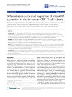
and exclusion criteria is shown in Fig. 1. Briefly, consecutive
patients were older than 18 years, had an Eastern Cooperative Oncology Group performance status of 0/1/2, and
signed inform consent. Exclusion criteria were: known alteration of immune system (active infections or autoimmune
disorder); treatment with G-CSF; contraindication to treatment with bevacizumab or its discontinuation; prior chemotherapy (CTx) for advanced disease, or adjuvant CTx less
than 6 months before enrollment onto study, cancer multiplicity. Choice of chemotherapy regimen was at the physicians’ discretion. Bevacizumab was administered at a dose of
5 mg/kg IV with the 2-week regimen or at a dose of 7.5 mg/
kg IV with the 3-week regimen. Patients’ responses to treatment and tumor measurements were evaluated with computer tomography scan by a staff radiologist according to
RECIST criteria. PFS was defined as the time from the beginning of treatment until the first observation of disease progression or death from any cause, while OS was defined as
the time from the beginning of treatment until death from
any cause. Patients were followed-up until death or loss to
follow-up. Survival rates were last updated in March 2018.
ORR was defined as the proportion of patients who have a
Bencsikova et al. BMC Cancer
(2019) 19:687
Page 3 of 9
Fig. 1 Study group definition. 1 Intended CTx regimen was chosen from among the following: CapeOX (oxaliplatin 130 mg/m2 IV day 1,
capecitabine 1000 mg/m2 twice daily per os (PO) for 14 days, repeat every 3 weeks); CapeIRI (irinotecan 250 mg/m2 day 1, capecitabine 1000 mg/
m2 twice daily PO for 14 days, repeat every 3 weeks); FOLFOX4 (oxaliplatin 85 mg/m2 intravenous (IV) day 1, Leucovorin 200 mg/m2 IV days 1 and
2, 5- fluorouracil 400 mg/m2 IV bolus on day 1 and 2, 5- fluorouracil 600 mg/m2 22-h continuous infusion days 1 and 2, repeat every 2 weeks);
FOLFIRI (irinotecan 180 mg/m2 IV day 1, Leucovorin 400 mg/m2 IV day 1, 5- fluorouracil 400 mg/m2 IV bolus day 1, then 5- fluorouracil 1200 mg/
m2 /d continuous infusion days 1 and 2, repeat every 2 weeks). Bevacizumab was administered on the first day of each cycle at a dose of 5 mg/
kg IV in combination with the 2-week regimen and at a dose of 7.5 mg/kg IV with the 3-week regimen. 2 KRAS status was not tested (not yet
performed or not ordered during the enrollment period) for mCRC patient management; KRAS testing was performed by ISO 15189-accredited
methods; specifically 2008 - December 2011 by real time PCR method using TheraScreen (DxS); January 2012 – May 2013 using the Cobas® KRAS
Mutation Test (Roche Diagnostics). 3 prior malignancy except for locally curable cancers such as basal or squamous cell skin cancer, superficial
bladder cancer, or carcinoma in situ of the prostate, cervix, or breast, curatively treated with no evidence of disease for ≥3 years. 4 active, known,
or suspected autoimmune disease requiring systemic treatment with immunosuppressive medication including chronic inflammatory bowel
disease (Crohn’s disease or ulcerative colitis). 5 active infection at the time of blood collection including clinically significant non-healing or
healing wound, ulcer. * exclusion criterion applicable if appears before the blood collection. ** exclusion criterion applicable if appears before the
achievement of objective clinical response
partial or complete response to treatment. Baseline characteristics of patients are summarized in Additional file 1:
Table S1.
Sample collection and lymphocyte count evaluation
Peripheral blood specimens were collected at initiation of
anti-VEGF treatment in a 2.6 mL S-Monovette® tube with
K3EDTA anticoagulant (Sarstedt, catalog number 04.1901) in
a phlebotomy room in close proximity to the laboratory where
analysis was performed. Blood specimens were mixed for
several minutes on a roller mixer. Immediately after that, absolute lymphocyte count was obtained from the complete
blood count by a differential analyzer Sysmex XE 5000 (Sysmex Corporation, Japan). Absolute lymphocyte count was
used for calculation of the absolute count of T cell subsets.
Flow cytometry – T cell subset quantification
Lymphocyte subsets were evaluated within 3 h of blood collection. For Treg detection as CD3+CD4+CD25+CD127−/low+
cells and CD4+ T cell detection, 50 μL of whole blood was
Bencsikova et al. BMC Cancer
(2019) 19:687
stained with a premixed cocktail of conjugated mAbs (Beckman Coulter) for the following markers, CD3-FITC (clone
UCHT1), CD25-PC5 (clone B1.49.9), CD4-PC7 (clone
13B8.2), and CD127-PE (clone R34.34) in concentrations according to manufacturer instructions. The gating strategy
for CD3+CD4+CD25+CD127−/low+ cells including details
on gating set-up and the analytical and statistical comparability of CD25+CD127−/low+ and CD25+FoxP3+ quantification approaches are shown in Additional file 1: Figure
S1. CD8+ cells were detected using 50 μL of whole blood
stained with tetraCHROME CD45-FITC/CD4-PE/CD8ECD/CD3-PC5 multi-color reagent (Beckman Coulter) in
concentrations according to the manufacturer instructions. After a 15 min staining for Tregs or CD8+ T-cells in
the dark, red blood cells were lysed for 15 min in the dark
by adding 600 μL of VersaLyse Lysing Solution (Beckman
Coulter, France). Cells were subsequently analyzed using a
Cytomics FC 500 flow cytometer, hardware compensation
and CXP software (Beckman Coulter, USA).
Statistical analysis
Wilcoxon two-sample two-tailed test was used to compare
continuous variables between the two groups in the Results
section, part I. Survival probabilities were estimated using
the Kaplan-Meier method in the Results section part II and
III. Log-rank test was used to assess the association of categorical variables with survival endpoints. Hazard ratios were
determined using Cox proportional hazard model. Logistic
regression was used to predict objective responses and to determine odds ratio. The need for adjustment by common
biomarkers was considered in the Results section part II and
III. The Cox model with interaction term was used to compare effects in subgroups in the Results section part III. Optimal cut points of continuous variables with respect to the
survival endpoints were determined using the conditional
hazard function which was estimated using smoothing techniques based on kernel methods [35]. Statistical comparison
of two Treg quantification approaches was performed using
Bland-Altman plot and Passing-Bablok regression in MS
Excel. Conditional hazard functions were estimated in
MATLAB, other analyses were performed in R, a language
and environment for statistical computing (R Core Team,
2013). Results with p < 0.05 were considered statistically
significant.
Results
Circulating Tregs, CD8+ CTLs and CD8:Treg ratio in
metastatic colorectal cancer patients in the context of
primary tumor sidedness and KRAS status
Relative and absolute numbers of circulating immune cells
were quantified in mCRC patients at the initiation of 1st line
anti-VEGF-based therapy and were evaluated in the context
of primary tumor sidedness and KRAS status. Regardless of
primary tumor sidedness, there was no difference in
Page 4 of 9
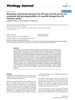
circulating Treg or CD8+ CTL count. A trend was observed
toward an increasing proportion of CD8+ CTLs in T cells
from proximal to distal tumor locations. Notably, KRAS wt
colorectal cancers exhibited a significantly higher proportion
of CD8+ CTLs among T cells but also higher Treg measures
(absolute count and the proportion of Tregs among CD4+
cells (Table 1, Fig. 2).
Circulating Tregs, CD8+ CTLs, CD8:Treg ratio, and clinical
outcome of 1st-line anti-VEGF-based therapy of mCRC
Median length of follow-up was 77.4 months. Median
PFS for the study group was 10.5 months (95% CI: 8.8–
16.3 months), median overall survival was 30.0 months
(95% CI: 23.3–38.5 months), and ORR was 55.6% (95%
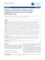
CI: 39.6–70.5%). Survival and response rate analysis was
performed for parameters clinically relevant for metastatic colorectal cancer, such as gender, age, M0 vs. M1,
number of metastatic sites, KRAS status, and primary
tumor sidedness (Fig. 3). Of those, age < 65 years was associated with shorter PFS and OS but not ORR (Fig. 3).
Levels of circulating immune cells at 1st-line anti-VEGF
therapy initiation were investigated in the context of
clinical outcome using the conditional hazard function
estimated by smoothing techniques (Additional file 1:
Figure S2). Cut-off levels for each parameter, dividing
cases to “low” and “high”, were established as shown in
Additional file 1: Figure S2 and subgroups defined by
levels of immune parameters were analyzed for PFS and
OS (Fig. 3). Of those, the baseline proportion of Tregs in
CD4+ cells was predictive for shorter PFS and OS and
worse ORR, and the baseline CD8:Treg ratio was predictive for longer PFS and OS. In the subgroup of mCRC
patients with < 6% frequency of Tregs among CD4+
cells, median PFS (mPFS) was 16.2 months, mOS was
38.5 months, and ORR was 76.4% compared to those
with a high frequency of circulating Tregs of ≥6% among
CD4+ cells which had a mPFS of 8.8 months, mOS of
22.3 months, and ORR of 36.8%. In the subgroup of
mCRC patients with a high CD8:Treg ratio of ≥10, mPFS
was 12.6 months and mOS was 37.8 months compared
to those with a ratio of circulating CD8:Treg of < 10
which had an mPFS of 8.1 months and mOS of 21.0
months (Additional file 1: Table S2).
Circulating Tregs, CD8+ CTLs and CD8:Treg ratio and the
clinical outcome of anti-VEGF-based therapy of mCRC in
the context of primary tumor sidedness
The association between number of circulating immune
cells and clinical outcome of mCRC therapy was further
analyzed in the context of primary tumor sidedness
(Fig. 4). The predictive value of the baseline proportion of
Tregs among CD4+ cells and the CD8:Treg ratio had the
same direction in primary right- and left-sided mCRC. In
addition to the strong association between high CD8:Treg
Bencsikova et al. BMC Cancer
(2019) 19:687
Page 5 of 9
Table 1 Medians of circulating immune cells in mCRC patient subgroups
mCRC
Lymphocytes (cells/μL)
+
1445
KRAS status
Primary tumor location
right c.
left c.
r.s./rectum
KRAS wt
1593
1469
1309
1521
KRAS mut
1312
CD3 in lymphocytes (%)
63
65
71
59
64
65
T cell count (cells/μL)
1042
1137
1151
894
1220
894
CD8+ in T cells (%)
44
38
44
48
45
CD8+ count (cells/μL)
380
372
511
401
558
Treg in lymphocytes (%)
1.9
1.7
2.0
2.0
2.3
Treg in CD4+ (%)
6.2
5.3
6.5
7.2
7.0
**
*
Treg count (cells/μL)
26.5
33.0
37.9
25.4
38.5
CD8:Treg
13.1
10.9
13.3
15.7
11.5
*
38
309
1.7
4.4
23.0
14.0
Stars indicate statistically significant difference in mCRC patients between respective subgroups: *p < 0.05, ** p < 0.005. c, colon; r.s., rectosigma
ratio and favorable clinical outcome in the entire study
group, the association between high CD8:Treg ratio and
longer overall survival was significantly higher in primary
right-sided mCRC (Fig. 4, Additional file 1: Figure S3) and
those with a high CD8:Treg ratio of ≥10 had a mPFS of
14.4 months and a mOS of 39.9 months compared to
those with a low ratio of circulating CD8:Treg of < 10
which had a mPFS 7.1 months and a mOS of 12.9 months
(Additional file 1: Table S2). In the subgroup of mCRC patients with primary tumors in the right colon, a significant
interaction between primary tumor sidedness and the
predictive value of absolute T cell count as well as the absolute CD8+ and CD4+ cell counts revealed an association
of poor PFS and OS with low baseline circulating absolute
T cells or CD8+ CTLs (Fig. 4, Additional file 1: Table S2
and Figure S3).
Discussion
Here we show that the baseline level of parameters derived from circulating Tregs, namely the Treg proportion among CD4+ T cells and the CD8:Treg ratio, at the
initiation of anti-VEGF-based therapy predicts treatment
Fig. 2 Circulating CTLs and Tregs in metastatic colorectal cancer patients in the context of primary tumor sidedness and KRAS mutation. p-values
refer to the level of circulating T cell subsets in KRAS wt vs. KRAS mut in the entire study group
Bencsikova et al. BMC Cancer
(2019) 19:687
Page 6 of 9
Fig. 3 Results of univariable analysis for progression-free, overall survival and objective response rate (ORR). Location: “right” = right colon, “left” =
left colon and rectum. ALC = absolute lymphocyte count
outcome in terms of both PFS and OS, and objective response rate. Our findings are in agreement with a study
by Roselli et al. by showing that a low baseline proportion
of Tregs in PBMC, but not any other clinical or laboratory
parameter evaluated, is associated with favorable outcome
in mCRC patients receiving 1st-line FOLFIRI plus bevacizumab [36]. Roselli et al. emphasized the unexplained lack
of association between clinical outcome and CD8+ T cells
[36] that we also observed when baseline circulating immune parameters from mCRC patients were analyzed irrespective of primary tumor sidedness. Nevertheless, and
based on our previous findings of poor clinical outcome
of mCRC patients with primary tumors in the right colon
[37] and the differential impact of KRAS status for 1st-line
anti-VEGF-based therapy in primary right vs. left-sided
mCRC [38], we analyzed circulating immune cells in the
context of primary tumor sidedness, revealing that the association of previously identified Treg-associated biomarkers, as well as a baseline number of circulating CD8+
T cells, with clinical outcome of 1st-line anti-VEGF-based
therapy is particularly strong in mCRC patients with primary tumor in the right colon.
The differential disease behavior of primarily right vs.
left-sided mCRC is substantiated by the prevalence of
distinct colorectal cancer subtypes within the colon and
rectum [39]. Based on the association of the immuneactivated, highly immunogenic CMS1 tumor subtype with
right-sided tumor location [39] on the one hand, and the
strong association of favorable circulating immune signature
(low Tregs, high CD8+ T cells, high CD8:Treg ratio) and favorable clinical outcome in primary right-sided mCRC on
the other, we propose that right-sided mCRC patients with
favorable circulating immune signature overlap with a subgroup of patients with immune-activated tumors that clearly
benefit from immunomodulatory anti-VEGF-based therapy.
Our hypothesis that immune characteristics in the TME are
reflected in the circulation is further supported by the finding
of an association of KRAS mutant status with reduction in
both CD8+ T cell count and number of Tregs. CMS2 and 3
subtypes are associated with reduced immune infiltration
and reactivity, and this immune quiescence is more profound
in KRAS-mutated tumors [40] and is likely mirrored in peripheral blood.
Due to the small size of study group, the cut-off levels of
immune cells stratifying prognostic subgroups may not be
accurate and should be validated in larger cohort of patients. Limited size of the study group also did not allow
multivariable analysis. A strength of this study is its long-
Bencsikova et al. BMC Cancer
(2019) 19:687
Page 7 of 9
Fig. 4 Results of Cox analyses for progression-free and overall survival according to primary tumor location. P-values correspond to test
significance of the interaction term (test of different effects of variables according to primary right- and left-sided mCRC). Location: “right” = right
colon, “left” = left colon and rectum
term follow-up. On the other hand, during the time period
when the study was designed, biomarkers such as
NRAS, BRAF, and MSI were just emerging in the clinical practice of colorectal cancer patient management
and unfortunately were not analyzed in the context of
circulating immune cells in mCRC treatment with bevacizumab. Thus, it remains to be investigated whether
the subset of patients with right-sided tumor and favorable circulating immune signature overlaps with the
MSI-H/CMS1 subset and may therefore be a good candidate for immunotherapy with checkpoint inhibitors.
Also, it remains to be addressed whether mCRC patients, particularly those with right-sided tumors with
an immunosuppressive circulating immune signature
(high Tregs, low CD8+ T cells and/or low CD8:Treg ratio) would benefit from the aggressive, triple combination chemotherapy regimen FOLFOXIRI [41].
Conclusions
Circulating immune parameters derived from the
baseline level of CD8 + CTLs and Tregs may predict
clinical outcome following 1st-line treatment with
the anti-VEGF angio/immunomodulatory agent bevacizumab and thereby identify mCRC patients, particularly within the primarily right-sided subgroup,
who have favorable outcome.
Bencsikova et al. BMC Cancer
(2019) 19:687
Additional files
Additional file 1: Table S1. Baseline characteristics of mCRC patients
included in the study. Figure S1. Gating strategy for
CD3+CD4+CD25+CD127−/low+ cells and the analytical comparability of a)
CD25+CD127−/low+ and b) CD25+FoxP3+ quantification approaches.
Statistical comparison of these approaches using c) Bland-Altman plot
and d) Passing-Bablok regression. Figure S2. Determination of the optimal cut points for circulating immune cells with respect to PFS and OS
using kernel estimates of conditional hazard functions. Table S2. Characteristics of clinical outcome (PFS and OS), proportion of Tregs in the
CD4+ cell subset, and the CD8: Treg ratio. Figure S3. Circulating immune
cells and clinical outcome of anti-VEGF-based therapy of mCRC in the
context of primary tumor sidedness. (DOCX 2640 kb)
Additional file 2: Spreadsheet with data generated and analyzed during
the study. (XLSX 20 kb)
Abbreviations
ALC: absolute lymphocyte count; CMS: Consensus molecular subtype;
CR: complete remission; CTLs: Cytotoxic CD8+ T cells; CTx: chemotherapy;;
iTregs: induced (or inducible) Tregs; IV: intravenous; mCRC: metastatic
colorectal cancer; NA: Not Available; NS: not specified; nTregs: natural Treg
cells; ORR: objective response rate; OS: overall survival; PD: progressive
disease; PFS: progression-free survival; PO: per os; PR: partial remission;
PS: performance status; SD: stable disease; TME: tumor microenvironment;
Tregs: Regulatory T cells
Acknowledgements
Not applicable.
Authors’ contributions
BB conceived of the study, participated in its design, performed patient
accrual, contributed to data interpretation, supervised data collection and
management, and drafted the manuscript. EB participated on the study
design, performed data analysis and statistical analysis, contributed to data
interpretation. IS performed statistical analysis, prepared figures and tables,
contributed to data interpretation, and drafted the manuscript. KP supervised
data collection, supervised laboratory testing, contributed to figure and table
preparation, and drafted the manuscript. LF contributed to data collection,
contributed to laboratory testing and laboratory data analysis. KG
contributed to data collection, contributed to laboratory testing, and drafted
the manuscript. RN contributed to data interpretation, reviewed and edited
the manuscript. DV contributed to data interpretation, reviewed and edited
the manuscript. RO performed patient accrual, contributed to data
interpretation, reviewed and edited the manuscript. MAS contributed to data
interpretation, reviewed and edited the manuscript. LZ-D conceived of the
study design, coordinated the study, contributed to data analysis and interpretation, drafted and finalized the manuscript. All authors read and approved the final manuscript.
Funding
The work was supported by the Czech Ministry of Health for projects AZV
16-31966A (data interpretation) and DRO 00209805 (design of the study,
writing the manuscript) and the Czech Ministry of Education, Youth and
Sports for projects LO1413 (sample and data analysis, writing the manuscript)
and LM2015089 (sample collection).
Availability of data and materials
All data generated and analysed during this study are included in this
published article (Additional file 2).
Ethics approval and consent to participate
The study was performed in compliance with the Declaration of Helsinki,
was approved by the Ethics Committee of Masaryk Memorial Cancer Institute
(MMCI, Brno, Czech Republic; reference number MOU/EK/131210) and
written informed consent was obtained from all patients.
Consent for publication
Not applicable.
Page 8 of 9
Competing interests
The authors declare that they have no competing interests.
Author details
1
Department of Comprehensive Cancer Care, Masaryk Memorial Cancer
Institute, Brno, Czech Republic. 2Regional Centre for Applied Molecular
Oncology, Masaryk Memorial Cancer Institute, Brno, Czech Republic.
3
Department of Laboratory Medicine, Masaryk Memorial Cancer Institute,
Brno, Czech Republic. 4Department of Oncological and Experimental
pathology, Masaryk Memorial Cancer Institute, Brno, Czech Republic.
Received: 8 October 2018 Accepted: 8 July 2019
References
1. Titu LV, Monson JR, Greenman J. The role of CD8(+) T cells in immune responses to
colorectal cancer. Cancer Immunol Immunother. 2002;51(5):235–47.
2. Chiba T, Ohtani H, Mizoi T, Naito Y, Sato E, Nagura H, Ohuchi A, Ohuchi K,
Shiiba K, Kurokawa Y, et al. Intraepithelial CD8+ T-cell-count becomes a
prognostic factor after a longer follow-up period in human colorectal
carcinoma: possible association with suppression of micrometastasis. Brit J
Cancer. 2004;91(9):1711–7.
3. Naito Y, Saito K, Shiiba K, Ohuchi A, Saigenji K, Nagura H, Ohtani H. CD8+ T
cells infiltrated within cancer cell nests as a prognostic factor in human
colorectal cancer. Cancer Res. 1998;58(16):3491–4.
4. Oberg A, Samii S, Stenling R, Lindmark G. Different occurrence of CD8+,
CD45R0+, and CD68+ immune cells in regional lymph node metastases
from colorectal cancer as potential prognostic predictors. Int J Color Dis.
2002;17(1):25–9.
5. Ohtani H. Focus on TILs: prognostic significance of tumor infiltrating
lymphocytes in human colorectal cancer. Cancer Immun. 2007;7:4.
6. Mlecnik B, Tosolini M, Kirilovsky A, Berger A, Bindea G, Meatchi T, Bruneval P,
Trajanoski Z, Fridman WH, Pages F, et al. Histopathologic-based prognostic
factors of colorectal cancers are associated with the state of the local
immune reaction. J Clin Oncol. 2011;29(6):610–8.
7. Galon J, Mlecnik B, Bindea G, Angell HK, Berger A, Lagorce C, Lugli A,
Zlobec I, Hartmann A, Bifulco C, et al. Towards the introduction of the
‘Immunoscore’ in the classification of malignant tumours. J Pathol. 2014;
232(2):199–209.
8. Taube JM, Klein A, Brahmer JR, Xu H, Pan X, Kim JH, Chen L, Pardoll DM,
Topalian SL, Anders RA. Association of PD-1, PD-1 ligands, and other
features of the tumor immune microenvironment with response to anti-PD1 therapy. Clin Cancer Res. 2014;20(19):5064–74.
9. Sakaguchi S, Miyara M, Costantino CM, Hafler DA. FOXP3+ regulatory T cells
in the human immune system. Nat Rev Immunol. 2010;10(7):490–500.
10. Whiteside TL. What are regulatory T cells (Treg) regulating in cancer and
why? Semin Cancer Biol. 2012;22(4):327–34.
11. Langier S, Sade K, Kivity S. Regulatory T cells: the suppressor arm of the
immune system. Autoimmun Rev. 2010;10(2):112–5.
12. Ling KL, Pratap SE, Bates GJ, Singh B, Mortensen NJ, George BD, Warren BF,
Piris J, Roncador G, Fox SB, et al. Increased frequency of regulatory T cells in
peripheral blood and tumour infiltrating lymphocytes in colorectal cancer
patients. Cancer Immun. 2007;7:7.
13. Sundstrom P, Stenstad H, Langenes V, Ahlmanner F, Theander L, Ndah TG,
Fredin K, Borjesson L, Gustavsson B, Bastid J, et al. Regulatory T cells from
Colon Cancer patients inhibit effector T-cell migration through an
adenosine-dependent mechanism. Cancer Immunol Res. 2016;4(3):183–93.
14. Ward-Hartstonge KA, Kemp RA. Regulatory T-cell heterogeneity and the
cancer immune response. Clin Transl Immunol. 2017;6(9):e154.
15. Luu M, Steinhoff U, Visekruna A. Functional heterogeneity of gut-resident
regulatory T cells. Clin Transl Immunol. 2017;6(9):e156.
16. Salama P, Phillips M, Grieu F, Morris M, Zeps N, Joseph D, Platell C, Iacopetta
B. Tumor-infiltrating FOXP3+ T regulatory cells show strong prognostic
significance in colorectal cancer. J Clin Oncol. 2009;27(2):186–92.
17. Zhuo C, Xu Y, Ying M, Li Q, Huang L, Li D, Cai S, Li B. FOXP3+ Tregs:
heterogeneous phenotypes and conflicting impacts on survival outcomes
in patients with colorectal cancer. Immunol Res. 2015;61(3):338–47.
18. Tosolini M, Kirilovsky A, Mlecnik B, Fredriksen T, Mauger S, Bindea G, Berger
A, Bruneval P, Fridman WH, Pages F, et al. Clinical impact of different classes
of infiltrating T cytotoxic and helper cells (Th1, th2, treg, th17) in patients
with colorectal cancer. Cancer Res. 2011;71(4):1263–71.
Bencsikova et al. BMC Cancer
(2019) 19:687
19. Verma C, Eremin JM, Robins A, Bennett AJ, Cowley GP, El-Sheemy MA, Jibril
JA, Eremin O. Abnormal T regulatory cells (Tregs: FOXP3+, CTLA-4+),
myeloid-derived suppressor cells (MDSCs: monocytic, granulocytic) and
polarised T helper cell profiles (Th1, Th2, Th17) in women with large and
locally advanced breast cancers undergoing neoadjuvant chemotherapy
(NAC) and surgery: failure of abolition of abnormal treg profile with
treatment and correlation of treg levels with pathological response to NAC.
J Transl Med. 2013;11:16.
20. Yamamoto T, Yanagimoto H, Satoi S, Toyokawa H, Hirooka S, Yamaki S, Yui
R, Yamao J, Kim S, Kwon AH. Circulating CD4+CD25+ regulatory T cells in
patients with pancreatic cancer. Pancreas. 2012;41(3):409–15.
21. Ihara F, Sakurai D, Horinaka A, Makita Y, Fujikawa A, Sakurai T, Yamasaki K, Kunii
N, Motohashi S, Nakayama T, et al. CD45RA(−)Foxp3(high) regulatory T cells
have a negative impact on the clinical outcome of head and neck squamous
cell carcinoma. Cancer Immunol Immunother. 2017;66(10):1275–85.
22. Jass JR. Classification of colorectal cancer based on correlation of clinical,
morphological and molecular features. Histopathology. 2007;50(1):113–30.
23. Budinska E, Popovici V, Tejpar S, D'Ario G, Lapique N, Sikora KO, Di Narzo AF, Yan P,
Hodgson JG, Weinrich S, et al. Gene expression patterns unveil a new level of
molecular heterogeneity in colorectal cancer. J Pathol. 2013;231(1):63–76.
24. Sadanandam A, Lyssiotis CA, Homicsko K, Collisson EA, Gibb WJ,
Wullschleger S, Ostos LC, Lannon WA, Grotzinger C, Del Rio M, et al. A
colorectal cancer classification system that associates cellular phenotype
and responses to therapy. Nat Med. 2013;19(5):619–25.
25. Dienstmann R, Vermeulen L, Guinney J, Kopetz S, Tejpar S, Tabernero J.
Consensus molecular subtypes and the evolution of precision medicine in
colorectal cancer. Nat Rev Cancer. 2017;17(2):79–92.
26. Popat S, Hubner R, Houlston RS. Systematic review of microsatellite
instability and colorectal cancer prognosis. J Clin Oncol. 2005;23(3):609–18.
27. Deschoolmeester V, Baay M, Lardon F, Pauwels P, Peeters M. Immune cells
in colorectal Cancer: prognostic relevance and role of MSI. Cancer
Microenviron. 2011;4(3):377–92.
28. Boissiere-Michot F, Lazennec G, Frugier H, Jarlier M, Roca L, Duffour J, Du
Paty E, Laune D, Blanchard F, Le Pessot F, et al. Characterization of an
adaptive immune response in microsatellite-instable colorectal cancer.
Oncoimmunology. 2014;3:e29256.
29. Bencsikova B, Bortlicek Z, Halamkova J, Ostrizkova L, Kiss I, Melichar B, Pavlik
T, Dusek L, Valik D, Vyzula R, et al. Efficacy of bevacizumab and
chemotherapy in the first-line treatment of metastatic colorectal cancer:
broadening KRAS-focused clinical view. BMC Gastroenterol. 2015;15:37.
30. Alfaro C, Suarez N, Gonzalez A, Solano S, Erro L, Dubrot J, Palazon A, HervasStubbs S, Gurpide A, Lopez-Picazo JM, et al. Influence of bevacizumab,
sunitinib and sorafenib as single agents or in combination on the inhibitory
effects of VEGF on human dendritic cell differentiation from monocytes.
British J Cancer. 2009;100(7):1111–9.
31. Osada T, Chong G, Tansik R, Hong T, Spector N, Kumar R, Hurwitz HI, Dev I,
Nixon AB, Lyerly HK, et al. The effect of anti-VEGF therapy on immature
myeloid cell and dendritic cells in cancer patients. Cancer Immunol
Immunother. 2008;57(8):1115–24.
32. Wada J, Suzuki H, Fuchino R, Yamasaki A, Nagai S, Yanai K, Koga K,
Nakamura M, Tanaka M, Morisaki T, et al. The contribution of vascular
endothelial growth factor to the induction of regulatory T-cells in malignant
effusions. Anticancer Res. 2009;29(3):881–8.
33. Terme M, Tartour E, Taieb J. VEGFA/VEGFR2-targeted therapies prevent the
VEGFA-induced proliferation of regulatory T cells in cancer.
Oncoimmunology. 2013;2(8):e25156.
34. Manzoni M, Rovati B, Ronzoni M, Loupakis F, Mariucci S, Ricci V, Gattoni E,
Salvatore L, Tinelli C, Villa E, et al. Immunological effects of bevacizumab-based
treatment in metastatic colorectal cancer. Oncology. 2010;79(3–4):187–96.
35. Selingerova I, Dolezelova H, Horova I, Katina S, Zelinka J. Survival of patients
with primary brain tumors: comparison of two statistical approaches. PLoS
One. 2016;11(2):e0148733.
36. Roselli M, Formica V, Cereda V, Jochems C, Richards J, Grenga I, Orlandi A,
Ferroni P, Guadagni F, Schlom J. The association of clinical outcome and
peripheral T-cell subsets in metastatic colorectal cancer patients receiving firstline FOLFIRI plus bevacizumab therapy. Oncoimmunology. 2016;5(7):e1188243.
37. Ostrizkova L, Petruzelka L, Hejduk K, Zdrazilova-Dubska L, Vocka M, Brancikova
D, Bencsikova B, Vyzula R, Obermannova R. Right-sided colon cancer is
associated with increased frequency of KRAS mutation and with a poor
outcome in patients with metastatic disease treated in the first line with
bevacizumab and chemotherapy. Annals Oncol. 2016;27(Suppl 2):114–5.
Page 9 of 9
38. Obermannova R, Ostrizkova L, Hejduk K, Zdrazilova-Dubska L, Vocka M,
Vyzula R, Bencsikova B, Petruzelka L. Right-sided versus left-sided primary
tumor location in patients with KRASmut metastatic colorectal cancer
(mCRC) treated with 1st-line anti-VEGF plus chemotherapy (CTx) - data from
the National Czech Registry. Annals Oncol. 2016;27(Suppl 9):168O.
39. Loree JM, Pereira AAL, Lam M, Willauer AN, Raghav K, Dasari A, Morris VK,
Advani S, Menter DG, Eng C, et al. Classifying colorectal Cancer by tumor
location rather than sidedness highlights a continuum in mutation profiles
and consensus molecular subtypes. Clin Cancer Res. 2018;24(5):1062–72.
40. Lal N, White BS, Goussous G, Pickles O, Mason MJ, Beggs AD, Taniere P,
Willcox BE, Guinney J, Middleton GW. KRAS mutation and consensus
molecular subtypes 2 and 3 are independently associated with reduced
immune infiltration and reactivity in colorectal Cancer. Clin Cancer Res.
2018;24(1):224–33.
41. Cremolini C, Loupakis F, Antoniotti C, Lupi C, Sensi E, Lonardi S, Mezi S,
Tomasello G, Ronzoni M, Zaniboni A, et al. FOLFOXIRI plus bevacizumab
versus FOLFIRI plus bevacizumab as first-line treatment of patients with
metastatic colorectal cancer: updated overall survival and molecular
subgroup analyses of the open-label, phase 3 TRIBE study. Lancet Oncol.
2015;16(13):1306–15.
Publisher’s Note
Springer Nature remains neutral with regard to jurisdictional claims in
published maps and institutional affiliations.