A multiple breast cancer stem cell model to predict recurrence of T1–3, N0 breast cancer
Bạn đang xem bản rút gọn của tài liệu. Xem và tải ngay bản đầy đủ của tài liệu tại đây (2.14 MB, 11 trang )
Qiu et al. BMC Cancer
(2019) 19:729
/>
RESEARCH ARTICLE
Open Access
A multiple breast cancer stem cell model to
predict recurrence of T1–3, N0 breast cancer
Yan Qiu1,2,3,4, Liya Wang5, Xiaorong Zhong6, Li Li1, Fei Chen1, Lin Xiao1, Fangyu Liu7, Bo Fu5, Hong Zheng6,
Feng Ye1,2,3* and Hong Bu1,2,3,4
Abstract
Background: Local or distant relapse is the key event for the overall survival of early-stage breast cancer after initial
surgery. A small subset of breast cancer cells, which share similar properties with normal stem cells, has been proven to
resist to clinical therapy contributing to recurrence.
Methods: In this study, we aimed to develop a prognostic model to predict recurrence based on the prevalence
of breast cancer stem cells (BCSCs) in breast cancer. Immunohistochemistry and dual-immunohistochemistry were
performed to quantify the stem cells of the breast cancer patients. The performance of Cox proportional hazard
regression model was assessed using the holdout methods, where the dataset was randomly split into two exclusive
sets (70% training and 30% testing sets). Additionally, we performed bootstrapping to overcome a possible biased error
estimate and obtain confidence intervals (CI).
Results: Four groups of BCSCs (ALDH1A3, CD44+/CD24−, integrin alpha 6 (ITGA6), and protein C receptor (PROCR))
were identified as associated with relapse-free survival (RFS). The correlated biomarkers were integrated as a prognostic
panel to calculate a relapse risk score (RRS) and to classify the patients into different risk groups (high-risk or low-risk).
According to RRS, 67.81 and 32.19% of patients were categorized into low-risk and high-risk groups respectively. The
relapse rate at 5 years in the low-risk group (2.67, 95% CI: 0.72–4.63%) by Kaplan-Meier method was significantly lower
than that of the high-risk group (19.30, 95% CI: 12.34–26.27%) (p < 0.001). In the multiple Cox model, the RRS was
proven to be a powerful classifier independent of age at diagnosis or tumour size (p < 0.001). In addition, we found
that high RRS score ER-positive patients do not benefit from hormonal therapy treatment (RFS, p = 0.860).
Conclusion: The RRS model can be applied to predict the relapse risk in early stage breast cancer. As such, high RRS
score ER-positive patients do not benefit from hormonal therapy treatment.
Keywords: Early stage breast cancer, Brest cancer stem cell, Relapse risk score, Prognosis
Background
More than 50% of patients with breast cancer are classified into the early-stage (T1–3N0M0) group [1]. Despite
systemic adjuvant therapy dramatically increasing the
clinical outcome of patients with early breast cancer, relapse still occurs in more than 20% of patients after surgery within 10 years [2]. Relapse, including recurrence
both at local or distant sites, is the main cause for patient deaths, and thus remains an unmet challenge for a
* Correspondence:
1
Laboratory of Pathology, West China Hospital, Sichuan University, Chengdu,
China
2
Key Laboratory of Transplant Engineering and Immunology, Ministry of
Health, West China Hospital, Sichuan University, Chengdu, China
Full list of author information is available at the end of the article
curative treatment of breast cancer. It is pivotal to identify patients at risk of relapse at early stages in hopes of
improving clinical outcomes, especially within the subgroup of node-negative females, defined as a relatively
indolent disease based on pathologic features. Recently,
several multigene assays have been developed for earlystage breast cancer patients [3]. Multigene assays are
able to provide more prognostic information than traditional parameters in several tumour types [4–11], and
several of them have been adopted by the oncology
guidelines for treatment. One example is 21-gene expression profiling, which has been widely accepted in
clinical practice [12].
© The Author(s). 2019 Open Access This article is distributed under the terms of the Creative Commons Attribution 4.0
International License ( which permits unrestricted use, distribution, and
reproduction in any medium, provided you give appropriate credit to the original author(s) and the source, provide a link to
the Creative Commons license, and indicate if changes were made. The Creative Commons Public Domain Dedication waiver
( applies to the data made available in this article, unless otherwise stated.
Qiu et al. BMC Cancer
(2019) 19:729
As reported, breast cancer is a tumour with high heterogeneity. Although recent advancements have further divided this heterogeneous disease into distinct subgroups
by gene expression profiling (GEP) assays, among other
methods, several intriguing findings revealed that a small
subset of cells isolated from different subgroups of breast
cancers exhibit remarkable similar biological behaviours.
These subset of cells were defined as cancer stem cells
(CSCs) and reported to be responsible for the heterogeneity. Accumulating evidence has proved that CSCs retain
the critical characteristics of normal stem cells, such as
ability self-renewal and the capacity of proliferation, which
contribute significantly to therapeutic resistance and
breast cancer relapse [13–17]. In addition, several articles
indicated that some CSCs might be derived from normal
stem cells, which suggested that normal mammary stem
cells might share similar identifying markers [18–20].
Mammary stem cell markers or combined markers have
been certified in different stages of stem cells in breast
cancer, including ALDH, CD44, CD24, ITGA6/EpCAM,
and PROCR. [21–26]. Some of these markers and combined markers (i.e., CD44+/CD24low ALDH+ and ITGA6+)
are considered to correlate with poor prognosis in breast
cancer [21, 27, 28], because they also identified a BCSC
subpopulation [14, 21, 26, 29]. In addition, it has been
suggested that ITGA6+/EpCAM+ mammary luminal progenitor cells were possible transformation targets in basallike breast cancers, which have close associations with
poor prognosis. In addition, it was reported that ITGA6
may define the mesenchymal population and is necessary
for CSC function [30–32]. PROCR was reported to be
highly expressed in myoepithelial cells of the mammary
gland. In a recent study, Wang D et al. identified PROCR
as a marker of multipotent mammary stem cells. They
found that PROCR-positive mammary cells exhibited epithelial-to-mesenchymal transition (EMT) characteristics,
and had high tumorigenesis ability in vivo, which suggested that PROCR-positive mammary cells might be one
of the progenitor populations for breast CSCs (BCSCs)
[24]. Furthermore, PROCR also promotes tumour metastasis in cancer cell lines [33, 34].
To explore the prognostic role of mammary stem cell
(MSCs) and BCSC markers, we have studied the ALDH
family (including ALDH1A1, ALDH1A3, ALDH3A1,
ALDH4A1, ALDH6A1, and ALDH7A1), PROCR, and
ITGA6/EpCAM. In a medium cohort of patients in previous studies, these findings revealed that ALDH1A3,
PROCR, ITGA6+, ITGA6+/EpCAM− and ITGA6−/
EpCAM+ were correlated with reduced RFS or overall
survival of these breast cancer patients [35–37]. In this
study, we defined these markers and CD44+/CD24low as
BCSC-associated markers and employed these biomarkers to label stem cells among patients with early
stage breast cancer. ALDH1A3, CD44+/CD24−, ITGA6,
Page 2 of 11
and PROCR were shown to be closely associated with
RFS. Then, they were integrated into the prognostic
panel to calculate an RRS. Patients were then divided
into two distinct risk groups, which effectively shows
promise in predicting prognosis and treatment. In
addition, several EMT transition associated markers,
proliferation factors and other clinicopathological parameters were also included in our study to improve the
efficiency of our model.
Materials and methods
Breast cancer patient dataset
Clinical information from 1036 patients with breast invasive ductal carcinoma (BIDC) diagnosed from 2006 to
2011 was collected from West China Hospital. After selection, 407 patients were enrolled into our study. All
the patients were adult females and were treated with
mastectomy or lumpectomy to negative margins and
with axillary lymph node dissection. Axillary nodes of
patients were observed to be without metastasis under
microscope. Patients with local invasion and distant metastasis identified initially were ineligible. Patients with
neoadjuvant chemotherapy were removed from our
study group to avoid its impact on the characteristics of
tumour cells in paraffin embedded tissues. Patients enrolled in the study were considered to be early-stage
BIDC and defined as entire datasets. The end-point of
follow-up was occurrence of local recurrence or distant
metastasis. Detailed information of this dataset is listed
in Additional file 4: Table S1.
Breast cancer stem cell biomarkers
BCSC-associated biomarkers were selected from literature as well as our previously confirmed biomarkers including CD44+/CD24−, ALDH1A3, EpCAM/ITGA6,
and PROCR, which showed prognostic value in BIDC
[21, 27, 28, 35–37].
Immunohistochemistry (IHC)
Single staining of CD44, CD24, EpCAM, ITGA6,
ALDH1A3, PROCR, Twist and Slug were performed
with the EnVision Staining System, while dual staining
of CD44/CD24 and EpCAM/ITGA6 were performed
with the EnVision G | 2 Doublestain System. The
haematoxylin and eosin (H&E) staining, as well as the
results of IHC staining were observed under bright field
microscopy. Pathological assessment of the tumours
were conducted by pathologists at West China Hospital
anonymously, including subtypes, histological grades,
oestrogen receptor (ER), progesterone receptor (PR),
and human epidermal growth factor receptor 2 (HER2)
etc. HER2 staining was analysed according to the guidelines of the American Society of Clinical Oncology. ER
and PR were analyzed by Allred system [38, 39]. The
Qiu et al. BMC Cancer
(2019) 19:729
scoring of BCSC-associated markers, such as ALHD1A3,
PROCR, ITGA6, CD44/CD24 and EpCAM/ITGA6 were
performed as follows: 0, 0% positive tumour cells; 1, 1 to
10% positive cells; 2, 11 to 50% positive cells; 3, 51 to
75% positive cells; and 4, 76 to 100% positive cells [27].
Scores of Twist and Slug were interpreted as follows: the
percentage (P) of positive cells (score 0 for 0%, 1 for
≤1%, 2 for 1–10%, 3 for 10–33%, 4 for 33–66%, and 5
for 66–100% positive cells) and the intensity (I) of staining (score 0 for negative, 1 for weak, 2 for moderate, and
3 for strong staining) were included. A Quick score was
generated. (Q = P*I; score range, 0–12) [40].
Detailed information and specificity of these antibodies
were shown in Additional file 5: Table S2, Additional file 1:
Figure S1, respectively.
Statistical analysis and model construction
The associations between relapse-free survival (RFS) and
the expression panel were analysed by the Cox proportional hazard regression model [41]. To investigate the
effectiveness of the BCSC-associated biomarker panel
for clinical outcome prediction, we assigned each patient
a risk score according to a linear combination of the expression level of BCSC-associated markers. The RRS for
sample i using the information from the significant bioP
markers was calculated as follows: RRS ¼ 4j¼1 WjÃSj:
In the above formula, Sj is the IHC score for biomarker
j, and Wj is the weight of the IHC score of biomarker j.
Weights were obtained by the coefficients derived from
the univariate Cox proportional hazard regression [42].
The RRS was calculated out by the receiver operating
characteristic curve (ROC, non-parametric test), which
identifies the cut-off value based on the maximum sums
of specificity and sensitivity in the ROC curve. Meanwhile, to investigate the association between the relapse
and other clinicopathological variables, univariate Cox
proportional hazard regression analysis was adopted
using clinicopathological factors (including age, tumour
size, histological grade, ER status, PR status and HER2
status), proliferation factors (Ki67), and EMT related factors (including Twist and Slug) in the dataset. The cutoff values of ER, PR, HER2 and Ki67 were 1, 1%, 1+/2+,
and 14%, respectively, according to the standards of clinical practice. For twist and slug, the final score was 0 to
12 as the cut-off value for the analyses to obtain significant results. Furthermore, multivariate Cox proportional
hazard regression analysis was applied to investigate
whether the predictive value of the panel was independent of other clinical variables.
The model was established using the and holdout
methods, an approach to out-of-sample evaluation,
where the dataset was randomly split into two exclusive
sets (70% training and 30% testing sets) [43]. The model
Page 3 of 11
was then trained on the training group and tested on the
testing group 10 times. Additionally, bootstrapping was
used to overcome a possible biased error estimate and
obtain confidence intervals (CI). We reported the 95%
CI of the coefficients, hazard ratio, and relapse rate for
each model. Statistical analyses were performed using
GraphPad Prism version 6 and R 3.4.0. To enroll more
effective biomarkers and clinicopathological factors into
further modelling, a p-value less than 0.1 was defined as
statistically significant in the univariate Cox Proportional
Analysis. Then, potential significant factors were enrolled into the multivariate Cox Proportional Analysis,
with the p-value less than 0.05 considered to be statistically significant. The detail was shown in Additional file 3:
Figure S3.
Results
Characteristics of patients and IHC results
The mean age of the patients was 49.3 ± 9.9 years. The
youngest patient was 23 years old while eldest one was
78 years old. Among the 407 patients, the median follow-up was 66 months, and relapse was observed in 42
(10.3%) patients during five years after diagnosis, consistent with results published in the literature. The characteristics of clinicopathological, proliferation, and EMT
related factors of the 407 patients are depicted in Table 1
and Additional file 4: Table S1. IHC staining was performed on slides of paraffin embedded blocks of those
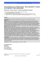
407 BIDC samples. Results are shown in Fig. 1. We also
performed IHC in tissues of patients with reductional
mammoplasty. The prevalence of BSCCs biomarkers in
reductional mammoplasty samples were shown in
Additional file 2: Figure S2.
Construction and validation of the RRS model
A univariate analysis was performed to test whether the
expression level of each BCSC-associated marker was related to differences of patient RFS. Among all the BCSC
related biomarkers, four biomarkers (ALDH1A3, CD44+/
CD24−, ITGA6+, and PROCR) were confirmed to be statistically correlated with patient RFS (Table 2). The RRS
formula according to the expression coefficient of those 4
BCSC-associated biomarkers for survival is listed as follows: RRS = 0.30× (score of ALDH) + 0.34× (score of
CD44+/CD24−) + 0.24× (score of ITGA6) + 0.56× (score
of PROCR). Therefore, patients were classified into highrisk and low-risk group individually using the optimal
RRS (RRS corresponding to the maximum sum of specificity and sensitivity in the ROC curve) as the cut-off value.
With the aid of the method described in the Materials and
Methods, the cut-off value was calculated to be 2.05.
Then, Kaplan-Meier analysis showed that the proportion of patients in the low-risk group who were free of
relapse at 5 years (97.68, 95% CI: 97.37–98.00%) was
Qiu et al. BMC Cancer
(2019) 19:729
Page 4 of 11
Table 1 Characteristics of Clinicopathological, Proliferation, and
EMT Related Factors of the 407 Patients
Clinicopathological Factors
Relapse or not(N,%)
No
Age
Tumor Size
Histological Grade
ER Status
PR Status
Yes
>40y
307
30 (8.90)
≤40y
58
12 (17.15)
≤2 cm
165
11 (6.25)
> 2 cm
200
31 (13.42)
Grade 1
19
0 (0.00)
Grade 2
126
18 (12.50)
Grade 3
220
24 (9.84)
≤1%(p)
113
12 (9.60)
> 1%(n)
252
30 (10.69)
≤1%(p)
135
16 (10.59)
> 1%(n)
230
26 (10.16)
Her2 Status
0/1+
253
31 (10.92)
3+
67
7 (9.46)
Menopausal status
Premenopausal
215
23 (9.66)
Postmenopausal
147
15 (9.26)
≤14%
127
11 (7.97)
> 14%
238
31 (11.52)
TS = 0
175
18 (9.33)
TS > 0
190
24 (11.21)
TS = 0
231
27 (10.47)
TS > 0
134
15 (10.07)
Mastectomy
333
40 (10.72)
Lumpectomy
32
2 (5.88)
Ki67
Twist
Slug
Surgery
p-value
(log-rank)
0.016
0.032
0.271
0.567
0.722
0.942
0.858
0.222
0.560
0.722
0.392
significantly higher than that in the high-risk group
(81.33, 95% CI: 80.50–82.16%) (p < 0.001) in the training
group. In another exclusive group (the testing group),
the proportion of patients in the low-risk group who
were free of relapse at 5 years (96.82, 95% CI: 95.88–
97.76%) was also higher than that in the high-risk group
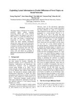
(82.13, 95% CI: 79.93–84.33%) (p < 0.001). Distributions
of risk score, relapse status and BCSC-associated biomarker expression of patients in the training group and
testing group is displayed in Table 3 and Fig. 2.
Among all the clinicopathological factors (including
age at diagnosis, tumour size, histological grade, ER status, PR status and HER2 status), proliferation factors
(Ki67), EMT related factors (including Twist and Slug),
age at diagnosis and tumour size were considered potential significant factors in the univariate survival analysis. These factors were then fully enrolled to the
multivariate Cox model with RRS. In a multiple Cox
model, RRS demonstrated significant predictive power
that was independent of tumour size and age at diagnosis in both the training group (p < 0.001) and testing
group (p = 0.014) (Table 4).
Assessment of the RRS model in the entire dataset
Assessment of the RRS model in univariate survival analysis
(Kaplan-Meier method)
To validate our findings, the RRS model was assessed in
the entire dataset (n = 407). By using the same cut-off
value of training groups, patients in the entire dataset
were classified into the high-risk group (n = 131) and
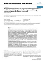
low-risk group (n = 276) (Fig. 3a). Patients with high risk
scores demonstrated significantly reduced RFS when
compared to those with low risk scores (log-rank test
p < 0.001) (Fig. 3b). The relapse rate at 5 years was
19.30% (95% CI: 12.34–26.27%) and 2.67% (95% CI:
0.72–4.63%) in the high-risk group and low-risk group,
respectively. Distributions of risk score, relapse status
and BCSC-associated biomarker expression of each patient in the entire datasets were then analysed (Fig. 3c).
Assessment of the RRS model in multivariate survival
analysis (cox proportional analysis)
In the entire dataset, the correlation between RFS and
clinicopathological factors (including age, tumour size,
histological grade, ER status, PR status and HER2 status), proliferation factors (Ki67), EMT related factors
(including Twist and Slug) was analysed by KaplanMeier method. Reduced RFS was only demonstrated in
patients with smaller tumour size (log-rank p = 0.032)
and younger age (log-rank p = 0.016) (Table 1). Then,
multivariate survival analyses were adopted to explore
the association between relapse and age as well as
tumour size. As a result, younger age, larger tumour size
and RRS were implied to be significant predictors of relapse (Table 5).
Hormone therapy benefit in different groups
Among the 407 patients, there were 282 ER-positive and
125 ER-negative patients. We found that our panel
worked in both of these two subgroups (Fig. 4a, b). In
the ER-positive group, all patients were treated with
chemotherapy, whereas only 89.72% (n = 253) of these
patients received hormone therapy. Our results demonstrated no difference for the RFS between those hormone-treated patients and non-treated patients in the
high-risk score group (p = 0.860 Fig. 4d). However, in
the low-risk score group, patients in the treated group
showed remarkably longer RFS than those in the nontreated group (p = 0.038, Fig. 4c), which indicated that
patients with a high-risk score may not benefit from the
traditional hormone therapy.
Discussion
An increasing number of females are diagnosed with
node negative invasive breast carcinomas. Even though
most of patients with early-stage breast cancer have a
favourable outcome, the 5-year rate of local relapse or
Qiu et al. BMC Cancer
(2019) 19:729
Page 5 of 11
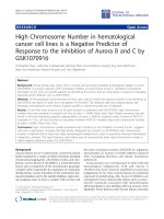
Fig. 1 IHC staining in early-stage BIDC patients. a Dual staining for CD44 (green arrow) and CD24 (yellow arrow); b Dual staining for EpCAM (green
arrow) and CD49 (yellow arrow); c-f Single staining for ALDH1A3 (cytoplasm), PROCR (membrane), Twist (nuclear) and Slug (nuclear), respectively
distant metastasis in our dataset is still up to 10.3%. As
metastatic diseases are challenging to cure, accurate
evaluation for prognosis and more efficacious treatments
are needed. In our present study, we developed and validated a novel prognostic model based on 4 BCSC-associated biomarkers to improve our accuracy of predicting
disease recurrence in patients with early stage BIDC
Table 2 Biomarkers Associated with Relapse in Training Group
by Univariate Cox Proportional Analysis
Biomarkers
Coefficient (Wj, 95% CI)
Hazard Radio(95% CI)a
ALDH1A3
0.30 (0.27–0.33)
1.35 (1.12–1.58)
CD44+/CD24−
0.34 (0.31–0.38)
1.41 (1.09–1.72)
ITGA6
0.24 (0.19–0.30)
1.27 (1.04–1.51)
PROCR+
0.56 (0.52–0.60)
1.75 (1.49–2.00)
+
CI confidence interval
a
(T1–3N0M0). The four biomarkers incorporated into our
predictive model have been shown to be involved in
stem cell ability in vivo and in vitro, including self-renewal ability and tumorigenic capacity, which could contribute greatly to metastasis of BIDC in vitro and in
vivo, or in tumour tissues [21–25, 44–46].
The holdout methods were adopted to establish our
RRS model, which assisted us to obtain a stable model
to calculate RRS in our study. Our model was further
validated in the entire dataset. The AUC value of ROC
curve is 0.781 which indicated that the RRS is a good
classifier for relapse among patients with early stage
breast cancer. The difference in the risk of relapse between patients with low risk scores and those with highrisk scores was large and statistically significant. There
are 276 (67.81%) patients who were classified in the lowrisk group, while only 32.19% of patients were included
Qiu et al. BMC Cancer
(2019) 19:729
Page 6 of 11
Table 3 Kaplan-Meier Estimation of the Rate of Recurrence at 5 Years, According to Recurrence-Score Risk Category
Percentage of patients (%)
Rate of recurrence at 5 years (%, 95 CI)a
p-value
Low-risk
67.54
2.32 (2.00–2.63)
< 0.001
High-risk
32.46
18.67 (17.84–19.50)
Low-risk
68.46
3.18 (2.24–4.12)
High-risk
31.54
17.87 (15.67–20.07)
RRS
Training set
Testing set
< 0.001
CI confidence interval
a
Fig. 2 Establishment and Validation of RRS of early-stage BIDC patients, a Kaplan-Meier analysis for RFS of early-stage BIDC patients in training
group. b Kaplan-Meier analysis for RFS of early-stage BIDC patients in testing group. c The distribution of the RRS, patients’ relapse status and
biomarker expression in training group. d The distribution of the RRS, patients’ relapse status and biomarker expression in the testing group. (We
conducted 10 times; Fig. 2 is only one example of them)
Qiu et al. BMC Cancer
(2019) 19:729
Page 7 of 11
Table 4 Multivariate Cox Proportional Analysis of Tumor Size,
age, and RRS in Relation to the Likelihood of Relapse
P-value
Hazard Radio (95% CI)
a
Table 5 Multivariate Cox Proportional Analysis of Age, Tumor
Size, and RRS in Relation to the Likelihood of Relapse in Entire
Dataset
Variable
Training group
p-value
Hazard Ratio (95%CI)a
Analysis without RRS
RRS (high vs. low)
< 0.001
6.75 (2.90–15.72)
Tumor size (> 2 cm vs. ≤2 cm)
0.037
2.72 (1.16–6.38)
Age (≤40y vs. >40y)
0.012
2.38 (1.21–4.69)
0.46 (0.20–1.05)
Tumor Size (> 2 cm vs. ≤2 cm)
0.022
2.22 (1.11–4.44)
2.22 (1.12–4.39)
Age (>40y vs. ≤40y)
0.098
Analysis with RRS
Testing group
RRS (high vs. low)
0.014
5.04 (1.52–16.81)
Age (≤40y vs. >40y)
0.022
Tumor size (> 2 cm vs. ≤2 cm)
0.177
3.33 (0.80–15.85)
Tumor Size (> 2 cm vs. ≤2 cm)
0.005
2.70 (1.34–5.41)
0.59 (0.15–2.41)
RRS (high vs. low)
< 0.001
5.92 (3.01–11.6)
Age (>40y vs. ≤40y)
0.316
CI confidence interval
a
in the high-risk group, and their rate of relapse at 5 years
was 19.30 and 2.67%, respectively. Therefore, the application of the RRS predictor provides a good estimate of the
risk of local or distant recurrence in individual patients.
We also enrolled other biomarkers in the univariate
survival analysis in the training set, such as age, tumour
size, histological grade, Ki67, and EMT related biomarkers. All those parameters have been reported to
CI confidence interval
a
play critical roles in accelerating the presence of distant
metastasis or local relapse [47, 48]. Despite the fact that
EMT has been reported to produce cells with stem celllike properties [49], we found that no parameter showed
significantly different RFS in different subgroups of
EMT related biomarkers. In this study, smaller tumour
size was validated as an independent factor protecting
patients from relapse. When the RRS was combined
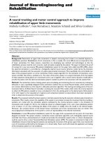
Fig. 3 Assessment of RRS of early-stage BIDC patients. a The ROC curves for RFS prediction. b Kaplan-Meier analysis for RFS of early-stage BIDC
patients. c The distribution of the RRS, patients’ relapse status and biomarker expression in early-stage BIDC
Qiu et al. BMC Cancer
(2019) 19:729
Page 8 of 11
Fig. 4 Kaplan-Meier analysis for RFS using RRS in the subgroups stratified by ER status and endocrine therapy. a Kaplan-Meier curves for earlystage BIDC patients with ER-positive status. b Kaplan-Meier curves for early-stage BIDC patients with ER-negative status. c Kaplan-Meier curves for
ER-positive patients with high risk scores stratified by endocrinotherapy. d Kaplan-Meier curves for ER-positive patients with low risk scores
stratified by endocrinotherapy
with data pertaining to tumour size to predict the risk of
relapse, the relapse score remained statistically significant in a multivariate analysis.
Due to poor compliance of our patients, in the ERpositive subgroups, only 89.72% of patients received
endocrine therapy systematically. The results indicated
that only patients with low risk responded well to endocrine therapy, while those with high risk showed no difference between the treated group and untreated group.
A previous study revealed that mesenchymal-like BCSCs
in hormone-sensitive luminal breast cancers were one of
the reasons for hormone-resistant [50]. Similar to above
finding, there was evidence suggesting that BCSCs
should be partially responsible for the endocrine-resistant capacity of breast cancer cells. This is due to the fact
that CSCs could only respond to treatment by virtue of
paracrine signalling pathway from adjacent differentiated
ER-positive tumour cells [51–54], which were probably
responsible for the endocrine-resistance in the high-risk
group.
The RRS not only offers an approach to predict therapeutic sensitivity but also provides a new perspective to
eliminate BCSCs in early stage breast cancer. As been
reported, BCSCs were not as sensitive to hormone therapy and conventional chemotherapy as non-BCSC tumours. Thus, targeting BCSCs clinically might enhance
the therapeutic sensitivity among patients with high
risk scores. The most promising CSC treatment strategies that target Notch, Hedgehog, Wnt and many
other BCSC self-renewal pathways provide a number of
opportunities for new clinical trials.20 In addition, the
strategy of “destemming” CSCs, including inducing
Qiu et al. BMC Cancer
(2019) 19:729
CSC differentiation or inhibiting self-renewal capacity
were also recommended [55]. Combination of BCSCtargeted therapy and traditional therapy may provide
our patients with high-risk scores more effective therapeutic strategies. However, the study of CSCs remains
an enigma, and further exploration is needed.
In terms of limitations, this study was a retrospective
analysis that selected patients who had not received neoadjuvant chemotherapy after resection in early stage
breast cancer, which may lead to a selection bias of patients with a relative lower risk of recurrence. However,
all our patients included in this study were T1–3N0M0 by
the TNM staging system, and the majority of them did
not receive neoadjuvant chemotherapy, according to the
NCCN guideline [12]. The total study size is modest in
absolute numbers, and some subgroup analyses may be
underpowered; however, this is one of the largest cohorts of well-characterized early stage breast cancer that
employed a BCSC biomarker panel as a prognosis
model. The shortcomings of this panel should not be ignored. First of all, though IHC staining is the most common method for semi-quantified the protein expression
level in carcinomous tissues, the subjectivity of evaluation of this method couldn’t be avoided. Secondly, the
selection of antibodies should be cautiously considered,
as their quality will affect the result of IHC staining directly. Performing immunofluorescence staining and qRT PCR may help us obtain a relative exact result; however, these two methods also have their disadvantages in
assessing BCSCs.
Conclusion
Though previous studies have combined different
BCSCs biomarkers for assessing prognosis in different
types of breast cancer, such as three-negative, HER2positive and metastatic breast cancer [56–59], no
BCSC-associated biomarkers have been combined to
form a model for evaluating the relapse risk of earlystage breast cancer. We propose that BCSCs could be
used as a panel in prognostic or predictive tests of
early-stage breast cancer. Here, we conducted a prospectively designed validation study of a multi-biomarker panel in a cohort of patients with early-stage
BIDC. In addition, this panel is promising for prediction of early-stage BIDC recurrence, the efficacy of
which warrants further validation in a large-scale cohort.
In addition, it reminds us that further consideration is
needed to explore new therapeutic managements for
high-risk patients with therapeutic resistance. In addition,
it is of practical significance that the panel only involves
the use of routine slides of the tumour tissues and five
antibodies, which is not as time-consuming and expensive
as other gene profiles.
Page 9 of 11
Additional files
Additional file 1: Figure S1. Different expression patterns of BSCCs
biomarkers expression pattern in external control and internal control
tissues. A. ALDH1A3 was shown positive in prostate cancer (external
control) and breast invasive ductal carcinoma (IDC, internal positive
control), and shown negative in lymphocytes (internal negative control);
B. PROCR was shown positive in intestine gland (external control) and
ductal carcinoma in situ (DCIS, internal positive control), and shown
negative in lymphocytes (internal negative control); C. CD44 was shown
positive in urothelium (external control) and IDC (internal positive
control), and shown negative in lymphocytes (internal negative control);
D. CD24 was shown positive in urothelium (external control) and IDC
(internal positive control), and shown negative in breast adenosis
(internal negative control); E. EpCAM was shown positive in intestine
gland (external control) and in breast adenosis (internal positive control),
and shown negative in lymphocytes (internal negative control); F. ITGA6
was shown positive in colorectal carcinoma (external control) and in IDC
(internal positive control), and shown negative in lymphocytes (internal
negative control). (JPG 5319 kb)
Additional file 2: Figure S2. The prevalence of BSCCs biomarkers in
reductional mammoplasty samples. A. Prevalence of ALDH1A3 in three in
reductional mammoplasty samples; B. Prevalence of PROCR in three in
reductional mammoplasty samples; C-D. Prevalence of CD44/CD24 in
three in reductional mammoplasty samples; E. Prevalence of EpCAM in
three in reductional mammoplasty samples; F. Prevalence of ITGA6 in
three in reductional mammoplasty samples. (JPG 4739 kb)
Additional file 3: Figure S3. Flow Chart for Construction of RRS model.
(JPG 293 kb)
Additional file 4: Table S1. The detailed information of end-point of
follow-up for local recurrence or distant metastasis. (XLSX 124 kb)
Additional file 5: Table S2. Antibodies used in the cohort of patients.
(DOCX 16 kb)
Abbreviations
BCSCs: Breast cancer stem cells; BIDC: Breast invasive ductal carcinoma;
CI: Confidence intervals; CSC: Cancer stem cell; EMT: Epithelial-tomesenchymal transition; ER: Oestrogen receptor; GEF: Gene expression
profiling; H&E: Haematoxylin and eosin; HER2: Human epidermal growth
factor receptor 2; IHC: Immunohistochemistry; PR: Progesterone receptor;
RFS: Relapse-free survival; ROC: Receiver operating characteristic curve;
RRS: Relapse risk score
Acknowledgements
Here, I’d like to express my appreciation to all those who help me in
writing and reviewing this manuscript. We specially thanked Dr. Bin Wei
and Dr. Ting Lei, who worked in west china hospital, for assisting us for
the IHC evaluation.
Authors’ contributions
Design for the study: FY and HB. Clinical data collection: YQ, XRZ and HZ.
Analysis and interpretation of data: LYW and BF. Clinical sample acquisition
and preparation: YQ LL, FC, LX and FYL. Supervision for the study: FY and HB.
Wrote, reviewed, and/or revised the manuscript: YQ, FY, and HB. All authors
read and approved the final manuscript.
Funding
This work was supported by Key Research and Development Project of
Department of Science & Technology in Sichuan Province (2017SZ0005) and
1.3.5 project for disciplines of excellence, West China Hospital, Sichuan
University (ZYGD18012) which were for excellent person who worked
excellently in the field of breast cancer.
Availability of data and materials
All data generated or analysed during this study are included in this
published article and its supplementary information files.
Qiu et al. BMC Cancer
(2019) 19:729
Ethics approval and consent to participate
Approval for the study was granted by the Clinical Test and Biomedical
Ethics Committee of West China Hospital Sichuan University (No. 2013–191).
And based on the third term in the ethic approval issued on Oct 14 of 2013
the need to obtain informed consent was waived.
Consent for publication
Not applicable.
Competing interests
The authors declare that they have no competing interests.
Author details
Laboratory of Pathology, West China Hospital, Sichuan University, Chengdu,
China. 2Key Laboratory of Transplant Engineering and Immunology, Ministry
of Health, West China Hospital, Sichuan University, Chengdu, China. 3Clinical
Research Center for Breast, West China Hospital, Sichuan University,
Chengdu, China. 4Department of Pathology, West China Hospital, Sichuan
University, Chengdu, China. 5Big Data Research Center, School of Computer
Science and Engineering, University of Electronic Science and Technology of
China, Chengdu, China. 6Laboratory of Molecular Diagnosis of Cancer &
Cancer Center, West China Hospital, Sichuan University, Chengdu, China.
7
West China School of Medicine, Sichuan University, Chengdu, China.
1
Received: 27 June 2018 Accepted: 23 April 2019
References
1. Iqbal J, Ginsburg O, Rochon PA, Sun P, Narod SA. Differences in breast
cancer stage at diagnosis and cancer-specific survival by race and ethnicity
in the United States. JAMA. 2015;313:165–73.
2. Kent C, Horton J, Blitzblau R, Koontz BF. Whose disease will recur after
mastectomy for early stage, node-negative breast cancer? A systematic
review. Clin Breast Cancer. 2015;15:403–12.
3. Verma A, Kaur J, Mehta K. Molecular oncology update: breast cancer gene
expression profiling. Asian J Oncol. 2015;1:65–72.
4. Buyse M, Loi S, van't Veer L, Viale G, Delorenzi M, Glas AM, d'Assignies MS,
Bergh J, Lidereau R, Ellis P, Harris A, Bogaerts J, Therasse P, Floore A, Amakrane
M, Piette F, Rutgers E, Sotiriou C, Cardoso F, Piccart MJ. TRANSBIG consortium.
Validation and clinical utility of a 70-gene prognostic signature for women
with node-negative breast cancer. J Natl Cancer Inst. 2006;98:1183–92.
5. Mook S, Schmidt MK, Viale G, Pruneri G, Eekhout I, Floore A, Glas AM,
Bogaerts J, Cardoso F, Piccart-Gebhart MJ, Rutgers ET, Van't Veer LJ. TRAN
SBIG consortium. The 70-gene prognosis-signature predicts disease
outcome in breast cancer patients with 1-3 positive lymph nodes in an
independent validation study. Breast Cancer Res Treat. 2009;116:295–302.
6. Gnant M, Sestak I, Filipits M, Dowsett M, Balic M, Lopez-Knowles E, Greil R,
Dubsky P, Stoeger H, Rudas M, Jakesz R, Ferree S, Cowens JW, Nielsen T,
Schaper C, Fesl C, Cuzick J. Identifying clinically relevant prognostic
subgroups of postmenopausal women with node-positive hormone
receptor-positive early-stage breast cancer treated with endocrine therapy:
a combined analysis of ABCSG-8 and ATAC using the PAM50 risk of
recurrence score and intrinsic subtype. Ann Oncol. 2015;26:1685–91.
7. Paik S, Shak S, Tang G, Kim C, Baker J, Cronin M, Baehner FL, Walker MG,
Watson D, Park T, Hiller W, Fisher ER, Wickerham DL, Bryant J, Wolmark N. A
multigene assay to predict recurrence of tamoxifen-treated, node-negative
breast cancer. N Engl J Med. 2004;351:2817–26.
8. Paik S, Tang G, Shak S, Kim C, Baker J, Kim W, Cronin M, Baehner FL, Watson
D, Bryant J, Costantino JP, Geyer CE Jr, Wickerham DL, Wolmark N. Gene
expression and benefit of chemotherapy in women with node-negative,
estrogen receptor-positive breast cancer. J Clin Oncol. 2006;24:3726–34.
9. Foekens JA, Atkins D, Zhang Y, Sweep FC, Harbeck N, Paradiso A, Cufer T,
Sieuwerts AM, Talantov D, Span PN, Tjan-Heijnen VC, Zito AF, Specht K,
Hoefler H, Golouh R, Schittulli F, Schmitt M, Beex LV, Klijn JG, Wang Y.
Multicenter validation of a gene expression-based prognostic signature in
lymph node-negative primary breast cancer. J Clin Oncol. 2006;24:1665–71.
10. Sotiriou C, Wirapati P, Loi S, Harris A, Fox S, Smeds J, Nordgren H, Farmer P,
Praz V, Haibe-Kains B, Desmedt C, Larsimont D, Cardoso F, Peterse H,
Nuyten D, Buyse M, Van de Vijver MJ, Bergh J, Piccart M, Delorenzi M. Gene
expression profiling in breast cancer: understanding the molecular basis of
histologic grade to improve prognosis. J Natl Cancer Inst. 2006;98:262–72.
Page 10 of 11
11. Filipits M, Rudas M, Jakesz R, Dubsky P, Fitzal F, Singer CF, Dietze O, Greil R,
Jelen A, Sevelda P, Freibauer C, Müller V, Jänicke F, Schmidt M, Kölbl H,
Rody A, Kaufmann M, Schroth W, Brauch H, Schwab M, Fritz P, Weber KE,
Feder IS, Hennig G, Kronenwett R, Gehrmann M, Gnant M. A new molecular
predictor of distant recurrence in ER-positive, HER2-negative breast cancer
adds independent information to conventional clinical risk factors. Clin
Cancer Res. 2011;17:6012–20.
12. National Comprehensive Cancer Network: Practice Guidelines in Oncology.
Invasive Breast Cancer, version 3. 2018. />guidelines/breast-invasive/.
13. Guo W. Concise review: breast cancer stem cells: regulatory networks, stem
cell niches, and disease relevance. Stem Cells Transl Med. 2014;3:942–8.
14. Al-Hajj M, Wicha MS, Benito-Hernandez A, Morrison SJ, Clarke MF.
Prospective identification of tumorigenic breast cancer cells. Proc Natl Acad
Sci U S A. 2003;100:3983–8.
15. Geng SQ, Alexandrou AT, Li JJ. Breast cancer stem cells: multiple capacities
in tumor metastasis. Cancer Lett. 2014;349:1–7.
16. Bozorgi A, Khazaei M, Khazaei MR. New findings on breast cancer stem cells:
a review. J Breast Cancer. 2015;18:303–12.
17. Smalley M, Piggott L, Clarkson R. Breast cancer stem cells: obstacles to
therapy. Cancer Lett. 2013;338:57–62.
18. Korkaya H, Wicha MS. HER2 and breast cancer stem cells: more than meets
the eye. Cancer Res. 2013;73:3489–93.
19. Reya T, Morrison SJ, Clarke MF, Weissman IL. Stem cells, cancer, and cancer
stem cells. Nature. 2001;414:105–11.
20. Singh SK, Hawkins C, Clarke ID, Squire JA, Bayani J, Hide T, Henkelman RM,
Cusimano MD, Dirks PB. Identification of human brain tumour initiating
cells. Nature. 2004;432:396–401.
21. Ginestier C, Hur MH, Charafe-Jauffret E, Monville F, Dutcher J, Brown M,
Jacquemier J, Viens P, Kleer CG, Liu S, Schott A, Hayes D, Birnbaum D, Wicha
MS, Dontu G. ALDH1 is a marker of normal and malignant human
mammary stem cells and a predictor of poor clinical outcome. Cell Stem
Cell. 2007;1:555–67.
22. Mannello F. Understanding breast cancer stem cell heterogeneity: time to
move on to a new research paradigm. BMC Med. 2013;11:169.
23. Luo M, Clouthier SG, Deol Y, Liu S, Nagrath S, Azizi E, Wicha MS. Breast
cancer stem cells: current advances and clinical implications. Methods Mol
Biol. 2015;1293:1–49.
24. Wang D, Cai C, Dong X, Yu QC, Zhang XO, Yang L, Zeng YA. Identification
of multipotent mammary stem cells by protein C receptor expression.
Nature. 2015;517:81–4.
25. Iqbal J, Chong PY, Tan PH. Breast cancer stem cells: an update. J Clin Pathol.
2013;66:485–90.
26. Oakes SR, Gallego-Ortega D, Ormandy CJ. The mammary cellular hierarchy
and breast cancer. Cell Mol Life Sci. 2014;71:4301–24.
27. Abraham BK, Fritz P, McClellan M, Hauptvogel P, Athelogou M, Brauch H.
Prevalence of CD44+/CD24−/low cells in breast cancer may not be
associated with clinical outcome but may favor distant metastasis. Clin
Cancer Res. 2005;11:1154–9.
28. Ali HR, Dawson SJ, Blows FM, Provenzano E, Pharoah PD, Caldas C. Cancer
stem cell markers in breast cancer: pathological, clinical and prognostic
significance. Breast Cancer Res. 2011;13:R118.
29. Honeth G, Schiavinotto T, Vaggi F, Marlow R, Kanno T, Shinomiya I,
Lombardi S, Buchupalli B, Graham R, Gazinska P, Ramalingam V, Burchell J,
Purushotham AD, Pinder SE, Csikasz-Nagy A, Dontu G. Models of breast
morphogenesis based on localization of stem cells in the developing
mammary lobule. Stem Cell Rep. 2015;4:699–711.
30. Goel HL, Gritsko T, Pursell B, Chang C, Shultz LD, Greiner DL, Norum JH,
Toftgard R, Shaw LM, Mercurio AM. Regulated splicing of the α6 integrin
cytoplasmic domain determines the fate of breast cancer stem cells. Cell
Rep. 2014;7:747–61.
31. Lim E, Vaillant F, Wu D, Forrest NC, Pal B, Hart AH, Asselin-Labat ML, Gyorki
DE, Ward T, Partanen A, Feleppa F, Huschtscha LI, Thorne HJ, kConFab, Fox
SB, Yan M, French JD, Brown MA, Smyth GK, Visvader JE, Lindeman GJ.
Aberrant luminal progenitors as the candidate target population for basal
tumor development in BRCA1 mutation carriers. Nat Med. 2009;15:907–13.
32. Turner NC, Reis-Filho JS. Basal-like breast cancer and the BRCA1 phenotype.
Oncogene. 2006;25:5846–53.
33. Beaulieu LM, Church FC. Activated protein C promotes breast cancer cell
migration through interactions with EPCR and PAR-1. Exp Cell Res. 2007;313:
677–87.
Qiu et al. BMC Cancer
(2019) 19:729
34. Spek CA, Arruda VR. The protein C pathway in cancer metastasis. Thromb
Res. 2012;129:S80–S4.
35. Qiu Y, Pu T, Li L, Cheng F, Lu C, Sun L, Teng X, Ye F, Bu H. The expression of
aldehyde dehydrogenase family in breast cancer. J Breast Cancer. 2014;17:54–60.
36. Yan Q, Zhong X, Zhang Z, Bing W, Feng Y, Hong B. Prevalence of protein C
receptor (PROCR) is associated with inferior clinical outcome in breast
invasive ductal carcinoma. Pathol Res Pract. 2017;213:1173–9.
37. Ye F, Zhong X, Qiu Y, Yang L, Wei B, Zhang Z, Bu H. ITGA6 can act as a
biomarker for local or distant recurrence in breast cancer. J Breast Cancer.
2017;20:142–9.
38. Wolff AC, Hammond ME, Hicks DG, Dowsett M, LM MS, Allison KH, Allred DC,
Bartlett JM, Bilous M, Fitzgibbons P, Hanna W, Jenkins RB, Mangu PB, Paik S,
Perez EA, Press MF, Spears PA, Vance GH, Viale G, Hayes DF, American Society
of Clinical Oncology; College of American Pathologists. Recommendations for
human epidermal growth factor receptor 2 testing in breast cancer: American
Society of Clinical Oncology/College of American Pathologists clinical practice
guideline update. Arch Pathol Lab Med. 2014;138:241–56.
39. Qureshi A, Pervez S. Allred scoring for ER reporting and it's impact in clearly
distinguishing ER negative from ER positive breast cancers. J Pak Med
Assoc. 2010;60:350–3.
40. Spizzo G, Obrist P, Ensinger C, Theurl I, Dünser M, Ramoni A, Gunsilius E, Eibl
G, Mikuz G, Gastl G. Prognostic significance of ep-CAM AND Her-2/neu
overexpression in invasive breast cancer. Int J Cancer. 2002;98:883–8.
41. Sun J, Chen X, Wang Z, Guo M, Shi H, Wang X, Cheng L, Zhou M. A
potential prognostic long non-coding RNA signature to predict metastasisfree survival of breast cancer patients. Sci Rep. 2015;5:16553.
42. Hu Z, Chen X, Zhao Y, Tian T, Jin G, Shu Y, Chen Y, Xu L, Zen K, Zhang C,
Shen H. Serum microRNA signatures identified in a genome-wide serum
microRNA expression profiling predict survival of non-small-cell lung cancer.
J Clin Oncol. 2010;28:1721–6.
43. Mohebian MR, Marateb HR, Mansourian M, Mañanas MA, Mokarian FA. A
hybrid computer-aided-diagnosis system for prediction of breast cancer
recurrence (HPBCR) using optimized ensemble learning. Comput Struct
Biotechnol J. 2017;15:75–85.
44. Sheridan C, Kishimoto H, Fuchs RK, Mehrotra S, Bhat-Nakshatri P, Turner CH,
Goulet R Jr, Badve S, Nakshatri H. CD44+/CD24- breast cancer cells exhibit
enhanced invasive properties: an early step necessary for metastasis. Breast
Cancer Res. 2006;8:R59.
45. Ye F, Qiu Y, Li L, Yang L, Cheng F, Zhang H, Wei B, Zhang Z, Sun L, Bu H.
The presence of EpCAM(−)/ITGA6(+) cells in breast cancer is associated with
a poor clinical outcome. J Breast Cancer. 2015;18:242–8.
46. Ghebeh H, Sleiman GM, Manogaran PS, Al-Mazrou A, Barhoush E, AlMohanna FH, Tulbah A, Al-Faqeeh K, Adra CN. Profiling of normal and
malignant breast tissue show CD44high/CD24low phenotype as a
predominant stem/progenitor marker when used in combination with epCAM/ITGA6 markers. BMC Cancer. 2013;13:289.
47. Cianfrocca M, Goldstein LJ. Prognostic and predictive factors in early-stage
breast cancer. Oncologist. 2004;9:606–16.
48. Bill R, Christofori G. The relevance of EMT in breast cancer metastasis:
correlation or causality? FEBS Lett. 2015;589:1577–87.
49. Mallini P, Lennard T, Kirby J, Meeson A. Epithelial-to-mesenchymal transition:
what is the impact on breast cancer stem cells and drug resistance. Cancer
Treat Rev. 2014;40:341–8.
50. Creighton CJ, Li X, Landis M, Dixon JM, Neumeister VM, Sjolund A, Rimm
DL, Wong H, Rodriguez A, Herschkowitz JI, Fan C, Zhang X, He X, Pavlick A,
Gutierrez MC, Renshaw L, Larionov AA, Faratian D, Hilsenbeck SG, Perou CM,
Lewis MT, Rosen JM, Chang JC. Residual breast cancers after conventional
therapy display mesenchymal as well as tumor-initiating features. Proc Natl
Acad Sci U S A. 2009;106:13820–5.
51. O'Brien CS, Howell SJ, Farnie G, Clarke RB. Resistance to endocrine therapy:
are breast cancer stem cells the culprits? J Mammary Gland Biol Neoplasia.
2009;14:45–54.
52. O'Brien CS, Farnie G, Howell SJ, Clarke RB. Breast cancer stem cells and their
role in resistance to endocrine therapy. Horm Cancer. 2011;2:91–103.
53. Stone A, Musgrove EA. Endocrine therapy: defining the path of least
resistance. Breast Cancer Res. 2014;16:101.
54. Arif K, Hussain I, Rea C, El-Sheemy M. The role of Nanog expression in
tamoxifen-resistant breast cancer cells. Onco Targets Ther. 2015;8:1327–34.
55. Wang T, Shigdar S, Gantier MP, Hou Y, Wang L, Li Y, Shamaileh HA, Yin W,
Zhou SF, Zhao X, Duan W. Cancer stem cell targeted therapy: progress amid
controversies. Oncotarget. 2015;6:44191–206.
Page 11 of 11
56. Yang F, Cao L, Sun Z, Jin J, Fang H, Zhang W, Guan X. Evaluation of breast
Cancer stem cells and Intratumor Stemness heterogeneity in triple-negative
breast Cancer as prognostic factors. Int J Biol Sci. 2016;12:1568–77.
57. Seo AN, Lee HJ, Kim EJ, Jang MH, Kim YJ, Kim JH, Kim SW, Ryu HS, Park IA,
Im SA, Gong G, Jung KH, Kim HJ, Park SY. Expression of breast cancer stem
cell markers as predictors of prognosis and response to trastuzumab in
HER2-positive breast cancer. Br J Cancer. 2016;114:1109–16.
58. Oon ML, Thike AA, Tan SY, Tan PH. Cancer stem cell and epithelialmesenchymal transition markers predict worse outcome in metaplastic
carcinoma of the breast. Breast Cancer Res Treat. 2015;150:31–41.
59. Giordano A, Gao H, Anfossi S, Cohen E, Mego M, Lee BN, Tin S, De
Laurentiis M, Parker CA, Alvarez RH, Valero V, Ueno NT, De Placido S, Mani
SA, Esteva FJ, Cristofanilli M, Reuben JM. Epithelial-mesenchymal transition
and stem cell markers in patients with HER2-positive metastatic breast
cancer. Mol Cancer Ther. 2012;11:2526–34.