Vasculogenic mimicry-associated ultrastructural findings in human and canine inflammatory breast cancer cell lines
Bạn đang xem bản rút gọn của tài liệu. Xem và tải ngay bản đầy đủ của tài liệu tại đây (2.33 MB, 9 trang )
Barreno et al. BMC Cancer
(2019) 19:750
/>
RESEARCH ARTICLE
Open Access
Vasculogenic mimicry-associated
ultrastructural findings in human and
canine inflammatory breast cancer cell lines
Lucía Barreno1, Sara Cáceres2, Ángela Alonso-Diez1, Ana Vicente-Montaña3, María Luisa García3, Mónica Clemente1,
Juan Carlos Illera2 and Laura Peña1*
Abstract
Background: Human inflammatory breast cancer (IBC) and canine inflammatory mammary cancer (IMC) are the
most lethal mammary cancers. An exacerbated angiogenesis and the existence of vasculogenic mimicry (VM) are
hallmarks of these tumors. The information regarding VM and ultrastructural characteristics of mammary cell lines is
scant.
Methods: In this study, IBC cell line SUM149 and IMC cell line IPC-366 in adherent (2D) and non-adherent (3D)
(mammospheres, cancer stem cells) conditions were analyzed by transmission and scanning electron microscopy
(TEM and SEM, respectively).
Results: The TEM revealed round to oval shape cells with microvilli on the surface, high numbers of peroxisomes in
close apposition to lipid droplets and some extracellular derived vesicles. The TEM and the SEM mammospheres
revealed group of cells clumping together with a central lumen (resembling a mammary acini). The cells joint are
tight junctions and zonula adherens. By SEM two cell morphologies were observed: spherical and flattened cells.
There was evidence endothelial-like cells (ELCs), which is characteristic for this disease, showing several or unique
cytoplasmic empty space. ELCs were more frequent in 3D than in 2D culture conditions and contained WeibelPalade cytoplasmic bodies, which are exclusive structures of endothelial cells.
Conclusions: Both cell lines, IPC-366 and SUM-149, shared ultrastructural characteristics, further supporting canine
IMC as a model for the human disease. To the best of our knowledge, this is the first study that demonstrate the
morphological differentiation of cultured cancer stem cells from cancer epithelial cell lines into endothelial-like cells,
confirming the vasculogenic mimicry phenomenon from an ultrastructural point of view.
Keywords: Vasculogenic mimicry, Inflammatory breast cancer, Mammospheres, Canine, Electron microscopy, Comparative
oncology
Background
Human inflammatory breast cancer (IBC) and canine
inflammatory mammary cancer (IMC) are the most
aggressive mammary neoplasms and are associated to
poor prognosis in both species [1–5]. The criterium for
histological diagnosis for IBC and IMC is the enormous
neoplastic embolization of dermal lymphatic vessels
which blockade lymphatic drainage originating the
* Correspondence:
1
Veterinary Clinical Hospital, Pathology Service, Complutense University of
Madrid, Madrid, Spain
Full list of author information is available at the end of the article
distinctive edema [4, 6–9]. The clinical form is characterized by a sudden presentation of erythema, firmness,
warmth and pain resembling an inflammatory process
and, therefore, this condition can be misdiagnosed with
a dermatitis or mastitis, especially if a mammary nodule
is absent [1, 2, 4–7]. Numerous epidemiologic, clinical
and histopathological characteristics are shared by IBC
and IMC, being the latter a good spontaneous animal
model for the study of IBC [5, 10, 11].
Characteristically, exacerbated angiogenesis, lymphangiogenesis, lymphangiotropism and vasculogenic mimicry (VM) are found in IBC and IMC [5, 9, 10, 12, 13].
© The Author(s). 2019 Open Access This article is distributed under the terms of the Creative Commons Attribution 4.0
International License ( which permits unrestricted use, distribution, and
reproduction in any medium, provided you give appropriate credit to the original author(s) and the source, provide a link to
the Creative Commons license, and indicate if changes were made. The Creative Commons Public Domain Dedication waiver
( applies to the data made available in this article, unless otherwise stated.
Barreno et al. BMC Cancer
(2019) 19:750
In order to grow and metastasize, tumors require a
proper oxygen and nutrients supply. The angiogenic
process (sprouting angiogenesis) is relatively complex and
it is regulated by numerous pro- and anti-angiogenic
factors, standing out the VEGF family and their receptors
[14]. VEGF-A is an angiogenic marker that is overexpressed in IBC/IMC and it is present in normal endothelial cells but also in neoplastic cells [10, 12]. According to
our previous study, both cell lines overexpress VEGF-A
and contributes to the exacerbated angiogenesis [15].
There is an intensive research going on in order to find
effective anti-angiogenesis drugs, and more than 300
angiogenesis inhibitors have been identified [16]. Unfortunately, the efficacy of angiogenesis inhibitors in cancer is
limited by resistance mechanisms that are poorly understood [17]. Furthermore, multiple studies have used angiogenesis inhibitors as adjuvant therapy and they have failed
to provide significant benefits to patients [18].
Angiogenesis is not an exclusive method to nourish
tumor tissues. Besides sprouting angiogenesis, that is induced by VEGF-A and is also found in non-neoplastic
tissues, two mechanisms of blood supply and metastasis
have been discovered in the last years to be exclusive of
highly aggressive neoplasms: vasculogenic mimicry (VM)
and vascular co-option (VCO) [17, 18]. In VM, cancer
stem cells induce tumor neovascularization by their
transformation into endothelial-like cells [19]. In VCO
cancer cells closely adhere preexisting blood vessels or
capillaries to obtain nutrients and oxygen and further
develop sprouting angiogenesis. Hypothetically, both VM
and VCO would explain the failure of antiangiogenic
therapies while VCO would be essential in the metastatic
growth [17, 18].
VM is the formation of vascular channels lined by
highly malignant neoplastic cells that gain endothelial
cells characteristics and are supposed to play an important role in the mechanisms of tumor invasion and metastasis [19–21]. Initially, vessels formed by VM are
lined by a mixed of tumor cells and endothelial cells that
gradually transform in tumor cells only. These VM
newly formed vessels connect with preexisting vessels
[19]. Hence, VM is an auspicious target for the developing of new anti-cancer therapy strategies. VM is prognostic characteristic in human oncology having patients
with VM a poor clinical outcome [18, 21].
VM is related to the presence of the so-called endothelial-like cells (ELCs) [9]. Endothelial cells store the
procoagulant glycoprotein von Willebrand Factor (vWF)
in elongate dense granules, known as Weibel-Palade
bodies (WPb) which are key for the identification of
endothelial cells by electron microscopy [22].
Several human IBC cell lines such as SUM149, have
been established in order to study the in vitro mechanisms of this special type of breast cancer [23, 24].
Page 2 of 9
Similarly, the IPC-366 is the unique canine IMC cell line
established [25] and has demonstrated to be a good
model in comparison with its human counterpart
SUM149 [15]. Human SUM149 and canine IPC-366 are
triple negative (ER-, PR-, HER2-) epithelial cell lines,
with high rates of cell growth in adherent (2D) and nonadherent (3D) conditions and metastatic capacity in
mice models [15]. The expression of CD146, a marker of
endothelial lineage stem cells, has been related in both
cell lines to the presence of VM, due to the existence of
CD146 positive endothelial-like cells lining the newlyformed VM channels [15]. Nevertheless, according to
some authors, these VM cells could not express endothelial cell markers [18, 20].
Mammospheres, clusters of mammary cell lines growing in 3D, are formed by breast cancer stem cells
(BCSC) [26] that constitute multipotent cells that have
the capacities of self-renewal, differentiation, unlimited
growth and can give rise to phenotypically different neoplastic subpopulations [27]. Mammospheres of SUM149
and IPC-366 cell lines exhibit a very similar immunophenotype for the expression of stem cells markers [15].
Microscopic study of 3D cultures and xenotransplanted
mice tumors from SUM149 and IPC-366 mammospheres have also revealed the presence of endotheliallike cells (ELCs) indicating that BCSC have the potential
to transform into ELCs in vitro and in vivo (VM) [15].
There is little information regarding ultrastructural characteristics of neoplastic mammary cell lines in adherent
conditions (2D) [28–30] and the ultrastructural characteristics of mammospheres (3D) are unknown [31–33].
To the best of our knowledge, there are no previous
studies on the ultrastructural features of ELCs neither in
cancer tissues nor cancer cell lines.
The aims of this study were to analyze by transmission
and scanning electron microscopy (TEM and SEM), the
human IBC cell line (SUM149) and the canine IMC cell
line (IPC-366) in adherent (2D) and non-adherent (3D)
conditions in order to compare the morphological characteristics of both cell lines for the better understanding
of their biology and to further support the IPC-366 cell
line as a good comparative model for human IBC. Another hypothesis to confirm, is the possible identification
of neoplastic epithelial cells showing ultrastructural
characteristics of endothelial cells.
Methods
Cell lines cultures in adherent conditions
SUM149 triple negative (ER−, PR−, HER-2−) human inflammatory breast carcinoma cell line was obtained from
Asterand, Plc. (Detroit, Michigan, USA) in 2015, was
maintained in Ham’s F-12 media supplemented with
10% fetal bovine serum (FBS) (Sigma Aldrich, Madrid,
Spain),1 μg mL−1 hydrocortisone, 5 μg mL−1 insulin and
Barreno et al. BMC Cancer
(2019) 19:750
Page 3 of 9
1% penicillin–streptomycin solution and 1% amphotericin B (Sigma Aldrich, Madrid, Spain). Triple negative
canine inflammatory mammary carcinoma cell line,
established and maintained in our laboratory [25], IPC366 (commercially available by Applied Biological Materials, ref. T8202) was cultured in Dulbecco’s modified
Eagle medium nutrient mixture F-12 Ham (DMEM/F12)
containing 10% (FBS), 1% penicillin streptomycin solution and 1% L-glutamine (Sigma Aldrich, Madrid, Spain).
Both cell lines were cultured in 25-cm2 culture flasks
and maintained in a humidified atmosphere of 5% carbon dioxide at 37∘C. The cell cultures were observed
daily by a phase-contrast microscopy to check cell viability and growth.
Scanning electron microscopy
Cell lines cultures in non-adherent conditions:
mammosphere formation assay
Results
In order to obtain the primary mammospheres, SUM149
and IPC-366 adherent cells were trypsinized, and the
resultant single cells were seeded in 6-well ultra-low
attachment plates (1×10⁴ and 2×10⁴ cells mL−1)(Corning; New York, NY, USA) [23, 26, 34] in serum-free
MEM supplemented with 20 ng mL−1 bFGF (basic fibroblast growth factor), 20ng mL−1 EGF (epidermal growth
factor) and 1× B27 (serum-free supplement) (Invitrogen,
Madrid, Spain) enriched media and incubated for 7 days.
Then, the mammospheres were stained with MTT [3-(4,
5-dimethylthiazolyl-2)-2, 5-diphenyltetrazolium bromide] (Invitrogen, Madrid, Spain) to improve visualization
before they were counted using a Gel-count colony
counter (Oxford Optronix, Oxford, UK). After 1 week of
culture, the first generation of mammospheres were harvested from the cultures and counted with a minimum
size of 50 μm. The resulting mammospheres were dissociated into single cells, re-cultured through passages and
counted every week.
Transmission electron microscopy
For the TEM, eight pellets were obtained (two for each
cell line and type of culture adherent and non-adherent)
and fixed with 2.5% glutaraldehyde (EMS) and 4% paraformaldehyde (EMS) solution. Then, the cells were incubated with 0.1 M Milloning´s buffer 4°C overnight,
treated with 2% osmium tetroxide (Panreac) and 3%
ferrocyanide (Panreac) solution (diluted in PBS) for 1h.
Subsequently, they were washed with distilled water and
dehydrated in acetones of increasing percentage (30, 50,
70, 80, and 100%). The samples were gradually infiltrated
in a Müllenhauer mixture resin (SPURR resin, TAAB),
and solidified at 60 8°C for 48h. The embedded cells
were ultrasectioned, observed and photographed at the
National Electron Microscopy Center (Madrid) by
means of a JEOL JEM 1010 transmission electron
microscope.
The mammospheres of each cell line contained in a 6well ultra-low attachment plate were fixed for 3 hours at
4ºC in 4 % paraformaldehide and 2,5% glutaraldehyde/0,
1 M Milloning’s buffer (pH 7.2). Cells were washed twice
in distilled water and post-fixed for 1 hour in buffered
1% osmium tetroxide. The samples were dehydrated in
an ascendant series of ethanol solution (30%, 50%, 70%,
80% and 100%). Finally, the samples were dried using a
critical point dryer (Leika EM CPD 300). The dried samples were sputtered with a 6 nm layer of gold using a
Quorum Q150RS. Observation and photographs were
made using a JEOL JSM 6400 scanning electron
microscope.
Transmission Electron Microscopy (TEM)
Cell lines cultures (SUM149 and IPC-366) in adherent
conditions (2D)
The pellets from the SUM149 and IPC-366 cell lines in
adherent cultures, shared very similar characteristics. Both
cell lines contained a majority of large individualized cells
and some groups of joint round to oval cells, showing several malignant features such as: marked anisocytosis and
anisocaryosis, with varying nuclear-cytoplasmic ratios, one
or two prominent nucleoli and some atypical mitoses. Binucleate and multinucleated cells were frequently observed.
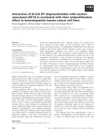
All cells exhibited numerous well developed “digit-like”
microvilli or cytoplasmic processes at the cytoplasmic
membrane, which did not contain actin or myosin filaments (Fig. 1).
Inside the cytoplasm, high numbers of clear lipid
droplets surrounded by numerous spheroid organelles
(150-250 nm), containing a slightly electron dense
matrix with fine granules (peroxisomes and microperoxisomes) were frequent. In some cases, peroxisomes contained an electron dense core (crystalline catalase/uric
acid oxidase) (Fig. 1).
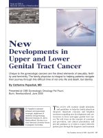
A hallmark finding in both cell lines was the presence
of cells with a large unique or small multiple coalescent
cytoplasmic clear empty spaces surrounded by cytoplasmic membrane with an elongated eccentric nucleus or
nuclei, resembling morphologically a single–endothelial
cell capillary vessel (endothelial-like cells, ELCs) (Fig. 2).
Cell lines cultures (SUM149 and IPC 366) in non-adherent
conditions (3D)
Ultrastructural features of SUM 149 and IPC-366 in
non-adherent cultures (mammospheres) were very similar, but differed from their adherent counterpart in the
presence of groups of cells (Fig. 3) and the existence of
more abundant endothelial–like cells (ELCs).
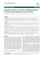
Higher magnification of the group of cells revealed the
intercellular junctions: D) tight-junctions and E) zonula
Barreno et al. BMC Cancer
(2019) 19:750
Page 4 of 9
Fig. 1 Transmission electron microscopy of SUM149 (a, b) and IPC-366 (c) in adherent conditions (2D). Large individualized round cells showing
cytoplasmic membrane processes (microvilli) and marked anisocaryosis and anisocytosis and prominent nucleoli. d, e IPC-366. Peroxisomes
(arrow) in close apposition to lipid droplets (asterisks). Original magnification; a, b × 6,000, c × 4,000, d × 12,000, e × 50,000
adherens. In tight junctions, also named zonula occludens, lateral cell cytoplasmic membranes of two adjoining cells come together and fuse with resultant
obliteration of the intercellular space. In zonula adherens, also named belt desmosome, the intercellular space
(approximately 200 A) is occupied by homogeneous, apparently amorphous material of low density, and there
are conspicuous bands of dense material in the subjacent
cytoplasmic matrix (Fig. 3). True desmosomes were not
observed.
In general, the cytoplasms contained abundant organelles
(mitochondria, Golgi apparatus (G), rough endoplasmic
reticulum (RER) abnormally distributed and frequently
swollen and degenerated. Nuclei were frequently irregular
Fig. 2 Transmission electron microscopy of IPC-366 (a, b) in adherent conditions (2D) and SUM149 (c, d, e, f) in non-adherent conditions
(mammospheres). a and b: Endothelial-like cells (ELCs) in formation. Multiple empty cytoplasmic spaces (arrows), with microvilli covered by
cytoplasmic membrane (insert) and nucleus margination. c, d and e: ELCs showing the characteristic morphology: a unique cytoplasmic empty
space and eccentric nucleus. f: ELC cytoplasm with Weibel-Palade bodies (arrows). Original magnification; a, d) × 6,000, b) × 10,000, c) × 3,000, e)
× 4,000, f) × 60,000
Barreno et al. BMC Cancer
(2019) 19:750
Page 5 of 9
Fig. 3 Transmission electron microscopy of IPC-366 (a, d, e) and SUM 149 (b, c, f) mammospheres. d is magnified in e. Groups of joined cells by tightjunctions (TJ) and belt desmosomes (zonula adherens, ZA). Rough endoplasmic reticulum (RER). Swollen and degenerate mitochondrias (M).
Autophagic vacuole (AFV). Membrane-derived vesicle (EV). Original magnification; a, b) X 4,000, c) X 6,000, d) X 30,000, e) X 100,000, f) X 60,000
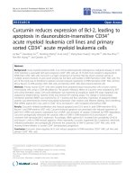
Fig. 4 Scanning electron microscopy of IPC-366 (a, b, c, e) and SUM149 (d, f) mammospheres. a Joint cells covered by numerous cytoplasmic
projections (microvilli). b Magnification of microvilli. c and d Spherical and flattened cells, respectively. e and f Mammary acini-like structures
Barreno et al. BMC Cancer
(2019) 19:750
and indented in shape, with predominant euchromatin and
less abundant heterochromatin, mostly attached to the
inner nuclear membrane. Abundant intermediate filaments,
up to 10 nm diameter, were also present. Scattered autophagic vacuoles with double membranes containing remains
of cellular organelles and abundant myeloid bodies were
present. Some neoplastic cells created and shed external
round membrane vesicles, identified as extracellular derived
vesicles (EVs), specifically exosomes (up to 50-60 nm in
diameter). Exosomes were detected in the cytoplasm, close
to the cell membranes or in the extracellular medium
encircled by cytoplasmic processes (Fig.3).
Some ELCs in mammospheres contained intracytoplasmic tubular elongated membrane-bound structures,
measuring up to 2000-3000 nm in length and 200 nm
thick, showing parallel alignment of internal striations
identified as Weibel-Palade bodies (WPb) (Fig. 2).
Scanning Electron Microscope (SEM)
Cell lines cultures (SUM149 and IPC-366) mammospheres
Mammospheres of both cell lines showed groups of cells
with multiple cytoplasmic projections over the surface.
Occasionally, these structures appeared arranged around
a lumen-like structure and less frequently the cells appeared isolated. There were two cellular shapes: rounded
and flattened cells. The surface of some cells seemed to
have extruded through the membrane boundary, originating plasma membrane blebs (Fig. 4).
Discussion
Human inflammatory breast cancer (IBC) and canine inflammatory mammary cancer (IMC) are comparable diseases [5, 10, 11]. IBC/IMC is a very aggressive type of
breast cancer with poor prognosis [1–5]. IBC/IMC has
specific carcinogenic mechanisms, including high rates
of metastasis and invasiveness that still are poorly
understood. In order to study the “inflammatory” phenotype from a mechanistic point of view, several IBC (i.e.
SUM 149) cell lines have been established [23, 24]. IPC366, a canine IMC cell line, has been demonstrated to
share similar characteristics with its human counterpart,
the IBC cell line SUM149 [15]. The literature regarding
ultrastructural features of mammary cell lines is scant
[28–30, 32]. To the best of our knowledge, this is the
first report in which human and canine inflammatory
mammary cell lines are ultrastructurally compared in adherent (2D) and non-adherent (3D) conditions. Few
studies refer the ultrastructural morphology of the IBC
mammospheres [31, 33].
In highly malignant neoplasms, the presence of vascular channels lined up by disregulated neoplastic cells has
been found and defined as vasculogenic mimicry (VM)
[35]. VM was firstly described in human melanoma [20]
and has been found to be frequent in IBC/IMC. VM has
Page 6 of 9
been identified in both cell lines (SUM149, IPC-366),
showing cells with endothelial-like morphology (ELCs)
[15]. SUM-149 and IPC-366 cells have the potential to
differentiate into endothelial-like cells (ELCs) in vitro
and in vivo [15]. The ability of cancer stem cells to
transform into endothelial cells has been previously reported [36]. In the present study, both cell lines, 2D and
3D, contained cells with a large unique cytoplasmic
empty space that marginated the nuclei to the periphery
resembling one capillary endothelial cells (endotheliallike cells, ELCs) [9]. Other cells had several small cytoplasmic empty spaces, interpreted as forming ELCs,
according to the previously published ultrastructural
morphology of endothelial cells in formation [37],
although their morphology has not been studied yet. By
SEM, two cellular shapes appeared: rounded and flattened cells. The latter ones are compatible with endothelial-like cells.
The present descriptive study can only address the
morphology of the cells, however, there are previous
studies on these two cell lines that support the molecular transformation of these cultured cells, with stem cells
phenotype, into ELCs [15, 25]. IPC-366 cells, including
ELCs, were intensely positive for COX-2 [25], which is
considered a marker for ELCs involved in VM [10, 38]
and a stem cell marker [39]. Moreover, SUM149 and
IPC-366 expressed CD146 [15], a cell adhesion molecule
specific marker for endothelial cell lineage [40]. Nevertheless, according to previous studies, it is possible that
the VM cells would not be able to express endothelial
cell markers [18, 20]. ELCs immunostaining with CD31
in IMC primary tumors, was inconclusive, and considered mostly negative [9]. The negative result of the ELCs
for CD31 is in agreement with previous similar studies in
human intraocular melanoma [20] and human IBC xenograft [41] . Furthermore, in several human clinical studies,
the presence of CD31+ cells in VM is controversial [18].
According to the present results, both cell lines can
acquire also unequivocal ultrastructural features of
endothelial cells, since some ELCs in mammospheres
exhibited Weibel-Palade cytoplasmic bodies (WPb). By
definition, WPb are specific endothelial cells cytoplasmic
structures that store von Willebrand factor (vWF) that is
required for correct hemostasis [42, 43]. WPb has also a
role in inflammation, vascular distention and angiogenesis [44]. Furthermore, vWF and WPb formation are
regulated by the RER and G complex [44]. Accordingly,
WPb often appeared in close apposition to RER and G
complex.
Excluding the ELCs, the rest of neoplastic cells of both
cell lines had similar morphological features as previously published in non IBC/IMC breast cancer cell lines
by means of transmission and scanning electron microscopy [28–33, 45].
Barreno et al. BMC Cancer
(2019) 19:750
The results of the present study revealed that both cell
lines have similar ultrastructural features; by transmission electron microscopy (TEM), in 2D and 3D cultures.
Both, SUM149 and IPC-366 cell lines were round to oval
cells with numerous surface microvilli, a high nuclearcytoplasmic ratio, marked anisocytosis and anisocaryosis,
abundant peroxisomes and the presence of frequent
highly malignant multinucleated cells and endotheliallike cells (ELCs). Although normal mammary epithelial
cells have cytoplasmic microvilli, it has been exhibited
by TEM and Scanning Electron Microscopy (SEM) that
both cell lines presented an exacerbated formation of
microvilli over the surface. This special feature represents a dramatic increase of the cell surface and could
be a reflection of a more malignant, efficient or abundant connection from the cells to the external medium
[46, 47] . The characteristic presence of euchromatin is
predominant in cancer cells and is attributable to the high
percentage of cells in DNA synthesis phase (S phase) [48].
An interesting finding observed in both cell lines was
the intracytoplasmic high number of peroxisomes closely
located to lipid droplets. Peroxisomes have an important
role in the lipid metabolism. These organelles contain
large amounts of oxidases that catalyze the oxidation of
long chain saturated fatty acids to acetyl- CoA [49, 50].
In general, great amount of peroxisomes are found in
cells that synthetize, metabolize or store lipids and/or
steroid hormones, such as cells of the adrenal gland cortex, Leydig-cells, corpus-luteum-cells, fat cells and epithelial cells of the gut [51]. A significant high content of
steroid hormones have been indicated in tumor samples
and serum of dogs with IMC [52–54]. Also, the secretion of steroid hormones (progesterone, estrone sulfate,
estradiol, androstenedione and testosterone) by SUM149
and IPC-366 in vitro cell lines has been recently described [55]. Thus, also could explain the high content
of cytoplasmic peroxisomes in SUM149 and IPC-366.
By TEM it was observed that cells of SUM149 and
IPC-366 mammospheres were frequently joined together
by tight junctions and belt desmosomes (zonula adherens). The cell to cell epithelial molecule adhesion Ecadherin is typically present in zonula adherens associated with intracellular actin microfilaments [56]. Interestingly, in contrast with other metastatic epithelial
cancers that loss E-cadherin, IBC typically overexpress
E-cadherin in the metastatic process [57, 58]. IBC cell
line SUM149 [59] and IMC cell line IPC-366 [25] also
overexpress E-cadherin. By SEM, both cell lines mammospheres showed groups of joined cells, and frequently
appeared as acini-like structures with a central lumen.
Extracellular derived vesicles (EVs) are membrane-limited vesicles that are released into the extracellular
microenvironment that are abnormally increased in cancer cells [60, 61]. Their role is still unknown; EVs
Page 7 of 9
contain diverse small molecules as proteins, lipids,
microRNAs, mRNA and DNA fragments [62] and participate in intercellular communication [63]. The knowledge about the EVs is rapidly expanding and they are
considered important as potential breast cancer biomarkers and therapeutic targets [64]. In cancer, EVs promote proliferation [65–67], migration [68], angiogenesis
[69], invasion and metastases [68], as well as induction
of epithelial-to-mesenchymal transition (EMT) [70]. In
the present study, abundant number of EVs in SUM149
and IPC-366 mammospheres were detected by TEM.
Additionally, by SEM, small round vesicles extruded on
the surface were observed; this structures are considered
compatible with EVs according to the size of the vesicles
(from 50 nm to 2 μm) and some of them were identified
as apoptotic bodies [71]. Stem cells are an abundant
source of EVs [61]. As previously reported, SUM149 and
IPC-366 cell lines in non-adherent (3D) cultures, exhibited similar immunophenotype for the expression of
stem cells markers. In veterinary medicine, very little is
known on cancer‐derived EVs. There is only a preliminary investigation on extracellular vesicles in canine and
feline mammary cancer [72]. Further studies are necessary to isolate, identify and characterize EVs from IBC/
IMC cell lines.
Conclusions
In summary, this investigation has provided evidence
that SUM-149 and IPC-366 share ultrastructural characteristics, supporting canine IMC as a model for the human disease. This study revealed for the first time, the
morphological differentiation of cultured cancer stem
cells from epithelial cell lines into endothelial- like cells,
showing ultrastructural characteristics of endothelial
cells and confirming the presence of the vasculogenic
mimicry phenomenon.
Abbreviations
2D: Adherent conditions; 3D: Non-adherent conditions; AFV: Autophagic
vacuole; BCSC: Breast cancer stem cell; bFGF: Basic fibroblast growth factor;
DMEM: Dulbeccos’s modified Eagle medium; EGF: Epidermal growth factor;
ELCs: Endothelial-like cells; EMT: Epithelial-to-mesenchymal transition;
ER: Estrogen receptor; EVs: Membrane-derived vesicles; FBS: Fetal bovine
serum; G: Golgi apparatus; HER2: Human epidermal growth factor receptor;
IBC: Inflammatory breast cancer; IMC: Inflammatory mammary cancer;
M: Mitochondria; MEM: Minimum Essential Medium; PBS: Phosphate-buffered
saline; PR: Progesterone receptor; RER: Rough endoplasmic reticulum;
SEM: Scanning electron microscopy; TEM: Transmission electron microscopy;
TJ: Tight junction; VCO: Vascular co-option; VM: Vasculogenic mimicry;
VWF: Von Willebrand Factor; WPb: Weibel- Palade body; ZA: Zonula adherens
Acknowledgements
The authors thank to Veterinary Clinical Hospital Pathology Service, Dept. of
animal Physiology and National Center of Electron Microscopy.
Authors’ contributions
LB: involvement in drafting the manuscript, design, interpretation of
ultrastructural images and data. SC: cellular lines maintenance and laboratory
procedures. AAD: involved in cellular lines maintenance and laboratory
procedures. AVM: process of samples for EM and acquisition of ultrastructural
Barreno et al. BMC Cancer
(2019) 19:750
images. MG: process of samples for EM and acquisition of ultrastructural
images. MC: laboratory data. JCI: involved in cellular lines maintenance and
laboratory procedures. LP: conception and design of the study, technical
procedure, acquisition of ultrastructural data and analysis. Elaboration of
manuscript. All authors have read and approved the manuscript.
Funding
Funding was provided by the Complutense University of Madrid to research
groups, specifically to the UCM Research group number 920694, and the
Spanish Ministry of Science and Education (research project no. SAF 2009–
10572). The funders had no role in study design, data collection and analysis,
decision to publish, or preparation of the manuscript.
Availability of data and materials
All samples and photographs are stored at the National Electron Microscopy
Center and the Dept. of Animal Medicine and Surgery, Veterinary School,
University Complutense of Madrid.
Ethics approval and consent to participate
This study deals with cell lines. Ethic Committee approval is not necessary.
Consent for publication
Not applicable.
Competing interests
The authors declare that no competing interests exist.
Author details
1
Veterinary Clinical Hospital, Pathology Service, Complutense University of
Madrid, Madrid, Spain. 2Department of animal Physiology, Complutense
University of Madrid, Madrid, Spain. 3National Center of Electron Microscopy,
Complutense University of Madrid, Madrid, Spain.
Received: 17 June 2019 Accepted: 18 July 2019
References
1. van Uden DJ, van Laarhoven HW, Westenberg AH, de Wilt JH, BlankenPeeters CF. Inflammatory breast cancer: an overview. Crit Rev Oncol
Hematol. 2015;93(2):116–26.
2. Dabi Y, Darrigues L, Pons K, Mabille M, Abd Alsamad I, Mitri R, et al.
Incidence of inflammatory breast cancer in patients with clinical
inflammatory breast symptoms. PLoS One. 2017;12(12):e0189385.
3. Woodward WA. Inflammatory breast cancer: unique biological and
therapeutic considerations. Lancet Oncol. 2015;16(15):e568–e76.
4. Perez Alenza MD, Tabanera E, Pena L. Inflammatory mammary carcinoma in
dogs: 33 cases (1995-1999). J Am Vet Med Assoc. 2001;219(8):1110–4.
5. Pena L, Perez-Alenza MD, Rodriguez-Bertos A, Nieto A. Canine inflammatory
mammary carcinoma: histopathology, immunohistochemistry and clinical
implications of 21 cases. Breast Cancer Res Treat. 2003;78(2):141–8.
6. Giordano SH, Hortobagyi GN. Inflammatory breast cancer: clinical progress and
the main problems that must be addressed. Breast Cancer Res. 2003;5(6):284–8.
7. Singletary SE, Cristofanilli M. Defining the clinical diagnosis of inflammatory
breast cancer. Semin Oncol. 2008;35(1):7–10.
8. Ueno NT, Espinosa Fernandez JR, Cristofanilli M, Overmoyer B, Rea D, Berdichevski
F, et al. International Consensus on the Clinical Management of Inflammatory
Breast Cancer from the Morgan Welch Inflammatory Breast Cancer Research
Program 10th Anniversary Conference. J Cancer. 2018;9(8):1437–47.
9. Clemente M, Perez-Alenza MD, Illera JC, Pena L. Histological,
immunohistological, and ultrastructural description of vasculogenic mimicry
in canine mammary cancer. Vet Pathol. 2010;47(2):265–74.
10. Clemente M, Sanchez-Archidona AR, Sardon D, Diez L, Martin-Ruiz A, Caceres
S, et al. Different role of COX-2 and angiogenesis in canine inflammatory and
non-inflammatory mammary cancer. Vet J. 2013;197(2):427–32.
11. Clemente M, Perez-Alenza MD, Pena L. Metastasis of canine inflammatory versus
non-inflammatory mammary tumours. J Comp Pathol. 2010;143(2-3):157–63.
12. Van der Auwera I, Van Laere SJ, Van den Eynden GG, Benoy I, van Dam P,
Colpaert CG, et al. Increased angiogenesis and lymphangiogenesis in
inflammatory versus noninflammatory breast cancer by real-time reverse
transcriptase-PCR gene expression quantification. Clin Cancer Res. 2004;
10(23):7965–71.
Page 8 of 9
13. Kleer CG, van Golen KL, Merajver SD. Molecular biology of breast cancer
metastasis. Inflammatory breast cancer: clinical syndrome and molecular
determinants. Breast Cancer Res. 2000;2(6):423–9.
14. Vasudev NS, Reynolds AR. Anti-angiogenic therapy for cancer: current progress,
unresolved questions and future directions. Angiogenesis. 2014;17(3):471–94.
15. Caceres S, Pena L, Lacerda L, Illera MJ, de Andres PJ, Larson RA, et al. Canine
cell line, IPC-366, as a good model for the study of inflammatory breast
cancer. Vet Comp Oncol. 2017;15(3):980–95.
16. Petrovic N. Targeting Angiogenesis in Cancer Treatments: Where do we
Stand? J Pharm Pharm Sci. 2016;19(2):226–38.
17. Frentzas S, Simoneau E, Bridgeman VL, Vermeulen PB, Foo S, Kostaras E,
et al. Vessel co-option mediates resistance to anti-angiogenic therapy in
liver metastases. Nat Med. 2016;22(11):1294–302.
18. Pinto MP, Sotomayor P, Carrasco-Avino G, Corvalan AH, Owen GI. Escaping
Antiangiogenic Therapy: Strategies Employed by Cancer Cells. Int J Mol Sci. 2016;17(9).
19. Ge H, Luo H. Overview of advances in vasculogenic mimicry - a potential
target for tumor therapy. Cancer Manag Res. 2018;10:2429–37.
20. Maniotis AJ, Folberg R, Hess A, Seftor EA, Gardner LM, Pe'er J, et al. Vascular
channel formation by human melanoma cells in vivo and in vitro:
vasculogenic mimicry. Am J Pathol. 1999;155(3):739–52.
21. Delgado-Bellido D, Serrano-Saenz S, Fernandez-Cortes M, Oliver FJ.
Vasculogenic mimicry signaling revisited: focus on non-vascular VE-cadherin.
Mol Cancer. 2017;16(1):65.
22. Valentijn KM, Sadler JE, Valentijn JA, Voorberg J, Eikenboom J. Functional
architecture of Weibel-Palade bodies. Blood. 2011;117(19):5033–43.
23. Klopp AH, Lacerda L, Gupta A, Debeb BG, Solley T, Li L, et al. Mesenchymal
stem cells promote mammosphere formation and decrease E-cadherin in
normal and malignant breast cells. PLoS One. 2010;5(8):e12180.
24. Fernandez SV, Robertson FM, Pei J, Aburto-Chumpitaz L, Mu Z, Chu K, et al.
Inflammatory breast cancer (IBC): clues for targeted therapies. Breast Cancer
Res Treat. 2013;140(1):23–33.
25. Caceres S, Pena L, de Andres PJ, Illera MJ, Lopez MS, Woodward WA, et al.
Establishment and characterization of a new cell line of canine
inflammatory mammary cancer: IPC-366. PLoS One. 2015;10(3):e0122277.
26. Wang R, Lv Q, Meng W, Tan Q, Zhang S, Mo X, et al. Comparison of
mammosphere formation from breast cancer cell lines and primary breast
tumors. J Thorac Dis. 2014;6(6):829–37.
27. Ponti D, Costa A, Zaffaroni N, Pratesi G, Petrangolini G, Coradini D, et al.
Isolation and in vitro propagation of tumorigenic breast cancer cells with
stem/progenitor cell properties. Cancer Res. 2005;65(13):5506–11.
28. Beneduci A, Chidichimo G, Tripepi S, Perrotta E. Transmission electron microscopy
study of the effects produced by wide-band low-power millimeter waves on MCF7 human breast cancer cells in culture. Anticancer Res. 2005;25(2A):1009–13.
29. Teodori L, Tagliaferri F, Stipa F, Valente MG, Coletti D, Manganelli A, et al.
Selection, establishment and characterization of cell lines derived from a
chemically-induced rat mammary heterogeneous tumor, by flow cytometry,
transmission electron microscopy, and immunohistochemistry. In Vitro Cell
Dev Biol Anim. 2000;36(3):153–62.
30. Else RW, Norval M, Neill WA. The characteristics of a canine mammary
carcinoma cell line, REM 134. Br J Cancer. 1982;46(4):675–81.
31. Morales J, Alpaugh ML. Gain in cellular organization of inflammatory breast
cancer: A 3D in vitro model that mimics the in vivo metastasis. BMC Cancer.
2009;9:462.
32. de Almeida SMV, da Silva L, de Lima LRA, Longato GB, Padilha RJR, Alves LC,
et al. Ultrastructural Assessment of 2-(acridin-9-ylmethylene)-Nphenylhydrazinecarbothioamide activity on human breast adenocarcinoma
cells. Micron. 2016;90:114–22.
33. Oktem G, Bilir A, Ayla S, Yavasoglu A, Goksel G, Saydam G, et al. Role of
intercellular communications in breast cancer multicellular tumor spheroids
after chemotherapy. Oncol Res. 2006;16(5):225–33.
34. Michishita M, Akiyoshi R, Yoshimura H, Katsumoto T, Ichikawa H, OhkusuTsukada K, et al. Characterization of spheres derived from canine mammary
gland adenocarcinoma cell lines. Res Vet Sci. 2011;91(2):254–60.
35. Qiao L, Liang N, Zhang J, Xie J, Liu F, Xu D, et al. Advanced research on
vasculogenic mimicry in cancer. J Cell Mol Med. 2015;19(2):315–26.
36. Alameddine RS, Hamieh L, Shamseddine A. From sprouting angiogenesis to
erythrocytes generation by cancer stem cells: evolving concepts in tumor
microcirculation. Biomed Res Int. 2014;2014:986768.
37. Quirici N, Soligo D, Caneva L, Servida F, Bossolasco P, Deliliers GL.
Differentiation and expansion of endothelial cells from human bone
marrow CD133(+) cells. Br J Haematol. 2001;115(1):186–94.
Barreno et al. BMC Cancer
(2019) 19:750
38. Markosyan N, Chen EP, Evans RA, Ndong V, Vonderheide RH, Smyth EM.
Mammary carcinoma cell derived cyclooxygenase 2 suppresses tumor
immune surveillance by enhancing intratumoral immune checkpoint
activity. Breast Cancer Res. 2013;15(5):R75.
39. Thanan R, Murata M, Ma N, Hammam O, Wishahi M, El Leithy T, et al.
Nuclear localization of COX-2 in relation to the expression of stemness
markers in urinary bladder cancer. Mediators Inflamm. 2012;2012:165879.
40. Tu T, Zhang C, Yan H, Luo Y, Kong R, Wen P, et al. CD146 acts as a novel
receptor for netrin-1 in promoting angiogenesis and vascular development.
Cell Res. 2015;25(3):275–87.
41. Shirakawa K, Wakasugi H, Heike Y, Watanabe I, Yamada S, Saito K, et al.
Vasculogenic mimicry and pseudo-comedo formation in breast cancer. Int J
Cancer. 2002;99(6):821–8.
42. Nightingale T, Cutler D. The secretion of von Willebrand factor from
endothelial cells; an increasingly complicated story. J Thromb Haemost.
2013;11(Suppl 1):192–201.
43. Rosnoblet C, Ribba AS, Wollheim CB, Kruithof EK, Vischer UM. Regulated von
Willebrand factor (vWf) secretion is restored by pro-vWf expression in a
transfectable endothelial cell line. Biochim Biophys Acta. 2000;1495(1):112–9.
44. Rondaij MG. Dynamics and Plasticity of Weibel-Palade Bodies in Endothelial
Cells. Arteriosclerosis, Thrombosis, and Vascular Biology. 2006;26(5):1002–7.
45. Jogalekar MP, Serrano EE. Morphometric analysis of a triple negative breast cancer
cell line in hydrogel and monolayer culture environments. PeerJ. 2018;6:e4340.
46. Ito E, Kudo R. Scanning electron microscopy of normal cells, dyskaryotic cells and
malignant cells exfoliated from the uterine cervix. Acta Cytol. 1982;26(4):457–65.
47. Lange K. Fundamental role of microvilli in the main functions of
differentiated cells: Outline of an universal regulating and signaling system
at the cell periphery. J Cell Physiol. 2011;226(4):896–927.
48. Tsuchiya S, Li F. Electron microscopic findings for diagnosis of breast
lesions. Med Mol Morphol. 2005;38(4):216–24.
49. Vamecq J, Cherkaoui-Malki M, Andreoletti P, Latruffe N. The human
peroxisome in health and disease: The story of an oddity becoming a vital
organelle. Biochimie. 2014;98:4–15.
50. Lodhi IJ, Semenkovich CF. Peroxisomes: a nexus for lipid metabolism and
cellular signaling. Cell Metab. 2014;19(3):380–92.
51. Kohlwein SD, Veenhuis M, van der Klei IJ. Lipid droplets and peroxisomes:
key players in cellular lipid homeostasis or a matter of fat--store 'em up or
burn 'em down. Genetics. 2013;193(1):1–50.
52. Illera JC, Perez-Alenza MD, Nieto A, Jimenez MA, Silvan G, Dunner S, et al.
Steroids and receptors in canine mammary cancer. Steroids. 2006;71(7):541–8.
53. Sanchez-Archidona AR, Jimenez MA, Perez-Alenza D, Silvan G, Illera JC, Pena L, et
al. Steroid pathway and oestrone sulphate production in canine inflammatory
mammary carcinoma. J Steroid Biochem Mol Biol. 2007;104(3-5):93–9.
54. Pena L, Silvan G, Perez-Alenza MD, Nieto A, Illera JC. Steroid hormone
profile of canine inflammatory mammary carcinoma: a preliminary study. J
Steroid Biochem Mol Biol. 2003;84(2-3):211–6.
55. Illera JC, Caceres S, Pena L, de Andres PJ, Monsalve B, Illera MJ, et al. Steroid
hormone secretion in inflammatory breast cancer cell lines. Horm Mol Biol
Clin Investig. 2015;24(3):137–45.
56. Hartsock A, Nelson WJ. Adherens and tight junctions: Structure, function
and connections to the actin cytoskeleton. Biochimica et Biophysica Acta
(BBA) - Biomembranes. 2008;1778(3):660–9.
57. Colpaert CG, Vermeulen PB, Benoy I, Soubry A, van Roy F, van Beest P, et al.
Inflammatory breast cancer shows angiogenesis with high endothelial proliferation
rate and strong E-cadherin expression. Br J Cancer. 2003;88(5):718–25.
58. Ye Y, Tellez JD, Durazo M, Belcher M, Yearsley K, Barsky SH. E-cadherin
accumulation within the lymphovascular embolus of inflammatory breast
cancer is due to altered trafficking. Anticancer Res. 2010;30(10):3903–10.
59. Smart CE, Morrison BJ, Saunus JM, Vargas AC, Keith P, Reid L, et al. In vitro
analysis of breast cancer cell line tumourspheres and primary human breast
epithelia mammospheres demonstrates inter- and intrasphere
heterogeneity. PLoS One. 2013;8(6):e64388.
60. Gyorgy B, Szabo TG, Pasztoi M, Pal Z, Misjak P, Aradi B, et al. Membrane
vesicles, current state-of-the-art: emerging role of extracellular vesicles. Cell
Mol Life Sci. 2011;68(16):2667–88.
61. Turturici G, Tinnirello R, Sconzo G, Geraci F. Extracellular membrane vesicles
as a mechanism of cell-to-cell communication: advantages and
disadvantages. Am J Physiology-Cell Physiol. 2014;306(7):C621–C33.
62. Xu R, Greening DW, Zhu HJ, Takahashi N, Simpson RJ. Extracellular vesicle
isolation and characterization: toward clinical application. J Clin Invest. 2016;
126(4):1152–62.
Page 9 of 9
63. Abels ER, Breakefield XO. Introduction to Extracellular Vesicles: Biogenesis,
RNA Cargo Selection, Content, Release, and Uptake. Cell Mol Neurobiol.
2016;36(3):301–12.
64. Sadovska L, Eglitis J, Line A. Extracellular Vesicles as Biomarkers and
Therapeutic Targets in Breast Cancer. Anticancer Res. 2015;35(12):6379–90.
65. Skog J, Wurdinger T, van Rijn S, Meijer DH, Gainche L, Sena-Esteves M, et al.
Glioblastoma microvesicles transport RNA and proteins that promote
tumour growth and provide diagnostic biomarkers. Nat Cell Biol. 2008;
10(12):1470–6.
66. Al-Nedawi K, Meehan B, Micallef J, Lhotak V, May L, Guha A, et al.
Intercellular transfer of the oncogenic receptor EGFRvIII by microvesicles
derived from tumour cells. Nat Cell Biol. 2008;10(5):619–24.
67. Keller S, Konig AK, Marme F, Runz S, Wolterink S, Koensgen D, et al.
Systemic presence and tumor-growth promoting effect of ovarian
carcinoma released exosomes. Cancer Lett. 2009;278(1):73–81.
68. Zaborowski MP, Balaj L, Breakefield XO, Lai CP. Extracellular Vesicles:
Composition, Biological Relevance, and Methods of Study. Bioscience. 2015;
65(8):783–97.
69. Svensson KJ, Kucharzewska P, Christianson HC, Skold S, Lofstedt T, Johansson
MC, et al. Hypoxia triggers a proangiogenic pathway involving cancer cell
microvesicles and PAR-2-mediated heparin-binding EGF signaling in
endothelial cells. Proc Natl Acad Sci U S A. 2011;108(32):13147–52.
70. Aga M, Bentz GL, Raffa S, Torrisi MR, Kondo S, Wakisaka N, et al. Exosomal
HIF1alpha supports invasive potential of nasopharyngeal carcinomaassociated LMP1-positive exosomes. Oncogene. 2014;33(37):4613–22.
71. Tanaka T, Shimada T, Akiyoshi H, Shimizu J, Zheng C, Yijyun L, et al.
Relationship between major histocompatibility complex class I expression
and prognosis in canine mammary gland tumors. J Vet Med Sci. 2013;
75(10):1393–8.
72. Sammarco A, Finesso G, Cavicchioli L, Ferro S, Caicci F, Zanetti R, et al.
Preliminary investigation of extracellular vesicles in mammary cancer of
dogs and cats: Identification and characterization. Vet Comp Oncol. 2018;
16(4):489–96.
Publisher’s Note
Springer Nature remains neutral with regard to jurisdictional claims in
published maps and institutional affiliations.