Malignant transformation of vaginal adenosis to clear cell carcinoma without prenatal diethylstilbestrol exposure: A case report and literature review
Bạn đang xem bản rút gọn của tài liệu. Xem và tải ngay bản đầy đủ của tài liệu tại đây (1.43 MB, 8 trang )
Pang et al. BMC Cancer
(2019) 19:798
/>
CASE REPORT
Open Access
Malignant transformation of vaginal
adenosis to clear cell carcinoma without
prenatal diethylstilbestrol exposure: a case
report and literature review
Lihong Pang1, Lei Li1* , Lan Zhu1, Jinghe Lang1 and Yalan Bi2
Abstract
Background: We report an extremely rare case of vaginal clear cell carcinoma, which originated from the
malignant transformation of vaginal adenosis without prenatal diethylstilbestrol (DES) exposure.
Case presentation: In this case, the patient was a Chinese woman with a history of two decades of intermittent
vaginal pain, sexual intercourse pain and vaginal contact bleeding. On September 1, 2011, when the patient was
39 years old, a vaginal biopsy revealed vaginal adenosis. After intermittent drug and laser treatment, her symptoms
did not improve. Four years later, on March 4, 2015, another vaginal biopsy for abnormal vaginal cytology revealed
atypical vaginal adenosis. After treatment with sirolimus, her symptoms and abnormal vaginal cytology results
persisted, and she underwent laparoscopic hysterectomy with bilateral salpingo-oophorectomy and excision of the
vaginal lesions. One year after the hysterectomy, on August 15, 2017, the vaginal cytology results suggested
atypical glandular cells, and a biopsy revealed vaginal clear cell carcinoma originating from the atypical vaginal
adenosis. A wide local resection of the vaginal lesions was performed, followed by concurrent chemoradiotherapy.
Regular follow-up over 16 months showed no evidence of the recurrence of vaginal adenosis or cancer.
Conclusions: Based on the evolution of a series of pathological evidence, we report the fourth case in the world of
vaginal clear cell carcinoma originating from vaginal adenosis without prenatal DES exposure. Wide local excision
with radiotherapy provided at least 16 months of disease-free survival.
Keywords: Vaginal adenosis, Vaginal clear cell carcinoma, Pathology, Cytology, Radiotherapy
Background
Vaginal adenosis is defined as the presence of residual
Mullerian ducts, which are considered remnants of the
accessory mesonephric duct from the embryonic period
[1], in the vaginal wall and superficial stroma of the
vagina after complete vaginal development [2]. The
persistence of Mullerian cells altered at the subcellular
level could form the basis for the development of carcinoma in later life with a history of maternal ingestion of
estrogens [3]. In November 1971, an association of the
use of diethylstilbestrol (DES) during pregnancy with the
* Correspondence:
1
Department of Obstetrics and Gynecology, Peking Union Medical College
Hospital, Peking Union Medical College & Chinese Academy of Medical
Science, Shuaifuyuan No. 1, Dongcheng District, Beijing 100730, China
Full list of author information is available at the end of the article
subsequent development of vaginal adenocarcinoma in
exposed offspring was announced [4]. Numerous studies
and databases have reported and registered cases of
vaginal and cervical clear cell carcinoma originating
from vaginal adenosis caused by DES. However, primary
vaginal clear cell carcinoma without prenatal DES exposure is very rare. To the best of our knowledge, there
have only been three cases of vaginal clear cell carcinoma due to the potential malignant transformation of
vaginal adenosis or atypical vaginal adenosis without
prenatal DES exposure in the English literature [5–7]. In
this study, we report the fourth case and review the relevant studies in the literature.
© The Author(s). 2019 Open Access This article is distributed under the terms of the Creative Commons Attribution 4.0
International License ( which permits unrestricted use, distribution, and
reproduction in any medium, provided you give appropriate credit to the original author(s) and the source, provide a link to
the Creative Commons license, and indicate if changes were made. The Creative Commons Public Domain Dedication waiver
( applies to the data made available in this article, unless otherwise stated.
Pang et al. BMC Cancer
(2019) 19:798
Case presentation
The patient in this report provided consent for its publication. The Institutional Review Board of Peking Union
Medical College Hospital approved this study. The patient
was a 45-year-old postmenopausal Han Chinese woman,
gravida 5, para 2, who presented with intermittent vaginal
pain, sexual intercourse pain and contact vaginal bleeding for
20 years. Her menstruation was regular with mild dysmenorrhea and a visual analog scale score of 4 of 10. Details of the
diagnosis and treatments are listed in Table 1. She had absolutely no prenatal exposure to DES or any other type of estrogen. DES was never introduced into the Chinese market,
and her parents stated that they did not have access to it during the Cold War, which was an era of prevalent DES use.
Discovery and treatment of vaginal adenosis (September
2011 to December 2015)
On September 1, 2011, at age 39, the patient underwent a
vaginal biopsy due to a vaginal ulcer found through physical
Page 2 of 8
examination. The pathological findings revealed vaginal
adenosis. After 3 months of treatment with tacrolimus, the
ulcerative lesion persisted. A biopsy of a 2-cm hypopigmented area of the medial right minor labia was performed, and
the pathological findings revealed chronic inflammation
with granulation formation. Later, two laser treatments
were performed for the vaginal adenosis and vulvar lesions,
and remission was achieved after the treatment. On March
4, 2013, the patient went to the outpatient clinic due to
aggravated vaginal pain. On physical examination, her
bilateral minor labia were slightly edematous with thinned
mucosa, but the vagina appeared normal. A cervical cytology test revealed a high-grade squamous intraepithelial
lesion (HSIL), and her high-risk human papillomavirus
(HPV) test result was negative. Subsequently, a cervical
biopsy and fractional curettage revealed grade I cervical
intraepithelial neoplasia and normal endometrium of the
late proliferative phase. No further surgical interventions
were performed, such as loop electrosurgical excision or
Table 1 Chronicle of the diagnosis and treatment. HPV, human papillomavirus
Date
Procedures of diagnosis and treatment
Pathological findings
September 1, 2011
Vaginal biopsy
Vaginal adenosis
December 16, 2011
Vulvar biopsy
Chronic inflammation; absence of focal epithelial
absence; granulation tissue formation
March 4, 2013
Cytology
A few atypical glandular cells and high-grade
squamous intraepithelial lesion
March 4, 2013
High-risk HPV test
Negative
April 10, 2013
Vaginal and cervical biopsy
chronic inflammation; cervical intraepithelial
neoplasia of grade I
May 10, 2013
Fractional curettage
Endometrium of late proliferative phase
March 4, 2015
Cytology
Atypical squamous cells: cannot exclude
high-grade squamous intraepithelial lesion
March 4, 2015
Vaginal biopsy
Vaginal adenosis; moderate atypical hyperplasia
of focal squamous epithelium
December 24, 2015
Cytology
A few atypical gland cells
December 24, 2015
High-risk HPV test
Negative
March 18, 2016
Cytology
Suspicious adenocarcinoma of cervix; atypical
squamous epithelial cells of vagina
March 3, 2016
Fractional curettage
A little cervical canal tissue and endometrium
of secretory phase
April 15, 2016
Vaginal biopsy
The serous papillary glands with active growth;
chronic inflammation
May 4, 2016
Hysterectomy with bilateral salpingoophorectomy,
and excision of vaginal lesions
Normal findings except atypical vaginal adenosis
in the vaginal wall
May 15, 2017
Cytology
A few atypical gland cells
May 15, 2017
Biopsy of vaginal stump
Serous papillary glands with active growth, which
suggested atypical adenosis
August 15, 2017
Excision of vaginal lesions
The mass of mid-anterior vaginal wall was
confirmed to be clear cell carcinoma
September 15, 2017
Wide local resection of vaginal lesions
Atypical vaginal adenosis with negative incision
margin
July 18, 2018
Biopsy of vulvar ulcer
Chronic inflammation of fibrous tissue and
squamous epithelium
Pang et al. BMC Cancer
(2019) 19:798
conization. She underwent 2 months of treatment with
sirolimus (rapamycin). On March 4, 2015, she came to the
hospital due to vaginal pain and an inability to have sexual
intercourse. A physical examination revealed that the lower
third of the vaginal mucosa was swollen with an erosive
lesion 0.5 cm in diameter. Her cervical cytology results
showed ASC-H (atypical squamous cells, cannot exclude
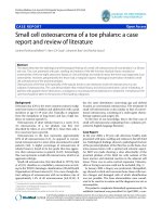
HSILs). A biopsy revealed vaginal adenosis with moderate
atypical hyperplasia of the focal squamous epithelium
(Fig. 1). She was treated with sirolimus for another two
months. The symptoms did not improve; she stopped
taking the medicine and was transferred to the unit of the
authors.
Discovery and treatment of atypical vaginal adenosis
(December 2015 to August 2017)
On December 24, 2015, the vaginal cytology results
showed suspicious adenocarcinoma and atypical squamous epithelial cells. Another biopsy of the visible
vaginal lesion suggested serous papillary glands with active growth. She underwent laparoscopic hysterectomy,
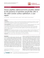
bilateral salpingo-oophorectomy, and excision of the vaginal lesions on May 4, 2016. The postoperative pathology revealed atypical vaginal adenosis (Fig. 2). Twelve
months after the hysterectomy, on May 15, 2017, her
physical examination revealed polypoid tissue on the
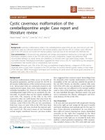
anterior vaginal wall, and vaginal biopsy revealed vaginal
atypical adenosis (Fig. 3).
Discovery and treatment of vaginal clear cell carcinoma
(August 2017 to December 2017)
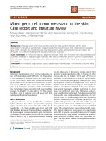
On August 15, 2017, excision of the visible vaginal lesions revealed clear cell carcinoma of the vagina (Fig. 4a,
Page 3 of 8
b) and coexisting lesions of atypical adenomyosis (Fig. 4c).
On September 15, 2017, she underwent wide local resection of the vagina, and the postoperative pathology results
showed atypical vaginal adenosis with a negative margin
and without residual carcinoma. Stage I vaginal clear cell
carcinoma was confirmed. She underwent brachytherapy
(30 Gy, five times) and concurrent cisplatin chemotherapy
from October to December 2017. Since the patient
refused external radiotherapy, concurrent cisplatin
chemotherapy was applied only once (60 mg, intravenous).
In October 2017, she provided samples for germline and
somatic sequencing using a multi-gene panel of 57 gene
mutations, including most genes involved in homologous
recombination (HR) and non-HR pathways, such as BRCA
1/2, RAD51C, PTEN, TP53, VHL, BAP1, SETD2, PBRM1,
and MTOR. No deleterious variants or variants of
unknown significance were discovered.
Follow-up (December 2017 to the present)
The patient participated in regular follow-up examinations. On July 18, 2018, she underwent a vulvar biopsy
because of a vulvar ulcer. The pathological findings revealed inflammation, which improved after treatment
with topical hormones. Her symptoms have since been
relieved. Her progression-free survival of vaginal cancer
reached 20 months in January 2019.
Discussion
Primary vaginal malignancies are very rare, accounting
for approximately 2% of all female genital malignancies
[8]. More than 80% of vaginal cancers are squamous cell
carcinomas [9]. Vaginal clear cell carcinoma is a rare
type of vaginal cancer that usually occurs in women
Fig. 1 Vaginal biopsy on March 4, 2015 revealed vaginal adenosis (hematoxylin and eosin staining, × 10)
Pang et al. BMC Cancer
(2019) 19:798
Page 4 of 8
Fig. 2 Excision of vaginal lesions on May 4, 2016 revealed atypical vaginal adenosis (hematoxylin and eosin staining, a, × 10; b, × 50)
whose mothers used DES during pregnancy [10]. However, there have only been three known cases of vaginal
clear cell carcinoma without prenatal DES exposure,
most likely due to the malignant transformation of vaginal adenosis or atypical vaginal adenosis (Table 2) [5–7].
In the report by Uehara et al. [5], a 54-year-old woman
complained of a 3-month history of genital bleeding, and
the examination revealed clear cell adenocarcinoma at
the anterior vagina, congenital anomalies of the
bicornuate uterus and vaginal septum, and left ureteral
agenesis. The patient was well without recurrence at 43
months after anterior pelvic exenteration. In the report
by Satou et al. [6], another patient died of disease 16
months after radical hysterectomy and chemotherapy. In
the report by Prasad et al. [7], the tumor, whose features
were found to be similar to those of small cell carcinomas
arising elsewhere in the female genital tract, was studied
by light and electron microscopy and immunohistochemistry; intracytoplasmic electron-dense neurosecretory-type
granules were observed, and immunohistochemistry revealed chromogranin A. The current report describes the
fourth case, in which a definite evolution from vaginal
adenosis to atypical vaginal adenosis and ultimately to
clear cell carcinoma was observed.
The exact pathogenesis of the malignant transformation of vaginal adenosis without prenatal DES exposure
is unknown. A study of clear cell carcinoma in women
exposed prenatally to DES revealed the presence of both
cervical ectropion and vaginal adenosis in all 20 specimens, and tubo-endometrial glands were intimately
related to the carcinoma in 18 of the 20 cases, suggesting that the tubo-endometrial epithelium, whether in the
ectocervix or vagina, serves as a source for the development of clear cell adenocarcinoma [11]. The frequency
with which atypical tubo-endometrial glands in the
vagina and cervix are associated with these carcinomas
and the proximity of the former to the latter provide
strong evidence that atypical vaginal adenosis and atypical
Fig. 3 Biopsy of vaginal stump on April 15, 2017 revealed atypical vaginal adenosis (hematoxylin and eosin staining, × 10)
Pang et al. BMC Cancer
(2019) 19:798
Page 5 of 8
Fig. 4 Excision of visible lesions in the mid-anterior vaginal wall on August 15, 2017 revealed clear cell carcinoma (hematoxylin and eosin
staining, a, × 10; b, × 20) and coexisting atypical vaginal adenosis (hematoxylin and eosin staining, c, × 4)
cervical ectropion of the tubo-endometrial type are precursors of clear cell adenocarcinoma [12]. Lewis et al. [13]
consistently found aneuploidy in 3 cases of invasive clear
cell carcinoma of the vagina, suggesting that the immediate precursor state should also be in the aneuploid range.
The 3 adenosis specimens, however, were in the normal
diploid to tetraploid range. Aside from the toxicity of DES
exposure, chemotherapeutic drugs may play a role in promoting the occurrence of vaginal adenosis and carcinoma.
Cases of vaginal adenosis after topical 5-fluorouracil
therapy for vaginal HPV-associated lesions [14] and vaginal adenosis together with clear cell carcinoma after 5fluorouracil treatment for condylomas [15] have been
reported. Congenital anomalies of the genitourinary tract
have been suspected as the cause of clear cell carcinoma
without DES exposure [5], which has been disputed [16].
Although there is a case report of adenocarcinoma originating from metanephric remnants [17], it is unlikely that it
originated from clear cell carcinoma because of the topographical dissimilarity [18]. Currently, objective findings
suggest that human prenatal epithelialization of the cervix
and vagina results in 3 morphogenetically determined
units [19], which may provide new insight into the histogenesis and transformation of vaginal adenosis.
In our report, before the discovery of adenosis, the
patient had undergone multiple medical and invasive
treatments, including treatment with tacrolimus and sirolimus, laser treatment and repeated biopsies. Whether
these medical regimens and procedures would prompt
the production of atypical adenosis or a transformation
to vaginal cancer requires further exploration. Although
there have been no reports on the relationship between
trauma or medical treatments, except for diethylstilbestrol, and the transformation of adenosis, an off-label and
unreasonable application of medicine should be avoided.
The natural history from vaginal adenosis to cancer
varies greatly. Most patients with vaginal adenosis have
no obvious symptoms. The lesions range widely, and
symptoms can manifest as postcoital hemorrhage, sexual
pain and a vaginal burning sensation [20]. In some cases,
vaginal palpation reveals submucous nodular or sandy
lesions 0.5–5 cm in diameter [20]. However, the main
clinical manifestations of vaginal cancer include irregular
vaginal bleeding, postpartum hemorrhage, postmenopausal hemorrhage and increased leucorrhea. The most
common type of local vaginal lesions is the papillary or
cauliflower type, followed by the ulcerative or infiltrative
type. Difficulty in sexual intercourse is a typical symptom
of advanced vaginal tumors.
Vaginal adenosis and clear cell carcinoma often occur
several years after exposure to DES in the uterine cavity.
Non-DES-induced vaginal adenosis has a reported incidence of approximately 10% in adult women. In the
present case, the patient’s mother did not use DES
during pregnancy since DES was never introduced into
the Chinese market. Vaginal clear cell carcinoma was
identified 6 years after the discovery of vaginal adenosis.
A consensus regarding the detection and diagnosis of
atypical vaginal adenosis and/or vaginal clear cell carcinoma is lacking. Cytology has been clinically valuable in
proving cases of vaginal adenosis and adenocarcinoma
[21]. Colposcopy with biopsy for abnormal vaginal and/
or cervical cytology results could reveal possible lesions,
as described in our report.
Laser therapy, cryotherapy and cautery can be used to
treat superficial and small lesions of vaginal adenosis
[22]. The lesions can also be coated topically with 10–
20% silver nitrate or potassium dichromate solution for
lesion necrosis and exfoliation. For a single localized
submucosal lesion, complete resection of the lesion can
be performed. For those with severe atypical hyperplasia
or malignant transformation, the principle of treatment
is the same as for those with vaginal cancer, despite a
lack of sufficient evidence [23]. On the other hand,
radiotherapy is the first choice for some patients with
early or advanced vaginal cancer [24]. Radiotherapy includes brachytherapy and external beam. The use of
brachytherapy in vaginal cancer imparts a benefit in
terms of disease-specific and overall survival [25, 26].
The treatment of vaginal cancer with a multichannel cylinder produces high local control [27]. Surgery is also
an option for patients with early-stage primary vaginal
cancer [28]. Patients with early-stage vaginal tumors
without deep infiltration may undergo radical hysterectomy, partial vaginal resection and pelvic lymphadenectomy. The margin of vaginal resection should be 2–3 cm
Age at diagnosis
of carcinoma
54 years
38 years
34 years
45 years
Reference
Uehara T et al.
[5]
Satou Y et al.
[6]
Prasad CJ [7]
Case in the
report
Yes
Yes
None
None
Atypical
adenosis
None
None
Didelphys uterus,
duplicated and
imperforated vagina
Bicornuate uterus and
vaginal septum and
left ureteral agenesis
Congenital anomalies
6 years
Not available
Not available
3 months
Courses from
adenosis to
carcinoma
Hysterectomy, wide local
resection of vaginal
lesion, and brachytherapy
Vaginectomy with
bilateral inguinal lymph
node dissection,
chemotherapy of cisplatin
and etoposide,
teletherapy,
brachytherapy
Radical hysterectomy and
chemotherapy
Anterior pelvic
exenteration
Treatment
16 months
Not available
Not available
43 months
Disease-free
survival
16 months
6 months
16 months
43 months
Overall survival
No
With tumor noted
at deep margins
of the vagina
Not available
No
Recurrence
No
Yes
Yes
No
Mortality
Table 2 Cases of vaginal clear cell carcinoma due to the potential malignant transformation of vaginal adenosis or atypical vaginal adenosis without prenatal DES exposure in
the English literature
Pang et al. BMC Cancer
(2019) 19:798
Page 6 of 8
Pang et al. BMC Cancer
(2019) 19:798
beyond the tumor. For vaginal midsegment tumors, in
addition to total vaginal hysterectomy, inguinal lymph
node or pelvic lymph node resection should be performed according to the size of the lesion and the
location of lymph node metastasis [29]. Total vaginal resection, including rectal resection or cystectomy (pelvic
exenteration), is necessary for treatment, but the operation is extremely complicated [30, 31]. The effect of
chemotherapy has been shown to be minimal. In the
case reported by Satou et al. [6], the patient survived
only 16 months after radical hysterectomy and chemotherapy. However, in the current case, several inappropriate therapy protocols were applied. Before being
transferred to our unit, the patient was treated with
tacrolimus and sirolimus, neither of which had definite
indications or resulted in symptomatic relief. Although
there have been several reports on the application of
tacrolimus for the treatment of erosive lichen planus
[32–34], these experiences are not applicable to the
treatment of adenosis.
In conclusion, we report the fourth case in the world
of vaginal clear cell carcinoma stemming from the malignant transformation of vaginal adenosis without prenatal DES exposure, with serial evidence of oncological
evolution. Wide local excision with radiotherapy provided at least 16 months of disease-free survival. Serial
follow-up examinations with vaginal cytology is essential
for patients with vaginal adenosis for the diagnosis of
atypical lesions and even cancer.
Abbreviations
ASC-H: Atypical squamous cells, cannot exclude high-grade squamous
intraepithelial lesions; DES: Prenatal diethylstilbestrol; HPV: Human papillomavirus;
HR: Homologous recombination; HSIL: High-grade squamous intraepithelial lesion
Acknowledgements
Not applicable.
Authors’ contributions
LL and LP planned and designed the analysis and contributed to the
acquisition of data. LZ and JL contributed to the acquisition of data,
interpretation of the analysis results and critical revision of the manuscript for
important intellectual content. YB reviewed and provided the pathological
outcomes. All authors have read and approved the final manuscript.
Funding
This study was supported by the Chinese Academy of Medical Sciences
Initiative for Innovative Medicine (CAMS-2017-I2M-1-002) and by the National
Science-technology Support Plan Projects (2015BAI13B04). The funders
played no role in the study design, data collection or analysis, decision to
publish, or manuscript preparation.
Availability of data and materials
The medical history of this patient, including detailed procedures for
diagnosis and treatment, are listed in Table 1 and described in the “Case
Presentation” section.
Ethics approval and consent to participate
The patient in this report provided consent for participation in the study.
The Institutional Review Board of Peking Union Medical College Hospital
approved this study.
Page 7 of 8
Consent for publication
The patient in this report provided consent for the publication of her
experiences in an anonymous style. A copy of the patient’s consent to
publication form is available to the Editor of the journal. All authors of this
report agree with and are greatly obliged to the Editorial Board of BMC
Cancer for the publication of this report.
Competing interests
The authors declare that they have no competing interests.
Author details
1
Department of Obstetrics and Gynecology, Peking Union Medical College
Hospital, Peking Union Medical College & Chinese Academy of Medical
Science, Shuaifuyuan No. 1, Dongcheng District, Beijing 100730, China.
2
Department of Pathology, Peking Union Medical College Hospital, Peking
Union Medical College & Chinese Academy of Medical Science, Beijing
100730, China.
Received: 6 February 2019 Accepted: 8 August 2019
References
1. Kranl C, Zelger B, Kofler H, Heim K, Sepp N, Fritsch P. Vulval and vaginal
adenosis. Br J Dermatol. 1998;139(1):128–31.
2. Kurman RJ, Carcangiu ML, Herrington CS, Young RH. WHO Classification of
Tumours of Female Reproductive Organs. 4th ed. Lyon: International
Agency for Research on Cancer (IARC); 2014.
3. Nordqvist SR, Fidler WJ Jr, Woodruff JM, Lewis JL Jr. Clear cell
adenocarcinoma of the cervix and vagina. A clinicopathologic study of 21
cases with and without a history of maternal ingestion of estrogens. Cancer.
1976;37(2):858–71.
4. Herbst AL, Ulfelder H, Poskanzer DC. Adenocarcinoma of the vagina.
Association of maternal stilbestrol therapy with tumor appearance in young
women. N Engl J Med. 1971;284(15):878–81.
5. Uehara T, Onda T, Sasajima Y, Sawada M, Kasamatsu T. A case of vaginal
clear cell adenocarcinoma complicated with congenital anomalies of the
genitourinary tract and metanephric remnant without prenatal
diethylstilbestrol exposure. J Obstet Gynaecol Res. 2010;36(3):681–5.
6. Satou Y, Takasu K. Clear cell adenocarcinoma in duplicated and
imperforated vagina with didelphys uterus. A case report. J Kyoto Pref Univ
Med. 1990;99:725–38.
7. Prasad CJ, Ray JA, Kessler S. Primary small cell carcinoma of the vagina
arising in a background of atypical adenosis. Cancer. 1992;70(10):2484–7.
8. Pingley S, Shrivastava SK, Sarin R, Agarwal JP, Laskar S, Deshpande DD,
Dinshaw KA. Primary carcinoma of the vagina: Tata memorial hospital
experience. Int J Radiat Oncol Biol Phys. 2000;46(1):101–8.
9. Lilic V, Lilic G, Filipovic S, Visnjic M, Zivadinovic R. Primary carcinoma of the
vagina. J BUON. 2010;15(2):241–7.
10. Marselos M, Tomatis L. Diethylstilboestrol: I, pharmacology, toxicology and
carcinogenicity in humans. Eur J Cancer. 1992;28A(6–7):1182–9.
11. Robboy SJ, Welch WR, Young RH, Truslow GY, Herbst AL, Scully RE.
Topographic relation of cervical ectropion and vaginal adenosis to clear cell
adenocarcinoma. Obstet Gynecol. 1982;60(5):546–51.
12. Robboy SJ, Young RH, Welch WR, Truslow GY, Prat J, Herbst AL, Scully RE.
Atypical vaginal adenosis and cervical ectropion. Association with clear cell
adenocarcinoma in diethylstilbestrol-exposed offspring. Cancer. 1984;54(5):
869–75.
13. Lewis JL Jr, Nordqvist SR, Richart RM. Studies of nuclear DNA in vaginal
adenosis and clear-cell adenocarcinoma. Am J Obstet Gynecol. 1973;115(6):
737–50.
14. Georgiev D, Karag'ozov I, Velev M, Makaveeva V. Three cases of vaginal
adenosis after topical 5-fluorouracil therapy for vaginal HPV-associated
lesions. Akush Ginekol (Sofiia). 2006;45(3):59–61.
15. Goodman A, Zukerberg LR, Nikrui N, Scully RE. Vaginal adenosis and clear
cell carcinoma after 5-fluorouracil treatment for condylomas. Cancer. 1991;
68(7):1628–32.
16. Ott MM, Rehn M, Muller JG, Gruss A, Martius J, Steck T, Muller-Hermelink HK.
Vaginal clear cell carcinoma in a young patient with ectopic termination of
the left ureter in the vagina. Virchows Arch. 1994;425(4):445–8.
Pang et al. BMC Cancer
(2019) 19:798
17. Shimao Y, Nabeshima K, Inoue T, Higo T, Wada T, Ikenoue T, Koono M.
Primary vaginal adenocarcinoma arising from the metanephric duct
remnant. Virchows Arch. 2000;436(6):622–7.
18. Kaminski PF, Maier RC. Clear cell adenocarcinoma of the cervix unrelated to
diethylstilbestrol exposure. Obstet Gynecol. 1983;62(6):720–7.
19. Reich O, Fritsch H. The developmental origin of cervical and vaginal
epithelium and their clinical consequences: a systematic review. J Low
Genit Tract Dis. 2014;18(4):358–60.
20. Han T, Jin Y, Li Y, Bi Y, Pan L. Clinicopathologic features and outcomes of
primary vaginal adenosis as a dermatologic and gynecologic burden: a
retrospective study. Medicine (Baltimore). 2018;97(49):e13470.
21. Chenoweth B, Rodney MB. Cytologic problems in diagnosis of endocervical
atypia in young females with and without maternal history of
diethylstilbestrol exposure. J Natl Med Assoc. 1978;70(12):925–30.
22. Cebesoy FB, Kutlar I, Aydin A. Vaginal adenosis successfully treated with
simple unipolar cauterization. J Natl Med Assoc. 2007;99(2):166–7.
23. Scurry J, Planner R, Grant P. Unusual variants of vaginal adenosis: a
challenge for diagnosis and treatment. Gynecol Oncol. 1991;41(2):172–7.
24. Hegemann S, Schafer U, Lelle R, Willich N, Micke O. Long-term results of
radiotherapy in primary carcinoma of the vagina. Strahlenther Onkol. 2009;
185(3):184–9.
25. Orton A, Boothe D, Williams N, Buchmiller T, Huang YJ, Suneja G, Poppe M,
Gaffney D. Brachytherapy improves survival in primary vaginal cancer.
Gynecol Oncol. 2016;141(3):501–6.
26. Murofushi KN, Kitamura N, Yoshioka Y, Sumi M, Ishikawa H, Oguchi M,
Sakurai H. A clinical evaluation of American brachytherapy society
consensus guideline for bulky vaginal mass in gynecological Cancer. Int J
Gynecol Cancer. 2018;28(7):1438–45.
27. Gebhardt BJ, Vargo JA, Kim H, Houser CJ, Glaser SM, Sukumvanich P,
Olawaiye AB, Kelley JL, Edwards RP, Comerci JT, et al. Image-based
multichannel vaginal cylinder brachytherapy for the definitive treatment of
gynecologic malignancies in the vagina. Gynecol Oncol. 2018;150(2):293–9.
28. Shrivastava SB, Agrawal G, Mittal M, Mishra P. Management of Vaginal
Cancer. Rev Recent Clin Trials. 2015;10(4):289–97.
29. Ozgul N, Basaran D, Boyraz G, Salman C, Yuce K. Radical hysterectomy and
Total abdominal Vaginectomy for primary vaginal Cancer. Int J Gynecol
Cancer. 2016;26(3):580–1.
30. Huang M, Iglesias DA, Westin SN, Fellman B, Urbauer D, Schmeler KM,
Frumovitz M, Ramirez PT, Soliman PT. Pelvic exenteration: impact of age on
surgical and oncologic outcomes. Gynecol Oncol. 2014;132(1):114–8.
31. Hockel M, Horn LC, Einenkel J. (Laterally) Extended Endopelvic Resection:
surgical treatment of locally advanced and recurrent cancer of the uterine
cervix and vagina based on ontogenetic anatomy. Gynecol Oncol. 2012;
127(2):297–302.
32. Helgesen AL, Gjersvik P, Jebsen P, Kirschner R, Tanbo T. Vaginal involvement in
genital erosive lichen planus. Acta Obstet Gynecol Scand. 2010;89(7):966–70.
33. Kortekangas-Savolainen O, Kiilholma P. Treatment of vulvovaginal erosive
and stenosing lichen planus by surgical dilatation and methotrexate. Acta
Obstet Gynecol Scand. 2007;86(3):339–43.
34. Lotery HE, Galask RP. Erosive lichen planus of the vulva and vagina. Obstet
Gynecol. 2003;101(5 Pt 2):1121–5.
Publisher’s Note
Springer Nature remains neutral with regard to jurisdictional claims in
published maps and institutional affiliations.
Page 8 of 8