Prognostic significance of 8-hydroxy-2′- deoxyguanosine in solid tumors: A metaanalysis
Bạn đang xem bản rút gọn của tài liệu. Xem và tải ngay bản đầy đủ của tài liệu tại đây (2.21 MB, 15 trang )
Qing et al. BMC Cancer
(2019) 19:997
/>
RESEARCH ARTICLE
Open Access
Prognostic significance of 8-hydroxy-2′deoxyguanosine in solid tumors: a metaanalysis
Xiangcheng Qing1*† , Deyao Shi1†, Xiao Lv1, Baichuan Wang1, Songfeng Chen2 and Zengwu Shao1*
Abstract
Background: High level of reactive oxygen species (ROS) has been detected in almost all cancers, which make it
become one of the best-characterized phenotypes in cancers. Though ROS plays an important role in tumors, the
degree of oxidative stress can be better evaluated by assessing stable metabolites of oxidative reactions because of
its high instability. 8-hydroxy-2′-deoxyguanosine (8-OHdG), a product of oxidative damage to 2′-deoxyguanosine, is
known as a useful marker for assessing oxidative DNA damage and has been a feature of carcinogenesis in several
researches. But the exact prognostic value of 8-OHdG expression in patients with cancer is still unclear.
Methods: A comprehensive search was performed in PubMed, Web of Science, EMBASE. Eligible studies were
included based on defined exclusion and inclusion criteria to perform a meta-analysis. STATA 14.0 was used to
estimate pooled hazard ratios (HRs) with 95% confidence interval (95% CI), the heterogeneity among studies and
publication bias to judge the prognostic value.
Results: A total of 2121 patients from 21 eligible studies were included in the meta-analysis. A significant
association was found between elevated 8-OHdG expression and poor OS (overall survival) in cancer patients
(pooled HR 1.921, 95% CI: 1.437–2.570); In the subgroup analysis, race of sample, cancer types, detection method of
8-OHdG, sample classification, detection location of 8-OHdG and paper quality (score more or less than 7) did not
alter the association between 8-OHdG expression and cancer prognosis. Furthermore, 8-OHdG expression was an
independent prognostic marker for overall survival in patients with cancer (pooled HR 2.110, 95% CI: 1.482–3.005)
using Cox multivariate analyses.
Conclusions: This meta-analysis found that highly expressed 8-OHdG in tumor tissues may be a predictor of
prognosis in most solid tumors. However, especially in breast cancer, low 8-OHdG expression is associated with
poor prognosis, which is partly because of the increased antioxidant mechanisms in breast cancer tissues. This
study demonstrates for the first time that 8-OHdG expression is associated with the prognosis of cancer patients. In
the future, whether the expression level of 8-OHdG can be used as a biomarker for the prognosis of all human
cancers requires more research.
Keywords: 8-OHdG, Meta-analysis, Prognosis, Solid tumor, Reactive oxygen species, DNA oxidative damage
* Correspondence: ;
†
Xiangcheng Qing and Deyao Shi contributed equally to this work as co-first
authors.
1
Department of Orthopaedics, Union Hospital, Tongji Medical College,
Huazhong University of Science and Technology, Wuhan 430022, China
Full list of author information is available at the end of the article
© The Author(s). 2019 Open Access This article is distributed under the terms of the Creative Commons Attribution 4.0
International License ( which permits unrestricted use, distribution, and
reproduction in any medium, provided you give appropriate credit to the original author(s) and the source, provide a link to
the Creative Commons license, and indicate if changes were made. The Creative Commons Public Domain Dedication waiver
( applies to the data made available in this article, unless otherwise stated.
Qing et al. BMC Cancer
(2019) 19:997
Background
Tumor cells constantly suffer various endogenous and environmental attacks, which make high level of reactive
oxygen species (ROS) be detected in almost all cancers
and become one of the best-characterized phenotypes [1–
3]. The role of ROS in cancer is a “doubled edged sword”.
ROS can serve as a carcinogenic factor through promoting
tumorigenesis, development and spread of cancers by activating or regulating signaling pathways that affect tumor
cell survival, proliferation and metastasis [4–6]. However,
high levels of ROS can also play a role in tumor suppression by inhibiting cell proliferation and inducing cell death
[7–9]. Many cancer treatments, such as radiotherapy and
certain chemotherapy agents, act through oxidative stress
pathways via the production of ROS to suppress tumor
growth and progression [10]. In order to prevent cell
death, cancer cells can scavenge reactive oxygen species to
adapt high levels of ROS and activate pro-tumorigenic signaling pathways, by upregulating antioxidant pathways
and regulatory factors [11–13].
Though ROS plays an important role in tumors, the degree of oxidative stress can be better evaluated by assessing
stable metabolites of oxidative reactions because of its high
instability. ROS can cause oxidative damage to doublestranded DNA directly, or to free bases in the cellular and
mitochondrial deoxynucleoside triphosphate (dNTP) pool
[14]. Among all the nucleobases, guanine is the most susceptible to oxidation by ROS [15]. Oxidative damage to 2′-deoxyguanosine produces 8-hydroxy-2′-deoxyguanosine (8OHdG). The formation of 8-OHdG on DNA can cause G:
C—T:A mispairing mutations, which are considered to have
a close relationship with the development and progression of
tumors, cell ageing and some degenerative diseases [16].
There is an increasing body of evidence indicating that
8-OHdG is a useful marker for assessing oxidative DNA
damage and has been a feature of carcinogenesis in several researches [17, 18]. High levels of 8-OHdG in tumors, blood samples or urine have been found in
various cancers and implicated as a promising marker
for predicting the prognosis of cancers [19–40]. However, the association of oxidative damage to DNA with
tumors still needs to be more extensively investigated
and most studies reported so far are limited in discrete
outcome and sample size. For these reasons we performed a quantitative meta-analysis and systematic review to gain better insight into the prognostic value of
8-OHdG expression in patients with cancer.
Methods
Search strategy
This analysis was conducted following the meta-analyses and
systematic reviews guidelines for prognosis-related tumor
marker researches [41, 42]. An electronic search of PubMed,
Web of Science, EMBASE was performed independently by
Page 2 of 15
two authors (XQ and DS) prior to May 15, 2018. Search
terms were used in all possible combinations as following: 7,
8-dihydro-8-oxodeoxyguanosine, 8-hydroxy-2′-deoxyguanosine, 8-hydroxy-2′- deoxyguanosine, 8-OHdG, 8OHdG, 8OH-dG,
8-OHG,
8-oxo-G,
8-oxo-dG,
8hydroxydeoxyguanosine, 8-oxo-guanine, 8-hydroxyguanine,
8-hydroxyguanosine, 8-oxo-2-deoxy guanosine, 8-oxo-7,8dihydro-2-deoxyguanosine, 8-oxo-7,8-dihydro- 2′-deoxyguanosine, 8-hydroxy-2-deoxyguanosine, 8-oxo-7,8-dihydro-2deoxyguanosine, tumor, cancer, sarcoma, carcinoma, neoplasm, malignancy, prognosis, mortality of metastasis, progression, development, outcome, survival, recurrence, clinical
significance. Conflicts were solved through group discussion.
Inclusion and exclusion criteria
Studies included in the present meta-analysis were independently reviewed by two investigators (XQ and DS) and
should meet the following criteria: (1) The prognostic data
of 8-OHdG in any type of human solid tumors needed to
be presented; (2) All cancer patients were diagnosed according to the gold standard for diagnosis, based on histopathological examinations; (3) 8-OHdG levels in tumors,
blood samples or urine were estimated in each study; (4)
The patients were divided into two groups according to
the levels of 8-OHdG; (5) Sufficient data should be provided to obtain hazard ratios (HR) for survival rates and
their 95% confidence intervals (95%CI). Studies were excluded from the present meta-analysis if one of the following criteria was met: (1) Case reports, reviews, metaanalysis, letters, editorials, comments, expert opinions or
any other reviews that didn’t contain raw data; (2) Full text
could not be obtained; (3) Researches on non-English
writing; (4) Repetitive publications; (5) No survival data or
data insufficient to be extracted and analyzed; (6) Survival
data was acquired based on animal studies and no followup of patients. Detailed inclusion and exclusion criteria of
each study are presented in Additional file 1: Table S1.
Data extraction and quality assessment
Data was extracted independently by the two researchers
(XQ and DS), and final consensus was reached through
discussion. Data were retrieved from each study including: author; year of publication; country of the population enrolled; ethnicity; tumor stage; sample size; study
design; follow-up data; survival data; survival analysis
methodology; expression levels, location and laboratory
methods of 8-OHdG; cut-off values; HR values and their
95% confidence intervals. Quality assessment of cohort
studies in this meta-analysis was performed using the
Newcastle-Ottawa scale (NOS) as recommended by the
Cochrane Non-Randomized Studies Methods Working
Group. Studies with score ≥ 7 were considered high
quality according to the NOS. Detailed NOS scores of
all included studies were shown in Table 1.
Japan
Finland
Italy
Ireland
Finland
USA
Matsumoto et al. 2003 [32]
Hintsala et al. 2016 [33]
Murtas et al. 2010 [34]
Sheridan et al. 2009 [35]
Karihtala et al. 2011 [36]
Maki et al. 2007 [37]
Hepatocellular
carcinoma
Breast cancer
Colorectal cancer
Melanoma
Melanoma
Hepatocellular
carcinoma
Colorectal cancer
Japan
Renal cell
carcinoma
Croatia
Matosevic et al. 2015 [31]
Ovarian cancer
Miyake et al.2004 [39]
Japan
Aman et al. 2017 [30]
Ovarian cancer
Breast cancer
Colorectal cancer
Bladder carcinoma
Ovarian cancer
Finland
Pylväs et al. 2011 [29]
144
72
53
68
72
105
30
79
113
46
121
73
138
95
84
145
79
252
Nonsmall-Cell Lung 99
cancer
Esophageal cancer
Ovarian cancer
Ovarian cancer
103
I-IV
I-IV
I-II
I-III
I-IV
I-II
NA
NA
I-IV
I-IV
I-IV
I-IV
I-IV
I-IV
I-IV
I-IV
I-IV
NA
I-IV
I-IV
NA
NA
NA
60
80
60
Over 150
Over 60
169
208
Over 125
112
100
300
82
60
Over 120
80
41
36
CSS
OS, DFS
DFS
CSS
OS
OS
CSS
CSS, RFS
OS
OS
OS
OS, DFS
OS
OS
OS
OS
OS, PFS
OS
OS
OS
High
High
High
Low
High
High
Low
High
High
High
High
Low
High
High
High
High
High
High
High
High
NA
Nuclei
Nuclei
NA
Nuclei
NA
percentage of positive Nuclei
tumor cells
median
positive > 5%
median
percentage of positive Nuclei
tumor cells
Fold change
IHC score 12
median
ELISA
ELISA
IHC
IHC
IHC
IHC
IHC
IHC
IHC
IHC
Nuclei
Nuclei
Nuclei
mean plus
one standard
deviation
median
NA
NA
percentage of positive NA
tumor cells
NA
NA
percentage of positive Nuclei
tumor cells
NA
percentage of positive NA
tumor cells
percentage of positive Cytoplasm
tumor cells
percentage of positive Nuclei
tumor cells
multivariate
multivariate
multivariate
univariate
multivariate
multivariate
multivariate
multivariate
univariate
multivariate
multivariate
univariate
univariate
multivariate
univariate
multivariate
NA
NA
multivariate
multivariate
univariate,
multivariate
NA
multivariate
multivariate
6
6
6
6
6
8
6
8
7
6
6
6
6
6
7
8
8
5
6
6
1
1,2
1
2
1
1
1
1,2
1
1
2
1
2
2
1
1,2
1,2
2
1
1
Location of Survival analysis NOS Method*
8-oxo-dG
score
percentage of positive Nuclei
tumor cells
Cut-off value
IHC, ELISA percentage of positive NA
tumor cells for IHC.
140 pg/mL for ELISA
IHC
LCEC
IHC
ELISA
IHC
ELISA
IHC
IHC
IHC
Sample Tumor Follow-up Outcome Expression Assay
size
stage (month)
measure associates
with poor
prognosis
(2019) 19:997
Pylväs-Eerola et al. 2015 [38] Finland
Croatia
Jakovcevic et al. 2015 [21]
USA
Shen et al. 2007 [26]
Finland
China
He et al. 2014 [25]
Poland
China
Xu et al. 2013 [24]
Dziaman et al. 2014 [20]
Thailand Hepatocellular
carcinoma
Ma-on et al. 2017 [23]
Soini et al. 2011 [27]
Finland
Karihtala et al. 2009 [19]
Hepatocellular
carcinoma
China
Li et al. 2012 [22]
Cancer Type
Region
Author
Table 1 Characteristics of studies included in the meta-analysis
Qing et al. BMC Cancer
Page 3 of 15
Finland
Sova et al. 2010 [40]
Breast cancer
Cancer Type
150
I-IV
NA
CSS
Low
IHC
Sample Tumor Follow-up Outcome Expression Assay
size
stage (month)
measure associates
with poor
prognosis
percentage
of positive
tumor cells
Cut-off value
Nuclei
multivariate
6
1
Location of Survival analysis NOS Method*
8-oxo-dG
score
OS overall survival, DFS disease free survival, PFS progression free survival, RFS recurrence free survival, CSS cancer specific survival, NOS Newcastle-Ottawa Scale, IHC Immunohistochemistry, ELISA Enzyme-linked
immunosorbent assay, LCEC Liquid chromatography electrochemistry, NA not available
*1 denoted as obtaining HRs directly from publications; 2 denoted as HRs were extracted and calculated from Kaplan-Meier curves
Region
Author
Table 1 Characteristics of studies included in the meta-analysis (Continued)
Qing et al. BMC Cancer
(2019) 19:997
Page 4 of 15
Qing et al. BMC Cancer
(2019) 19:997
Statistical analysis
The meta-analysis was performed as previously described
[43]. In the present study, statistical analysis and graphical
representation were performed using Stata version 14.0
(Stata Corporation, College Station, TX, USA). Pooled
HRs and ORs with 95%CIs were used to evaluate the association between 8-OHdG expression and prognosis. HRs
or ORs with 95%CIs can be directly obtained from most
included studies or estimated from the existing data using
methods as previously described [41]. An HR > 1 indicates
a worse outcome of patient with high 8-OHdG expression,
while an HR < 1 implied a worse survival for patients with
decreased 8-OHdG expression. The test for heterogeneity
of combined HRs was carried out using a χ2 based
Cochran Q test and Higgins I2 statistic. I2 values > 50% indicated heterogeneity among studies. If there existed heterogeneity, a random-effect model, subgroup analysis and
meta regression by factors contributing to heterogeneity
would be carried out. Influence analyses was performed to
examine the effect of each study on the overall pooled results. The presence of publication bias was evaluated by
using funnel plots, Begg’s test and Egger’s test. P values <
0.05 were considered statistically significant.
Results
Included studies and characteristics
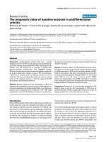
Based on our searching strategy, a total of 3537 articles
were identified from PubMed (n = 915), Web of Science
(n = 1319) and EMBASE (n = 1303). After removing duplicates, 1665 articles were left. Furthermore, 1607 of
the remaining articles were excluded according to the titles and abstracts. Finally, a total of 21 relevant articles
were included in this meta-analysis after a more careful
full-text reading. The detailed screening process is
shown in Fig. 1.
Fig. 1 The flow diagram of the meta analysis
Page 5 of 15
Among the 21 studies, a total of 2121 patients were included, with mean sample size of 101 patients (range 30
to 252). The period of these studies ranged from 2003 to
2017. The regions represented in the studies include
various countries around Europe, Asia and America, of
which the race contains both Caucasoid and Mongoloid.
Eight different types of cancer were evaluated. Most
studies analyzed the expression level of 8-OHdG by IHC
or ELISA, while there was one study unitizing liquid
chromatography electrochemistry. Overall survival (OS),
cancer-specific survival (CSS), recurrence-free survival
(RFS), disease-free survival (DFS) and progression-free
survival (PFS) were estimated as survival outcomes in
the studies. RFS, DFS and PFS were merged into the
event-free survival (EFS) group for analysis. Cox multivariable analyses were performed in 17 studies. Further
detailed characteristics of each study are presented in
Table 1.
Overall survival (OS) based on different 8-OHdG expression levels was reported in 8 types of solid tumors
from 15 of the 21 included studies with a total of 1596
patients. Elevated 8-OHdG was significantly associated
with poor OS in these patients (pooled HR 1.921,
95%CI: 1.437–2.570) (Fig. 2a), while significant heterogeneity was found in these studies (Tau2 = 0.2298; χ2 =
53.52, df = 16, p < 0.0001; I2 = 70.1%). Since obvious heterogeneity was observed, subgroups analysis was performed by factors of the race of sample, cancer types,
detection method of 8-OHdG, detection location of 8OHdG, sample classification and research quality (Fig. 3).
Detailed results of subgroup analysis were demonstrated
in Table 2. Despite the subgroup of hepatocellular carcinoma (Cancer Types) and the subgroup of cytoplasm
(Detection location of 8-OHdG), the significant association between 8-OHdG expression and poor OS could
Qing et al. BMC Cancer
(2019) 19:997
Page 6 of 15
Fig. 2 Meta-analysis of the pooled HRs of OS with elevated 8-OHdG expression in cancer patients. a All studies included. b Study of Jakovcevic
et al. excluded
Qing et al. BMC Cancer
(2019) 19:997
Page 7 of 15
Fig. 3 Subgroup analysis of the pooled HRs of OS by various factors. a Subgroup analysis of HRs of OS by factor of race. b Subgroup analysis of
HRs of OS by factor of cancer types. c Subgroup analysis of HRs of OS by factor of detection method of 8-OHdG. d Subgroup analysis of HRs of
OS by factor of detection location of 8-OHdG. e Subgroup analysis of HRs of OS by factor of research quality. f Subgroup analysis of HRs of OS by
factor of sample classification
be observed in each subgroup. We further performed
meta-regression with the covariates including above factors to explore the source of heterogeneity. From the result we found that p<0.05 was only observed in the
subgroup of breast cancer (Cancer types) covariate,
which implied that the subgroup of breast cancer may
be the major source of heterogeneity. The study of
Jakovcevic et al. enrolled patients with breast cancer and
drew a conclusion that negative 8-OHdG expression was
a poor prognostic biomarker, which was contrary to the
other researches. It could be a consequence caused by
cancer specificity. We discussed this point in the discussion part below.
Base on the above result of meta-regression, we
excluded the study of Jakovcevic et al. and still found
significant association between elevated 8-OHdG expression and poor OS in cancer patients (pooled HR 2.022,
95% CI: 1.540–2.641) with reduced heterogeneity (I2 =
65.5%) (Fig. 2b). Furthermore, as shown in Fig. 4, influence analysis was carried out for purpose of ensuring the
stability of the result. No obvious change of the pooled
HR and 95% CIs could be observed after excluding any
study from the whole studies. In aspect of the publication bias, Begg’s test and Egger’s linear regression test
were performed. The Begg’s tests proved that there was
no evidence of publication bias (p = 0.053) while the
Egger’s test showed there was significant publication bias
(p = 0.007) (Fig. 5a and Fig. 5b). Thus “Trim and fill”
analysis was conducted and the result estimated that 8
studies evaluating the association between expression of
8-OHdG and overall survival of cancer patients were
remaining unpublished. The result of filled meta-analysis
was pooled HR 1.545, 95% CI: 1.179–2.026, which exhibited that the significant association between elevated 8OHdG expression and poor OS in cancer patients maintained unchanged (Fig. 6a).
Among the 21 included studies, four studies reported
event-free survival (EFS) in 489 patients. A close
relationship was observed between elevated 8-OHdG
expression and EFS (pooled HR 1.612, 95% CI: 1.121–
2.310, I2 = 78.7%) (Fig. 7a). However, due to the limited
number of included studies, appraisal of publication bias
was not performed.
There were 5 studies reported the association between
8-OHdG expression and cancer-specific survival (CSS),
corresponding to hepatocellular carcinoma, melanoma,
renal cell carcinoma and breast cancer, including a total
of 495 patients. After summarizing the results, we found
Qing et al. BMC Cancer
(2019) 19:997
Page 8 of 15
Table 2 Subgroup analysis of pooled HR of OS by various factors with elevated 8-OHdG expression
Subgroup analysis
No. of studies
No. of patients
Pooled HR (95%CI)
Meta regression (p -value)
Heterogeneity
I2
p -value
Race
Caucasoid
12
1129
1.962 [1.341–2.870]
0.907
66.6%
0.001
Mongoloid
5
467
1.862 [1.117–3.104]
–
80.2%
< 0.001
2
156
2.853 [0.673–12.089]
0.727
84.2%
0.012
Cancer types
Hepatocellular carcinoma
Ovarian carcinoma
6
424
1.867 [1.190–2.930]
0.464
58.0%
0.036
Colorectal cancer
4
330
1.637 [0.850–3.153]
0.352
71.5%
0.014
Esophageal cancer
1
144
3.400 [2.055–5.624]
0.982
–
–
Nonsmall-Cell Lung cancer
1
99
3.330 [1.588–6.982]
–
–
–
Melanoma
1
46
1.470 [1.019–2.121]
0.367
–
–
Breast cancer
1
145
0.100 [0.017–0.583]
0.019
–
–
Bladder cancer
1
252
3.130 [1.298–7.548]
0.950
–
–
Detection method of 8-OHdG
IHC
12
1157
1.787 [1.246–2.563]
0.646
74.1%
< 0.001
ELISA
4
360
2.386 [1.167–4.881]
0.947
71.0%
0.016
LCEC
1
79
2.510 [1.018–6.187]
–
–
Sample classification
Tissue
14
1412
1.792 [1.307–2.458]
–
73.6%
< 0.001
Plasma or urine
3
268
3.042 [1.676–5.519]
0.006
0.0%
0.856
Nuclei
10
1019
1.927 [1.321–2.810]
0.596
71.5%
< 0.001
Cytoplasm
1
138
0.759 [0.454–1.268]
0.118
–
–
Not mentioned
6
439
2.345 [1.429–3.848]
–
57.6%
0.038
NOS score ≥ 7
5
499
1.658 [1.002–2.743]
0.526
82.7%
< 0.001
NOS score < 7
12
1097
2.104 [1.456–3.040]
–
60.6%
0.003
Detection location of 8-OHdG
research quality
there was no significant association between 8-OHdG
expression and CSS (pooled HR 0.793, 95%CI: 0.344–
1.828, I2 = 81.0%) (Fig. 7b). We need to point out that
this result is contrasted to the other results above.
A total of 11 studies including 1243 patients used
Cox multivariate analysis to assess whether 8-OHdG
expression could be an independent prognostic factor
for OS of cancer patients. Elevated 8-OHdG as an independent factor for poor prognosis was found alone
in nine of them. The results of Cox multivariate analyses in these 11 studies showed that 8-OHdG expression was an independent prognostic factor for overall
survival (pooled HR 2.110, 95% CI:1.482–3.005), and
heterogeneity was still observed among studies
(Tau2 = 0.2339; χ2 = 35.73, df = 10, p < 0.0001; I2 =
72.0%). (Fig. 8).
As for the publication bias, the Begg’s test (p = 0.276)
and Egger’s test (p = 0.031) showed opposite conclusion.
(Fig. 5c and Fig. 5d) Thus we applied the “Trim and fill”
analysis to confirm our result. There were 3 studies
evaluating whether 8-OHdG expression could be an independent prognostic factor for OS remaining unpublished. The result of filled meta-analysis was pooled HR
1.793, 95% CI: 1.242–2.436, which confirmed that elevated 8-OHdG could be an independent factor for poor
prognosis of overall survival after the “Trim and fill”
analysis. (Fig. 6b).
Discussion
Cancer is a major public health problem worldwide and
is the second leading cause of death in the United States
[44]. The 5-year survival of many cancers is still quite
low. For most types of cancers, the pathological staging
is a gold standard to predict its prognosis. However, patients with the same tumor stage often exhibit quite different clinical outcomes, which suggests that this
conventional method is unable to precisely predict the
prognosis of cancer patients. Therefore, new potential
Qing et al. BMC Cancer
(2019) 19:997
Page 9 of 15
Fig. 4 Influence analysis of the included studies for OS. No obvious change of the pooled HRs and 95% confidence intervals was observed after
excluding any included study
Fig. 5 Plot of publication bias analysis. a Begg’s test and (b) Egger’s test for analysis of the association between 8-OHdG expression and OS. c
Begg’s test and (d) Egger’s test graph for analysis of the independent role of 8-OHdG expression for OS
Qing et al. BMC Cancer
(2019) 19:997
Page 10 of 15
Fig. 6 Plot of the “Trim and fill” analysis. a Analysis of the association between 8-OHdG expression and OS. b Analysis of the independent role of
8-OHdG expression for OS
biomarkers for prognosis and diagnosis are urgently
needed to improve the prognosis of cancer patients.
From the important role of oxidative stress in cancer
treatment, progression and metastasis, we infer that it
may also be particularly important in cancer prognosis.
However, ROS is so instable that it’s not easy to be precisely detected and the degree of oxidative stress can be
better assessed by detecting its stable metabolites. 8OHdG, a typical biomarker of oxidative stress, can originate from 8-oxo-dGTP in the nucleotide pool, or by
direct oxidation of guanine base in DNA. MTH1 (MutT
Homolog 1) with 8-oxo-dGTP hydrolyzing activity,
OGG1 (8-oxoguanine DNA glycosylas) with 8-OHdG
DNA glycosylase activity and MUTYH (MutY homolog)
with adenine DNA glycosylase activity, all play roles in
minimizing 8-oxoG accumulation in cellular DNAs [45].
Thus, the levels of 8-OHdG measured in tumor tissues
may be representative of the DNA oxidative damagerepair ability of the cell and an intermediate biomarker
of the extent of accumulated intratumoral oxidative
DNA damage [26]. High levels of 8-OHdG in tumors,
blood samples or urine have been found in various cancers and implicated as a promising marker for predicting
the prognosis of cancers [19–40]. Nevertheless, the exact
relationship between DNA oxidative damages and tumors is still unknown. To the best of our knowledge,
this is the first meta-analysis performed to obtain a comprehensive insight into the prognostic value of 8-OHdG
in solid tumors.
In our meta-analysis, we examined 21 independent
studies enrolling a total of 2121 cancer patients. After
systematic review of these studies, we discovered that 8OHdG was highly expressed in various types of tumors
except a few specific tumors such as breast cancer. By
Qing et al. BMC Cancer
(2019) 19:997
Page 11 of 15
Fig. 7 a Meta-analysis of the pooled HRs of EFS with elevated 8-OHdG expression in cancer patients. b meta-analysis of the pooled HRs of CSS
with elevated 8-OHdG expression in cancer patients
combining the survival data obtained from these studies,
we found that high 8-OHdG expression was a biomarker
for poor prognosis for overall survival in most solid cancer patients.
Because obvious heterogeneity was observed among
studies, we performed s subgroup analysis, meta regression analysis and influence analysis to examine the
source of heterogeneity and the stability of the pooled
Qing et al. BMC Cancer
(2019) 19:997
Page 12 of 15
Fig. 8 Meta-analysis of the independent role of elevated 8-OHdG in OS in cancer patients
result. In subgroup analysis, we still found that high 8OHdG expression was associated with poor overall survival in most subgroups. The factors such as race of
sample, cancer types, detection method of 8-OHdG, detection location of 8-OhdG, sample classification and research quality would not influence the pooled result.
Meta regression analysis found that the subgroup of
breast cancer would be the major source of heterogeneity. After excluding the corresponding study, we could
still find significant association between elevated 8OHdG expression and poor OS in cancer patients with
reduced heterogeneity. In addition, influence analysis
was performed and confirmed the stability of our pooled
result. Furthermore, through summarizing the data from
studies using Cox multivariate analysis, we found that 8OHdG could be an independent prognostic risk factor
for overall survival. Besides, by collecting the survival
data of cancer recurrence or progression, we found that
elevated 8-OHdG expression was associated with eventfree survival of cancer patients. However, the number of
these studies was relatively limited, which made the conclusion not so convincing as above. It should be noted
that there were three studies reporting the association
between 8-OHdG expression and prognosis of breast
cancer patients. One was analyzed with overall survival
data and the other two were cancer specific survival
data. All of the three studies reported that negative or
weak 8-OHdG expression was associated with poor survival of breast cancer patients. These results were contrasted with the other studies and the pooled result.
There are several potential mechanisms behind the
different association of 8-OHdG levels and tumor prognosis in breast cancer. To deal with the threats posed by
high ROS production, tumor cells evolve lots of antioxidant mechanisms, which would prevent ROS from interacting with DNA or directly eliminate 8-OHdG, thus
decreasing the expression level of 8-OHdG in tumor tissues. For example, transcription factor NF-E2-related
factor 2 (Nrf2), the main inductor of multiple antioxidant enzymes, has been revealed to be highly expressed
in various cancer cells [33, 46–49]. Nrf2 up-regulation
and consequent antioxidant enzyme induction may lead
to low expression level of 8-OHdG and counteract the
negative effect of ROS, which would promote cancers
progression and potentially metastasis. This may explain
why patients with low 8-oxodG levels have worse prognosis in breast cancer patients [36, 40]. This mechanism
was also demonstrated in melanoma [33].
In our study, a few limitations should be pointed out.
First, the cut-off values of high and low 8-OHdG
Qing et al. BMC Cancer
(2019) 19:997
expression were different among studies. Most were set
to be the median, while some of them were set by different standards. Second, as for the race of included
patients, there were only Caucasoid and Mongoloid, the
representativeness of our results could be limited. Third,
several HRs could not be directly obtained from the
publications. Data extracted and calculated through
survival curves might not be precise enough. Fourth, the
association between 8-OHdG expression and clinicopathological characteristics could not be analyzed due to
the insufficient data. Therefore, larger-scale, multicenter,
and high-quality studies are highly necessary to further
confirm our findings. Fifth, although we have confirmed
that all the antibodies used in involved studies were
mouse original and commercial antibodies, it’s definite
that different clones may target different parts of the
interest protein, which may possibly be a source of heterogeneity. Furthermore, it is necessary to discuss those
different samples with various detecting laboratory
methods to evaluate 8-OHdG. Because there hasn’t been
a golden standard technique for detecting 8-OHdG,
different samples (shown in Table 1) were used in the
included studies. Although high-pressure liquid chromatography measurements are preferred by some investigators, it is a technically difficult method, takes a long
time, and has some limitations (further 8-OHdG lesions
can be artificially produced during DNA extraction and
sample preparation) [50]. Excretion of 8-OHdG with
urine represents the average rate of oxidative stress/
DNA damage in the whole body. High urinary levels of
oxidized DNA-derived metabolites have been reported
in several pathological conditions [51], which indicate
that it can not precisely represent the exact levels of 8OHdG and DNA oxidative damages in tumor tissues.
These might represent a potential source of heterogeneity. However, subgroup analysis and meta-regression
using different laboratory methods with different biological samples (cancerous tissues, plasma or urine) for
the measurement of 8-OHdG showed they were not the
major source of heterogeneity. Another potential reason
why obvious heterogeneity was observed in the current
meta-analysis may be partially due to the different locations of 8-OHdG detected in the included studies. 8OHdG is a major product of ROS damages to DNA and
mainly located in nuclei. In order to localize the 8OHdG, most included studies analyzed the expression
levels of 8-OHdG using immunohistochemical method.
However, there are also some limitations in immunohistochemistry, such as it can be only used as a method of
semi-quantitative analysis and results in different studies
are evaluated according to different standards and cutoff values. Nevertheless, in consistent with different biological samples, subgroup analysis and meta-regression
in different locations of 8-OHdG (nuclei, cytoplasm or
Page 13 of 15
not mentioned) for the measurement of 8-OHdG
showed they were also not the major source of heterogeneity. Given the above, further studies with uniform
standards of detection assay and analysis method to
evaluate the expression levels of 8-OHdG are required
to elucidate the role of 8-OHdG in human cancers.
Conclusion
This meta-analysis found that highly expressed 8-OHdG
in tumor tissues may be a predictor of prognosis in most
solid tumors. However, especially in breast cancer, low
8-OHdG expression is associated with poor prognosis,
which is partly because of the increased antioxidant
mechanisms in breast cancer tissues. This study demonstrates for the first time that 8-OHdG expression is associated with the prognosis of cancer patients. In the
future, whether the expression level of 8-OHdG can be
used as a biomarker for the prognosis of all human cancers requires more research.
Supplementary information
Supplementary information accompanies this paper at />1186/s12885-019-6189-9.
Additional file 1: Table S1. Inclusion and exclusion criteria.
Abbreviations
8-OHdG: 8-hydroxy-2′-deoxyguanosine; 95% CI: 95% confidence interval;
CSS: Cancer-specific survival; DFS: Disease-free survival;
dNTP: deoxynucleoside triphosphate; EFS: Event-free survival; ELISA: Enzymelinked immunosorbent assay; HRs: Hazard ratios; IHC: Immunohistochemistry;
LCEC: Liquid chromatography electrochemistry; MTH1: MutT Homolog 1;
MUTYH: MutY homolog; NA: Not available; NOS: Newcastle-Ottawa Scale;
Nrf2: NF-E2-related factor 2; OGG1: 8-oxoguanine DNA glycosylase;
OS: Overall survival; PFS: Progression-free survival; RFS: Recurrence-free
survival; ROS: Reactive oxygen species
Acknowledgments
Thanks for the scientific research training program for young talents from
Union Hospital.
Authors’ contributions
XQ and DS made equal contributions to research design, the acquisition, analysis or
interpretation of data and to drafting the paper or revising it critically. XL made
contributions to analysis or interpretation of data and to drafting the paper or
revising it critically; ZS, the co-corresponding author, made contributions to research
design, revising the paper and approval of the submitted and final versions. All of
other authors took part in the research design. BW and SC made contributions to
the acquisition, analysis of data. All authors have read and approved the final submitted manuscript.
Funding
This study was supported by the National Key Research and Development
Program of China (Grant No.2016YFC1100100) and Scientific Research
Training Program for Young Talents from Union Hospital, Tongji Medical
College, Huazhong University of Science and Technology, both of which
played an important role in the design of the study and collection, analysis,
and interpretation of data and writing the manuscript.
Availability of data and materials
The datasets supporting the conclusions of this article are included within
the article.
Qing et al. BMC Cancer
(2019) 19:997
Ethics approval and consent to participate
This work did not require any written patient consent. The ethics committee
of the Union Hospital, Tongji Medical College, Huazhong University of
Science and Technology approved this work.
Consent for publication
Not applicable.
Competing interests
The authors declare that they have no competing interests.
Author details
1
Department of Orthopaedics, Union Hospital, Tongji Medical College,
Huazhong University of Science and Technology, Wuhan 430022, China.
2
Department of Orthopaedics, The First Affiliated Hospital of Zhengzhou
University, Zhengzhou City 450052, China.
Received: 20 June 2018 Accepted: 23 September 2019
References
1. Liou GY, Storz P. Reactive oxygen species in cancer. Free Radic Res. 2010;
44(5):479–96.
2. Magder S. Reactive oxygen species: toxic molecules or spark of life. Crit
Care. 2006;10(1):208.
3. Schieber M, Chandel NS. ROS function in redox signaling and oxidative
stress. Curr Biol. 2014;24(10):R453–62.
4. Weinberg F, Hamanaka R, Wheaton WW, et al. Mitochondrial metabolism
and ROS generation are essential for Kras-mediated tumorigenicity. Proc
Natl Acad Sci U S A. 2010;107(19):8788–93.
5. Wallace DC. Mitochondria and cancer. Nat Rev Cancer. 2012;12(10):685–98.
6. Weinberg F, Chandel NS. Reactive oxygen species-dependent signaling
regulates cancer. Cell Mol Life Sci. 2009;66(23):3663–73.
7. Moon DO, Kim MO, Choi YH, Hyun JW, Chang WY, Kim GY. Butein induces
G (2)/M phase arrest and apoptosis in human hepatoma cancer cells
through ROS generation. Cancer Lett. 2010;288(2):204–13.
8. Khan HY, Zubair H, Ullah MF, Ahmad A, Hadi SM. A prooxidant mechanism
for the anticancer and chemopreventive properties of plant polyphenols.
Curr Drug Targets. 2012;13:1738–49.
9. Qing X, Shao Z, Lv X, Pu F, Gao F, Liu L, Shi D. Anticancer effect of (S)crizotinib on osteosarcoma cells by targeting MTH1 and activating reactive
oxygen species. Anti-Cancer Drugs. 2018;29:341–52.
10. Lee JD, Cai Q, Shu XO, Nechuta SJ. The role of biomarkers of oxidative
stress in breast cancer risk and prognosis: a systematic review of the
epidemiologic literature. J Women's Health (Larchmt). 2017;26(5):467–82.
11. Schafer ZT, Grassian AR, Song L, et al. Antioxidant and oncogene rescue of
metabolic defects caused by loss of matrix attachment. Nature. 2009;
461(7260):109–13.
12. Chen W, Sun Z, Wang XJ, et al. Direct interaction between Nrf2 and
p21(Cip1/WAF1) upregulates the Nrf2-mediated antioxidant response. Mol
Cell. 2009;34(6):663–73.
13. Tai DJ, Jin WS, Wu CS, et al. Changes in intracellular redox status influence
multidrug resistance in gastric adenocarcinoma cells. Exp Ther Med. 2012;
4(2):291–6.
14. Ichikawa J, Tsuchimoto D, Oka S, et al. Oxidation of mitochondrial
deoxynucleotide pools by exposure to sodium nitroprusside induces cell
death. DNA Repair (Amst). 2008;7(3):418–30.
15. Nakabeppu Y, Ohta E, Abolhassani N. MTH1 as a nucleotide pool sanitizing
enzyme: friend or foe. Free Radic Biol Med. 2017;107:151–8.
16. Bowerman B. Cell biology. Oxidative stress and cancer: a beta-catenin
convergence. Science. 2005;308(5725):1119–20.
17. Halliwell B. Why and how should we measure oxidative DNA damage in
nutritional studies? How far have we come. Am J Clin Nutr. 2000;72(5):
1082–7.
18. Nakae D, Kobayashi Y, Akai H, et al. Involvement of 8-hydroxyguanine
formation in the initiation of rat liver carcinogenesis by low dose levels of
N-nitrosodiethylamine. Cancer Res. 1997;57(7):1281–7.
19. Karihtala P, Soini Y, Vaskivuo L, Bloigu R, Puistola U. DNA adduct 8hydroxydeoxyguanosine, a novel putative marker of prognostic significance
in ovarian carcinoma. Int J Gynecol Cancer. 2009;19(6):1047–51.
Page 14 of 15
20. Dziaman T, Banaszkiewicz Z, Roszkowski K, et al. 8-Oxo-7,8-dihydroguanine
and uric acid as efficient predictors of survival in colon cancer patients. Int J
Cancer. 2014;134(2):376–83.
21. Jakovcevic D, Dedic-Plavetic N, Vrbanec D, Jakovcevic A, Jakic-Razumovic J.
Breast cancer molecular subtypes and oxidative DNA damage. Appl
Immunohistochem Mol Morphol. 2015;23(10):696–703.
22. Li S, Wang X, Wu Y, et al. 8-Hydroxy-2′-deoxyguanosine expression predicts
hepatocellular carcinoma outcome. Oncol Lett. 2012;3(2):338–42.
23. Ma-On C, Sanpavat A, Whongsiri P, et al. Oxidative stress indicated by
elevated expression of Nrf2 and 8-OHdG promotes hepatocellular
carcinoma progression. Med Oncol. 2017;34(4):57.
24. Xu X, Wang Y, Guo W, et al. The significance of the alteration of 8-OHdG in
serous ovarian carcinoma. J Ovarian Res. 2013;6(1):74.
25. He H, Zhao Y, Wang N, Zhang L, Wang C. 8-Hydroxy-2′-deoxyguanosine
expression predicts outcome of esophageal cancer. Ann Diagn Pathol. 2014;
18(6):326–8.
26. Shen J, Deininger P, Hunt JD, Zhao H. 8-Hydroxy-2′-deoxyguanosine (8-OHdG) as a potential survival biomarker in patients with nonsmall-cell lung
cancer. Cancer. 2007;109(3):574–80.
27. Soini Y, Haapasaari KM, Vaarala MH, Turpeenniemi-Hujanen T, Kärjä V,
Karihtala P. 8-hydroxydeguanosine and nitrotyrosine are prognostic
factors in urinary bladder carcinoma. Int J Clin Exp Pathol. 2011;4(3):
267–75.
28. Karihtala P, Kauppila S, Puistola U, Jukkola-Vuorinen A. Absence of the DNA
repair enzyme human 8-oxoguanine glycosylase is associated with an
aggressive breast cancer phenotype. Br J Cancer. 2012;106(2):344–7.
29. Pylväs M, Puistola U, Laatio L, Kauppila S, Karihtala P. Elevated serum 8OHdG is associated with poor prognosis in epithelial ovarian cancer.
Anticancer Res. 2011;31(4):1411–5.
30. Aman M, Ohishi Y, Imamura H, et al. Expression of protease-activated
receptor-2 (PAR-2) is related to advanced clinical stage and adverse
prognosis in ovarian clear cell carcinoma. Hum Pathol. 2017;64:156–63.
31. Matosevic P, Klepac-Pulanic T, Kinda E, Augustin G, Brcic I, Jakic-Razumovic
J. Immunohistochemical expression of 8-oxo-7,8-dihydro-2′-deoxyguanosine
in cytoplasm of tumour and adjacent normal mucosa cells in patients with
colorectal cancer. World J Surg Oncol. 2015;13:241.
32. Matsumoto K, Satoh Y, Sugo H, et al. Immunohistochemical study of the
relationship between 8-hydroxy-2′-deoxyguanosine levels in noncancerous
region and postoperative recurrence of hepatocellular carcinoma in
remnant liver. Hepatol Res. 2003;25(4):435–41.
33. Hintsala HR, Jokinen E, Haapasaari KM, et al. Nrf2/Keap1 pathway and
expression of oxidative stress lesions 8-hydroxy-2′-deoxyguanosine and
Nitrotyrosine in melanoma. Anticancer Res. 2016;36(4):1497–506.
34. Murtas D, Piras F, Minerba L, et al. Nuclear 8-hydroxy-2′-deoxyguanosine as
survival biomarker in patients with cutaneous melanoma. Oncol Rep. 2010;
23(2):329–35.
35. Sheridan J, Wang LM, Tosetto M, et al. Nuclear oxidative damage correlates
with poor survival in colorectal cancer. Br J Cancer. 2009;100(2):381–8.
36. Karihtala P, Kauppila S, Soini Y. Arja-Jukkola-Vuorinen. Oxidative stress and
counteracting mechanisms in hormone receptor positive, triple-negative
and basal-like breast carcinomas. BMC Cancer. 2011;11:262.
37. Maki A, Kono H, Gupta M, et al. Predictive power of biomarkers of oxidative
stress and inflammation in patients with hepatitis C virus-associated
hepatocellular carcinoma. Ann Surg Oncol. 2007;14(3):1182–90.
38. Pylväs-Eerola M, Karihtala P, Puistola U. Preoperative serum 8hydroxydeoxyguanosine is associated with chemoresistance and is a
powerful prognostic factor in endometrioid- type epithelial ovarian cancer.
BMC Cancer. 2015;15:493.
39. Miyake H, Hara I, Kamidono S, Eto H. Prognostic significance of oxidative
DNA damage evaluated by 8-hydroxy-2′-deoxyguanosine in patients
undergoing radical nephrectomy for renal cell carcinoma. Urology. 2004;
64(5):1057–61.
40. Sova H, Jukkola-Vuorinen A, Puistola U, Kauppila S, Karihtala P. 8Hydroxydeoxyguanosine: a new potential independent prognostic factor in
breast cancer. Br J Cancer. 2010;102(6):1018–23.
41. McShane LM, Altman DG, Sauerbrei W, Taube SE, Gion M, Clark GM.
REporting recommendations for tumor MARKer prognostic studies
(REMARK). Nat Clin Pract Urol. 2005;2(8):416–22.
42. Altman DG, McShane LM, Sauerbrei W, Taube SE. Reporting
recommendations for tumor marker prognostic studies (REMARK):
explanation and elaboration. BMC Med. 2012;10:51.
Qing et al. BMC Cancer
(2019) 19:997
43. Shi D, Wu F, Gao F, Qing X, Shao Z. Prognostic value of long non-coding
RNA CCAT1 expression in patients with cancer: a meta-analysis. PLoS One.
2017;12:e0179346.
44. Siegel RL, Miller KD, Jemal A. Cancer statistics, 2018. CA Cancer J Clin. 2018;
68(1):7–30.
45. Nakabeppu Y. Cellular levels of 8-oxoguanine in either DNA or the
nucleotide pool play pivotal roles in carcinogenesis and survival of cancer
cells. Int J Mol Sci. 2014;15(7):12543–57.
46. Zhang C, Wang HJ, Bao QC, et al. NRF2 promotes breast cancer cell
proliferation and metastasis by increasing RhoA/ROCK pathway signal
transduction. Oncotarget. 2016;7(45):73593–606.
47. Zhong Y, Zhang F, Sun Z, et al. Drug resistance associates with activation of
Nrf2 in MCF-7/DOX cells, and wogonin reverses it by down-regulating Nrf2mediated cellular defense response. Mol Carcinog. 2013;52(10):824–34.
48. Li CQ, Kim MY, Godoy LC, Thiantanawat A, Trudel LJ, Wogan GN. Nitric
oxide activation of Keap1/Nrf2 signaling in human colon carcinoma cells.
Proc Natl Acad Sci U S A. 2009;106:14547–51.
49. Isohookana J, Haapasaari KM, Soini Y, Karihtala P. Keap1 expression has
independent prognostic value in pancreatic adenocarcinomas. Diagn
Pathol. 2015;10:28.
50. Himmetoglu S, Dincer Y, Ersoy YE, Bayraktar B, Celik V, Akcay T. DNA oxidation
and antioxidant status in breast cancer. J Investig Med. 2009;57(6):720–3.
51. Di MA, Turnu L, Porro B, et al. 8-Hydroxy-2-deoxyguanosine levels and heart
failure: a systematic review and meta-analysis of the literature. Nutr Metab
Cardiovasc Dis. 2017;27(3):201–8.
Publisher’s Note
Springer Nature remains neutral with regard to jurisdictional claims in
published maps and institutional affiliations.
Page 15 of 15