Local dissemination of osteosarcoma observed after massage therapy: A case report
Bạn đang xem bản rút gọn của tài liệu. Xem và tải ngay bản đầy đủ của tài liệu tại đây (3.91 MB, 6 trang )
Miwa et al. BMC Cancer
(2019) 19:993
/>
CASE REPORT
Open Access
Local dissemination of osteosarcoma
observed after massage therapy: a case
report
Shinji Miwa1,2* , Michi Kamei3, Satoru Yoshida3, Satoshi Yamada1, Hisaki Aiba1, Hiroyuki Tsuchiya2 and
Takanobu Otsuka1
Abstract
Background: Limited evidence is available regarding the dissemination of tumor tissues due to compression
during massage therapy, a routine procedure in patients with various symptoms in Asian countries.
Case presentation: A 12-year-old male presented at a massage clinic with pain and swelling of his left knee, which
worsened the same night. Consistent with conventional osteosarcoma, radiography revealed cortical bone
destruction, osteoblastic changes, and periosteal reactions. Magnetic resonance imaging revealed a tumor in the
distal femur, an extraskeletal mass, and an infiltrative lesion in the intramuscular and neurovascular areas
surrounding the distal femur; this was considered as hemorrhage and dissemination of the tumor tissue. 18Fluorinelabelled fluorodeoxyglucose-positron emission tomography and computed tomography revealed multiple
metastases in the spine, liver, and lung. Consistent with osteosarcoma, histopathological examination revealed
tumor cell proliferation with extensive pleomorphism and mitoses. Despite undergoing chemotherapy, radiation
therapy, and hip disarticulation, the patient died due to multiple metastases 13 months after the initial diagnosis.
Conclusions: The present case suggests association of massage therapy with the local dissemination of tumor
tissues, although influence of massage therapy on metastatic lesions remains unclear. Massage therapists should be
aware of the possibility for dissemination of hidden malignancies due to the procedure.
Keywords: Osteosarcoma, Dissemination, Massage therapy
Background
Despite it being the most common primary malignancy of
the bone in adolescents and young adults, the incidence of
osteosarcoma is only 5–7 cases/million/year [1]. Standard
treatment modalities for osteosarcoma include preoperative chemotherapy, tumor resection with surgical margin,
and postoperative chemotherapy. Prior to the introduction
of chemotherapy, long term survival rates were < 20% [2,
3]; however, chemotherapy has significantly improved outcomes [4–6]. The current 5-year survival rate in patients
with osteosarcoma is approximately 60–70% [7, 8]. Furthermore, limb-sparing surgery has become the standard
* Correspondence:
1
Department of Orthopedic Surgery, Graduate School of Medical Science,
Nagoya City University, Nagoya, Japan
2
Department of Orthopedic Surgery, Graduate School of Medical Science,
Kanazawa University, Kanazawa, Japan
Full list of author information is available at the end of the article
surgical procedure since the introduction of chemotherapy, and 85–97% of patients with osteosarcoma have reportedly undergone limb-sparing surgery [9, 10].
Osteosarcoma most commonly affects the distal femur
[11], and patients with osteosarcoma of the distal femur
sometimes present with knee pain. The discrepancy between the lesion site and symptoms may lead to delayed
diagnosis and inadequate treatments. Particularly in
Asian countries, massage therapy is used for a variety of
health-related purposes [12, 13]. Patients with malignancies sometimes receive massage therapy to alleviate
symptoms including pain, swelling, and numbness. On
the other hand, compression of tumor tissue may cause
infiltration and metastasis although there is no clear evidence to support this process. Here we present a case
suggesting the influence of compression of osteosarcoma
© The Author(s). 2019 Open Access This article is distributed under the terms of the Creative Commons Attribution 4.0
International License ( which permits unrestricted use, distribution, and
reproduction in any medium, provided you give appropriate credit to the original author(s) and the source, provide a link to
the Creative Commons license, and indicate if changes were made. The Creative Commons Public Domain Dedication waiver
( applies to the data made available in this article, unless otherwise stated.
Miwa et al. BMC Cancer
(2019) 19:993
Page 2 of 6
on local dissemination of tumor tissue and discuss the
effect of massage on the clinical course of tumor lesions.
Case presentation
A 12-year-old male presented at a massage clinic with pain
and swelling in his left knee, which worsened the same
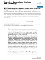
night. At the orthopedic clinic, radiography performed
on the following day revealed cortical bone destruction,
osteoblastic changes, and periosteal reactions, consistent
with conventional osteosarcoma (Fig. 1). For further examination and treatment, the patient was referred to our hospital 5 days after the massage therapy. Magnetic resonance
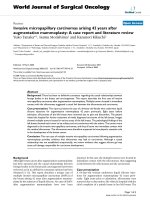
imaging (MRI) revealed iso-signal intensity on T1-weighted
images and high-signal intensity on T2-weighted images of
the left distal femur; it also revealed an extraskeletal mass
(Fig. 2). Furthermore, MRI revealed diffuse signal alteration
in the muscles and the neurovascular areas surrounding the
lesion in the distal femur; hence, hemorrhage and dissemination of the tumor were considered (Fig. 2). Consistent with
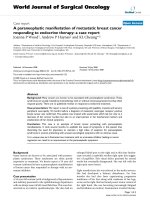
osteosarcoma, open biopsy followed by histopathological
examination revealed tumor cell proliferation with extensive pleomorphism and mitoses (Fig. 3). Seventeen days
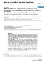
after the massage therapy, computed tomography revealed
multiple metastatic lesions in the lung and liver (Fig. 4).
Thoracic MRI revealed multiple metastases in the thoracic
spine (Fig. 5). 18Fluorine-labeled fluorodeoxyglucosepositron emission tomography revealed tumor metastasis
in the femur and multiple metastases in the thoracic and
lumbar spine, liver, and pelvis (Fig. 6). The patient underwent chemotherapy comprising ifosfamide, carboplatin, pirarubicin, etoposide, doxorubicin, and methotrexate (Fig. 7).
During the second course of chemotherapy, paraplegia due
to spinal metastases developed and progressed. After eight
courses of chemotherapy, the metastatic lesions in the lung
and liver reduced in size (Fig. 8), although considerable
Fig. 1 Radiograph before chemotherapy. Sclerotic lesion with
periosteal reaction was observed in the distal femur
primary tumor growth was observed (Fig. 9). Subsequently,
the patient received hip disarticulation 6 months after the
initial diagnosis, and he then underwent radiation therapy
for metastatic lesions in the liver and sacrum. However,
metastatic lesion growth was observed, and the patient died
due to multiple metastases 13 months after the initial
diagnosis.
Fig. 2 Magnetic resonance imaging (MRI) prior to chemotherapy. MRI revealed extraskeletal mass of distal femur (black arrow), and a lesion
thought to be hemorrhage and dissemination of tumor tissues (arrow) were observed
Miwa et al. BMC Cancer
(2019) 19:993
Page 3 of 6
Fig. 3 Histology. Hematoxylin and eosin staining showed proliferation of tumor cells with extensive pleomorphism and mitoses, which
was consistent with osteosarcoma
Discussion and conclusions
Despite the weak evidence regarding its efficacy, massage
therapy is widely used to mitigate various types of chronic
pain symptoms and to promote return to normal function
[14–19]. Indeed, a randomized trial showed that therapeutic
massage provides relief from intense pain, improves mood
status, and offers muscle relaxation in patients with metastatic bone pain [20]. The possibility that direct compression
of a tumor may induce metastasis and dissemination has
been considered, although there is little evidence. Therefore,
compression due to Esmarch’s bandages and tourniquets
are contraindications for tumors in the extremities [21, 22].
Hayashi et al. investigated the association of tumor compression and lymph node metastasis in a mouse model of fibrosarcoma [23]; in vivo fluorescence imaging of the
fibrosarcoma cells labeled with a fluorescent protein showed
that pressure-dependent compression of the tumor tissue
increased the number of tumor cells that shed into the
lymph duct. An in vivo study using GFP-labeled osteosarcoma cells demonstrated that massage increases tumor volume as well as metastases in the lymph node and lung [13].
In a retrospective study conducted in Taiwan, 70 of 134
patients (52%) with osteosarcoma underwent alternative
medical treatment including massage therapy before their
initial visit to the hospital [12]. A remarkable difference was
observed in the 5-year overall survival rate–58% in patients
treated with massage therapy versus 92% in those not
treated massage therapy. However, these results were confounded because prior to the hospital visit, there was a significantly higher incidence of metastatic lung lesions upon
initial diagnosis (51% in the massage group vs 19% in the
non-massage group) and higher rate of tumor recurrence
(29% in the massage group vs 6% in the non-massage
group). Another retrospective study showed that massage
therapy decreased overall survival and increased incidence
of local recurrence and metastases [13]. Thus, due to the
Fig. 4 Computed tomography (CT) prior to chemotherapy. Metastatic lesions in the lung and liver were observed (arrow)
Miwa et al. BMC Cancer
(2019) 19:993
Fig. 5 Magnetic resonance imaging. Metastases were observed at
the Th4 and Th 12 vertebrae (arrow)
Page 4 of 6
fragility of the tumor tissue compared with normal tissue,
compression during massage is thought to destroy tissues
and rupture tumor vessels. Dissemination of tumor tissue
due to hemorrhage renders it difficult to perform limb salvage surgery, thereby impacting survival. In the present case,
the association of massage therapy with the dissemination of
osteosarcoma cannot be determined because lack of MRI
before massage therapy. However, the diffuse signal alteration in the muscles and the neurovascular areas surrounding the tumor observed by MRI is consistent with a cause of
the severe pain after massage therapy. Therefore, the present
case suggests the local dissemination of tumor tissue due to
compression of the osteosarcoma, although the influence of
massage therapy on metastatic disease remains unclear. Although massage therapy alleviates several symptoms and
brings relief, massage therapists should be aware of the possibility that their massage can disseminate hidden malignancies. In conclusion, the present case suggests the
dissemination of tumor tissue due to massage therapy,
which while creating awareness regarding this rare but most
common malignant bone tumor in youth also cautions massage therapists to be aware of the condition and the
outcomes.
Fig. 6 18Fluorine-labeled fluorodeoxyglucose–positron emission tomography. Multiple metastatic lesions were observed in the liver, spine, and pelvis
Miwa et al. BMC Cancer
(2019) 19:993
Page 5 of 6
Fig. 7 Treatment courses. IC: ifosfamide (2.65 g/m2 daily for 3 days) and carboplatin (560 mg/m2 on Day 1); THP-EI: Pirarubicin (50 mg/m2 on Day
1), etoposide (125 mg/m2 at Day 1 and Day 4), and ifosfamide (1500 mg/m2 daily for 4 days); A: doxorubicin (25 mg/m2 daily for 3 days); M:
methotrexate (12 g/m2); A: doxorubicin (30 mg/m2 daily for 3 days)
Fig. 8 Computed tomography (CT) after chemotherapy. Reductions in the tumor volumes of metastatic lesions were observed in the lung and liver
Fig. 9 Magnetic resonance imaging after chemotherapy. Significant increase in the tumor size was observed in the distal femur
Miwa et al. BMC Cancer
(2019) 19:993
Abbreviations
18
F-FDG: 18Fluorine-labelled fluorodeoxyglucose; CT: Computed tomography;
GFP: Green fluorescent protein; MRI: Magnetic resonance imaging;
PET: Positron emission tomography
Acknowledgments
Not applicable.
Authors’ contributions
All listed authors substantially contributed to the following aspects of the
manuscript: SM, SY, HA, MK, SY, TO, and HT participated in diagnosing and
treating the patient and in acquisition of data. SM, SY, HA, MK, SY, and TO
collected the findings and drafted the manuscript. SM and HT revised the
manuscript. All authors read and approved the final manuscript.
Funding
Not applicable.
Availability of data and materials
To protect privacy and respect confidentiality, no raw data have been made
available in any public repository. The datasets used and/or analyzed during
the current study available from the corresponding author on reasonable
request.
Ethics approval and consent to participate
A family of the patient signed a letter of informed consent to allow his data
to be stored, as required by Nagoya City University Hospital.
Consent for publication
Written informed consent was obtained from the patient and his parents for
the publication of this case report and any accompanying images. A copy of
the written consent form is available for review by the Editor of this journal.
Competing interests
The authors declare that they have no competing interests.
Author details
1
Department of Orthopedic Surgery, Graduate School of Medical Science,
Nagoya City University, Nagoya, Japan. 2Department of Orthopedic Surgery,
Graduate School of Medical Science, Kanazawa University, Kanazawa, Japan.
3
Department of Neonatology and Pediatrics, Graduate School of Medical
Science, Nagoya City University, Nagoya, Japan.
Page 6 of 6
10. Ayerza MA, Farfalli GL, Aponte-Tinao L, Muscolo DL. Does increased rate of
limb-sparing surgery affect survival in osteosarcoma? Clin Orthop Relat Res.
2010;468(11):2854–9.
11. Bielack SS, Kempf-Bielack B, Delling G, Exner GU, Flege S, Helmke K, Kotz R,
Salzer-Kuntschik M, Werner M, Winkelmann W, et al. Prognostic factors in
high-grade osteosarcoma of the extremities or trunk: an analysis of 1,702
patients treated on neoadjuvant cooperative osteosarcoma study group
protocols. J Clin Oncol. 2002;20(3):776–90.
12. Wu PK, Chen WM, Lee OK, Chen CF, Huang CK, Chen TH. The prognosis for
patients with osteosarcoma who have received prior manipulative therapy.
J Bone Joint Surg Br. 2010;92(11):1580–5.
13. Wang JY, Wu PK, Chen PC, Yen CC, Hung GY, Chen CF, Hung SC, Tsai SF, Liu
CL, Chen TH, et al. Manipulation therapy prior to diagnosis induced primary
osteosarcoma metastasis--from clinical to basic research. PLoS One. 2014;
9(5):e96571.
14. Cohen SP, Hooten WM. Advances in the diagnosis and management of
neck pain. BMJ. 2017;358:j3221.
15. Gross A, Langevin P, Burnie SJ, Bedard-Brochu MS, Empey B, Dugas E, FaberDobrescu M, Andres C, Graham N, Goldsmith CH, et al. Manipulation and
mobilisation for neck pain contrasted against an inactive control or another
active treatment. Cochrane Database Syst Rev. 2015;9:CD004249.
16. Furlan AD, Giraldo M, Baskwill A, Irvin E, Imamura M. Massage for low-back
pain. Cochrane Database Syst Rev. 2015;9:CD001929.
17. Marletta G, Canfora A, Roscani F, Cernicchiaro L, Cutrera M, Russo M, Artioli
G, Sarli L. The complementary medicine (CAM) for the treatment of chronic
pain: scientific evidence regarding the effects of healing touch massage.
Acta Biomed. 2015;86(Suppl 2):127–33.
18. Martin ML, Hernandez MA, Avendano C, Rodriguez F, Martinez H. Manual
lymphatic drainage therapy in patients with breast cancer related
lymphoedema. BMC Cancer. 2011;11:94.
19. Batalha LM, Mota AA. Massage in children with cancer: effectiveness of a
protocol. J Pediatr. 2013;89(6):595–600.
20. Jane SW, Chen SL, Wilkie DJ, Lin YC, Foreman SW, Beaton RD, Fan JY, Lu
MY, Wang YY, Lin YH, et al. Effects of massage on pain, mood status,
relaxation, and sleep in Taiwanese patients with metastatic bone pain: a
randomized clinical trial. Pain. 2011;152(10):2432–42.
21. Sarkar MR, Kinzl L. Use of tourniquet with or without Esmarch bandage.
Orthop Traumatol. 1999;7(3):230–7.
22. Younger AS, Kalla TP, McEwen JA, Inkpen K. Survey of tourniquet use in
orthopaedic foot and ankle surgery. Foot Ankle Int. 2005;26(3):208–17.
23. Hayashi K, Jiang P, Yamauchi K, Yamamoto N, Tsuchiya H, Tomita K, Moossa
AR, Bouvet M, Hoffman RM. Real-time imaging of tumor-cell shedding and
trafficking in lymphatic channels. Cancer Res. 2007;67(17):8223–8.
Received: 10 June 2019 Accepted: 6 October 2019
Publisher’s Note
References
1. Ottaviani G, Jaffe N. The epidemiology of osteosarcoma. Cancer Treat Res.
2009;152:3–13.
2. Janeway KA, Grier HE. Sequelae of osteosarcoma medical therapy: a review
of rare acute toxicities and late effects. Lancet Oncol. 2010;11(7):670–8.
3. Bernthal NM, Federman N, Eilber FR, Nelson SD, Eckardt JJ, Eilber FC, Tap
WD. Long-term results (>25 years) of a randomized, prospective clinical trial
evaluating chemotherapy in patients with high-grade, operable
osteosarcoma. Cancer. 2012;118(23):5888–93.
4. Rosen G, Nirenberg A. Neoadjuvant chemotherapy for osteogenic sarcoma:
a five year follow-up (T-10) and preliminary report of new studies (T-12).
Prog Clin Biol Res. 1985;201:39–51.
5. Rosen G. Preoperative (neoadjuvant) chemotherapy for osteogenic sarcoma:
a ten year experience. Orthopedics. 1985;8(5):659–64.
6. Rosen G, Marcove RC, Caparros B, Nirenberg A, Kosloff C, Huvos AG. Primary
osteogenic sarcoma: the rationale for preoperative chemotherapy and
delayed surgery. Cancer. 1979;43(6):2163–77.
7. Liu ZL, Wang G, Peng AF, Luo QF, Zhou Y, Huang SH. Fatty acid synthase
expression in osteosarcoma and its correlation with pulmonary metastasis.
Oncol Lett. 2012;4(5):878–82.
8. Anninga JK, Gelderblom H, Fiocco M, Kroep JR, Taminiau AH, Hogendoorn
PC, Egeler RM. Chemotherapeutic adjuvant treatment for osteosarcoma:
where do we stand? Eur J Cancer. 2011;47(16):2431–45.
9. Grimer RJ. Surgical options for children with osteosarcoma. Lancet Oncol.
2005;6(2):85–92.
Springer Nature remains neutral with regard to jurisdictional claims in
published maps and institutional affiliations.