Expression and clinical significance of PDL1 and BRAF expression in nasopharyngeal carcinoma
Bạn đang xem bản rút gọn của tài liệu. Xem và tải ngay bản đầy đủ của tài liệu tại đây (1.14 MB, 8 trang )
Cao et al. BMC Cancer
(2019) 19:1022
/>
RESEARCH ARTICLE
Open Access
Expression and clinical significance of PDL1 and BRAF expression in nasopharyngeal
carcinoma
Yabing Cao1* , Kin Iong Chan2, Gungli Xiao1, Yanqun Chen1, Xibin Qiu1, Hu Hao1, Sao Chi Mak1 and Tongyu Lin3
Abstract
Background: The prognostic value of programmed death-ligand 1 (PD-L1) and BRAF expression in nasopharyngeal
carcinoma (NPC) is not well-defined. In this study we investigated alterations in PD-L1, BRAF and EGFR by using
immunohistochemistry analysis in a cohort of consecutively enrolled NPC patients.
Methods: A retrospective review of 154 NPC patients form our previous study (BMC Cancer. 2013; 13:226) were
conducted. Survival and prognostic impacts were analyzed based on PD-L1, BRAF and EGFR expression levels.
Results: One hundred fifty four patients were included in this study. PD-L1 expression was detected in 87.7% of
patients; 14.3% had 1–5% PD-L1 expression, 47.4% had 5–49% expression while 26% had ≥50% expression Higher
PD-L1 expression was significantly associated with shorter PFS and OS. The median PFS was 25 months (95% CI
15.7–34.3 months) and OS was 35 months (95% CI 22.60–47.4 months) for patients with PD-L1 expression ≥50%;
both median PFS and OS were not yet reached for patients with PD-L1 expression < 50%. PFS was significantly
higher in BRAF mutation positive patients (5-year PFS: 55.1% vs. 30.8%, P = 0.044).
Conclusion: Tumor PD-L1 expression and BRAF mutation are associated with poor outcomes in patients with NPC.
This study was retrospectively registered in ClinicalTrials.gov (NCT03989297) on 2019-6-18.
Keywords: Nasopharyngeal carcinoma, Programmed death-ligand 1, BRAF, Prognosis
Background
Nasopharyngeal carcinoma (NPC) is rare in most parts
of the world but is one of the more common types of
cancer in southern China. In 2015, it was estimated that
the incidence of NPC was 60.6 per 100,000 in China
with a mortality rate of 34.1 per 100,000 [1, 2]. The main
treatment for NPC is radiotherapy or chemoradiotherapy [3], and the 5-year survival rate is about 85% [4].
Even with best available treatment, about 30% of patients
relapse with local recurrence or metastasis [5]. The
prognosis for patients with recurrent or primary metastatic NPC is poor with a median progression free survival of 19.4 months [6]. Evidently, novel approaches and
better therapies are needed for the treatment of NPC.
Biomarkers that can reliably predict the prognosis of
patients are important. In a previous study, we found
* Correspondence:
1
Department of Oncology, Kiang Wu Hospital, Macau, SAR, China
Full list of author information is available at the end of the article
that gender and age were strong independent prognostic
factors for NPC [7]. Specifically, younger and male patients were more likely to have distant metastases and
exhibit poorer overall survival and progression-free survival rates compared to other NPC patients treated in
our center [7]. A more recent study identified a prognostic gene expression-based signature that predicts distant
metastasis in locoregionally advanced NPC [8].
In addition to prognostic biomarkers, predictive biomarkers that can identify patients who are likely to benefit
from a particular therapy can help guide treatment selection.
NPC is characterized by lymphocyte infiltration, including T
cells and cytotoxic tumor-infiltrating T lymphocytes [9].
Since immune checkpoint inhibitors can activate cytotoxic
T cells to attack cancer cells, patients with lymphocyte-rich
cancer types (such as EBV-positive NPC) may benefit more
from immunotherapy [10, 11]. Tumor programmed deathligand 1 (PD-L1) expression levels have also been suggested
to be of predictive value for treatment efficacy in some
© The Author(s). 2019 Open Access This article is distributed under the terms of the Creative Commons Attribution 4.0
International License ( which permits unrestricted use, distribution, and
reproduction in any medium, provided you give appropriate credit to the original author(s) and the source, provide a link to
the Creative Commons license, and indicate if changes were made. The Creative Commons Public Domain Dedication waiver
( applies to the data made available in this article, unless otherwise stated.
Cao et al. BMC Cancer
(2019) 19:1022
cancer types [12–15]. However, the clinical significance of
PD-L1 expression in NPC is controversial due to conflicting
data amongst studies [16–19].
BRAF is one of downstream of EGFR pathway molecule
[20], and BRAF (V600E) mutation is rarely reported in
previous study [21]. In other solid tumors such as melanoma and non-small cell lung cancer, BRAF inhibitors were
approved for patients with BRAF mutation positive.
In the present study we aim to evaluate the clinical
significance of PD-L1, BARF and EGFR expressions in
the tumor cells of a cohort of NPC patients. Separate
data from this cohort of patients have been reported in a
previous publication [7].
Methods
Patient selection
Consecutive patients who were pathologically diagnosed
with NPC between 2006 and December 2010 at the Kiang
Wu Hospital (Macau SAR of China) and for whom freshfrozen tissue samples were available were included. The
clinicopathologic information of all patients was collected,
including sex, age, tumor stage, pathologic type, and treatment methods and outcomes. Tumor stage was classified
according to the International Union Against Cancer and
American Joint Committee on Cancer staging system for
NPC, seventh edition. Fresh nasopharyngeal tissue samples were obtained from all patients. The protocol was approved by the institutional review board of the Kiang Wu
Hospital (KWH 2016–014).
Treatment and outcome
All patients received standard treatment including radiation therapy with or without chemotherapy. Briefly, the
intensity modulated radiotherapy technique technology
were utilized for radiation. Chemotherapy were given for
patients based on their tumor stage and the decision by
each patient’s physician. Chemotherapy regimen was
based on NCCN guidelines.
We defined progression-free survival (PFS) as time
from date of treatment to the date of disease progression
or death from any causes, whichever came first. Overall
survival (OS) was defined as the time from date of treatment to the time of death.
Immunohistochemistry for PD-L1, BRAF, and EGFR
expression
PD-L1, BRAF and EGFR expressions in the tumor cells was
evaluated using immunohistochemistry. Four mm-thick sections were prepared from paraffin-embedded specimens of
the NPC tumor. The sections were deparaffinized in xylene
followed by 95% ethanol. After rehydration, sections were
pretreated in a microwave oven at 95 °C for 15 min in citrate
buffer (pH 6.0) for antigen retrieval. Next, endogenous
Page 2 of 8
peroxidase activity was blocked with 4% Block ACE Powder
in H2O at 37 °C for 10 min.
Immunohistochemistry (IHC) was carried out by
benchmark XT automated stainer
PD-L1 protein was detected by using PD-L1 (SP263)
rabbit monoclonal antibody with Ultraview detection system (Ventana, Tucson, Arizona). Reference to the interpretation guide of Ventana PD-L1 (SP263) assay staining
of non-small cell lung cancer, the tumor cells was counted
if any intensity of the staining result demonstrating in
membrane with a discontinuous, circumferential or basolateral pattern or rarely in peri-nuclear dot-like body.
BRAF V600E protein was detected by using BRAF
V600E (VE1) mouse monoclonal primary antibody and
the OptiView DAB IHC Detection Kit (Ventana, Tucson,
Arizona). The immunostaining result was interpreted as
positive if any intensity of cytoplasmic staining.
EGFR mutation specific antibodies were detected by
using EGFR mutation specific rabbit monoclonal antibodies against del E746-A750 (6B6, dilution:1:50; Cell
Signaling Technology, Inc., Boston, MA, USA) and
L858R (43B2, dilution:1:10; Cell Signaling Technology,
Inc). The immunoreactions were detected by OptiView
DAB IHC Detection Kit (Ventana, Tucson, Arizona).
The immunostaining results were interpreted as positive if any intensity on cytoplasmic and/or membrane
staining.
Assessment of PD-L1, BRAF, and EGFR expression
PD-L1, BRAF mutation, and EGFR mutation expression
in tumor cells were evaluated in a blind fashion without
knowledge of any existing clinical characteristics. Any
staining within the tumor cell membrane or cytoplasm
was considered positive. Grading was based on staining
ratio of the tumor cells, ≥50% of tumor cells positive
was scored as 3; ≥5 to < 50% (5–49%) of tumor cells
expressed positive as 2; ≥1 to < 5% (1–5%) of tumor cells
expressed positive as 1; negative as 0.
BRAF and EGFR mutation expression were categorized as negative or positive.
Statistical analysis
Fisher’s exact test or the chi-squared test was performed
to examine the association between PD-L1 expression
and the oncogenic mutations versus various clinicopathological features, as appropriate. The PD-L1 expression
was evaluated as a categorical variable (0, 1–5%, 5–49%
and ≥ 50% expression). Survival curves were plotted
using the Kaplan-Meier method and compared using a
log-rank test. The prognostic impact of relevant clinicopathological variables including PD-L1 expression in the
pulmonary metastatic tumors was evaluated using the
Cox proportional hazards regression models and hazard
Cao et al. BMC Cancer
(2019) 19:1022
ratios (HRs). To assess the prognostic value of high PDL1 expression, variables with P < 0.2 in the univariate
analysis were entered into the multivariate analysis, and
variables with P < 0.05 were included in a final model
with backward elimination methods. A two-sided Pvalue< 0.05 was considered statistically significant. Statistical analyses were performed using the SPSS version
20.0 software package (SPSS Inc., Chicago, IL, USA).
Page 3 of 8
Table 1 Patient demographics and disease characteristics
N = 154
Characteristic
Cases
Percentage (%)
Age (Years)
Median
60
Range
26–83
< 60
71
46.1
≥60
83
53.9
Male
116
75.2
Female
38
24.8
I–II
67
43.5
III–IV
86
56.5
Chemoradiation
124
80.5
Radiation Only
31
19.5
0–1
131
85.0
≥2
24
15.0
Yes
76
49.4
No
78
50.6
Relationship between PD-L1, BRAF mutation, and EGFR
mutation expression with patient characteristics
0%
17
11.0
1–5%
22
14.3
PD-L1 expression was detected in 87.6% of biopsy tissue.
PD-L1 expression was 0% in 11.0% of patients, 1–5% in
14.3% of patients, 5–49% in 47.4% of patients and ≥ 50%
in 26% of patients. There was no difference in PD-L1 expression between genders or age groups. However, there
was significantly higher expression of PD-L1 among patients with disease recurrence or metastasis (P = 0.001).
There was also a significantly higher expression of BRAF
mutation among patients with disease recurrence or metastasis (P = 0.035). There was no significant association
between PD-L1 expression levels, BRAF V600E mutation
and EGFR 19del mutation with age, sex or disease stage.
Most of the tumor tissues that expressed PD-L1 were
BRAF V600E mutation negative (P = 0.002). There was
no significant association between PD-L1 expression
levels and EGFR 19del mutation (P = 0.161).
5–49%
73
47.4
≥50
40
26.0
Unknown
2
1.3
Negative
139
90.3
Positive
13
8.4
Unknown
2
1.3
Negative
149
96.8
Positive
3
1.9
Unknown
2
1.3
Negative
151
98.1
Positive
0
0.0
Unknown
3
1.9
Results
Sex
Patient characteristics
A total of 154 patients were included in the analysis.
The baseline characteristics of patients are shown in
Table 1. Median age was 60 years (range 26–83 years).
The majority of patients were male (75.2%). All patients
were diagnosed with non-keratinizing undifferentiated
carcinoma according to the WHO histological classification. The median and maximum follow-up duration was
76 months and 145 months, respectively. The last day of
follow-up was in January 2019. Seventy six patients
(49.4%) had tumor recurrence or metastasis. None of the
patients received anti-PD-L1 antibody treatment because
anti-PD-L1 antibody treatment was not available in
Macau during the follow-up period.
Stage
Treatment
ECOG
Progression
Expression of PD-L1, BRAF and EGFR
Fig. 1
PD-L1
BRAF V600E
EGFR 19del
EGFR L858R
Prognostic impact on progression free survival and
overall survival
Values are presented as number (%) unless otherwise stated
Percentages may not sum to exactly 100 due to rounding
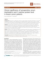
PD-L1 expression was significantly associated with overall survival. Higher expressions of PD-L1 were associated
with shorter PFS (P < 0.001, Fig. 2a) and reduced OS
(P < 0.001, Fig. 2b). The 5-year PFS rates for patients
with PD-L1 expression 0%, 1–5%, 5–49% and ≥ 50%
were 75.5, 72.7, 55.9 and 24.8%, respectively (P < 0.001).
The median PFS was 25 months (95% CI 15.7–34.3
months) for patients with PD-L1 expression ≥50%, and
not yet reached for patients with PD-L1 expression <
Cao et al. BMC Cancer
(2019) 19:1022
Page 4 of 8
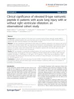
Fig. 1 Representative immunostaining of programmed death-ligand 1 (PD-L1), BRAF V600E mutation and EGFR 19del and L858R mutations
(magnification, × 200). Anti-PD-L1 antibody (clone SP263) is validated using placenta as a positive control. HE staining of NPC tissue is presented
in A. PD-L1 expression in NPC biopsy tissues was graded as 0% (b), 1–5% (c), 5–49% (d), and ≥ 50% (e and f). BRAF V600E and EGFR staining
not shown
50%. The overall median PFS for all patients was 84
months. The 5-year OS rate for patients with PD-L1 expression 0%, 1–5%, 5–49% and ≥ 50% were 85.7, 72.7,
68.3 and 35.0%, respectively (P < 0.001). The median OS
was 35 months (95% CI 22.60–47.4 months) for patients
with PD-L1 expression ≥50%, and not yet reached for
patients with PD-L1 expression < 50%. The overall
median OS for all patients was 96 months (95% CI 60.2–
131.8 months). Consistent with our previous report,
female patients had a favorable prognosis than male patients (P = 0.009, figure not show).
PFS was significantly different between BRAF mutation negative and positive patients (5-year PFS: 55.1% vs.
30.8%, P = 0.044; Fig. 2c). However, OS rates did not differ significantly between BRAF mutation negative and
positive patients (5-year OS rate: 61.9% vs. 46.2%, P =
0.075; Fig. 2d).
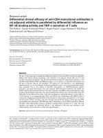
The results of prognostic factor analysis for survival
using Cox proportional hazards regression model are
shown in Fig. 3. Univariate analysis showed that high PDL1 expression and the female gender were significantly associated with a shorter OS. Multivariate analysis indicated
that high PD-L1 expression, along with gender is associated with shorter OS and thus poorer prognosis. There
was no interaction between PD-L1 expression and gender
(Table 2). The presence of BRAF V600E mutation was associated with disease progression (P = 0.035; Table 2).
Discussion
It is known that PD-L1 expression is upregulated on
various tumor cell lines. NPC is an EBV-associated cancer. Previous studies demonstrated that EBV-related latent membrane protein 1 (LMP1) and interferon-gamma
(IFN-γ) may upregulate PD-L1 in NPC [22, 23] and NK/
T cell lymphoma [24]. Expression of viral proteins, such
as EBV nuclear antigen-1 or LMP1 and 2 in NPC cells
can elicit a virus-specific immune response in patients
with NPC. LMP1 expression and IFN-γ activation can
synergistically induce the expression of PD-L1 in NPC
cells [22]. Expression of PD-L1 can also be upregulated by
tumor-infiltrating lymphocytes (TILs), which is associated
with impaired effector function (cytokine production and
cytotoxic efficacy against tumor cells) and poor outcomes
in NPC [25]. In our study, we found positive PD-L1 expression in 87.7% of patients with NPC; 14.3% had 0–1%
PD-L1 expression, 47.4% had 1–49% expression while
26% had ≥50% expression. This is consistent with other
studies which reported PD-L1 expression in 89–95% of
NPC tumors, with 50% or more malignant cells being PDL1 positive in the majority of these tumors [26].
Activation of PD-1 pathway can lead to T cell exhaustion. Thus, the PD-1/PD-L1 axis is crucial in regulating
anti-tumor immunity. In this study, we performed a retrospective analysis on 154 consecutive patients who were
homogeneously treated with IMRT. Our findings demonstrate that high PD-L1 expression is a poor prognostic factor for NPC patients. Best progression free survival was
seen in the PD-L1 expression negative group, with a 5year PFS rate of 75.2%. For patients with positive PD-L1
expression, the PFS rate reduces as expression levels increase; 5-year PFS rates were 72.7, 55.9 and 24.8% for patients with PD-L1 expression 1–5%, 5–49% and ≥ 50%,
respectively. The 5-year OS rate for patients with PD-L1
expression 0%, 1–5%, 5–49% and ≥ 50% were 85.7, 72.7,
68.3 and 35.0%, respectively. Our data are consistent to
those recently published by Ben-Betzalel et al. [19, 27–29]
Cao et al. BMC Cancer
(2019) 19:1022
a
b
c
d
Fig. 2 Progression free survival (PFS) and overall survival (OS) for all
patients. PFS (a) and OS (b) by PD-L1 expression levels. PFS (c) and
OS (d) by BRAF V600E mutation
Page 5 of 8
who found similar association of PD-L1 expression with
poor survival. However, other studies have reported favorable prognosis with increased PD-L1 expression [17, 30],
while others found no relation between PD-L1 expression
and survival [31, 32].
There many reasons behind these inconsistent findings. First, some studies included a mixed patient population, which consists of patients with NPC patients as
well as those with other types of head and neck squamous cancer. Second, not all studies used commercially
available clones of PD-L1 antibodies. SP263 and 22C3
(Dako), and SP142 (Ventana) have been shown to pass
the Western Blot and immunohistochemical validation.
In prior comparison trials, it was shown that 22C3 and
SP263 were closely aligned in tumor cell staining, but
SP142 stains less tumor cells [33]. Third, the follow up
period of some of the studies were too short for PFS and
OS analysis. Our study focused on NPC patients and
with an extended follow-up period of 13 years. Since the
percentage of PD-L1-positive cells can vary due to different antibody clones and immunostaining methods, finding the best cutoff value with the highest clinical
significance is crucial in such studies. We used the
SP263 antibody with the standard cut off value of 1 and
5%, which is frequently used for lung cancer and other
cancer types [34]. Inevitably, whenever an IHC-based
biomarker is considered, questions will arise regarding
the reproducibility of the staining of the tissue and
consistency in interpretation of the test by pathologists.
In future, multicenter, international standardization efforts could address many of these questions and help develop one “standardized” assay to analyse additional
immunotherapy-related predictive markers [35].
BRAF mutations have been identified in melanoma
and colorectal cancer, but is rarely reported in NPC
[36]. BRAF mutations are associated with poorer survival
in patients with melanoma [37], but the significance of
BRAF mutations among NPC patients has not been
thoroughly investigated. For the first time, we report that
the BRAF V600E mutation was significantly associated
with disease progression and PFS. In this study, 13 of
154 patients (8.4%) were BRAF V600E mutation positive.
The 5-year PFS of BRAF V600E mutation positive and
negative patients were 55.1 and 30.8%, respectively.
Using multivariate analysis, PD-L1 expression and
gender were independent prognostic factors for overall
survival. This confirmed our previous study, that female
patients had a favorable prognosis than male patients.
PD-L1 expression is the most extensively studied biomarker with respect to predicting the efficacy of anti–PD1 or anti–PD-L1 therapies. A positive correlation between
PD-L1 expression and treatment efficacy has been reported in the study of nivolumab [38] and pembrolizumab
for NPC [39]. In our center, 70 patients with NPC have
Cao et al. BMC Cancer
(2019) 19:1022
Page 6 of 8
Fig. 3 Forest plot of hazard ratio (HR) for overall survival (OS) by independent prognostic factors
received PD-1 therapy, 13 received nivolumab monotherapy, 29 received pembrolizumab monotherapy and 28 received pembrolizumab combined with chemotherapy. An
internal analysis of these patients revealed that PD-L1
positive tumor cell with high CD8 positive tumor infiltrates correlated with objective response to PD-L1 inhibitor (data not published).
Our study has some limitations. Our study lacks EBV loading data as the EBV DNA test was not routinely carried out
during the period that patients received treatment. EBV expression is an import contributor here and may increase PDl1 expression [24, 40]. Secondly, patients in this study did
not receive PD-1 or PD-L1 targeted therapy as these were
not available in Macau during the follow-up period. Therefore, we were unable to explore the correlation between PDL1 expression and the efficacy of immunotherapy.
Conclusion
Our results suggest that high tumor PD-L1 expression and
BRAF V600E mutation are associated with poor outcomes in
patients with NPC. PD-L1 expression was found to be a significant prognostic factor, and high PD-L1 expression may
be of prognostic value for disease progression and survival.
Table 2 Association between clinical parameters and expression of PD-L1, BRAF and EGFR proteins
Characteristic
PD-L1 (N)
BRAF V600E (N)
EGFR 19Del (N)
0
1–5
5–49
> 50
P-value
Negative
Positive
P-value
Negative
Positive
P-value
< 60
10
11
36
12
0.127
64
5
0.411*
67
2
0.431*
≥ 60
7
11
37
28
75
8
82
1
Male
14
17
53
30
104
10
112
2
Female
3
5
20
10
35
3
37
1
I-II
7
9
36
14
60
6
III-IV
10
13
37
26
79
7
Yes
4
6
32
33
65
10
No
13
16
41
7
74
3
Age
Sex
0.776
0.602*
0.581*
Stage
0.517
0.529*
65
1
84
2
73
2
76
1
0.599*
Progression
*: Fisher’s exact test
Chi-squared test was used in variables without *
0.001
0.035*
0.490*
Cao et al. BMC Cancer
(2019) 19:1022
Abbreviations
PD-L1: programmed death-ligand 1 (PD-L1); NPC: nasopharyngeal carcinoma
(NPC); EGFR: epidermal growth factor receptor; PFS: progression-free survival;
OS: overall survival; IHC: Immunohistochemistry; LMP-1: latent membrane
protein 1; EBV: Epstein–Barr virus
Acknowledgements
None.
Authors’ contributions
YBC and TYL conceived of the presented idea. YBC and JIC carried out the
experiment. GLX, YQC, XBQ, SCM and HH collected data. YBC verified the
analytical methods. Both YBC and TYL contributed to the final version of the
manuscript. All authors have read and approved the manuscript.
Page 7 of 8
8.
9.
10.
11.
12.
Funding
This study is funded by The Science and Technology Development Fund
(FDCT) of Macau, grant number is 019/2016/AFJ. This funding source had no
role in study design, data collection and analysis, decision to publish, or
preparation of the manuscript.
13.
14.
Availability of data and materials
The datasets used and analyzed during the current study are available from
the corresponding author on reasonable request.
15.
Ethics approval and consent to participate
The protocol was approved by the institutional review board of the Kiang
Wu Hospital (KWH 2016–014).
Declarations: The need for consent to participate was waived by the
institutional review board of the Kiang Wu Hospital on June 21, 2019.
Consent for publication
No details, images, or videos relating to an individual person was included in
this paper therefore written informed consent for the publication of these
details was not obtained from any person.
16.
17.
18.
Competing interests
The authors declare that they have no competing interests.
Author details
1
Department of Oncology, Kiang Wu Hospital, Macau, SAR, China.
2
Department of Pathology, Kiang Wu Hospital, Macau, SAR, China.
3
Department of Oncology, Sun Yat-Sen University Cancer Center,
Guangzhou, China.
19.
20.
21.
Received: 25 June 2019 Accepted: 18 October 2019
22.
References
1. Chen W, Zheng R, Baade PD, Zhang S, Zeng H, Bray F, et al. Cancer statistics
in China, 2015. CA Cancer J Clin. 2016;66(2):115–32.
2. Chen W, Xia C, Zheng R, Zhou M, Lin C, Zeng H, et al. Disparities by
province, age, and sex in site-specific cancer burden attributable to 23
potentially modifiable risk factors in China: a comparative risk assessment.
Lancet Glob Health. 2019;7(2):e257–e69.
3. Blanchard P, Lee A, Marguet S, Leclercq J, Ng WT, Ma J, et al. Chemotherapy
and radiotherapy in nasopharyngeal carcinoma: an update of the MAC-NPC
meta-analysis. Lancet Oncol. 2015;16(6):645–55.
4. Yang L, Xia L, Wang Y, He S, Chen H, Liang S, et al. Development and
external validation of nomograms to predict the risk of skeletal metastasis
at the time of diagnosis and skeletal metastasis-free survival in
nasopharyngeal carcinoma. BMC Cancer. 2017;17(1):628.
5. Lee AW, Ma BB, Ng WT, Chan AT. Management of Nasopharyngeal
Carcinoma: current practice and future perspective. J Clin Oncol. 2015;
33(29):3356–64.
6. Zhang L, Huang Y, Hong S, Yang Y, Yu G, Jia J, et al. Gemcitabine plus
cisplatin versus fluorouracil plus cisplatin in recurrent or metastatic
nasopharyngeal carcinoma: a multicentre, randomised, open-label, phase 3
trial. Lancet. 2016;388(10054):1883–92.
7. Xiao G, Cao Y, Qiu X, Wang W, Wang Y. Influence of gender and age on the
survival of patients with nasopharyngeal carcinoma. BMC Cancer. 2013;13:226.
23.
24.
25.
26.
27.
28.
29.
Tang XR, Li YQ, Liang SB, Jiang W, Liu F, Ge WX, et al. Development and
validation of a gene expression-based signature to predict distant
metastasis in locoregionally advanced nasopharyngeal carcinoma: a
retrospective, multicentre, cohort study. Lancet Oncol. 2018;19(3):382–93.
Ono T, Azuma K, Kawahara A, Sasada T, Matsuo N, Kakuma T, et al.
Prognostic stratification of patients with nasopharyngeal carcinoma based
on tumor immune microenvironment. Head Neck. 2018;40(9):2007–19.
Cho YA, Yoon HJ, Lee JI, Hong SP, Hong SD. Relationship between the
expressions of PD-L1 and tumor-infiltrating lymphocytes in oral squamous
cell carcinoma. Oral Oncol. 2011;47(12):1148–53.
Mattox AK, Lee J, Westra WH, Pierce RH, Ghossein R, Faquin WC, et al. PD-1
expression in head and neck squamous cell carcinomas derives primarily
from functionally Anergic CD4(+) TILs in the presence of PD-L1(+) TAMs.
Cancer Res. 2017;77(22):6365–74.
Dang TO, Ogunniyi A, Barbee MS, Drilon A. Pembrolizumab for the
treatment of PD-L1 positive advanced or metastatic non-small cell lung
cancer. Expert Rev Anticancer Ther. 2016;16(1):13–20.
Doroshow DB, Sanmamed MF, Hastings K, Politi K, Rimm DL, Chen L,
et al. Immunotherapy in non-small cell lung Cancer: facts and hopes.
Clin Cancer Res. 2019.
Koemans WJ, Chalabi M, van Sandick JW, van Dieren JM, Kodach LL. Beyond
the PD-L1 horizon: in search for a good biomarker to predict success of
immunotherapy in gastric and esophageal adenocarcinoma. Cancer Lett.
2019;442:279–86.
Taube JM, Klein A, Brahmer JR, Xu H, Pan X, Kim JH, et al. Association of PD1, PD-1 ligands, and other features of the tumor immune
microenvironment with response to anti-PD-1 therapy. Clin Cancer Res.
2014;20(19):5064–74.
Li YF, Ding JW, Liao LM, Zhang ZL, Liao SS, Wu Y, et al. Expression of
programmed death ligand-1 predicts poor outcome in nasopharyngeal
carcinoma. Mol Clin Oncol. 2017;7(3):378–82.
Liu YJ, Tsang NM, Hsueh C, Yeh CJ, Ueng SH, Wang TH, et al. Low PD-L1
Expression Strongly Correlates with Local Recurrence in Epstein-Barr VirusPositive Nasopharyngeal Carcinoma after Radiation-Based Therapy. Cancers
(Basel). 2018;10(10).
Yang WF, Wong MCM, Thomson PJ, Li KY, Su YX. The prognostic role of PDL1 expression for survival in head and neck squamous cell carcinoma: a
systematic review and meta-analysis. Oral Oncol. 2018;86:81–90.
Zhou Y, Shi D, Miao J, Wu H, Chen J, Zhou X, et al. PD-L1 predicts poor
prognosis for nasopharyngeal carcinoma irrespective of PD-1 and EBV-DNA
load. Sci Rep. 2017;7:43627.
Fernandez-Medarde A, Santos E. Ras in cancer and developmental diseases.
Genes Cancer. 2011;2(3):344–58.
Zhang JW, Qin T, Hong SD, Zhang J, Fang WF, Zhao YY, et al. Multiple
oncogenic mutations related to targeted therapy in nasopharyngeal
carcinoma. Chin J Cancer. 2015;34(4):177–83.
Fang W, Zhang J, Hong S, Zhan J, Chen N, Qin T, et al. EBV-driven LMP1
and IFN-gamma up-regulate PD-L1 in nasopharyngeal carcinoma:
implications for oncotargeted therapy. Oncotarget. 2014;5(23):12189–202.
Yoshizaki T, Kondo S, Endo K, Nakanishi Y, Aga M, Kobayashi E, et al. Modulation
of the tumor microenvironment by Epstein-Barr virus latent membrane protein 1
in nasopharyngeal carcinoma. Cancer Sci. 2018;109(2):272–8.
Bi XW, Wang H, Zhang WW, Wang JH, Liu WJ, Xia ZJ, et al. PD-L1 is
upregulated by EBV-driven LMP1 through NF-kappaB pathway and
correlates with poor prognosis in natural killer/T-cell lymphoma. J Hematol
Oncol. 2016;9(1):109.
Hsu MC, Hsiao JR, Chang KC, Wu YH, Su IJ, Jin YT, et al. Increase of
programmed death-1-expressing intratumoral CD8 T cells predicts a poor
prognosis for nasopharyngeal carcinoma. Mod Pathol. 2010;23(10):1393–403.
Chen BJ, Chapuy B, Ouyang J, Sun HH, Roemer MG, Xu ML, et al. PD-L1
expression is characteristic of a subset of aggressive B-cell lymphomas and
virus-associated malignancies. Clin Cancer Res. 2013;19(13):3462–73.
Zhao L, Liao X, Hong G, Zhuang Y, Fu K, Chen P, et al. Mismatch repair
status and high expression of PD-L1 in nasopharyngeal carcinoma. Cancer
Manag Res. 2019;11:1631–40.
Zhang J, Fang W, Qin T, Yang Y, Hong S, Liang W, et al. Co-expression of
PD-1 and PD-L1 predicts poor outcome in nasopharyngeal carcinoma. Med
Oncol. 2015;32(3):86.
Qu Y, Wang D, Yang L, Liu HY, Cui W, Che YQ. Expression and clinical
significance of programmed death ligand 1 in nasopharyngeal carcinoma.
Mol Clin Oncol. 2018;9(1):75–81.
Cao et al. BMC Cancer
(2019) 19:1022
30. Lee VH, Lo AW, Leung CY, Shek WH, Kwong DL, Lam KO, et al. Correlation
of PD-L1 expression of tumor cells with survival outcomes after radical
intensity-modulated radiation therapy for non-metastatic nasopharyngeal
carcinoma. PLoS One. 2016;11(6):e0157969.
31. Chang AMV, Chiosea SI, Altman A, Pagdanganan HA, Ma C. Programmed
death-ligand 1 expression, microsatellite instability, Epstein-Barr virus, and
human papillomavirus in nasopharyngeal carcinomas of patients from the
Philippines. Head Neck Pathol. 2017;11(2):203–11.
32. Larbcharoensub N, Mahaprom K, Jiarpinitnun C, Trachu N, Tubthong N,
Pattaranutaporn P, et al. Characterization of PD-L1 and PD-1 expression and
CD8+ tumor-infiltrating lymphocyte in Epstein-Barr virus-associated
nasopharyngeal carcinoma. Am J Clin Oncol. 2018;41(12):1204–10.
33. Lantuejoul S, Damotte D, Hofman V, Adam J. Programmed death ligand 1
immunohistochemistry in non-small cell lung carcinoma. J Thorac Dis. 2019;
11(Suppl 1):S89–S101.
34. Marchetti A, Barberis M, Franco R, De Luca G, Pace MV, Staibano S, et al.
Multicenter comparison of 22C3 PharmDx (Agilent) and SP263 (Ventana)
assays to test PD-L1 expression for NSCLC patients to be treated with
immune checkpoint inhibitors. J Thorac Oncol. 2017;12(11):1654–63.
35. Kerr KM, Tsao MS, Nicholson AG, Yatabe Y, Wistuba II, Hirsch FR, et al.
Programmed death-ligand 1 immunohistochemistry in lung Cancer: in what
state is this art? J Thorac Oncol. 2015;10(7):985–9.
36. Chou CC, Chou MJ, Tzen CY. PIK3CA mutation occurs in nasopharyngeal
carcinoma but does not significantly influence the disease-specific survival.
Med Oncol. 2009;26(3):322–6.
37. Houben R, Becker JC, Kappel A, Terheyden P, Brocker EB, Goetz R, et al.
Constitutive activation of the Ras-Raf signaling pathway in metastatic
melanoma is associated with poor prognosis. J Carcinog. 2004;3:6.
38. Ma BBY, Lim WT, Goh BC, Hui EP, Lo KW, Pettinger A, et al. Antitumor
activity of Nivolumab in recurrent and metastatic nasopharyngeal
carcinoma: an international, multicenter study of the Mayo Clinic phase 2
consortium (NCI-9742). J Clin Oncol. 2018;36(14):1412–8.
39. Hsu C, Lee SH, Ejadi S, Even C, Cohen RB, Le Tourneau C, et al. Safety and
antitumor activity of Pembrolizumab in patients with programmed deathligand 1-positive nasopharyngeal carcinoma: results of the KEYNOTE-028
study. J Clin Oncol. 2017;35(36):4050–6.
40. Anastasiadou E, Stroopinsky D, Alimperti S, Jiao AL, Pyzer AR, Cippitelli C,
et al. Epstein-Barr virus-encoded EBNA2 alters immune checkpoint PD-L1
expression by downregulating miR-34a in B-cell lymphomas. Leukemia.
2019;33(1):132–47.
Publisher’s Note
Springer Nature remains neutral with regard to jurisdictional claims in
published maps and institutional affiliations.
Page 8 of 8