CYR61, a potential biomarker of tumor inflammatory response in epithelial ovarian cancer microenvironment of tumor progress
Bạn đang xem bản rút gọn của tài liệu. Xem và tải ngay bản đầy đủ của tài liệu tại đây (815.28 KB, 8 trang )
Shi et al. BMC Cancer
(2019) 19:1140
/>
RESEARCH ARTICLE
Open Access
CYR61, a potential biomarker of tumor
inflammatory response in epithelial ovarian
cancer microenvironment of tumor
progress
Jun Shi1,2, Rongfen Huo3, Ningli Li3, Haichuan Li3, Tianhang Zhai3, Huidan Li3, Baihua Shen3, Jing Ye1,2,
Ruojin Fu1,2 and Wen Di1,2*
Abstract
Background: Recent studies have found that inflammatory response is involved in the pathogenesis of ovarian
cancer. Advanced ovarian cancer is often presented with ascites that is rich in cytokines, inflammatory factors or
cancer cells. Therefore, it is important to study the microenvironment of ascites in order to further clarify the
occurrence and progression of ovarian cancer. As a pro-inflammatory factor, the Cyr61 expression patterns are
inconsistent in human tumors. Although it has been reported that Cyr61 is related to the progression of ovarian
cancer, its specific mechanism is not yet clear. This study sought to evaluate the Cyr61 levels of ascites, serum and
different tissues of ovarian cancer to explore the potential association of Cyr61with the tumor-associated
inflammatory microenvironment of EOC.
Methods: Tumor specimens were procured from patients with ovarian serous cystadenocarcinoma and ovarian
serous cystadenoma. Cyr61 and IL-6 levels of serum or ascites were determined by ELISA (Enzyme-Linked
ImmunoSorbent Assay), while Cyr61 expressions of different ovarian tumor tissues were evaluated by IHC
(Immunohistochemistry). Then the correlation of Cyr61 level in ascites with clinicopathologic features was analyzed.
And other laboratory data were obtained from medical records.
Results: Both in ascites and serum, significantly higher Cyr61 levels were found in ovarian serous
cystadenocarcinoma. In malignant ascites, higher Cyr61 level of ovarian serous cystadenocarcinoma was more
closely associated with FIGO stage, initial tumor size > 10 cm and the residual tumor size. And the increased IL-6
level was linearly related to Cyr61 level. Moreover, the serum levels of Cyr61, IL-6 and CRP in advanced stage of
ovarian cancer were much higher than those in early stage. Lastly, the IHC data demonstrate that Cyr61 expression
of ovarian serous adenocarcinoma was higher than that of ovarian serous cystadenoma, but it was lower than the
paired metastatic lesions.
Conclusions: As a pro-inflammatory factor, increased ascites Cyr61 level is associated with FIGO stage, initial tumor
size > 10 cm and the residual tumor size. Moreover, serum Cyr61 may be used as a potential marker for EOC
inflammatory response. Finally, Cyr61 may be involved in the process of tumor metastasis and progression by
producing IL-6 and CRP in the EOC inflammatory microenvironment.
Keywords: Epithelial ovarian cancer, Cyr61, Tumor-associated inflammatory microenvironment, Tumor progression
* Correspondence:
1
Department of Obstetrics and Gynecology, Renji Hospital, School of
Medicine, Shanghai Jiaotong University, Shanghai 200127, China
2
Shanghai Key Laboratory of Gynecologic Oncology, Shanghai 200127,
People’s Republic of China
Full list of author information is available at the end of the article
© The Author(s). 2019 Open Access This article is distributed under the terms of the Creative Commons Attribution 4.0
International License ( which permits unrestricted use, distribution, and
reproduction in any medium, provided you give appropriate credit to the original author(s) and the source, provide a link to
the Creative Commons license, and indicate if changes were made. The Creative Commons Public Domain Dedication waiver
( applies to the data made available in this article, unless otherwise stated.
Shi et al. BMC Cancer
(2019) 19:1140
Background
Epithelial ovarian cancer (EOC) is the most lethal
gynecological cancer [1]. Due to its unclear pathogenesis
and lack of early detection method, about 75% of EOC
patients have advanced-stage at initial diagnosis, and the
effect of treatment and prognosis are both not good.
Therefore, further exploration of the mechanism of EOC
onset and later peritoneal metastasis is of great significance for finding new earlier diagnostic biomarkers and
new target of blocking tumor metastasis.
In modern tumor biology, it is well known that tumor
microenvironment is a key factor in malignant tumor
development and metastasis. Moreover, studies have
shown that the tumor-associated inflammatory microenvironment constructed by a variety of inflammatory
factors secreted by tumors and stromal cells (such as fibroblasts), regulates the growth, invasion and metastasis
of tumor cells, and ultimately directly determine the malignant properties of tumor cells [2–6]. It is reported
that the inflammatory response involved in the pathogenesis of ovarian cancer, for the malignant ascites containing a large number of exfoliated cancerous cells,
which may become a source of cancer cells metastasis
and peritoneal implantation [7–9]; high concentrations
of pro-inflammatory cytokines such as IL-6, IL-8 contained in ascites can promote cancer cell growth and
metastasis, which all can accelerate the progress of the
disease, reducing the treatment effect and worsening the
prognosis [10–12]. However, with further research, some
other factors should be found to be more decisive in the
formation and maintain of inflammatory microenvironment, and might play very important role in tumor
growth by promoting secretion of some inflammatory
cytokines.
Cyr61 (cysteine-rich protein 61) is the first identified member of the CCN family, also known as CCN1. It is a 40 kDa
secreted matrix protein and is known to play an important
role in cell proliferation, adhesion, inducing angiogenesis and
other important physiological activities [13–16]. Moreover,
Cyr61 has been reported recently to participate in tumor development, promoting vascular proliferation or increasing
tumor cell proliferation and migration [17–19]. What’s more,
Cyr61 may mainly promote secretion of IL-6, IL-8, pro-IL-1β
et al. to enhancing inflammation and tissue damage as a
novel pro-inflammatory cytokine [20–24]. In human tumor
inflammatory microenvironment, IL-6 has also been proved
to stimulate the migration and invasion of cancer cells of
breast cancer, pancreatic cancer and osteosarcoma [25–27].
Similarly, some studies about ovarian cancer have found that
IL-6 also promotes the development of tumor, which is
closely related to the prognosis [28, 29]. So Cyr61 may be a
protagonist in tumor inflammatory microenvironment.
For ovaries, there is a dynamic inflammatory reaction
in each ovulation cycle. And recent studies have shown
Page 2 of 8
that the incidence of ovarian cancer is closely related to
the wound repair caused by continuous ovulation. So it
is of great significance to explore the inflammatory response involved in the formation and maintain of
ovarian cancer microenvironment for the early diagnosis and appropriate treatment. Nevertheless, whether
Cyr61 plays a pivotal role in the inflammation microenvironment processes of ovarian carcinoma development has not been explored yet. In this study, the
Cyr61 expression patterns in serum, ascites and tissue
of EOC were evaluated. At the same time, the correlation of Cyr61 with IL-6, other inflammatory markers
and clinicopathologic features was analyzed respectively
to explore the potential association of Cyr61with EOC
progression in the tumor-associated inflammatory
response.
Methods
Patient samples
Between January 2014 and December 2016, tumor tissue,
ascites (or peritoneal lavage fluid) and peripheral blood
samples were obtained from 66 patients with ovarian
serous cystadenocarcinoma (mean age: 58.24 ± 0.99
years) and 18 patients with ovarian serous cystadenoma
(mean age: 43.06 ± 2.16 years) of the Department of and
Obstetrics and Gynecology, Renji Hospital, School of
Medicine, Shanghai Jiao Tong University, Shanghai,
China.
Two experienced pathologists reviewed the paraffin
pathology respectively. Stage is based on the 2014 International Federation of Gynecology and Obstetrics
(FIGO) criteria. The exclusion criteria were inadequate
follow-up data, chemotherapy before operation and
combined with inflammatory or immune disease. Ascites
fluid was obtained at the time of initial surgery and centrifuged at 1000 g for 15 min. The peripheral blood samples were taken on the morning before the operation.
Ascites supernatants and all serum samples were stored
at − 80 °C until assayed. Tissue specimens were snapfrozen in liquid nitrogen.
This study was approved by ethics committee of Renji
Hospital, School of Medicine, Shanghai Jiao Tong University
and it was in compliance with the Helsinki Declaration. All
the patients gave written informed consent for participation
in the study.
Enzyme linked Immunosorbent assay (ELISA)
ELISA kit (Cyr61, Cat Log#: DY4055; IL-6, Cat Log#:
HS600B; R&D System, MN, USA) for quantitatively detecting serum and ascites of Cyr61, IL-6 were used according to the manufacturer’s instructions. Briefly, the
samples were added in duplicate to the wells of the
microtiter plate coated with an antibody against Cyr61
or IL-6 with horseradish peroxidase-conjugate. Then,
Shi et al. BMC Cancer
(2019) 19:1140
absorbance at the 450 nm in each microwell was measured using spectrophotometer. Each cytokine analysis
was simultaneously performed on all patients and control serum, thereby avoiding a possible defrosting/refreezing bias.
Page 3 of 8
product of the intensity score multiplied by the percentage
score, was classified as follows: 0 for negative; 1–3 for
weak; 4–7 for moderate; and 8–12 for strong.
Statistical analysis
Laboratory analyses
Laboratory data were obtained from medical records; Blood
samples were originally taken using standard procedures
and analyzed in the course of routine treatment. Blood routine examination was quantified by Sysmex kit. C-reactive
protein (CRP) was quantified by Aristo (AR51200) kit.
Immunohistochemical (IHC) stain and data analysis
Ovarian serous cystadenoma and serous cystadenocarcinoma tissues were fixed in 4% paraformaldehyde, embedded in paraffin and sectioned. The Cyr61 expression
was determined by immunohistochemistry assay. Briefly,
the tissue samples were stained with mouse anti-human
Cyr61 mAb at a concentration of 1:200 followed by HRP
conjugated goat anti-mouse secondary antibody according to previous reports [30, 31]. Cyr61 expression evaluated by two independent observers though examining
the Cyr61-stained tissue, and a consensus score was determined for each specimen.
A positive reaction was scored into 4 grades, according
to the intensity of the staining: 0, +, ++, and +++. The percentages of Cyr61-positive cells were also scored into 5
categories: 0 (≤5%), 1 (6–25%), 2 (26–50%), 3 (51–75%)
and 4 (76–100%). The final score, calculated as the
Data were presented as mean ± SD or n (%), differences
between groups were analyzed by unpaired Student’s t
test. Comparisons of categorical variables were conducted using x2 testing. For all statistical analyses, 2tailed P < 0.05 was considered statistically significant. All
statistical analyses were performed using the Statistical
Package for the Social Sciences, version 13.0 (SPSS Inc.,
Chicago, IL, USA) or GraphPad Prism 4.0 (GraphPad
Software, San Diego, CA).
Results
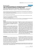
Higher Cyr61 level was found in ascites than in serum of
ovarian serous cystadenocarcinoma
Both in ascites and serum, significantly higher Cyr61 levels
were found in the malignant ovarian tumor (Fig. 1).
In ascites, the Cyr61 level of ovarian serous cystadenocarcinoma (n = 66) and serous cystadenoma (n = 18) was
1624.33 ± 191.92 cf. 230.11 ± 25.63 pg/ml respectively
(p < 0.001); in serum, the Cyr61 level was 77.21 ± 4.81 cf.
13.32 ± 3.14 pg/ml, correspondingly (p < 0.001).
Moreover, the same patient with ovarian serous cystadenocarcinoma, ascites of Cyr61 level was much higher
than its serum level.
Fig. 1 Expression levels of Cyr61 in ascites and serum of ovarian benign and malignant tumor Cyr61 levels in ascites and serum of ovarian serous
adenocarcinoma patients (n = 66) were significantly higher than those of ovarian serous cystadenoma patients (n = 18). And the ascites Cyr61 level was
much higher than that of serum
Shi et al. BMC Cancer
(2019) 19:1140
Page 4 of 8
High ascites Cyr61 level associated with clinicopathologic
features of ovarian serous cystadenocarcinoma
Tumor ascites microenvironment may reflect the tumor
characteristics and its progress. So ascites Cyr61 was analyzed to clear its relationships with the clinicopathologic features of ovarian serous cystadenocarcinoma.
Multiple regression analysis showed that.
Ascites Cyr61 level was more closely associated with
FIGO stage (p = 0.001), initial tumor size > 10 cm (p = 0.002)
and the residual tumor size (p = 0.025). But there was no
correlation with the tumor histological grade (p = 0.539),
total ascites volume (p = 0.124), ascites contains tumor cells
(p = 0.124), vascular invasion (p = 1.756) and lymph node
metastasis (p = 1.475) (Table 1).
High ascites IL-6 level is associated with Cyr61 in the
inflammatory microenvironment of ovarian serous
cystadenocarcinoma
As a new pro-inflammatory factor, Cyr61 can regulate the
expression of cytokines in inflammatory environment and
it was correlated with tumor stage. So the ascites Cyr61
and IL-6 levels of ovarian serous cystadenocarcinoma
were detected, respectively.
Ascites Cyr61 and IL-6 levels of advanced stage were
(2199.86 ± 116.24 pg/ml, 3227.42 ± 147.82 pg/ml) both
higher than those of early stage (778.98 ± 47.25 pg/ml,
1422.32 ± 74.69 pg/ml) (Fig. 2A).
And the increase of IL-6 level in ascites was linearly
related to Cyr61 level (Fig. 2B).
Expressions of serum Cyr61 and inflammatory markers in
different stages of ovarian cancer
Inflammation is a reaction in the process of tumor development. We further examined the expression patterns
of Cyr61, IL-6, CRP and neutrophil percentage in peripheral blood of patient with early or advanced stage of
ovarian serous cystadenocarcinoma.
In addition to Cyr61 and IL-6, CRP serum level in advanced ovarian cancer was significantly higher than
those in the early stage. However, the proportion of neutrophils of the advanced stage patients was a little higher
than that in early stage, but there was no statistical difference (Table 2).
Cyr61 expression patterns in ovarian serous tumor
18 cases of ovarian serous cystadenoma, 66 cases of
ovarian serous adenocarcinoma and 20 cases of its
paired metastatic lesions were evaluated using IHC to
confirm Cyr61expression patterns in ovarian caner (Fig. 3
and Table 3).
Cyr61 expression positive rate (≥4 scores) of ovarian
serous cystadenoma was significantly lower than the
ovarian cancer (p < 0.01). Further, the positive rate of its
paired metastatic lesions of was higher than the primary
adenocarcinoma (p < 0.05).
Discussion
The latest research results showed that there were six characteristics of the pre-metastasis microenvironment including immunosuppression, inflammatory response, enhanced
angiogenesis and permeability, lymphangiogenesis, organotropy and reprogramming. It indeed indicated that the inflammatory reaction was an indispensable part of tumor
progress [32]. Further, recent studies have confirmed that
inflammatory microenvironment is an essential environment for tumor cells survive. In the inflammatory microenvironment, different extracellular matrix, inflammatory
factors and stromal cells interact with tumor cells to
Table 1 Correlation of the clinicopathologic features and ascites Cyr61 level of ovarian serous adenocarcinoma
Clinicopathologic factors
FIGO stage
Initial tumor size (cm)
Ascites volume (ml)
Residual tumor (cm)
Lymphatic invasion
Vascular invasion
Ascites tumor cells
Categorization
Number (%)
Cyr61 (pg/ml)
P value
0.001
I-II
14 (21.1)
698.74 ± 87.12
III-IV
52 (78.9)
2054.23 ± 132.09*
< 10
45 (68.2)
910.81 ± 98.31
≥10
21 (31.8)
2125.66 ± 154.38*
< 500
9 (13.6)
1416.41 ± 102.37
≥500
57 (86.4)
1760.65 ± 154.64
<1
54 (81.8)
1031.64 ± 78.31
≥1
12 (18.2)
2097.30 ± 174.69*
–
60 (90.9)
1936.22 ± 191.37
+
6 (9.1)
2128.37 ± 478.26
–
62 (93.9)
2117.11 ± 201.39
+
4 (6.1)
2403.74 ± 345.21
–
57 (86.4)
1613.62 ± 119.27
+
9 (13.6)
1736.28 ± 114.30
0.002
0.124
0.025
1.475
1.756
0.124
Shi et al. BMC Cancer
(2019) 19:1140
Page 5 of 8
Fig. 2 Cyr61 and IL-6 levels in ascites of different (the early or advanced) stage of ovarian cancer patient and the correlation. In the patient with
ovarian serous adenocarcinoma, ascites Cyr61 and IL-6 levels of the advanced stage (n = 52) were both higher than those of the early stage (n = 14).
And the increased IL-6 expression was linearly related to Cyr61 level in malignant ascites
promote tumor proliferation and metastasis [33–37]. The
malignant ascites of ovarian cancer is a huge tumor microenvironment with its complex composition, which can enhance the ability of tumor to deteriorate [38–40].
Since previous studies have shown that the incidence
of ovarian cancer is closely related to periodic ovulation
and the continuous repair of ovarian surface tissues
damage, it was found that the inflammatory microenvironment had a direct regulatory effect on ovarian cancer
development. For various inflammatory reactions are accompanied with the process of the ovulation, all sorts of
secreted cytokines and chemokine may form the microenvironment together, and it can promote the activation
of oncogenes and cell carcinogenesis. So it can reveal
the early events of ovarian cancer and the biological
characteristics of the tumor cells. On the other hand, to
be an ovarian cancer cell, it is also affected daily by
physiological cycle and corresponding inflammatory
changes, accelerating tumor progression. Therefore, the
inflammatory microenvironment is essential for ovarian
cancer [41–44].
As a novel pro-inflammatory factor, Cyr61 has been
found to paly a key promoter to maintain the inflammatory microenvironment in some inflammatory and
autoimmune diseases. Recently, it is interesting that
“interstitium” is found to be one of the largest human
organs. It is linked together to form a network supported by a strong, flexible protein network, filled
with fluids in the human body. As a “highway”, full
of the flowing fluids in the body, “interstitum” may
help cancer cells metastasis [45]. Therefore, as one of
the important interstitial proteins, Cyr61might act as
a mediator in the inflammatory microenvironment of
tumor. In order to analyze the role of Cyr61 play in
the inflammatory microenvironment of ovarian cancer, we detected the Cyr61 level in ascites and serum
of patients with ovarian serous adenocarcinoma. The
Cyr61 level not only in ascites but also in serum of
ovarian serous adenocarcinoma was higher than that
of ovarian serous cystadenoma.
What’s more, about ovarian serous adenocarcinoma,
Cyr61 level of ascites was higher than that of serum,
which fully demonstrated that Cyr61 might be one of
the important components in malignant ascites. And
multiple regression analysis showed that Cyr61 level in
ascites of ovarian serous adenocarcinoma only related to
the initial tumor size, FIGO stage and surgical residual
tumor size, which indicates that the increased Cyr61
level closely associated with tumor proliferation and metastasis. Further, IHC was used to analyze the expression
of Cyr61 in ovarian serous adenocarcinoma tissues at
different stages of progression. The results showed that
the Cyr61 expression of metastatic tumor lesions (peritoneal foci) was higher than that of the primary lesions,
which may signify Cyr61 playing a vital role in peritoneal
metastasis.
As it is well known that IL-6 acts as promoter of tumor
development and metastasis by composing inflammatory
Table 2 Inflammatory markers in peripheral blood of ovarian cancer patient with different stages
Stage
Cyr61 (pg/ml)
IL-6 (pg/ml)
CRP (mg/L)
Neutrophils (%)
Early stage
52.55 ± 13.93
67.23 ± 17.43
7.77 ± 1.10
67.45 ± 2.37
Advanced stage
120.76 ± 22.35
212.47 ± 40.27
23.18 ± 4.48
73.56 ± 2.21
P value
0.031
0.015
0.004
1.247
Shi et al. BMC Cancer
(2019) 19:1140
Page 6 of 8
Fig. 3 Cyr61 expression patterns in the different tissues of ovarian tumor by immunohistochemistry. a Benign ovarian cyst (ovarian serous
cystadenoma) showed weak Cyr61 expression. b High grade of ovarian serous adenocarcinoma showed moderate degree Cyr61 expression. c and d
High grade of ovarian serous adenocarcinoma of the same patient of primary and paired metastatic site showed the moderate and strong degree
Cyr61 expression, respectively
environment [46, 47]. To be the up-stream factor, Cyr61
was reported that it really promoted IL-6 secretion in the
microenvironment of some inflammatory and autoimmune diseases. To explore the hypothesis that Cyr61
might also promote IL-6 production by tumor cells, then
together with other factors, forming a tumor-associated
inflammatory microenvironment to promote tumorigenesis, we examined the IL-6 level of ascites in ovarian serous adenocarcinoma patients and analyzed its correlation
with Cyr61. The results showed that the levels of Cyr61
and IL-6 in advanced stage were significantly higher than
those in early stage, which indicated that the later of the
disease, the higher of Cyr61 and IL-6 levels. Further analysis showed that elevated level of IL-6 was positively correlated with elevated Cyr61 level, suggesting a synergistic
effect between them in the development of tumor inflammation microenvironment.
Latest research suggested that there must be a series of
special inflammatory response during the initial stage of
the human tumor. Clinically, there are some commonly
used biomarkers in the body’s inflammatory response, for
example, white blood cells, neutrophils, CRP [48] and so
on. Among them, neutrophils are the earliest to reach the
site of inflammatory response [49]. It is very interesting
that Cyr61 (early event) could recruit neutrophils to target
sites by up-regulation IL-8 production by tissue cells at
the site of inflammation. However, whether Cyr61 can
mediate cytokines production and neutrophil infiltration
in tumor tissues cells has not been reported yet. So we analyzed the changes of Cyr61, CRP and the percentage neutrophils in the peripheral blood of ovarian tumor. In
addition to Cyr61 and IL-6, CRP serum level in advanced
ovarian serous adenocarcinoma was significantly higher
than those in early stage, but there was no correlation with
Cyr61level. And the increasing trend of the proportion
neutrophils was consistent with that of Cyr61 level. The
higher level of Cyr61was, the higher percentage neutrophils became. Thus it can be seen that Cyr61may mediate
Table 3 Correlation of the pathological features and Cyr61 expression positive rate
Pathological type
Cyr61 expression
Ovarian serous cystadenoma (n = 18)
Ovarian serous adenocarcinoma (n = 66)
Paired Metastatic site (n = 20)
Negative (0)
Weak (1–3)
Moderate (4–7)
Strong (8–12)
Positive rate
9
8
1
0
5.56
G1/G2 (n = 7)
0
3
4
0
57.14**
G3 (n = 59)
0
6
38
15
89.83**
0
0
1
19
95.00*
Positive rate: including moderate and strong of the intensity score (≥4)
**: Ovarian serous cystadenoma cf. Ovarian serous adenocarcinoma (p < 0.01);
*: Paired Metastatic site cf. Ovarian serous adenocarcinoma (p < 0.05)
Shi et al. BMC Cancer
(2019) 19:1140
neutrophil infiltration and CRP product in advanced ovarian serous adenocarcinoma just like it acts in the inflammation or autoimmune diseases. However, the specific
signaling pathway remains to be studied in the near
future.
Conclusion
Our study has systematically analyzed the important role of
Cyr61 as a tumor related inflammatory factor in promoting
the development of ovarian cancer microenvironment.
As a pro-inflammatory matrix protein, Cyr61 expression is increasing in ovarian serous adenocarcinoma, increasing in the advanced stage, and increasing in its
metastatic tumor, which suggested that Cyr61 is closely
associated with the development and metastasis of EOC.
More importantly, Cyr61 may paly regulation action in
the upstream for tumor inflammatory microenvironment
formation and maintain, especially for IL-6 expression.
And as a new tumor associated inflammatory marker,
Cyr61 might be a potential target and biomarker in the
diagnosis, treatment and prediction of EOC, but it needs
to be further study.
Abbreviations
EOC: Epithelial ovarian cancer; FIGO: Federation of Gynecology and
Obstetrics; ELISA: Enzyme Linked Immunosorbent Assay; CRP: C-reactive
protein
Acknowledgements
Not applicable.
Authors’ contributions
JS developed the idea, performed the experiments, analyzed the data and
prepared the manuscript. RH, HL, TZ and HL provided technical assiatance.
BS, JY and RF collected specimens and the clinical data. WD and NL both
initially conceived the idea and participated in the experimental design and
manuscript preparation. All authors read and approved the final manuscript.
Authors’ information
WD is the professor and chief man of Shanghai Key Laboratory of
Gynecologic Oncology and the department of of Obstetrics and Gynecology,
Renji Hospital, School of Medicine, Shanghai Jiaotong University.
JS, JY and RF are the doctors of department of Obstetrics and Gynecology,
Renji Hospital, School of Medicine, Shanghai Jiaotong University.
NL is the professor of Shanghai Institute of Immunology, Shanghai Jiao Tong
University School of Medicine.
RH and BS are the laboratory technicians of Shanghai Institute of
Immunology, Shanghai Jiao Tong University School of Medicine.
HL and TZ are the graduate students of Shanghai Institute of Immunology,
Shanghai Jiao Tong University School of Medicine.
Funding
Not applicable.
Availability of data and materials
The data was collected and saved in hospital’s medical history management
center. Due to the legitimate protection of patients’ privacy, our information
is not available on public or any private websites, but is available from the
corresponding author on reasonable request.
Ethics approval and consent to participate
The Ethics Committee of the Renji Hospital approved all the tumor
specimens used for this study. All the participating patients in this study
have signed the informed consent forms.
Page 7 of 8
Consent for publication
Not applicable.
Competing interests
The authors declare that they have no competing interests .
Author details
1
Department of Obstetrics and Gynecology, Renji Hospital, School of
Medicine, Shanghai Jiaotong University, Shanghai 200127, China. 2Shanghai
Key Laboratory of Gynecologic Oncology, Shanghai 200127, People’s
Republic of China. 3Shanghai Institute of Immunology, School of Medicine,
Shanghai Jiao Tong University, Shanghai 200025, China.
Received: 18 May 2018 Accepted: 31 October 2019
References
1. Siegel RL, Miller KD, Jemal A. Cancer statistics, 2018. CA Cancer J Clin. 2018
Jan;68(1):7–30.
2. Coussens LM, Werb Z. Inflammation and cancer. Nature. 2002;420(6917):860–7.
3. Kulbe H, Thompson R, Wilson JL, Robinson S, Hagemann T, Fatah R, et al.
The inflammatory cytokine tumor necrosis factor- α generates an autocrine
tumor-promoting network in epithelial ovarian cancer cells. Cancer Res.
2007;67(2):585–92.
4. Joyce J. A, Pollard J.W. “Microenvironmental regulation of metastasis”. Nat
Rev Cancer 2009; 9(4): 239–252.
5. Kulbe H, Chakravarty P, Leinster DA, Charles KA, Kwong J, Thompson RG,
et al. A dynamic inflammatory cytokine network in the human ovarian
cancer microenvironment. Cancer Res. 2012;72(1):66–75.
6. Hussian S. P, Harris C.C. “Inflammation and cancer: an ancient link with
novel potentials”. Int J Cancer 2007; 121(11): 2373–2380.
7. Mills GB, May C, McGill M, Roifman CM, Mellors A. A putative new growth
factor in ascitic fluid from ovarian cancer patients: identification,
characterization, and mechanism of action. Cancer Res. 1988;48(5):1066–71.
8. Freedman RS, Deavers M, Liu J, Wang E. Peritoneal inflammation – a
microenvironment for epithelial ovarian cancer (EOC). J Transl Med. 2004;2(1):23.
9. Fang L, Xinjuan K, Qian D, Jin Y, Yuhu S. Evaluation of tumor markers for the
differerntial diagnosis of benign and malignant ascites. Ann Hepatol. 2014;
13(3):357–63.
10. Matte I, Lane D, Laplante C, Rancourt C, Piche A. Profiling of cytokines in
human epithelial ovarian cancer ascites. Am J Cancer Res. 2012;2(5):566–80.
11. Lane D, Matte I, Rancourt C, Piche A. Prognostic significance of IL-6 and IL-8
ascites levels in ovarian cancer patients. BMC Cancer. 2011;11(1):210.
12. Lane D, Matte I, Garde-Granger P, Laplante C, Carignan A, Rancourt C, et al.
Inflammation-regulating factors in ascites as predictive biomarkers of drug
resistance and progression-free survival in serous epithelial ovarian cancers.
BMC Cancer. 2015;15(1):492.
13. Heng EC, Huang Y, Black SA Jr, Trackman PC. CCN2, connective tissue growth
factor, stimulates collagen deposition by gingival fibroblasts via module 3 and
alpha6- and beta1 integrins. J Cell Biochem. 2006;98(2):409–20.
14. Chen N, Chen CC, Lau LF. Adhesion of human skin fibroblasts to Cyr61 is
mediated through integrin alpha 6 beta 1 and cell surface heparin sulfate
proteoglycans. J Biol Chem. 2000;275(32):24953–61.
15. Grzeszkiewicz TM, Lindner V, Chen N, Lam SC, Lau LF. The angiogenic factor
cysteine-rich 61 (CYR61, CCN1) supports vascular smooth muscle cell adhesion
and stimulates chemotaxis through integrin alpha (6) beta (1) and cell surface
heparin sulfate proteoglycans. Endocrinology. 2002;143(4):1441–50.
16. Schober JM, Chen N, Grzeszkiewicz TM, Jovanovic I, Emeson EE, Ugarova TP,
Ye RD, Lau LF, Lam SC. Identification of integrin alpha (M) beta (2) as an
adhesion receptor on peripheral blood monocytes for Cyr61 (CCN1) and
connective tissue growth factor (CCN2): immediate-early gene products
expressed in atherosclerotic lesions. Blood. 2002;99(12):4457–65.
17. Tsai MS, Bogart DF, Castaneda JM, Li P, Lupu R. Cyr61 promotes breast
tumorigenesis and cancer progression. Oncogene. 2002;21(53):8178–85.
18. Xie D, Yin D, Tong X, O’Kelly J, Mori A, Miller C, et al. Cyr61 is overexpressed
in gliomas and involved in integrin-linked kinase-mediated Akt and βcatenon-TCF/Lef signaling pathways. Cancer Res. 2004;64(6):1987–96.
19. Zhou D, Herrick DJ, Rosenbloom J, Chaqour B. Cyr61 mediates the
expression of VEGF, αv-integrin, and α-actin genes through cytoskeletally
based mechanotransduction mechanisms in bladder smooth muscle cell. J
Appl Physiol. 2005;98(6):2344–54.
Shi et al. BMC Cancer
(2019) 19:1140
20. Lin J, Li N, Chen H, Liu C, Yang B, Ou Q. Serum CYR61 is associated with
clinical disease activity and inflammation in patients with systemic lupus
erythematosus. Medicine (Baltimore). 2015;94(19):e834.
21. Choi JS, Kim KH, Lau LF. The matricellular protein CCN1 promotes mucosal
healing in murine colitis through IL-6. Mucosal Immunol. 2015;8(6):1285–96.
22. Lin J, Zhou Z, Huo R, Xiao L, Ooyang G, Wang L, Sun Y, Shen B, Li D, Li N.
Cyr61 induces IL-6 production by fibroblast-like synoviocytes promoting Th17
differentiation in rtheumatoid arthritis. J Immunol. 2012;188(11):5776–84.
23. Wu P, Ma G, Zhu X, Gu T, Zhang J, Sun Y, Xu H, Huo R, Wang B, et al. Cyr61/
CCN1 is involved in the pathogenesis of psoriasis vulgaris via promoting IL8 production by keratinocytes in a JNK/NF-kB pathway. Clin Immunol. 2017;
174:53–62.
24. Zhu X, Xiao L, Huo R, Zhang J, Lin J, Xie J, Sun S, He Y, Sun Y, Zhou Z, Shen
B, Li N. Cyr61 is involved in neutrophil infiltration in joints by inducing IL-8
production by fibroblast-like synoviocytes in rheumatoid arthritis. Arthritis
Res Ther. 2013;15(6):R187.
25. Harris LG, Pannell LK, Singh S, Samant RS, Shevde LA. Increased vascularity
and spontaneous metastasis of breast cancer by hedgehog signaling
mediated upregulation of Cyr61. Oncogene. 2012;31(28):3370–80.
26. D’Antonio KB, Toubaji A, Albadine R, Mondul AM, Platz EA, Netto GJ,
Getzenberg RH. Extracellular matrix associated protein CYR61 is linked to
prostate cancer development. J Urol. 2010;183(4):1604–10.
27. Hou CH, Lin FL, Hou SM, Liu JF. Cyr61 promotes epithelial-mesenchymal
transition and tumor metastasis of osteosarcoma by Raf-1/MEK/ERK/Elk-1/
TWIST-1 signaling pathway. Mol Cancer. 2014;13:236.
28. Lo CW, Chen MW, Hsiao M, Wang S, Chen CA, Hsiao SM, et al. IL-6 transsignaling in formation and progression of malignant ascites in ovarian
cancer. Cancer Res. 2011;71(2):424–34.
29. Nonna K, Kevin HE, Anm Nazmul HK, Kassondra SG, Kelly LS, et al. Cytokine
profiling of ascites at primary surgery identifies an interaction of tumor
necrosis factor-α and interleukin-6 in predicting reduced progression-free
survival in epithelial ovarian cancer. Gynecol Oncol. 2015;138(2):352–7.
30. Sun Y, Zhang J, Zhou Z, Wu P, Huo R, Wang B, Shen Z, Li H, Zhai T, Shen B,
Chen X, Li N. CCN1, a pro-inflammatory factor, aggravates psoriasis skin
lesions bu promoting keratinocyte activation. J Invest Dermatol. 2015;
135(11):2666–75.
31. Lin J, Huo R, Wang L, Zhou Z, Sun Y, Shen B, Wang R, Li N. A novel antiCyr61 antibody inhibits breast cancer growth and metastasis in vivo. Cancer
Immunol Immunother. 2012;61(5):677–87.
32. Yang L, Cao X. Characteristics and significance of pre-metastatic niche.
Cancer Cell. 2016;30(5):668–81.
33. Farajzadeh VS, Keshavarz-Fathi M, Silvestris N, Argentiero A, Rezaei N. The
role of inflammatory cytokines and tumor associated macrophages (TAMs)
in microenvironment of pancreatic cancer. Cytokine Growth Factor Rev.
2018;39:46–61.
34. Liao Z, Tan ZW, Zhu P, Tan NS. Cancer-associated fibroblasts in tumor
microenvironment-Accomplices in tumor malignancy. Cell Immunol.
2018; Jan 31.
35. Yang L, Lin PC. Mechanisms that drive inflammatory tumor
microenvironment, tumor heterogeneity, and metastatic progression. Semin
Cancer Biol. 2017;47:185–95.
36. Voronov E, Apte RN. Targeting the tumor microenvironment by intervention
in interleukin-1 biology. Curr Pharm Des. 2017;23(32):4893–905.
37. Blank S, Nienhüser H, Dreikhausen L, Sisic L, Heger U, Ott K, Schmidt T.
Inflammatory cytokines are associated with response and prognosis in
patients with esophageal cancer. Oncotarget. 2017;8(29):47518–32.
38. Ahmed N, Stenvers KL. Getting to know ovarian cancer ascites:
opportunities for targeted therapy-based translational research. Front Oncol.
2013;3:256.
39. Reinart S, Schumann T, Finkernagel F, Wortmann A, Jansen JM, Meissner W,
et al. Mixed-polarization phenotype of ascites-associated macrophages in
human ovarian carcinoma: correlation of CD163 expression, cytokine levels
and early relapse. Int J Cancer. 2014;134(1):32–42.
40. Dijkgraaf EM, Heusinkveld M, Tummers B, Vogelpoel LT, Goedemans R, Jha
V, et al. Chemotherapy alters monocyte differentiation to favour generation
of cancer-supporting M2 macrophages in the tumor microenvironment.
Cancer Res. 2013;73(8):2480–92.
41. Kotsopoulos J, Lubinski J, Gronwald J, Cybulski C, Demsky R, Neuhausen SL,
Kim-Sing C, Tung N, et al. Factors influencing ovulation and the risk of
ovarian cancer in BRCA1 and BRCA2 mutation carriers. Int J Cancer. 2015;
137(5):1136–46.
Page 8 of 8
42. Richards JS. From follicular development and ovulation to ovarian cancers:
an unexpected journey. Vitam Horm. 2018;107:453–72.
43. Sapoznik S, Bahar-Shany K, Brand H, Pinto Y, Gabay O, Glick-Saar E, Dor C,
et al. Activation-induced cytidine deaminase links ovulation-induced
inflammation and serous carcinogenesis. Neoplasia. 2016;18(2):90–9.
44. Cardenas C, Alvero AB, Yun BS, Mor G. Redefining the origin and evolution
of ovarian cancer: a hormonal connection. Endocr Relat Cancer. 2016;23(9):
R411–22.
45. Benias PC, Wells RG, Sackey-Aboagye B, Klavan H, Reidy J, Buonocore D,
Miranda M, Kornacki S, Wayne M, Carr-Locke DL, Theise ND. Structure and
distribution of an unrecognized interstitium in human tissues. Sci Rep. 2018;
8(1):4947.
46. So KA, Min KJ, Hong JH, Lee JK. Interleukin-6 expression by interactions
between gynecologic cancer vells and human mesenchymal stem cells
promotes epithelial-mesenchymal transition. Int J Oncol. 2015;47:1451–9.
47. Bachelot T, Ray-Coquard I, Menetrier-Caux C, Rastkha M, Duc A, Blay JY.
Prognostic value of serum levels of interleukin 6 and of serum and plasma
levels of vascular endothelial growth factor in hormone-refractory
metastatic breast cancer patients. Br J Cancer. 2003;88(11):1721–6.
48. Maccio A, Lai P, Santona MC, Pagliara L, Melis GB, Mantovani G. High serum
levels of soluble IL-2 receptor, cytokines, and C reactive protein correlate
with impairment of T cell response in patients with advanced epithelial
ovarian cancer. Gynecol Oncol. 1998;69(3):248–52.
49. Gregory AD, Houghton AM. Tumor-associated neutrophils: new targets for
cancer therapy. Cancer Res. 2011;71(7):2411–6.
Publisher’s Note
Springer Nature remains neutral with regard to jurisdictional claims in
published maps and institutional affiliations.