Circulating tumour DNA (ctDNA) as a biomarker in metachronous melanoma and colorectal cancer- a case report
Bạn đang xem bản rút gọn của tài liệu. Xem và tải ngay bản đầy đủ của tài liệu tại đây (714.71 KB, 5 trang )
Calapre et al. BMC Cancer
(2019) 19:1109
/>
CASE REPORT
Open Access
Circulating tumour DNA (ctDNA) as a
biomarker in metachronous melanoma and
colorectal cancer- a case report
Leslie Calapre1†, Lydia Warburton2*† , Michael Millward2,3 and Elin S. Gray4
Abstract
Background: Circulating tumour DNA (ctDNA) has emerged as a promising blood-based biomarker for monitoring
disease status of patients with advanced cancers. The presence of ctDNA in the blood is a result of biological
processes, namely tumour cell apoptosis and/or necrosis, and can be used to monitor different cancers by targeting
cancer-specific mutation.
Case presentation: We present the case of a 67 year old Caucasian male that was initially treated with BRAF
inhibitors followed by anti-CTLA4 and then anti-PD1 immunotherapy for metastatic melanoma but later developed
colorectal cancer. The kinetics of ctDNA derived from each cancer type were monitored targeting BRAF V600R
(melanoma) and KRAS G13D (colon cancer), specifically reflected the status of the patient’s tumours. In fact, the
discordant pattern of BRAF and KRAS ctDNA was significantly correlated with the clinical response of melanoma to
pembrolizumab treatment and progression of colorectal cancer noted by PET and/or CT scan. Based on these
results, ctDNA can be used to specifically clarify disease status of patients with metachronous cancers.
Conclusions: Using cancer-specific mutational targets, we report here for the first time the efficacy of ctDNA to
accurately provide a comprehensive outlook of the tumour status of two different cancers within one patient. Thus,
ctDNA analysis has a potential clinical utility to delineate clinical information in patients with multiple cancer types.
Keywords: BRAF, Melanoma, Circulating tumor DNA, Colon cancer, Survivorship, Case report
Background
In recent years, tumour-derived cell free DNA (ctDNA)
has emerged as a promising biomarker of disease status
for metastatic cancer [1–3]. Plasma ctDNA are short nucleic acid fragments (~ 166 bp) thought to be released in
the systemic circulation as a result of tumour cell apoptosis and/or necrosis [4, 5]. Previous studies have shown
that ctDNA carries genetic information from the entire
tumour genome and can therefore provide insights into
clonal heterogeneity and evolution of all solid cancers
present at any one time [6, 7]. As analysis of ctDNA can
be tailored for different cancers by targeting specific mutations, it provides detailed information via a minimally
invasive ‘liquid biopsy’, eliminating the morbidity
* Correspondence:
†
Leslie Calapre and Lydia Warburton contributed equally to this work.
2
Department of Medical Oncology, Sir Charles Gairdner Hospital, Nedlands,
WA, Australia
Full list of author information is available at the end of the article
associated with serial sampling of tumours for monitoring patients with any advanced solid cancers.
Various studies in breast, lung and colorectal cancers
have demonstrated the potential clinical application of
ctDNA analysis at each stage of cancer management: early
diagnosis [5, 8], molecular profiling [6, 9–11], prognostication [5, 12, 13], detection of residual disease [14, 15], monitoring response and clonal evolution [16–20]. In
melanoma, several studies have also shown the efficacy of
utilising ctDNA for monitoring patients with BRAF mutant
tumours, particularly in the context of treatment response
and identification of mechanisms of resistance to BRAF inhibitors [7, 21–26]. These studies provide credence to the
utility of ctDNA for patient monitoring only in the context
of singular cancer. To date, ctDNA remains unutilised in
clinical management of patients with multiple tumour types
and/or those metachronous cancers where new primary tumours arise that are unrelated to the original malignancy.
In this case study, we demonstrated the efficiency of ctDNA
© The Author(s). 2019 Open Access This article is distributed under the terms of the Creative Commons Attribution 4.0
International License ( which permits unrestricted use, distribution, and
reproduction in any medium, provided you give appropriate credit to the original author(s) and the source, provide a link to
the Creative Commons license, and indicate if changes were made. The Creative Commons Public Domain Dedication waiver
( applies to the data made available in this article, unless otherwise stated.
Calapre et al. BMC Cancer
(2019) 19:1109
to delineate the different status of both melanoma and
colorectal cancers in a single patient.
Case presentation
A 67-year old male was investigated in our institution in
2012 for weight loss and abdominal pain. He was otherwise fit and well, with no significant comorbid medical
history. He was not on any regular medications, had no
known allergies and had no significant family history.
Computed tomography (CT) revealed moderate ascites
and a large splenic mass. Fine needle splenic aspirate
was non-diagnostic and therefore a therapeutic/diagnostic splenectomy was performed. Metastatic melanoma
was confirmed histologically, and further testing confirmed a BRAF V600R mutation via Sanger sequencing.
In July 2014, he commenced dabrafenib and trametinib
treatment for progressive disease but suffered unacceptable toxicity, which led to the cessation of the combined
targeted therapies.
At progression the patient was subsequently treated
with four doses of ipilimumab (3 mg/kg three weekly)
but was found to have disease progression on the first
response assessment CT scan. Confirmed progression
in lung metastases and the intra-abdominal nodal disease led to commencement of anti PD-1 therapy
Page 2 of 5
(pembrolizumab 2 mg/kg three weekly) in March 2015
(week 2, Fig. 1). He completed 28 cycles (week 94) of
pembrolizumab and achieved a complete metabolic
response on PET at six months in all the previously
identified metastatic sites. He tolerated treatment well
with vitiligo as the sole side effect.
However, PET at 32 weeks identified a new FDG avid
lesion within the sigmoid colon. This was investigated
with colonoscopy and tissue biopsy confirmed a low
grade sigmoid adenocarcinoma. He proceeded to a subtotal colectomy, ilio-sigmoid anastomosis and lymph
node dissection in January 2016 (week 46). Histopathology confirmed a stage III (T4N1M0 AJCC 7th edition)
low grade sigmoid adenocarcinoma with 3/33 lymph
nodes involved. The tumour had no mismatch repair deficiency. Molecular analysis using next generation sequencing via the Illumina Trusight tumour panel
showed the primary tumour to be KRAS p. G13D mutant, NRAS and BRAF wild type. Post-operative CEA
measurements were negative.
Adjuvant chemotherapy for colon cancer was offered
but the patient decided to continue with pembrolizumab
for metastatic melanoma and declined chemotherapy. In
November 2016, eleven months after curative resection
of primary colorectal cancer, para-aortic nodes enlarged
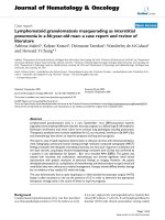
Fig. 1 ctDNA analysis can discriminate the status of different tumours in a patient with both melanoma and colorectal cancer. Levels of BRAF and
KRAS ctDNA (green) inform of the status of melanoma and colon cancer respectively. Clinical partial response (PR) and complete response (CR)
annotations are indicative of melanoma response to pembrolizumab as measured by RECIST on CT imaging. PET scan images associated with
four different timepoints with differential BRAF and KRAS ctDNA levels
Calapre et al. BMC Cancer
(2019) 19:1109
marginally and became intensely FDG avid on PET despite on-going pembrolizumab. Biopsy of an enlarging
para-aortic node at approximately week 88 confirmed
metastatic colorectal cancer. Molecular analysis of the
colorectal metastasis confirmed KRAS p. G13D mutation. It is of note that the patient had no other sites of
disease progression and remained in complete response
from metastatic melanoma, which led to cessation of
pembrolizumab treatment.
The recurrence was unresectable and the patient was
offered palliative FOLFOX chemotherapy with bevacizumab (B) but chose to undergo observation with three
monthly clinical and radiological reviews. His imaging
demonstrated RECIST (Response Evaluation Criteria in
Solid Tumours) stable disease for 18 months and he then
progressed with new liver and lung lesions. He has
recently commenced B-FOLFOX chemotherapy with response assessment pending.
Page 3 of 5
with the exception of the blood sample collected at week
64 that was negative for ctDNA. In this case, real time
knowledge of detectable ctDNA following curative bowel
resection, implying residual microscopic disease, and the
negative BRAF mutant melanoma ctDNA may have
influenced and ultimately changed the clinician and patients decision from not having adjuvant chemotherapy,
to receiving it.
Increased KRAS mutant ctDNA was further observed at
week 70 (6 c/mL), which provided an early indication of
disease progression. Prior to the cessation of pembrolizumab corresponding to the melanoma complete response,
KRAS mutant ctDNA levels was at its peak (14 c/mL). A
final ctDNA assessment at week 119, revealed that BRAF
V600R ctDNA continues to be undetectable which is consistent with sustained complete response of melanoma.
Nevertheless, ctDNA for KRAS G13D remained high (7 c/
ml) suggesting possible radiologically undetectable progression of the patient’s untreated colon cancer.
ctDNA screening and monitoring
In parallel to the imaging scans, the patient was monitored for melanoma and colorectal cancer by tracking
BRAF p.V600R and KRAS p.G13D mutations in ctDNA
respectively. Blood samples were collected in EDTA and
Streck tubes. Plasma was separated within 24 h by centrifugation at 300 g for 20 min, followed by a second
centrifugation at 4700 g for 10 min, and then stored at
-80 °C until extraction. Cell-free DNA (cfDNA) was
extracted from 5 ml of plasma using the QIAamp Circulating Nucleic Acid Kit (Qiagen) as per the manufacturer’s instructions. Analysis of plasma ctDNA was
carried out using an in-house BRAF p.V600R assays [27]
and a commercial KRAS p.G13D (Bio-Rad) for droplet
digital PCR (ddPCR). Protocols used for ddPCR analysis
were as previously described [21, 28] and ctDNA levels
were calculated based on the number of copies per millilitres of plasma (c/mL).
Plasma analysis demonstrated the presence of BRAF
V600R ctDNA at baseline prior to initiating pembrolizumab, which became undetectable at subsequent followup (weeks 2–10.) The patient achieved sustained partial
response to pembrolizumab (week 18–49) by CT and
complete metabolic response by PET scan, which was
supported by his corresponding ctDNA data (Fig. 1). As
predicted, the patient’s blood sample at the time of colorectal cancer diagnosis (week 36) had detectable KRAS
mutant ctDNA (2 c/mL). Retrospective analysis of the
previous blood samples revealed detectable levels of
KRAS mutant ctDNA prior to immunotherapy (3 c/mL),
suggesting that colorectal cancer may have already been
present at the time of stage IV melanoma diagnosis.
Subsequent plasma samples (weeks 2–49) were also
found to have detectable KRAS mutant ctDNA, albeit at
consistently low levels that ranged from 2 to 4 c/ml,
Discussion and conclusion
This case study highlights the evolving role of ctDNA in
detecting metachronous cancers. Development of new
primary tumours that are unrelated to the original malignancy have become a significant adverse effect in
patients with metastatic cancers who achieved sustained
or durable disease control in response to targeted and/or
systemic therapy. Thus, oncologists should be vigilant to
the possibility that patients are at continued risk for new
and separate malignancies.
Previous studies have demonstrated the clinical utility
of ctDNA as a biomarker of disease status in patients
with metastatic cancers, particularly in the context of
singular tumour types. However, to date there has been
no report of the clinical utility of ctDNA to delineate
status of different tumour types within patients with
multiple cancers. In this case study, we demonstrated
the efficiency of ctDNA, by targeting tumour-specific
mutations to specifically inform treatment response and
tumour status in a patient with both melanoma and
colorectal cancer. Given the increased risk of cancer patients to develop other malignant tumours, this study
supports the potential clinical use of ctDNA for profiling
of other emerging lesions and identification of their origin. Plasma ctDNA may be useful for accurate stratification of treatment response in patients with two or more
different tumour types, providing better perspective of
disease status for more informed treatment options. In
this setting, pan-cancer ctDNA testing can aid on the
early detection of metachronous cancers.
The low melanoma derived ctDNA at baseline may be
the result of partial disease control by the previous ipilimumab therapy, although not evident by the CT scan
performed. The immediate drop and undetectable levels
Calapre et al. BMC Cancer
(2019) 19:1109
of BRAF mutant ctDNA during pembrolizumab treatment indicated the response of the patient’s melanoma
tumour to this treatment. On the other hand, colon cancer derived ctDNA (KRAS) was also detectable at baseline, and given that it is at similar concentrations as
BRAF, the patient’s melanoma and colorectal tumour
burden may be relatively similar. Studies have shown
that ctDNA is readily detectable in early stages colon
cancer patients [29, 30] which may explain the detectability of colon cancer ctDNA in this patient. Nevertheless, we like to note that we observed fluctuations of the
level of KRAS mutant ctDNA at the time of pembrolizumab treatment. Interestingly, the KEYNOTE-164 clinical
trial has demonstrated durable anti-tumour activity of
pembrolizumab in colorectal cancer patients, particularly
those with high microsatellite instability (MSI) [31]. We
hypothesise that pembrolizumab may have exerted some
level of control on the colorectal tumour. However, further investigation is needed, particularly identifying the
MSI status of the patient, to determine if he may have
benefited from pembrolizumab treatment. The variability on detection of ctDNA across multiple cancers and
tumour locations, also remains a topic of investigation in
the field of liquid biopsy research.
In conclusion, ongoing close surveillance of melanoma
patients who achieved complete response to BRAF
inhibition and/or immune-checkpoint inhibitors is paramount to monitor potential recurrent disease. Emergent
of new malignant lesions in this population should be
regarded as a metastasis only after detailed evaluation,
including a biopsy where feasible; otherwise there is a
possibility of missing a secondary malignancy. Plasma
ctDNA may aid in clarifying disease status of patients
with metachronous cancer.
Abbreviations
AJCC: American joint committee on cancer; B: Bevacizumab;
CEA: Carcinoembryonic antigen; ctDNA: Circulating tumour DNA; CTLA4: Cytotoxic T-lymphocyte antigen 4; ddPCR: Droplet digital polymerase
chain reaction; FDG-PET/CT: Fluorine 18 fluorodeoxyglucose- Positron
emission tomography/ Computed tomography; FOLFOX: Folinic acid,
fluorouracil and oxaliplatin chemotherapy; PD- 1: Programmed death antigen
1; RECIST: Response evaluation criteria in solid tumours
Acknowledgements
N/A
Consent to publication
A copy of written consent is available for BMC if required.
Authors’ contributions
All authors read and approved the final manuscript. LW and LC both made
substantial contribution to the conception/design of the report. They both
collected, analyzed and interpreted the patient data including clinical data,
ctDNA and outcome. LW and LC both equally provided major contribution
in drafting, revising and writing the manuscript. EZ and MM assisted in
acquisition of the data and participated in revising and critically appraising
the report. All authors read and approved the final manuscript.
Page 4 of 5
Funding
Lydia Warburton was supported by a WA Cancer and Palliative Care network
fellowship in the form of a fellowship salary. The declared funding body
played no part in the design of the study, collection, analysis, interpretation
of data nor the writing of the manuscript.
Availability of data and materials
All data analyzed for this case report has been presented within the
manuscript. Data sharing is not applicable to this article as no datasets were
generated or analysed during the current study.
Ethics approval and consent to participate
Written informed consent was obtained from patients under approved
Human Research Ethics Committee protocols from Edith Cowan University
(No. 2932) and Sir Charles Gairdner Hospital (No.2007–123). De-identification
of images was performed and written consent for publication attained.
Competing interests
The authors declare that they have no competing interests.
Author details
1
School of Medical Science, Edith Cowan University, Joondalup, WA,
Australia. 2Department of Medical Oncology, Sir Charles Gairdner Hospital,
Nedlands, WA, Australia. 3School of Medicine and Pharmacology, The
University of Western Australia, Crawley, Western Australia, Australia. 4School
of Biomedical Science, University of Western Australia, Crawley, WA, Australia.
Received: 17 April 2019 Accepted: 5 November 2019
References
1. Calapre L, et al. Circulating tumour DNA (ctDNA) as a liquid biopsy for
melanoma. Cancer Lett. 2017;404:62–9.
2. Wan JCM, et al. Liquid biopsies come of age: towards implementation of
circulating tumour DNA. Nat Rev Cancer. 2017;17(4):223–38.
3. Heitzer E, Ulz P, Geigl JB. Circulating tumor DNA as a liquid biopsy for
cancer. Clin Chem. 2015;61(1):112–23.
4. Canzoniero JV, Park BH. Use of cell free DNA in breast oncology. Biochimica
et Biophysica Acta (BBA) - Reviews on Cancer. 2016;1865(2):266–74.
5. Bettegowda, C., et al., Detection of circulating tumor DNA in early- and latestage human malignancies. Sci Transl Med, 2014. 6(224): p. 224ra24.
6. Murtaza M, et al. Multifocal clonal evolution characterized using circulating
tumour DNA in a case of metastatic breast cancer. Nat Commun. 2015;6:8760.
7. Wong SQ, et al. Circulating tumor DNA analysis and functional imaging
provide complementary approaches for comprehensive disease monitoring
in metastatic melanoma. JCO Precision Oncology. 2017;1:1–14.
8. Newman AM, et al. An ultrasensitive method for quantitating circulating
tumor DNA with broad patient coverage. Nat Med. 2014;20(5):548–54.
9. De Mattos-Arruda L, et al. Capturing intra-tumor genetic heterogeneity by
de novo mutation profiling of circulating cell-free tumor DNA: a proof-ofprinciple. Ann Oncol. 2014;25(9):1729–35.
10. Jamal-Hanjani M, et al. Detection of ubiquitous and heterogeneous
mutations in cell-free DNA from patients with early-stage non-small-cell
lung cancer. Ann Oncol. 2016;27(5):862–7.
11. De Mattos-Arruda L, et al. Cerebrospinal fluid-derived circulating tumour
DNA better represents the genomic alterations of brain tumours than
plasma. Nat Commun. 2015;6:8839.
12. Dawson S-J, et al. Analysis of circulating tumor DNA to monitor metastatic
breast cancer. N Engl J Med. 2013;368(13):1199–209.
13. Forshew, T., et al., Noninvasive identification and monitoring of cancer
mutations by targeted deep sequencing of plasma DNA. Science translational
medicine, 2012. 4(136): p. 136ra68-136ra68.
14. Tie J, et al. Circulating tumor DNA as an early marker of therapeutic
response in patients with metastatic colorectal cancer. Ann Oncol. 2015;
26(8):1715–22.
15. Garcia-Murillas, I., et al., Mutation tracking in circulating tumor DNA predicts
relapse in early breast cancer. Science translational medicine, 2015. 7(302): p.
302ra133-302ra133.
16. Murtaza M, et al. Non-invasive analysis of acquired resistance to cancer
therapy by sequencing of plasma DNA. Nature. 2013;497(7447):108–12.
Calapre et al. BMC Cancer
(2019) 19:1109
17. Wang W, Song Z, Zhang Y. A comparison of ddPCR and ARMS for detecting
EGFR T790M status in ctDNA from advanced NSCLC patients with acquired
EGFR-TKI resistance. Cancer Med. 2017;6(1):154–62.
18. Zheng D, et al. Plasma EGFR T790M ctDNA status is associated with clinical
outcome in advanced NSCLC patients with acquired EGFR-TKI resistance. Sci
Rep. 2016;6:20913.
19. Sundaresan TK, et al. Detection of T790M, the acquired resistance EGFR
mutation, by tumor biopsy versus noninvasive blood-based analyses. Clin
Cancer Res. 2016;22(5):1103–10.
20. Van Emburgh BO, et al. Acquired resistance to EGFR-targeted therapies in
colorectal cancer. Mol Oncol. 2014;8(6):1084–94.
21. Gray ES, et al. Circulating tumor DNA to monitor treatment response and
detect acquired resistance in patients with metastatic melanoma.
Oncotarget. 2015;6(39):42008–18.
22. Sanmamed MF, et al. Quantitative cell-free circulating BRAFV600E mutation
analysis by use of droplet digital PCR in the follow-up of patients with
melanoma being treated with BRAF inhibitors. Clin Chem. 2015;61(1):297–304.
23. Girotti MR, et al. Application of sequencing, liquid biopsies, and patientderived Xenografts for personalized medicine in melanoma. Cancer Discov.
2016;6(3):286–99.
24. Gonzalez-Cao M, et al. BRAF mutation analysis in circulating free tumor DNA
of melanoma patients treated with BRAF inhibitors. Melanoma Res. 2015;
25(6):486–95.
25. Schreuer M, et al. Quantitative assessment of BRAF V600 mutant circulating
cell-free tumor DNA as a tool for therapeutic monitoring in metastatic
melanoma patients treated with BRAF/MEK inhibitors. J Transl Med. 2016;14:95.
26. Lee J, et al. Circulating tumour DNA predicts response to anti-PD1
antibodies in metastatic melanoma. Ann Oncol. 2017;28.
27. Reid AL, et al. Detection of BRAF-V600E and V600K in melanoma circulating
tumour cells by droplet digital PCR. Clin Biochem. 2015;48.
28. Calapre L, et al. Locus-specific concordance of genomic alterations between tissue
and plasma circulating tumor DNA in metastatic melanoma. Mol Oncol. 2018.
29. Bettegowda C, et al. Detection of circulating tumor DNA in early-and latestage human malignancies. Sci Transl Med. 2014;6.
30. Tie, J., et al., Circulating tumor DNA analysis detects minimal residual disease
and predicts recurrence in patients with stage II colon cancer. Sci Transl Med,
2016. 8(346): p. 346ra92.
31. Le, D.T., et al., KEYNOTE-164: Pembrolizumab for patients with advanced
microsatellite instability high (MSI-H) colorectal cancer. Journal of Clinical
Oncology, 2018. 36(15_suppl): p. 3514–3514.
Publisher’s Note
Springer Nature remains neutral with regard to jurisdictional claims in
published maps and institutional affiliations.
Page 5 of 5