Báo cáo khoa học: " Perforated mixed carcinoid-adenocarcinoma in transverse colon and at gastroenterostomy site: case report" pot
Bạn đang xem bản rút gọn của tài liệu. Xem và tải ngay bản đầy đủ của tài liệu tại đây (294.85 KB, 3 trang )
CAS E REP O R T Open Access
Perforated mixed carcinoid-adenocarcinoma
in transverse colon and at gastroenterostomy
site: case report
Enver İhtiyar
1*
, Özgül Paşaoğlu
2
, Serdar Erkasap
1
, Barış R Karakaş
1
, Fatih N Yaşar
1
Abstract
Goblet cell carcinoid of the large intestine is a rare neoplasm, usually located in ascending colon and rectum.
A 60-year-old male patient underwent surgery after the diagnosis of acute abdomen. Exploratory laparotomy
revealed perforation with a diameter of 1 cm at the site of the previously performed gastroenterostomy and dilata-
tion of the right colic flexure, secondary to a solid obstructive mass located in the mid-portion of transverse colon.
Histopathological investigation of the biopsies, taken from the gastroenterostomy site and the tumor, revealed
mixed carcinoid-adenocarcinoma with carcinoid component, predominantly composed of goblet cells. Three cycles
of FOLFOX-4 protocol was administered. Following respiratory distress secondary to pulmonary metastasis, the
patient’s condition deteriorated and subsequently died in the fourth postoperative month. Our aim with this paper
is to point out that more cases should be reported for more effective diagnosis, histopathological study, clinical
investigation, treatment and prognosis of this specific neoplasm.
Background
Goblet cell carcinoid (GCC) of the large intestine is a
rare neoplasm, usually located in ascending colon and
rectum. Histologically, it is similar to goblet cell carci-
noid of the appendix [1]. GCC has both endocrine and
glandular differentiation. D ual differentiation probably
arises from a pluripotent intestinal stem cell instead of
two different mature cells. The mean age for diagnosing
GCC of the appendix is 58.89 years with equal represen-
tation in both genders. Regional and systemic metastasis
is common at initial diagnosis. These tumors perform
aggressive behavior with tendency for metastasis and
wide local dissemination [2]. Lesions are treate d accord-
ing to the same conventional oncologic approach to
adenocarcinoma [3]. We present here, a 60 year-old
male patient, who diagnose as mixed carcinoid-adeno-
carcinoma located in transverse colon and at gastroen-
terostomy site.
Case
A 60 year- old male patient presented with complains of
nausea, vomiting, abdominal distension, and no dis-
charge for three days. He also had intermittent cramp-
ing abdominal pain, mainly located in the upper left
abdominal quadrant. He had a history of prior gastric
surgery, performed 26 years ago, for peptic ulcer disease.
His vital signs included temperature of 36.4°C, blood
pressure of 100/80 mmHg, pulse rate of 60 beats/min,
respiratory rate of 22 breaths/min. On physical examina-
tion, the scar of the midline incision was inspected and
the abdomen was distended and tender to palpation
with guarding. Routine hematological and biochemical
investigations were within normal limits except for
raised total leucocytes count (32,000/mm³). Serum carci-
noembryonic antigen (CEA) and cancer antigen (CA)
19-9 levels were not elevated on the postoperative 3rd
day of the follow-up. Plain X-ray of abdomen rev ealed
few fluid levels and free gas in subphrenic spaces
whereas the abdominal ultrasonography showed no find-
ing but diffuse intestinal gas. The patient underwent
surg ery after the diagnosis of acute abdomen was made.
Exploratory laparotomy revealed perforation with a dia-
meter of 1 cm at the site of the previously performed
gastroenterostomy and dilatation of the right colic
* Correspondence:
1
Department of General Surgery, Eskişehir Osmangazi University, School of
Medicine 26480, Eskişehir, Turkey
Full list of author information is available at the end of the article
İhtiyar et al. World Journal of Surgical Oncology 2010, 8:110
/>WORLD JOURNAL OF
SURGICAL ONCOLOGY
© 2010 İİhtiyar et al; licensee BioMed Central Ltd. T his is an Open Acces s article distributed under the terms of the Creative Commons
Attribution License ( 2.0), which perm its unrestricted use, distribution, and reproduction in
any medium, provided the original work is properly cited.
flexure, secondary to a solid obstructive mass located in
the mid-portion of transverse colon. There were no
metastatic liver lesions whereas metastatic lymph nodes
were detected in mesocolon. The gastroenterostomy was
recons tructed after anastomosis and the mid se gment of
the transverse colon with approximately 5-6 cm margins
on either side of the tumor was resected.
Histopathological investigation of the biopsies, taken
from the gastroenterostomy site and the tumor, revealed
mixed carcinoid-adenocarcinoma with carcinoid compo-
nent, predominantly composed of goblet cells. Ulcero-
vegetative mass in the transverse colon with the size of
5 × 5 × 1.5 cm, infiltrating the intestinal serosa, and
three tissue samples, each measuring approximately
2.5×1.5×0.3cm,takenfromthegastroenterostomy
site were microscopically similar and had the character-
istics of mixed carcinoid-adenocarcinoma with carcinoid
component, predominantly composed of goblet cells
(Figure 1). Tumor invasion in all layers of the transverse
colon and the gastroenterostomy site are accompanied
by perforation. Immunohistochemical stains showed that
neoplastic cells were positive for neuron-specific enolase
(NSE), synaptophysin and E-cadherin and negative for
chromogranin. Ten metastatic lymph nodes were
detected in mesocolon. At three months postoperatively
the needle biopsy specimen of the liver revealed
metastasis.
The 24 hours urine vanillylmandelic acid (VMA) l evel
was within normal range o n the postoperative 5th week
of the follow- up. In-111 octreotide scintigraphy detected
increased uptake in the region of the para-aortic lymph
node, compatible with a lesion which had the expression
of somatostatin receptors. A bone scan was performed
using 20 mCi of Tc-99 m MDP, and uncovered no evi-
dence of abnormality. Three cycles of FOLFOX-4
protocol was administered by the medical oncology
department. He was hospitalized three months after the
operation because of poor health status. Ultrasonography
of the liver showed an inhomogeneous echo texture,
hyperechoic nodules with peripheral hypoechoic halos
and the largest lesion with size of 3 × 2.8 × 2.4 cm was
localized in the anterosuperior portion of the right lobe.
A needle biopsy of the liver was positive for metastasis of
the carcinoma. Following respiratory distress secondary
to pulmonary metastasis, his health situation got worse
and subsequently died in the fourth postoperative month.
In additionally, the patient was questioned about any
symptoms of the carcinoid syndrome, which includes
flushing, diarrhea, wheezing etc. pre-operatively once the
post op diagnosis was made and post-operatively. He did
not encounter any symptoms of the carcinoid syndrome.
Discussion
Since more than 30 years ago, a new variant type of
epithelial tumor of appendix has been recognized. This
tumor is repo rted under diff erent names including gob-
let cell carcinoid (GCC), adenocarcinoid, mucinous car-
cinoid, intermediate type of carcinoid, crypt cell
carcinoma, amphicrine (endo-exocrine) neoplasia, com-
posite tumor and microglandular carcinoma. All names
except GCC have been omitted from the current World
Health Organization (WHO) classification [2]. Subbus-
wamy et al described the first report of GCC in 1974
[4]. The histology and biology appears to be intermedi-
ate between carcinoid tumors and adenocarcinomas.
This tumor appears to combine features of epithelial
and carcinoid neoplasms and in addition the surface
mucosal epithelium is not neoplastic. Histopathological
features such as in creased number of Paneth cells,
increased amount of mucin secretion and presence of
pancreatic polypeptide may predict a more aggressive
behavior [2].
Mucocarcinoids also called mucinous or adenocarci-
noids, show a quite different histological appearance
from carcinoids and endocrine cell carcinomas. The
tumor is com posed predominantly of s mall clumps,
strands, or glandular collections of mucin-producing
cells looking like goblet cells or signet-ring cells, and
intermingled with endocrine cells in a variable number
and occasionally with Pan eth’s cells. The admixed endo-
crine cells comprise a variety of cell types, such as
somatostatin-containing D cells, serotonin-containing
end ocrine cells and enterochromaffin-like cells contain-
ing histamine. They are often sparse and, in about 10%
of cases, difficu lt to find. The tumor was originally con-
sidered to be a variant of a carcinoid. The frequent pau-
city of endocrine cells and more a ggressive clinical
nature are not consistent with such speculation. Muco-
carcinoids are a variant of adenocarcinomas showing
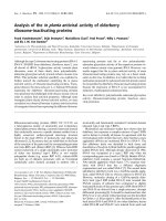
Figure 1 Carcinoid component of mixed carcinoid-
adenocarcinoma is composed mainly of goblet cells. (H.E.; × 200).
İhtiyar et al. World Journal of Surgical Oncology 2010, 8:110
/>Page 2 of 3
differentiation to both mucin- producing cells and endo-
crine cells. They occur most frequently in the appendix,
but rarely in stomach. Ito et al reported only one case
treated in 10 years period [5].
Even if there are many questions about histogenesis of
tumors with mixed differentiation, it is hypothesized
that these neoplastic lesions may probably arise from a
single pluripotent stem cell as well as different mature
cells [6]. Histologically, these tumors are divided into
three subtypes: mixed (composite) tumors, collision
tumors and amphicrine tumors. In mixed tumors, the
two elements typically merge and intermingle, and in
some areas transitions can be seen, such that the two
components can be difficult to distinguish. A carcinoid
component should compose at least one third of the
tumor cell population in a composite tumor. In collision
tumors, the two elements should be in intimate contact
without intermixture of individual cell types. Amphi-
crine tumors differ from the above tumor types in that
endocrine and nonendocrine epithelial cell constituents
are present within the same cell [ 7]. These tumors
behave more like adenocarcinomas than carcinoids. Two
cases with mixed carcinoid-adenocarcinoma, for the first
time, were reported by Moyana et al in 1988 [8].
Conclusions
This case with the characteristics of mixed carcinoid-
adenocarcinoma with carcinoid component, predom i-
nantly composed of goblet cells, is reported because o f
its rarity and points out that more d ata should be col-
lected to develop our knowledge about diagnosis, histo-
pathological and clinical features, prognosis, and
conventional treatment of this neoplasm.
Consent
Written informed consent was obtained from the family
of the deceased patient for publication of this case
report and accompanying images. A copy of the wri tten
consent is available for review by the Editor-in-Chief of
this journal.
Author details
1
Department of General Surgery, Eskişehir Osmangazi University, School of
Medicine 26480, Eskişehir, Turkey.
2
Department of Pathology, Eskişehir
Osmangazi University, School of Medicine 26480, Eskişehir, Turkey.
Authors’ contributions
Eİ performed the operation and helped manuscript preparation particularly
in describe the findings and the follow-up, revised and edited most of the
manuscript. ÖP performed the histopathological and immunohistochemical
analyses of all surgical specimens, provided the figure of the microscopic
appearance of the tumor, helped literature search, and corrected the final
draft. SE helped with the editing of the manuscript and literature search.
BRK and NFY were involved in preparation of initial draft and literature
search. All authors read and approved the final manuscript for public ation.
Competing interests
The authors declare that they have no competing interests.
Received: 4 July 2010 Accepted: 22 December 2010
Published: 22 December 2010
References
1. Copper HS: Intestinal neoplasms. In Anderson’s pathology. 10 edition.
Edited by: Damjanov I, Linder J. St. Louis: Mosby; 1996:1576-1578.
2. Pahlavan PS, Kanthan R: Goblet cell carcinoid of the appendix. World J
Surg Oncol 2005, 3:36.
3. Bullard KM, Rothenberger DA: Colon, rectum, and anus. In Schwartz’s
Principles of Surgery. 8 edition. Edited by: Brunicardi FC. New York: McGraw-
Hill, Medical Pub Div; 2005:1055-1117.
4. Subbuswamy SG, Gibbs NM, Ross CF, Morson BC: Goblet cell carcinoid of
the appendix. Cancer 1974, 34:338-344.
5. Ito H, Tahara E: Endocrine cell tumor of the stomach. In Gastric cancer.
Edited by: Nishi M, Ichikawa M, Nakajima T, Maruyama K, Tahara E. Tokyo:
Springer-Verlag; 1993:151-167.
6. Cooper HS: Intestinal neoplasms. In Sternberg’s Diagnostic Surgical
Pathology. 4 edition. Edited by: Mills SE, Carter D, Green son JK, Oberman
HA, Reuter V, Stoler MH. Philadelphia: Lippincott Williams 2004:1543-1602.
7. Jiao YF, Nakamura S, Arai T, Sugai T, Uesugi N, Habano W, et al: Adenoma,
adenocarcinoma and mixed carcinoid-adenocarcinoma arising in a small
lesion of the colon. Pathol Int 2003, 53:457-62.
8. Moyana TN, Qizilbash AH, Murphy F: Composite glandular-carcinoid
tumors of the colon and rectum. Report of two cases. Am J Surg Pathol
1988, 12:607-11.
doi:10.1186/1477-7819-8-110
Cite this article as: İhtiyar et al.: Perforated mixed carcinoid-
adenocarcinoma in transverse colon and at gastroenterostomy site:
case report. World Journal of Surgical Oncology 2010 8:110.
Submit your next manuscript to BioMed Central
and take full advantage of:
• Convenient online submission
• Thorough peer review
• No space constraints or color figure charges
• Immediate publication on acceptance
• Inclusion in PubMed, CAS, Scopus and Google Scholar
• Research which is freely available for redistribution
Submit your manuscript at
www.biomedcentral.com/submit
İhtiyar et al. World Journal of Surgical Oncology 2010, 8:110
/>Page 3 of 3