Prediction of lymph node metastasis by tumor-infiltrating lymphocytes in T1 breast cancer
Bạn đang xem bản rút gọn của tài liệu. Xem và tải ngay bản đầy đủ của tài liệu tại đây (970.77 KB, 13 trang )
Takada et al. BMC Cancer
(2020) 20:598
/>
RESEARCH ARTICLE
Open Access
Prediction of lymph node metastasis by
tumor-infiltrating lymphocytes in T1 breast
cancer
Koji Takada1, Shinichiro Kashiwagi1* , Yuka Asano1, Wataru Goto1, Rika Kouhashi1, Akimichi Yabumoto1,
Tamami Morisaki1, Masatsune Shibutani2, Tsutomu Takashima1, Hisakazu Fujita3, Kosei Hirakawa1,2 and
Masaichi Ohira1,2
Abstract
Background: Lymph node metastasis is more likely in early-stage breast cancer with lower tumor-infiltrating
lymphocyte (TIL) density. Therefore, we investigated the correlation between TILs and lymph node metastasis in cT1
breast cancer patients undergoing surgery and the usefulness of TILs in predicting sentinel lymph node metastasis
(SLNM) in cT1N0M0 breast cancer.
Methods: We investigated 332 breast cancer patients who underwent surgery as the first-line treatment after
preoperative diagnosis of cT1. A positive diagnosis of SLNM as an indication for axillary clearance was defined as
macrometastasis in the sentinel lymph node (SLN) (macrometastasis: tumor diameter > 2 mm). Semi-quantitative
evaluation of lymphocytes infiltrating the peritumoral stroma as TILs in primary tumor biopsy specimens prior to
treatment was conducted.
Results: For SLN biopsy (SLNB), a median of 2 (range, 1–8) SLNs were pathologically evaluated. Sixty cases (19.4%)
of SLNM (macrometastasis: 46, micrometastasis: 16) were observed. Metastasis was significantly greater in breast
cancers with tumor diameter > 10 mm than in those with diameter ≤ 10 mm (p = 0.016). Metastasis was significantly
associated with lymphatic invasion (p < 0.001). These two clinicopathological factors correlated with SLNM even in
patients diagnosed with cN0 (tumor size; p = 0.017, lymphatic invasion; p = 0.002). Multivariate analysis for SLNM
predictors revealed lymphatic invasion (p = 0.008, odds ratio [OR] = 2.522) and TILs (p < 0.001, OR = 0.137) as
independent factors.
Conclusions: Our results suggest a correlation between lymph node metastasis and tumor immune-microenvironment in
cT1 breast cancer. TIL density may be a predictor of SLNM in breast cancer without lymph node metastasis on preoperative
imaging.
Keywords: Breast cancer, Tumor-infiltrating lymphocytes, Tumor immune-microenvironment, Lymph node metastasis,
Sentinel lymph node
* Correspondence:
1
Department of Breast and Endocrine Surgery, Osaka City University
Graduate School of Medicine, 1-4-3 Asahi-machi, Abeno-ku, Osaka 545-8585,
Japan
Full list of author information is available at the end of the article
© The Author(s). 2020 Open Access This article is licensed under a Creative Commons Attribution 4.0 International License,
which permits use, sharing, adaptation, distribution and reproduction in any medium or format, as long as you give
appropriate credit to the original author(s) and the source, provide a link to the Creative Commons licence, and indicate if
changes were made. The images or other third party material in this article are included in the article's Creative Commons
licence, unless indicated otherwise in a credit line to the material. If material is not included in the article's Creative Commons
licence and your intended use is not permitted by statutory regulation or exceeds the permitted use, you will need to obtain
permission directly from the copyright holder. To view a copy of this licence, visit />The Creative Commons Public Domain Dedication waiver ( applies to the
data made available in this article, unless otherwise stated in a credit line to the data.
Takada et al. BMC Cancer
(2020) 20:598
Background
Breast cancer frequently metastasizes to the axillary
lymph nodes, and the status of axillary lymph nodes metastasis is a prognostic factor in early breast cancer. Sentinel lymph node (SLN) biopsy (SLNB) is commonly
used for pathological evaluation even if axillary lymph
node metastasis is not detected on imaging. SLNB is
considered a minimally invasive method based on the results of previously reported randomized controlled trials
[1, 2]. However, in recent years, SLNB is being considered excessively invasive for breast cancer patients with
a small primary tumor because it is unlikely to have metastasized [3]. Therefore, clinical trials that omit SLNB
for cN0 breast cancer patients diagnosed by ultrasonography (US) are underway [4, 5]. One of the prospective
randomized trials targeted cT1 breast cancer patients
and the other trial targeted small primary tumor that
could be resected with breast-conserving surgery. However, to summarize the previous reports, the SLN metastasis (SLNM) rate in T1 breast cancer was 18.8–29.6%,
which is substantial [6–10]. These studies have additionally reported various predictors of SLNM.
The tumor microenvironment, comprising cancerassociated fibroblastic cells, angiogenic vascular cells,
and infiltrating immune cells, is strongly involved in
cancer invasion and metastasis [11, 12]. Among these
cells, lymphocytes around tumors, the so-called “tumorinfiltrating lymphocytes (TILs)”, are used as a simple indicator of tumor-related immune response. It has been
suggested that TILs may also affect cancer invasion and
metastasis [11]. However, in breast cancer, TILs are
strongly affected by the subtype of breast cancer. Hormone receptor-negative breast cancers such as human
epidermal growth factor receptor 2 (HER2)-enriched
breast cancer (HER2-enriched BC) and triple-negative
breast cancer (TNBC) are known to have higher TIL
density than hormone receptor-positive breast cancers
[13, 14].
Therefore, we hypothesized that lymph node metastasis is likely to occur in breast cancer with lower TIL
density. If this hypothesis is correct, we can also
hypothesize that TILs could be a predictor of SLNM.
Since the tumor size is a strong predictor of SLNM, and
a prospective randomized trial that omit SLNB for
cT1N0 breast cancer patients is in progress, we investigated the correlation between TILs and lymph node metastasis in cT1 breast cancer patients undergoing surgery
along with the usefulness of TILs in predicting SLNM
for cT1N0M0 breast cancer in this study.
Methods
Patients
In this study, we included 332 breast cancer patients
who had undergone surgery as the first-line treatment
Page 2 of 13
after preoperative diagnosis of cT1 from April 2007 to
October 2015 at Osaka City University Hospital. In all
patients, breast cancer was diagnosed pathologically by
core-needle biopsy (CNB) or vacuum-assisted biopsy
(VAB). The expressions of estrogen receptor (ER), progesterone receptor (PgR), HER2, and Ki67 in the biopsy
tissue was determined immunohistologically. Subsequently, we classified breast cancer based on the results
of immunohistological staining as follows: HER2enriched BC (ER-, PgR-, and HER2+); TNBC (negative
for ER, PgR, and HER2); hormone receptor (HR) +
HER2 + BC (hormone receptor and HER2-positive breast
cancer; ER+ and/or PgR+, and HER2+); and HR +
HER2-BC (hormone receptor-positive and HER2negative breast cancer; ER+ and/or PgR+, and HER2-).
Based on previous reports, the cutoff value for Ki67 was
considered to be 14% [15]. US, computed tomography
(CT), and bone scintigraphy were performed to rule out
distant metastasis. All patients underwent mastectomy
or breast-conserving surgery. In patients in whom axillary lymph node metastasis was suspected on imaging,
axillary lymph node dissection was performed. In contrast, in patients in whom metastasis to the lymph nodes
was not suspected, SLNB was performed. The SLN was
identified using a combination of radioisotope and dye
methods, as per previous reports [16, 17]. SLNs were
sliced into 2-mm-thick slices and pathologically examined for metastases [18, 19]. SLNM was classified according to previous reports; (Macrometastasis: tumor
diameter > 2 mm. Micrometastasis: tumor diameter > 0.2
mm, ≤2 mm, or < 200 tumor cells. Isolated tumor cells:
tumor diameter < 0.2 mm or < 200 tumor cells) [20].
Histopathological evaluation of TIL density
Histopathological evaluation of TIL density was performed in the biopsy specimens. The definition and evaluation of TIL were based on the International TILs
working group 2014 guideline, which calculates the average density of the infiltrating lymphocytes within the
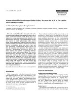
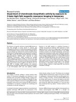
tumor stroma in five randomly selected fields [21]. We defined 4 classes or scores according to TIL density according to previous reports; (score 3; > 50%, score 2; > 10–
50%, score 1; ≤10%, or score 0; absent) (Fig. 1) [22, 23].
Statistical analysis
Statistical analyses were performed using JMP software
package (SAS, Tokyo, Japan). To compare the distribution of TIL density according to the state of lymph node
metastasis, we performed Student’s t test. Pearson’s chisquare test was used to evaluate the correlation between
two groups based on clinicopathological features. Odds
ratios (ORs) and 95% confidence intervals (CIs) were
calculated using logistic regression analysis. Multivariable analysis was performed using the multivariable
Takada et al. BMC Cancer
(2020) 20:598
Page 3 of 13
Fig. 1 Histopathologic analysis for tumor-infiltrating lymphocyte (TIL) density was performed on a single full-face hematoxylin and eosin-stained
tumor section. TIL density scores were defined as 3, 2, 1, and 0 if the area of stroma with lymphoplasmacytic infiltration around the invasive
tumor cell nests was > 50% (a); > 10–50% (b); ≤10% (c); and absent (d), respectively
Table 1 Clinicopathological features of 332 patients who had surgery after being diagnosed with cT1N0-2 M0 breast cancer,
including 319 cT1N0M0 breast cancer
Parameters
Number of all patients
(n = 332) (%)
Number of cN0 patients
(n = 319) (%)
Age at operation (years old)
median 59 (range, 29–79)
median 59 (range, 29–79)
Tumor size (mm)
median 13 (range, 4–20)
median 13 (range, 4–20)
Clinical lymph node metastasis cN0 / cN1 / cN2
319 (96.1%) / 11 (3.3%) / 2 (0.6%)
–
Estrogen receptor Negative / Positive
59 (17.8%) / 273 (82.2%)
57 (17.9%) / 262 (82.1%)
Progesterone receptor Negative / Positive
130 (39.2%) / 202 (60.8%)
125 (39.2%) / 194 (60.8%)
HER2 Negative / Positive
306 (92.2%) / 26 (7.8%)
295 (92.5%) / 24 (7.5%)
Ki67 ≤ 14% / > 14%
206 (62.0%) / 126 (38.0%)
196 (61.4%) / 123 (38.6%)
Intrinsic subtype HR + HER2-BC / HR
+ HER2 + BC / HER2enriched BC / TNBC
265 (79.8%) / 11 (3.3%) / 15
255 (79.9%) / 10 (3.1%) / 14
(4.5%) / 41 (12.4%)
(4.4%) / 40 (12.6%)
Lymphatic invasion ly0 / ly1
229 (69.0%) / 103 (31.0%)
224 (70.2%) / 95 (29.8%)
Venous invasion v0 / v1
318 (95.8%) / 14 (4.2%)
306 (95.9%) / 13 (4.1%)
Nuclear grade 1 / 2 / 3
164 (49.4%) / 129 (38.9%) / 39
158 (49.5%) / 125 (39.2%) / 36
(11.7%)
(11.3%)
Pathological lymph node metastasis
pN0 / pN1mic / pN1a / pN2
257 (77.4%) / 16 (4.8%) / 54
257 (80.6%) / 16 (5.0%) / 46
(16.3%) / 5 (1.5%)
(14.4%) / 0 (0.0%)
TILs (score) 0 / 1 / 2 / 3
29 (8.7%) / 243 (73.2%) / 57
25 (7.8%) / 235 (73.7%) / 56
(17.2%) / 3 (0.9%)
(17.6%) / 3 (0.9%)
HER2: human epidermal growth factor receptor 2. HR + HER2-BC: hormone receptor-positive and HER2 negative breast cancer (ER+ and/or PgR+, and HER2-). HR +
HER2 + BC: hormone receptor-positive and HER2 positive breast cancer (ER+ and/or PgR+, and HER2+). HER2 enriched BC: human epidermal growth factor
receptor 2-enriched breast cancer (ER-, PgR-, and HER2+). TNBC: triple negative breast cancer (ER-, PgR-, and HER2-). TILs: tumor- infiltrating lymphocytes
115 (42.1%)
> 60
219 (80.2%)
> 10.0
223 (81.7%)
50 (18.3%)
164 (60.1%)
109 (39.9%)
107 (39.2%)
> 14%
ly0
201 (73.6%)
28 (47.5%)
19 (32.2%)
40 (67.8%)
6 (10.2%)
53 (89.8%)
50 (84.7%)
9 (15.3%)
38 (64.4%)
21 (35.6%)
50 (84.7%)
9 (15.3%)
55 (93.2%)
4 (6.8%)
31 (52.5%)
28 (47.5%)
pN1a or 2
(n = 59)
<
158 (72.8%)
0.001
69 (31.8%)
0.316 148 (68.2%)
–
0.461 –
–
0.715 –
159 (73.3%)
0.538 58 (26.7%)
214 (98.6%)
0.577 3 (1.4%)
172 (79.8%)
0.016 45 (20.7%)
87 (40.1%)
0.144 130.(59.9%)
25 (52.1%)
11 (22.9%)
37 (77.1%)
–
–
–
–
37 (77.1%)
11 (22.9%)
48 (100.0%)
0 (0.0%)
44 (91.7%)
4 (8.3%)
25 (52.1%)
23.(47.9%)
pN1a or 2
(n = 48)
HR + HER2-BC (n = 265)
p
pN0 or 1mic
value (n = 217)
–
–
0.005 7
(77.8%)
6
(66.7%)
0.225 3
(33.3%)
–
–
–
5
(55.6%)
0.586 4
(44.4%)
–
0 (0.0%)
2 (100.0%)
0 (0.0%)
1 (50.0%)
1 (50.0%)
1 (50.0%)
2 (100.0%)
0 (0.0%)
–
–
–
–
1 (50.0%)
1 (50.0%)
9
2 (100.0%)
(100.0%)
0.413 0 (0.0%)
7
(77.8%)
0.045 2
(22.2%)
4
(44.4%)
0.128 5
(55.6%)
pN1a or 2
(n = 2)
HR + HER2 + BC (n = 11)
p
pN0
value (n = 9)
–
–
–
–
0.425 8 (72.7%)
10
(90.9%)
0.338 1 (9.1%)
–
–
–
0.887 –
–
1.000 –
11
(100.0%)
0.461 0 (0.0%)
4 (36.4%)
0.887 7(63.6%)
0 (0.0%)
4 (100.0%)
0 (0.0%)
–
–
–
–
–
–
–
–
4 (100.0%)
0 (0.0%)
1 (25.0%)
3 (75.0%)
pN1a or 2
(n = 4)
HER2enriched BC (n = 15)
p
pN0 (n =
value 11)
–
–
–
–
–
–
–
0.013 28
(77.8%)
14
(38.9%)
0.533 22
(61.1%)
–
–
–
–
–
1.000 29
(80.6%)
7 (19.4%)
20
(55.6%)
0.680 16 (44.4)
2 (40.0%)
2 (40.0%)
3 (60.0%)
–
–
–
–
–
–
–
–
5 (100.0%)
0 (0.0%)
4 (80.0%)
1 (20.0%)
pN1a or 2
(n = 5)
TNBC (n = 41)
p
pN0 (n =
value 36)
0.074
0.962
–
–
–
–
0.279
0.299
p
value
(2020) 20:598
Lymphatic invasion
166 (60.8%)
20 (7.3%)
253 (92.7%)
226 (82.8%)
47 (17.2%)
≤ 14%
Ki67
Positive
Negative
HER2
Positive
Negative
Hormone receptor
Positive
Negative
Progesterone receptor
Positive
Negative
Estrogen receptor
54 (19.8%)
≤ 10.0
Tumor size (mm)
158 (57.9%)
≤ 60
Age (years old)
pN0 or 1mic
(n = 273)
Parameters All intrinsic subtype (n = 332)
Table 2 Correlation between lymph node metastasis and clinicopathological features in cT1 breast cancer patients undergoing surgery
Takada et al. BMC Cancer
Page 4 of 13
72 (26.4%)
9 (3.3%)
v1
30 (11.0%)
3
261 (95.6%)
42 (71.2%)
17 (28.8%)
6 (10.2%)
53 (89.8%)
9 (15.3%)
50 (84.7%)
5 (8.5%)
54 (91.5%)
31 (52.5%)
pN1a or 2
(n = 59)
<
11 (5.1%)
0.001
206 (94.9%)
27 (12.4%)
0..082 190 (87.6%)
14 (6.5%)
0.356 203 (93.5%)
8 (3.7%)
0.073 209 (96.3%)
59 (27.2%)
34 (70.8%)
14 (29.2%)
4 (8.3%)
44 (91.7%)
6 (12.5%)
42 (87.5%)
5 (10.4%)
43 (89.6%)
23 (47.9%)
pN1a or 2
(n = 48)
HR + HER2-BC (n = 265)
p
pN0 or 1mic
value (n = 217)
1 (50.0%)
0 (0.0%)
0 (0.0%)
2 (100.0%)
0 (0.0%)
<
0 (0.0%) 0 (0.0%)
0.001
9
2 (100.0%)
(100.0%)
2
(22.2%)
0.423 7
(77.8%)
0 (0.0%)
0.151 9
2 (100.0%)
(100.0%)
0 (0.0%)
0.051 9
2 (100.0%)
(100.0%)
2
(22.2%)
pN1a or 2
(n = 2)
HR + HER2 + BC (n = 11)
p
pN0
value (n = 9)
11
(100.0%)
1.000 0 (0.0%)
6 (54.5%)
0.461 5 (45.5%)
4 (36.4%)
1.000 7 (63.6%)
1 (9.1%)
1.000 10
(90.9%)
3 (27.3%)
3 (75.0%)
1 (25.0%)
1 (25.0%)
3 (75.0%)
3 (75.0%)
1 (25.0%)
0 (0.0%)
4 (100.0%)
4 (100.0%)
pN1a or 2
(n = 4)
HER2enriched BC (n = 15)
p
pN0 (n =
value 11)
35
(97.2%)
0.086 1 (2.8%)
19
(52.8%)
0.310 17
(47.2%)
12
(33.3%)
0.185 24
(66.7%)
0 (0.0%)
0.533 36
(100.0%)
8 (22.2%)
3 (60.0%)
2 (40.0%)
1 (20.0%)
4 (80.0%)
0 (0.0%)
5 (100.0%)
0 (0.0%)
5 (100.0%)
3 (60.0%)
pN1a or 2
(n = 5)
TNBC (n = 41)
p
pN0 (n =
value 36)
0.003
0.169
0.125
1.000
p
value
HER: human epidermal growth factor receptor. HR + HER2-BC: hormone receptor-positive and HER2 negative breast cancer (ER+ and/or PgR+, and HER2-). HR + HER2 + BC: hormone receptor-positive and HER2 positive
breast cancer (ER+ and/or PgR+, and HER2+). HER2 enriched BC: human epidermal growth factor receptor 2-enriched breast cancer (ER-, PgR-, and HER2+). TNBC: triple negative breast cancer (ER-, PgR-, and HER2-).
TILs: tumor- infiltrating lymphocytes
1–3
0
12 (4.4%)
54 (19.8%)
2, 3
TILs (score)
219 (80.2%)
0, 1
TILs (score)
243 (89.0%)
1, 2
Nuclear grade
264 (96.7%)
v0
Venous invasion
ly1
pN0 or 1mic
(n = 273)
Parameters All intrinsic subtype (n = 332)
Table 2 Correlation between lymph node metastasis and clinicopathological features in cT1 breast cancer patients undergoing surgery (Continued)
Takada et al. BMC Cancer
(2020) 20:598
Page 5 of 13
115 (42.1%)
> 60
219 (80.2%)
> 10.0
223 (81.7%)
50 (18.3%)
164 (60.1%)
109 (39.9%)
107 (39.2%)
> 14%
ly0
201 (73.6%)
23 (50.0%)
16 (34.8%)
30 (65.2%)
4 (8.7%)
42 (91.3%)
39 (84.8%)
7 (15.2%)
30 (65.2%)
16 (34.8%)
39 (84.8%)
7 (15.2%)
43 (93.5%)
3 (6.5%)
25 (54.3%)
21 (45.7%)
pN1a or 2
(n = 46)
pN0 or 1mic
(n = 217)
0.002 158 (72.8%)
69 (31.8%)
0.567 148 (68.2%)
–
0.749 –
–
0.735 –
159 (73.3%)
0.506 58 (26.7%)
214 (98.6%)
0.606 3 (1.4%)
172 (79.3%)
0.017 45 (20.7%)
87 (40.1%)
20 (52.6%)
10 (26.3%)
28 (73.7%)
–
–
–
–
29 (76.3%)
9 (23.7%)
38 (100.0%)
0 (0.0%)
35 (92.1%)
3 (7.9%)
21 (55.3%)
17 (44.7%)
pN1a or 2
(n = 38)
HR + HER2-BC (n = 255)
0.124 130 (59.9%)
p
value
pN0
(n = 9)
0 (0.0%)
1 (100.0%)
0 (0.0%)
0.(0.0%)
0.012 7
(77.8%)
6
(66.7%)
0.500 3
(33.3%)
–
–
–
–
5
(55.6%)
0.694 4
(44.4%)
1 (100.0%)
1 (100.0%)
0 (0.0%)
–
–
–
–
1 (100.0%)
0 (0.0%)
9
1 (100.0%)
(100.0%)
0.466 0 (0.0%)
7
(77.8%)
0.062 2
(22.2%)
4
(44.4%)
1 (100.0%)
pN1a or 2
(n = 1)
HR + HER2 + BC (n = 10)
0.081 5
(55.6%)
p
value
3 (100.0%)
0 (0.0%)
–
–
–
–
–
–
–
–
3 (100.0%)
0 (0.0%)
0.598 8 (72.7%) 0 (0.0%)
10
(90.9%)
0.490 1 (9.1%)
–
–
–
–
–
0.389 –
–
1.000 –
11
(100.0%)
0.598 0 (0.0%)
4 (36.4%) 1 (33.3%)
0.389 7 (63.6%) 2 (66.7%)
pN1a or 2
(n = 3)
HER2enriched BC (n = 14)
p
pN0 (n =
value 11)
3 (75.0%)
1 (25.0%)
0.024 28
(77.8%)
14
(38.9%)
0.588 22
(61.1%)
–
–
–
–
–
–
–
–
29
(80.6%)
2 (50.0%)
2 (50.0%)
2 (50.0%)
–
–
–
–
–
–
–
–
4 (100.0%)
1.000 7 (19.4%) 0 (0.0%)
20
(55.6%)
0.923 16
(44.4%)
pN1a or 2
(n = 4)
TNBC (n = 40)
p
pN0 (n =
value 36)
0.224
0.667
0.332
0.455
p
value
(2020) 20:598
Lymphatic invasion
166 (60.8%)
20 (7.3%)
253 (92.7%)
226 (82.8%)
47 (17.2%)
≤ 14%
Ki67
Positive
Negative
HER2
Positive
Negative
Hormone receptor
Positive
Negative
Progesterone receptor
Positive
Negative
Estrogen receptor
54 (19.8%)
≤ 10.0
Tumor size (mm)
158 (57.9%)
≤ 60
Age (years old)
pN0 or 1mic
(n = 273)
Parameters All intrinsic subtype (n = 319)
Table 3 Correlation between lymph node metastasis and clinicopathological features in cT1N0M0 breast cancer patients undergoing SLNB
Takada et al. BMC Cancer
Page 6 of 13
72 (26.4%)
9 (3.3%)
v1
30 (11.0%)
3
54 (19.8%)
2, 3
261 (95.6%)
1–3
33 (71.7%)
13 (28.3%)
5 (10.9%)
41 (89.1%)
6 (13.0%)
40 (87.0%)
4 (8.7%)
42 (91.3%)
23 (50.0%)
pN1a or 2
(n = 46)
59 (27.2%)
pN0 or 1mic
(n = 217)
206 (94.9%)
< 0.001 11 (5.1%)
27 (12.4%)
0.128 190 (87.6%)
14 (6.5%)
0.689 203 (93.5%)
8 (3.7%)
27 (71.1%)
11 (28.9%)
3 (7.9%)
35 (92.1%)
3 (7.9%)
35 (92.1%)
4 (10.5%)
34 (89.5%)
18 (47.4%)
pN1a or 2
(n = 38)
HR + HER2-BC (n = 255)
0.124 209 (96.8%)
p
value
2
(22.2%)
pN0
(n = 9)
0 (0.0%)
pN1a or 2
(n = 1)
HR + HER2 + BC (n = 10)
0 (0.0%)
9 (100.0)
< 0.001 0 (0.0%)
2
(22.2%)
0.422 7
(77.8%)
0 (0.0%)
1 (100.0%)
0 (0.0%)
0 (0.0%)
1 (100.0%)
0 (0.0%)
0.742 9
1 (100.0%)
(100.0%)
0 (0.0%)
0.066 9
1 (100.0%)
(100.0%)
p
value
0 (0.0%)
3 (100.0%)
11
(100.0%)
1.000 0 (0.0%)
2 (66.7%)
1 (33.3%)
6 (54.5%) 1 (33.3%)
0.598 5 (45.5%) 2 (66.7%)
4 (36.4%) 3 (100.0%)
1.000 7 (63.6%) 0 (0.0%)
1 (9.1%)
1.000 10
(90.9%)
3 (27.3%) 3 (100.0%)
pN1a or 2
(n = 3)
HER2enriched BC (n = 14)
p
pN0 (n =
value 11)
35
(97.2%)
0.047 1 (2.8%)
19
(52.8%)
0.515 17
(47.2%)
12
(33.3%)
0.051 24
(66.7%)
0 (0.0%)
0.588 36
(100.0%)
3 (75.0)
1 (25.0%)
1 (25.0%)
3 (75.0%)
0 (0.0%)
4 (100.0%)
0 (0.0%)
4 (100.0%)
8 (22.2%) 2 (50.0%)
pN1a or 2
(n = 4)
TNBC (n = 40)
p
pN0 (n =
value 36)
0.053
0.292
0.168
1.000
p
value
SLNB: sentinel lymph node biopsy. HER: human epidermal growth factor receptor. HR + HER2-BC: hormone receptor-positive and HER2 negative breast cancer (ER+ and/or PgR+, and HER2-). HR + HER2 + BC: hormone
receptor-positive and HER2 positive breast cancer (ER+ and/or PgR+, and HER2+). HER2 enriched BC: human epidermal growth factor receptor 2-enriched breast cancer (ER-, PgR-, and HER2+). TNBC: triple negative
breast cancer (ER-, PgR-, and HER2-). TILs: tumor- infiltrating lymphocytes
12 (4.4%)
0
TILs (score)
219 (80.2%)
0, 1
TILs (score)
243 (89.0%)
1, 2
Nuclear grade
264 (96.7%)
v0
Venous invasion
ly1
pN0 or 1mic
(n = 273)
Parameters All intrinsic subtype (n = 319)
Table 3 Correlation between lymph node metastasis and clinicopathological features in cT1N0M0 breast cancer patients undergoing SLNB (Continued)
Takada et al. BMC Cancer
(2020) 20:598
Page 7 of 13
Takada et al. BMC Cancer
(2020) 20:598
Page 8 of 13
Table 4 Correlation between TILs and clinicopathological features in cT1N0M0 breast cancer patients undergoing SLNB
Parameters
tumor- infiltrating lymphocytes (n = 319)
Score 0 (n = 25)
Score 1–3 (n = 294)
p value
Score 0, 1 (n = 260)
Score 2, 3 (n = 59)
p value
Age (years old)
≤ 60
10 (40.0%)
169 (57.5%)
> 60
15 (60.0%)
125 (42.5%)
≤ 10.0
1 (4.0%)
56 (19.0%)
> 10.0
24 (96.0%)
238 (81.0%)
Negative
3 (12.0%)
54 (18.4%)
Positive
22 (88.0%)
240 (81.6%)
Negative
9 (36.0%)
116 (39.5%)
Positive
16 (64.0%)
178 (60.5%)
Negative
3 (12.0%)
51 (17.3%)
Positive
22 (88.0%)
243 (82.7%)
Negative
24 (96.0%)
271 (92.2%)
Positive
1 (4.0%)
23 (7.8%)
≤ 14%
19 (76.0%)
177 (60.2%)
170 (65.4%)
26 (44.1%)
> 14%
6 (24.0%)
177 (39.8%)
0.119
90.(34.6%)
33 (55.9%)
0.002
ly0
19 (56.0%)
210 (71.4%)
0.105
182 (70.0%)
42 (71.2%)
0.857
ly1
11 (44.0%)
84 (28.6%)
78 (30.0%)
17 (28.8%)
0.091
144 (55.4%)
35 (59.3%)
116 (44.6%)
24 (40.7%)
49 (18.8%)
8 (13.6%)
211 (81.2%)
51 (86.4%)
29 (11.2%)
28 (47.5%)
231 (88.8%)
31 (52.5%)
88 (33.8%)
37 (62.7%)
172 (66.2%)
22 (37.3%)
27 (10.4%)
27 (45.8%)
233 (89.6%)
32 (54.2%)
245 (94.2%)
50 (84.7%)
15 (5.8%)
9 (15.35)
0.582
Tumor size (mm)
0.059
0.339
Estrogen receptor
0.425
< 0.001
Progesterone receptor
0.734
< 0.001
Hormone receptor
0.494
< 0.001
HER2
0.487
0.013
Ki67
Lymphatic invasion
Venous invasion
v0
25 (100.0%)
281 (95.6%)
252 (96.9%)
54 (91.5%)
v1
0 (0.0%)
13 (4.4%)
8 (3.1%)
5 (8.5%)
1, 2
24 (96.0%)
259 (88.1%)
236 (90.8%)
47 (79.7%)
3
1 (4.0%)
35 (11.9%)
0.230
24 (9.2%)
12 (20.3%)
219 (84.2%)
54 (91.5%)
< 0.00121
41 (15.8%)
5 (8.5%)
0.283
0.058
Nuclear grade
0.015
Pathological lymph node metastasis
pN0 / pN1mic
12 (48.0%)
261 (88.8%)
pN1a / pN2
13 (52.0%)
33 (11.2%)
0.150
TILs tumor- infiltrating lymphocytes, SLNB sentinel lymph node biopsy, HER human epidermal growth factor receptor
logistic regression model. P-values less than 0.05 were
considered significant.
of the investigational nature of this study and provided
their written, informed consent.
Results
Ethics statement
Clinicopathological features
This study was conducted at Osaka City University,
Osaka, Japan, and conducted in accordance with the
Declaration of Helsinki. The study protocol was approved by the Ethics Committee of Osaka City University (approve number: #926). All patients were informed
Table 1 shows the clinicopathological features of 332 patients with cT1N0-2 M0 breast cancer who underwent
surgery and 319 patients with cT1N0M0 breast cancer
who underwent SLNB. Therefore, 13 patients (3.9%)
were diagnosed with axillary lymph node metastases on
Takada et al. BMC Cancer
(2020) 20:598
Page 9 of 13
Fig. 2 Comparison of tumor-infiltrating lymphocyte (TIL) density by differences in lymph node metastasis by box-plot diagrams in cT1 breast cancer:
all (a), HR + HER2-BC (b), HR + HER2 + BC (c), HER2-enriched BC (d), triple-negative breast cancer (e). Correlation was performed by Student’s t test
imaging investigation (cN1: 11 patients (3.3%), cN2: 2
patients(0.6%)).In both groups, the median age was 59
(range, 29–79) years, and the median tumor diameter
was 13 mm (range, 4.0–20.0 mm). In patients with
cT1N0M0 breast cancer, 262 patients (82.1%) were positive for ER, 194 (60.8%) were positive for PgR, and 24
(7.5%) were positive for HER2. High Ki67 expression
was observed in 123 patients (38.8%). The following results were demonstrated by the intrinsic subtypes: HR +
HER2-BC: 255 patients (79.9%), HR + HER2 + BC: 10 patients (3.1%), HER2-enriched BC 14 patients (4.4%),
TNBC: 40 patients (12.5%). Pathologically, lymphatic invasion was observed in 95 patients (29.8%), and venous
invasion in 13 patients (4.1%). Regarding the nuclear
Fig. 3 Comparison of tumor-infiltrating lymphocyte (TIL) density by differences in lymph node metastasis by box-plot diagrams in cT1N0M0 breast
cancer patients undergoing SLNB: all (a), HR + HER2-BC (b), HR + HER2 + BC (c), HER2-enriched BC (d), triple-negative breast cancer (e). Correlation was
performed by Student’s t test
Takada et al. BMC Cancer
(2020) 20:598
Page 10 of 13
grade, only 36 patients (11.3%) were diagnosed with
grade 3. These results did not differ significantly when
compared with the entire group of cT1 patients undergoing surgery.
For SLNB, a median of 2 (range, 1–8) SLNs were identified and evaluated pathologically. There were 60 cases
(19.4%) of SLNM (macrometastasis: 46 cases, micrometastasis: 16 cases). The intrinsic subtype of all breast cancers with micrometastasis was HR + HER2-BC. All
patients who underwent axillary dissection due to lymph
node metastasis on radiological examination had pathological metastasis to the lymph nodes.
When TIL densities were examined in the biopsied tissues, in cN0 cases, 25 patients (7.8%) had score 0, 235
(73.7%) had score 1, 56 (17.6%) had score 2, and three
(0.9%) had score 3. In the 13 cases in which lymph node
metastasis was detected by imaging, four patients had
score 0, eight had score 1, and one had score 2.
Correlation between clinicopathological features and
lymph node metastasis
The correlations between clinicopathological features
and lymph node metastasis are listed in Table 2. Metastasis was significantly higher in breast cancers with
tumor diameter > 10 mm than in those with diameter ≤
10 mm (p = 0.016). Additionally, metastasis was significantly associated with lymphatic invasion (p < 0.001).
These two clinicopathological factors correlated with
SLNM even in patients diagnosed with cN0 (tumor size;
p = 0.017, lymphatic invasion; p = 0.002) (Table 3).
Correlation between clinicopathological features and TILs
We examined the correlation between clinicopathological features and TILs in cN0 breast cancer cases
(Table 4). When the patients were divided into TIL
density score 0–1 and score 2–3, that is, a cut-off value
of 10% was used for division into the higher group and
lower group, the lower group correlated with the following clinicopathological factors; ER positive (p < 0.001),
PgR positive (p < 0.001), HER2 negative (p = 0.013), Ki67
high (p = 0.002), nuclear grade high (p = 0.015). However, if the patients were divided into TIL density score
0 and score 1–3, that is, by the presence or absence of
TIL density, correlation with these clinicopathological
factors was not observed. When examined by intrinsic
subtype, in HR + HER2-BC, patients with TILs density
score 0 were significantly more aged (p = 0.035) and had
a larger tumor size (p = 0.020) than in patients with TILs
density score 1–3 (Supplementary Table 1). In HER2enriched BC, the frequency of venous invasion was significantly higher in patients with TILs density score 0
than in patients with TILs density score 1–3 (p = 0.011).
However, SLNM was significant in breast cancer with
absent TIL density (p < 0.001). When examined by intrinsic subtypes, HR + HER-2 BC and HER2-enriched BC
significantly correlated with SLNM, and TNBC also
showed a similar tendency (HR + HER2-BC: p < 0.001,
HER2-enriched BC: p = 0.047, TNBC: p = 0.053) (Table
3).
TIL density was significantly lower in patients with
lymph node metastasis than in those without it in all
cT1 patients (p = 0.018) (Fig. 2). When examined by intrinsic subtype, there was no significant difference between the subtypes. Moreover, no significant difference
was observed in all cases when focusing on cN0 cases
(p = 0.061) (Fig. 3).
Based on these results, multivariate analysis for SLNM
predictors revealed that lymphatic invasion (p = 0.008,
OR = 2.522) and TILs (p < 0.001, OR = 0.137) were independent factors for prediction of SLNM (Table 5).
Table 5 Univariate and multivariate analysis with sentinel lymph node metastasis for cT1N0M0 breast cancer
Parameters
Univarite analysis
Multivarite analysis
Odd ratio
95% CI
p value
Age at operation (years old) ≤ 60 vs > 60
1.636
0.873–3.065
0.124
Tumor size (mm) ≤ 10.0 vs > 10.0
3.534
1.056–11.825
0.017
Estrogen receptor Negative vs Positive
1.249
0.528–2.955
0.606
Progesterone receptor Negative vs Positive
1.246
0.648–2.395
0.506
Hormone receptor Negative vs Positive
1.159
0.488–2.748
0.735
HER2 Negative vs Positive
1.205
0.392–3.700
0.749
Ki67 ≤ 14% vs > 14%
0.827
0.430–1.590
0.567
Lymphatic invasion ly0 vs ly1
2.792
1.476–5.282
0.002
Venous invasion v0 vs v1
2.794
0.823–9.481
0.124
Nuclear grade 1, 2 vs 3
1.215
0.475–3.105
0.689
TILs 0, 1 vs 2, 3
0.495
0.187–1.311
0.128
TILs 0 vs 1–3
0.117
0.049–0.277
< 0.001
CI confidence intervals, HER2 human epidermal growth factor receptor 2, TILs tumor- infiltrating lymphocytes
Odd ratio
95% CI
p value
2.639
0.888–11.346
0.085
2.522
1.280–4.973
0.008
0.137
0.055–0.335
< 0.001
Takada et al. BMC Cancer
(2020) 20:598
Discussion
Numerous studies have reported predictors of SLNM.
Although some studies have reported age [6–9], site [6,
10, 24], ER positivity [7, 24], PgR positivity [8, 24], HER2
positivity [25] as predictors of SLNM, the most commonly reported predictors are tumor size [6–10, 24, 25],
lymphatic invasion [6–8, 24, 25], and pathological nuclear grade [6–10, 24, 25]. In our study, the SLNM rate
was similar to previous reports, and tumor size and
lymphatic invasion were found to be predictive factors.
However, intrinsic subtype and nuclear grade were not
found to be predictors in our study. In recent years, it
has been known that the pathological response to preoperative chemotherapy is a predictor of prognosis [26–
29]. Based on these reports, preoperative chemotherapy
is actively administered in HER2-positive breast cancer
and TNBC because the treatment response is greater
than that in hormone receptor-positive breast cancer. As
a result, the number of patients who underwent surgery
primarily for HER2-positive breast cancer or TNBC was
considered to be the reason for conducting this study.
After defining the cut-off value for TIL density as 10%,
as previously reported, hormone-positive breast cancer
was observed to have lower TIL density while hormonenegative breast cancer or HER2-positive breast cancer
were observed to have higher TIL density in this study
[13, 14]. When the correlation between TILs and clinicopathological factors was examined, in HR + HER2-BC,
the correlations between TILs and tumor size or age
were shown. Regarding the tumor size, it has recently
been reported that the microenvironment around the
cancer changes depending on the local progression [30].
According to the report, not only CD8 + lymphocytes
that suppress cancer progression but also FOXP3positive lymphocytes that promote cancer progression
are reduced. In other words, as cancer progresses, immune escape may begin to occur, and metastases are
likely to occur accordingly. Regarding age, we have previously reported that young breast cancer patients tend
to have higher TILs density (date not shown). That may
have influenced the results in this time. This study suggests that the tumor immune-microenvironment is involved in lymph node metastasis. Our hypothesis was
that the TIL density may be a predictor of SLNM. The
correlation between TILs and lymph node metastasis
has been reported in gastric cancer, melanoma, and
breast cancer [31–33]. A study on breast cancer examined 76 patients who underwent surgery first and 96 patients who underwent preoperative chemotherapy, and it
reported that there was a correlation between TILs and
lymph node metastasis in both groups. Interestingly,
Caziuc evaluated not only SLNs but also axillary lymph
nodes in cases of additional axillary lymph node dissection due to SLNM. However, detailed analysis of the
Page 11 of 13
subtypes that could affect TIL density was not conducted, and no detailed data were provided on the relationship between TILs and clinicopathological factors.
Furthermore, no relationship was found between any
clinicopathological features other than TILs and lymph
node metastasis. Accordingly, this report did not examine clinicopathological factors other than TILs, which
are predictors of lymph node metastasis. However, our
research is significant because we examined the correlation between TILs and clinicopathological factors such
as all the subtypes and performed multivariate analysis
to determine the predictors of SLNM, including TILs.
We are aware that our study has some limitations.
Firstly, there were few HER2-positive breast cancer and
TNBC patients, as we have stated earlier. Furthermore,
there were a few cases with distant metastases along
with a primary lesion of less than 20 mm that were excluded from our study. However, some studies have reported that TIL density is predictive of chemotherapy
response [34, 35]. Therefore, if SLNB was omitted even
if the SLN had metastasized in cN0 breast cancer with
high TIL density, postoperative chemotherapy would be
expected to have a high therapeutic effect and not affect
the prognosis.
Conclusions
Our study suggests a correlation between lymph node
metastasis and the tumor immune-microenvironment in
cT1 breast cancer cases. Moreover, TIL density may be a
predictor of SLNM in breast cancer patients without
lymph node metastasis on preoperative imaging.
Supplementary information
Supplementary information accompanies this paper at />1186/s12885-020-07101-y.
Additional file 1: Supplementary Table 1. Correlation between TILs
and clinicopathological features in cT1N0M0 breast cancer patients
undergoing SLNB by intrinsic subtype.
Abbreviations
BC: Breast cancer; CI: Confidence intervals; CT: Computed tomography;
ER: Estrogen receptor; HER2: Human epidermal growth factor receptor 2;
HR: Hormone receptor; OR: Odds ratio; PgR: Progesterone receptor;
SLN: Sentinel lymph node; SLNB: Sentinel lymph node biopsy;
SLNM: Sentinel lymph node metastasis; TILs: Tumor-infiltrating lymphocytes;
TNBC: Triple-negative breast cancer; US: Ultrasonography; VAB: Vacuumassisted biopsy
Acknowledgements
We thank Yayoi Matsukiyo and Tomomi Okawa (Department of Breast and
Endocrine Surgery, Osaka City University Graduate School of Medicine) for
helpful advice regarding data management.
Authors’ contributions
KT participated in the design of the study and drafted the manuscript. SK
participated in the design of the study and manuscript editing. YA, WG, RK,
AY, TM, MS and TT helped with study data collection and manuscript
preparation. HF helped with study data collection and participated in its
Takada et al. BMC Cancer
(2020) 20:598
design. KH and MO conceived the study, and participated in its design and
coordination and helped to draft the manuscript. All authors have read and
approved the final manuscript.
Funding
This study was supported in part by Grants-in Aid for Scientific Research
(KAKENHI, Nos. 17 K10559 and 19 K18067) from the Ministry of Education, Science, Sports, Culture and Technology of Japan. The funders had no role in
the design of the study and collection, analysis, and interpretation of data
and in writing the manuscript.
Availability of data and materials
The datasets used and/or analyzed during the current study are available
from the corresponding author on reasonable request.
Page 12 of 13
8.
9.
10.
11.
12.
Ethics approval and consent to participate
A written informed consent to participate in the study was obtained from
each subject in accordance with the declaration of Helsinki principles. Each
patient or the patient’s family was fully informed of the investigational
nature of this study and provided their written, informed consent. The study
protocol was approved by the Ethics Committee of Osaka City University
(approve number #926).
13.
14.
Consent for publication
Not applicable.
15.
Competing interests
The authors declare that they have no competing interests.
16.
Author details
1
Department of Breast and Endocrine Surgery, Osaka City University
Graduate School of Medicine, 1-4-3 Asahi-machi, Abeno-ku, Osaka 545-8585,
Japan. 2Department of Gastrointestinal Surgery, Osaka City University
Graduate School of Medicine, 1-4-3 Asahi-machi, Abeno-ku, Osaka 545-8585,
Japan. 3Department of Scientific and Linguistic Fundamentals of Nursing,
Osaka City University Graduate School of Nursing, 1-5-17 Asahi-machi,
Abeno-ku, Osaka 545-0051, Japan.
17.
18.
Received: 20 April 2020 Accepted: 22 June 2020
19.
References
1. Veronesi U, Paganelli G, Viale G, Luini A, Zurrida S, Galimberti V, Intra M,
Veronesi P, Maisonneuve P, Gatti G, et al. Sentinel-lymph-node biopsy as a
staging procedure in breast cancer: update of a randomised controlled
study. Lancet Oncol. 2006;7(12):983–90.
2. Krag DN, Anderson SJ, Julian TB, Brown AM, Harlow SP, Costantino JP,
Ashikaga T, Weaver DL, Mamounas EP, Jalovec LM, et al. Sentinel-lymphnode resection compared with conventional axillary-lymph-node dissection
in clinically node-negative patients with breast cancer: overall survival
findings from the NSABP B-32 randomised phase 3 trial. Lancet Oncol. 2010;
11(10):927–33.
3. Jozsa F, Ahmed M, Baker R, Douek M. Is sentinel node biopsy necessary in
the radiologically negative axilla in breast cancer? Breast Cancer Res Treat.
2019;177(1):1–4.
4. Gentilini O, Botteri E, Dadda P, Sangalli C, Boccardo C, Peradze N, Ghisini R,
Galimberti V, Veronesi P, Luini A, et al. Physical function of the upper limb after
breast cancer surgery. Results from the SOUND (sentinel node vs. observation
after axillary ultra-souND) trial. Eur J Surg Oncol. 2016;42(5):685–9.
5. Reimer T, Stachs A, Nekljudova V, Loibl S, Hartmann S, Wolter K, Hildebrandt
G, Gerber B. Restricted axillary staging in clinically and Sonographically
node-negative early invasive breast Cancer (c/iT1-2) in the context of breast
conserving therapy: first results following commencement of the
intergroup-sentinel-mamma (INSEMA) trial. Geburtshilfe Frauenheilkd. 2017;
77(2):149–57.
6. Capdet J, Martel P, Charitansky H, Lim YK, Ferron G, Battle L, Landier A, Mery
E, Zerdoub S, Roche H, et al. Factors predicting the sentinel node
metastases in T1 breast cancer tumor: an analysis of 1416 cases. Eur J Surg
Oncol. 2009;35(12):1245–9.
7. Reyal F, Rouzier R, Depont-Hazelzet B, Bollet MA, Pierga JY, Alran S, Salmon
RJ, Fourchotte V, Vincent-Salomon A, Sastre-Garau X, et al. The molecular
20.
21.
22.
23.
24.
25.
26.
subtype classification is a determinant of sentinel node positivity in early
breast carcinoma. PLoS One. 2011;6(5):e20297.
Viale G, Zurrida S, Maiorano E, Mazzarol G, Pruneri G, Paganelli G,
Maisonneuve P, Veronesi U. Predicting the status of axillary sentinel lymph
nodes in 4351 patients with invasive breast carcinoma treated in a single
institution. Cancer. 2005;103(3):492–500.
Ding J, Jiang L, Wu W. Predictive value of Clinicopathological characteristics
for sentinel lymph node metastasis in early breast Cancer. Med Sci Monit.
2017;23:4102–8.
Zhang Y, Li J, Fan Y, Li X, Qiu J, Zhu M, Li H. Risk factors for axillary lymph
node metastases in clinical stage T1-2N0M0 breast cancer patients.
Medicine (Baltimore). 2019;98(40):e17481.
Soysal SD, Tzankov A, Muenst SE. Role of the tumor microenvironment in
breast Cancer. Pathobiology. 2015;82(3–4):142–52.
Hanahan D, Coussens LM. Accessories to the crime: functions of cells
recruited to the tumor microenvironment. Cancer Cell. 2012;21(3):309–22.
Ohtani H, Mori-Shiraishi K, Nakajima M, Ueki H. Defining lymphocytepredominant breast cancer by the proportion of lymphocyte-rich stroma
and its significance in routine histopathological diagnosis. Pathol Int. 2015;
65(12):644–51.
Stanton SE, Adams S, Disis ML. Variation in the incidence and magnitude of
tumor-infiltrating lymphocytes in breast Cancer subtypes: a systematic
review. JAMA Oncol. 2016;2(10):1354–60.
Cheang MC, Chia SK, Voduc D, Gao D, Leung S, Snider J, Watson M, Davies S,
Bernard PS, Parker JS, et al. Ki67 index, HER2 status, and prognosis of patients
with luminal B breast cancer. J Natl Cancer Inst. 2009;101(10):736–50.
McMasters KM, Tuttle TM, Carlson DJ, Brown CM, Noyes RD, Glaser RL,
Vennekotter DJ, Turk PS, Tate PS, Sardi A, et al. Sentinel lymph node biopsy
for breast cancer: a suitable alternative to routine axillary dissection in multiinstitutional practice when optimal technique is used. J Clin Oncol. 2000;
18(13):2560–6.
Kashiwagi S, Onoda N, Asano Y, Kurata K, Noda S, Kawajiri H, Takashima T,
Ohsawa M, Kitagawa S, Hirakawa K. Ambulatory sentinel lymph node biopsy
preceding neoadjuvant therapy in patients with operable breast cancer: a
preliminary study. World J Surg Oncol. 2015;13:53.
Lee A, Krishnamurthy S, Sahin A, Symmans WF, Hunt K, Sneige N.
Intraoperative touch imprint of sentinel lymph nodes in breast carcinoma
patients. Cancer. 2002;96(4):225–31.
Khanna R, Bhadani S, Khanna S, Pandey M, Kumar M. Touch imprint
cytology evaluation of sentinel lymph node in breast cancer. World J Surg.
2011;35(6):1254–9.
Houvenaeghel G, Nos C, Mignotte H, Classe JM, Giard S, Rouanet P, Lorca
FP, Jacquemier J, Bardou VJ, Groupe des Chirurgiens de la Federation des
Centres de Lutte Contre le C. Micrometastases in sentinel lymph node in a
multicentric study: predictive factors of nonsentinel lymph node
involvement--Groupe des Chirurgiens de la Federation des Centres de Lutte
Contre le Cancer. J Clin Oncol. 2006;24(12):1814–22.
Salgado R, Denkert C, Demaria S, Sirtaine N, Klauschen F, Pruneri G, Wienert
S, Van den Eynden G, Baehner FL, Penault-Llorca F, et al. The evaluation of
tumor-infiltrating lymphocytes (TILs) in breast cancer: recommendations by
an international TILs working group 2014. Ann Oncol. 2015;26(2):259–71.
Kashiwagi S, Asano Y, Goto W, Takada K, Takahashi K, Noda S, et al. Use of
tumor-infiltrating lymphocytes (TILs) to predict the treatment response to
eribulin chemotherapy in breast cancer. PLoS One. 2017;12(2):e0170634.
Ono M, Tsuda H, Shimizu C, Yamamoto S, Shibata T, Yamamoto H, et al.
Tumor-infiltrating lymphocytes are correlated with response to neoadjuvant
chemotherapy in triple-negative breast cancer. Breast Cancer Res Treat.
2012;132(3):793–805.
Qiu PF, Liu JJ, Wang YS, Yang GR, Liu YB, Sun X, Wang CJ, Zhang ZP. Risk
factors for sentinel lymph node metastasis and validation study of the
MSKCC nomogram in breast cancer patients. Jpn J Clin Oncol. 2012;42(11):
1002–7.
Klar M, Foeldi M, Markert S, Gitsch G, Stickeler E, Watermann D. Good
prediction of the likelihood for sentinel lymph node metastasis by using the
MSKCC nomogram in a German breast cancer population. Ann Surg Oncol.
2009;16(5):1136–42.
Rastogi P, Anderson SJ, Bear HD, Geyer CE, Kahlenberg MS, Robidoux A,
Margolese RG, Hoehn JL, Vogel VG, Dakhil SR, et al. Preoperative
chemotherapy: updates of National Surgical Adjuvant Breast and bowel
project protocols B-18 and B-27. J Clin Oncol. 2008;26(5):778–85.
Takada et al. BMC Cancer
(2020) 20:598
27. von Minckwitz G, Untch M, Blohmer JU, Costa SD, Eidtmann H, Fasching PA,
Gerber B, Eiermann W, Hilfrich J, Huober J, et al. Definition and impact of
pathologic complete response on prognosis after neoadjuvant
chemotherapy in various intrinsic breast cancer subtypes. J Clin Oncol. 2012;
30(15):1796–804.
28. Cortazar P, Zhang L, Untch M, Mehta K, Costantino JP, Wolmark N, Bonnefoi
H, Cameron D, Gianni L, Valagussa P, et al. Pathological complete response
and long-term clinical benefit in breast cancer: the CTNeoBC pooled
analysis. Lancet. 2014;384(9938):164–72.
29. Bonnefoi H, Litiere S, Piccart M, MacGrogan G, Fumoleau P, Brain E, Petit T,
Rouanet P, Jassem J, Moldovan C, et al. Pathological complete response
after neoadjuvant chemotherapy is an independent predictive factor
irrespective of simplified breast cancer intrinsic subtypes: a landmark and
two-step approach analyses from the EORTC 10994/BIG 1-00 phase III trial.
Ann Oncol. 2014;25(6):1128–36.
30. Eleni P, Minas S, Maria M, Evangelos T, Haralambos K, Eleni K. A
standardized evaluation method for FOXP3+ Tregs and CD8+ T-cells in
breast carcinoma: association with breast carcinoma subtypes, Stage and
Prognosis. Anticancer Res. 2019;39(3):1217–32.
31. Kim JY, Kim CH, Lee Y, Lee JH, Chae YS. Tumour infiltrating lymphocytes are
predictors of lymph node metastasis in early gastric cancers. Pathology.
2017;49(6):589–95.
32. Azimi F, Scolyer RA, Rumcheva P, Moncrieff M, Murali R, McCarthy SW, Saw
RP, Thompson JF. Tumor-infiltrating lymphocyte grade is an independent
predictor of sentinel lymph node status and survival in patients with
cutaneous melanoma. J Clin Oncol. 2012;30(21):2678–83.
33. Caziuc A, Schlanger D, Amarinei G, Dindelegan GC. Can tumor-infiltrating
lymphocytes (TILs) be a predictive factor for lymph nodes status in both
early stage and locally advanced breast cancer? J Clin Med. 2019;8(4):545.
34. Adams S, Gray RJ, Demaria S, Goldstein L, Perez EA, Shulman LN, Martino S,
Wang M, Jones VE, Saphner TJ, et al. Prognostic value of tumor-infiltrating
lymphocytes in triple-negative breast cancers from two phase III
randomized adjuvant breast cancer trials: ECOG 2197 and ECOG 1199. J Clin
Oncol. 2014;32(27):2959–66.
35. Loi S, Michiels S, Salgado R, Sirtaine N, Jose V, Fumagalli D, KellokumpuLehtinen PL, Bono P, Kataja V, Desmedt C, et al. Tumor infiltrating
lymphocytes are prognostic in triple negative breast cancer and predictive
for trastuzumab benefit in early breast cancer: results from the FinHER trial.
Ann Oncol. 2014;25(8):1544–50.
Publisher’s Note
Springer Nature remains neutral with regard to jurisdictional claims in
published maps and institutional affiliations.
Page 13 of 13