Effect of fenugreek seed and leaves on some hematological and biochemical parameters in CCl4-induced liver injury
Bạn đang xem bản rút gọn của tài liệu. Xem và tải ngay bản đầy đủ của tài liệu tại đây (266.57 KB, 10 trang )
Int.J.Curr.Microbiol.App.Sci (2017) 6(4): 2328-2337
International Journal of Current Microbiology and Applied Sciences
ISSN: 2319-7706 Volume 6 Number 4 (2017) pp. 2328-2337
Journal homepage:
Original Research Article
/>
Effect of Fenugreek Seed and Leaves on Some Hematological and Biochemical
Parameters in CCl4-induced Liver Injury
Firdaws A. AL-Mashhadani*
Food technology Dep., Agriculture College, Salahaddin University, Erbil, Iraq
*Corresponding author
ABSTRACT
Keywords
Fenugreek,
CCL4 –
Induced
liver injury.
Article Info
Accepted:
20 March 2017
Available Online:
10 April 2017
This study was carried out to evaluate the effect of fenugreek plant on CCL4 –induced
liver injury by following the hematological and biochemical parameters. To achieve this
purpose forty male albino rats were used and divided to four groups. The first group
represented control group which received normal diet and intraperitoneal injection with oil
(0.5ml/kg). The second group represented the CCL (1ml/kg) model. The third group
received 200 mg/kg fenugreek leaves extract by gavage. The forth group received 500
mg/kg fenugreek seed extract by gavage. The fenugreek seed and leave extracts treated
group showed significant differences in AST, ALT, ALP, direct bilirubin, MDA, GSH,
liver SOD, WBC, LYM and PLT when compared to CCl4 treated rats. These results
indicate that these plants can be used as a good source of antioxidant and hepatic
protective activities as well as a good antibiotic agent against some pathogenic bacteria.
The methanolic extract of fenugreek seeds with different concentrations in ml inhibited the
growth of the pathogenic E. coli, Staphylococcus aureus and Bacillus subtilis bacteria
more than the aqueous extract for the fenugreek leaves and seeds.
Introduction
Medicinal plants are important part of health
care. Large varieties of plants (more than
1200) are available with known therapeutic
effects (Kipkore et al., 2014). Approximately
70–80% people worldwide depend on
medicinal plants to cure various human
ailments including viral diseases (Wang and
Liu, 2013). Moreover, herbal drugs have
gained much importance due to their easily
adaptability, low cost and fewer side reactions
on patients (Edziri et al., 2011).
Natural antioxidants can protect the body
against the adverse effects of CCl4 and some
other toxins (Kader et al., 2014, Amini et al.,
2012). Medicinal plants have been used to
treat various disorders throughout the history
of human life, but the use of synthetic drugs
was highly prevalent since the middle of last
century (Sewell and Rafieian-Kopaei, 2014).
With the rapid detection of their adverse side
effects of synthetic drugs on public health, the
trend is increasing for application of
medicinal plants as alternatives to synthetic
ones (Bahmani et al., 2014a,b).
Fenugreek (Trigonella foenum graecum Linn)
is an annual herb that belongs to the family
Leguminosae. The seeds of fenugreek are
commonly used in the Middle East and South
Asia as a spice in food preparation and used
as traditional medicines in diabetes, high
2328
Int.J.Curr.Microbiol.App.Sci (2017) 6(4): 2328-2337
cholesterol,
inflammations
and
gastrointestinal ailments (Basu et al., 2010;
Belguith-Hadriche et al., 2010).
Liver diseases are one of the major causes of
mortality and morbidity worldwide, druginduced liver toxicity is a major cause of
hepatic dysfunction (Abboud and Kaplowitz,
2007). Oxidative stress is considered as a
mechanism in contributing to the initiation
and progression of hepatic damage in a
variety of liver disorders. Cell damage occurs
when there is an excess of reactive species
derived from oxygen and nitrogen or
deficiency of antioxidants (Girish and
Pradhan, 2008a). Oxidative stress, involving
enhanced generation of reactive oxygen
species (ROS), has been implicated in the
etiology of many human diseases.
Antioxidants capable of neutralizing ROS and
their actions are considered beneficial. In this
context, natural dietary components with
antioxidant activities could be important
(Bandyopadhyay et al., 1999; Yamamoto,
2000).
Among environmental toxins, carbon
tetrachloride (CCl4) dedicated most of
conducted studies to itself (Olagunju et al.,
2009).
Fenugreek has a good antimicrobial property
because. It contains certain bioactive
components such as volatile oils, alkaloids,
mucilage. All these components in Fenugreek
adds on to its antibacterial activity. They
contain
multiple
constituents
with
antimicrobial activity including phenols,
quinones, flavones, tannins, terpenoids, and
alkaloids (24).
Aim of this study was to study the antioxidant
activity of fenugreek plant and its hepatic
protective activity and to determine the
oxidative stress and antioxidant markers and
some hematological parameters in CCL4
treated rat groups. Also the aim of this study
is to evaluate the effect of ethanolic and
aqueous extracts of the seeds and leaves of
fenugreek against various pathogenic bacteria
growth.
Materials and Methods
Materials
Plant preparation
A Fenugreek (Trigonella foenum graecum)
seeds and leaves sample were collected from
the local market of Baghdad. Dry fenugreek
seed and leaves were cleaned and ground into
small pieces by a blender and 70 % ethanol
was used extraction by soxhelt extraction
method for six hours.
The extracts were combined, and evaporated
to dryness under reduced pressure at 60 Co by
a rotary evaporator. Extracts were placed in
dark bottle, and stored at -4 C° until further
analysis. The extract was suspended in
distilled water for hepato protective studies
(Bukhari et al., 2008).
Experimental animals
Forty male albino rats (Rattus norvegicus),
weighing about 250 – 350gm were used.
The animals were given standard rat diet
chow and housed in plastic cages bedded with
wooden chips in a room with controlled
temperature of 24±3ºC, 12/12 hours light/dark
schedule in an animal house belong to
Biology department, College of Science,
Salahaddin University-Erbil.
Standard chaw ingredients included (wheat
66.6%,soya 25.6%, oil sun flower 4.4%, lime
stone 1.5%, salt 0.63%, methionine 0.158%,
Lysine 0.24%, choline chloride 0.062% and
trace elements 0.05%)
2329
Int.J.Curr.Microbiol.App.Sci (2017) 6(4): 2328-2337
Experimental Design
The experimental rats were divided randomly
to 4 groups. This experiment was carried out
for four weeks as explained below:
(less than 0.5cm 3 thicknesses) then kept in
formalin, while the other part stored at
refrigerators until homogenized for estimation
of SOD, HYP and GSH.
Tissue homogenate
Group 1: Control rats (n=10)
The rats of this group were given olive oil
intraperitoneally (0.5 ml/kg body weight) for
four weeks.
Group 2: CCl4 treated rats (n=10)
The rats of this group were given CCl4
intraperitoneally 1ml/kg b.w. (1:1 in olive oil)
for four weeks
Liver washed with cold saline. Pieces of each
tissue used for homogenization by 20 mM
cold phosphate buffer saline (pH 7.4).The
liver tissues homogenized (10%w/v) using
handheld glass homogenizer (Chowdhury et
al., 2013). Homogenates were centrifuged at
6000 rpm for10 minutes. The supernatants
were collected and stored at -80Co until
assayed.
Estimation of glutathione in liver tissue
Group 3: Fenugreek (n=10)
The rats of this group were given CCl4
intraperitoneally 1ml/kg b.w. (1:1 in olive oil)
and fenugreek seeds extract 500 mg/kg
dissolved in distilled water and given to rats
by gavage daily for four weeks.
Group 4: Fenugreek leaves (n=10)
The rats of this group were given CCl4
intraperitoneally 1ml/kg b.w. (1:1 in olive oil)
and Fenugreek leaves extract 200 mg/kg
dissolved in water and given to rats by gavage
daily for four weeks
The procedure of (Moron et al., 1979) was
followed with some modification. Weighting
1 gm of liver tissue and homogenate by using
handled homogenizer with 10 ml of cold tris
buffer solution. One ml of tissue homogenate
was added to 0.25ml of 25% trichloroacetic
acid. After centrifugation for 5 minutes at
3000rpm 0.2 ml of supernatant was taken in a
test tube, adding one ml o.15mole imidazole
solution then adding 1.7ml distilled water and
o.1ml 5.5(DTNB) solution finally absorbance
was read at 412nm after 3minutes of adding
DTNB.
Tissue preparation
The concentration of GSH was calculated
according to the absorbance of blank (B), test
(T) and standard (S) solutions by the
following equation:
Anesthesia, dissecting, liver and kidney
removing
GSH conc. (μmol/mg of tissue) = *conc.
Standard * 100 (3.1)
All animals were anesthetized with Ketamine
hydrochloride 80mg/Kg (Trittau, Germany)
and Xylazin 12mg/Kg (Interchem, Holland).
The liver was removed then divided into two
equal parts, one part cut into small pieces
Determination of liver tissue superoxide
dismutase
Methods
Liver samples were washed with 0.9% NaCl
to remove red blood cells. The tissue was then
2330
Int.J.Curr.Microbiol.App.Sci (2017) 6(4): 2328-2337
blotted dry and weighed followed by
homogenization in 200 μl buffer (0.05 M
potassium phosphate and 0.1 mM EDTA, pH
7.8) and centrifuged at 15,000xg for 30 min at
4˚C. The supernatant was used for
determination of SOD. Superoxide dismutase
was measured using the Superoxide
Dismutase assay kit provided by Elabscience
(Elabscience, WuHan P.R.C).
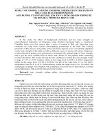
The concentration of SOD was determined by
competitive-ELISA method.
The concentration of SOD in the samples is
then determined by comparing the OD of the
samples to the standard curve (Figure 1).
Blood collection
At the end of the treatment period, blood
samples were collected from anesthetized rats
through cardiac puncture. The collected blood
samples were immediately placed into test
tube and centrifuged and the sera were stored
at -80Co (Sanyo – Ultra – Low Temperature,
Japan) until assayed. While, for hematological
analysis blood were collected in EDTA tube.
Hematological analysis
White blood cell (WBC) count, LYM and
PLT count were determined automatically by
using automated hematology analyzer
(Sysmex model: K-1000, Japan).
procedures were used to compare between
means of different groups. Data are
represented as the mean±standard error
(M±SE). Graphpad prism program, version
6.01, computer program was used for
statistical analysis. P<0.05 was considered
statistically significant.
Citations
and
references were managed by Endnote X 7
(Endnote software, Thomson Reutter,
Canada)
The Antibacterial Effects of Leave and
Seed Watery and Alcoholic Extracts:The inhibitory of many concentrations of
leave and seeds was carried out to determine
the lowest concentration needed to inhibit
visible
bacterial
growth
by
fixed
concentration of experimental isolates of
bacteria after an overnight incubation. The
inhibition value of was confirmed based on
the inhibition and growth observed on the
agar plate which had been carried out as
follow:
Leave and seeds in different weights (0.01,
0.02, 0.1, 0.2 and 0.5) gm were added to
freshly prepared growth media in 250 ml
Erlenmeyer flasks containing 100 ml sterile
Nutrient agar, these media poured in sterilized
petri dish and inoculated with 1ml of suitable
dilution incubated at 37C for 24hr. The test
was carried out in triplicate and the mean
value was calculated (AL-Bayaty et al.,
2011).
Determination of Liver Function Paramet
Alkaline
Phosphatase,
Aspartate
Aminotransferase, Alanine Aminotransferase
and bilirubin were achieved automatically by
using full automated (COBAS Integra
400plus-roche, Germany).
Statistical analysis
One way analysis of variance followed by
Newman-Keuls post hoc test comparison
Antibacterial Activity of Leave and Seed
Watery and Alcoholic Extracts by Well
Diffusion Agar Leave and seeds 0.01 g, 0.02
g, was analyzed for inhibition activities
against tested bacteria by agar –well diffusion
Muller-Hinton agar seeded with bacterial
isolates. The inoculums were prepared by
adding (5) isolated colonies grown on
Nutrient agar plate to (5) ml of nutrient broth
and incubated at 37C0 for 18 hrs. and
compared with (0.5) Mcfarland tube. A sterile
2331
Int.J.Curr.Microbiol.App.Sci (2017) 6(4): 2328-2337
swabs was used to obtain an inoculums was
streaked on Muller-Hinton agar plate and left
to dry. Wells (5) mm were hollowed out in
agar using a sterile cork borer, a volume of
(50) μl of tested extracts compounds were
dropped separately in each well, and
incubated at 37 0C for 24 hrs.; inhibition zone
around the wells were measured and recorded
in millimeter after subtraction 5 mm (well
diameter).
Results and Discussion
Effect of fenugreek leaves and seed on liver
function tests in carbon tetrachloride
treated rats
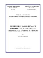
Table (1) shows the effects of fenugreek
leaves and seeds on the liver function tests in
CCl4 treated rats. The results of this study
showed variations in the level of liver
function tests in CCl4 treated rats. The ALP
level was significantly decreased in control
(P<0.05) and fenugreek group (P<0.01),
modified Harvard style but there were no
statistical difference of ALP level in
fenugreek leave group when compared to the
CCl4 treated rats,. Also, AST levels were
significantly decreased (P<0.001) in control,
both of fenugreek groups when compared to
the CCl4 treated rats.
Moreover, it revealed that in all treated
groups, serum ALT levels were decreased
significantly (P≤ 0.001) compared with CCl4
treated rats. With respect to direct bilirubin
level, control, also both of fenugreek treated
groups were significantly decreased (P≤
0.001) compared to CCl4 treated rats.
Results of the current data showed the
increase in ALP, AST, ALT and bilirubin
levels in CCl4 treated groups are in agreement
with (Girish and Pradhan, 2012). The
mechanism of hepatic damage by CCl4 is
well documented by Buege and Aust (1978)
they were reported that CCl4 is metabolized
by Cytochrome P450 enzyme to (CCl3). This
in turn reacts with molecular oxygen and gets
converted to trichloromethyl peroxy radical.
This radical forms covalent bonds with
sulfhydryl groups of several membrane
molecules like GSH leading to their depletion
and causes lipid peroxidation. The lipid
peroxidation initiates a cascade of reactions
leading to liver necrosis. Liver damage is
detected by measuring the activities of liver
function marker enzymes like AST, ALT and
ALP, which are released into the blood from
damaged cells. They are also indicators of
liver damage (Meera et al., 2009).
Our results showed that extract of fenugreek
can prevent the CCl4 induced toxicity in the
liver by significantly reduction of AST, ALT,
ALP and direct bilirubin levels, these results
are in agreement with (Meera et al., 2009)
they achieved that the normalization of the
above enzyme levels in rat liver with the plant
drugs estabilishes the hepato protective effect
of T. foenum-graecum which may be able to
induce accelerated regeneration of liver cells
reducing the leakage of these enzymes into
the blood. The results indicated that fenugreek
significantly prevented the increased liver
function marker enzyme activity induced by
CCl4, indicating an improvement of the
functional status of the liver by the fenugreek.
Effect of Fenugreek seed and leave extracts
on the some hematological parameters in
carbon tetrachloride treated rats
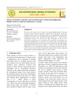
The results showed (Table 2) that WBC count
significantly decreased in fenugreek seeds (P≤
0.001), but there were no statistical
differences in control, fenugreek leaves when
compared with CCl4 treated rats. Moreover,
number of LYM significantly decreased in
fenugreek seeds (P≤ 0.05), while there were
no significant differences in control,,
fenugreek leaves when compared with CCl4
2332
Int.J.Curr.Microbiol.App.Sci (2017) 6(4): 2328-2337
treated group. Furthermore, the PLT count
significantly decreased in control, fenugreek
(P≤ 0.01), and, fenugreek leaves (P≤ 0.05)
when compared with CCl4 treated rats.
The present study showed that the rats treated
with fenugreek significantly decreased WBC,
LYM and PLT when compared with CCl4
treated rats.
Effect of fenugreek seed and leave extracts on
the liver super oxide dismutase and liver
glutathione levels in carbon tetrachloride
treated rats
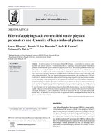
As shown in table (3), the level of liver GSH
in fenugreek groups significantly increased
(P≤ 0.001), but there was no statistical
difference of liver GSH level in control when
compared to CCl4 treated group. Also, liver
SOD significantly increased in control (P≤
0.001), fenugreek seeds and leaves (P≤ 0.05)
Glutathion (GSH) is the most important of the
sulfur-containing non-enzymatic antioxidant
molecules. GSH can also conjugate with free
radicals directly, earmarking them for renal
excretion, which is especially important for
dealing with the products of hepatic
cytochrome P450 enzyme activity. The
sulfhydryl (–SH) portion of the GSH can be
used to reduce a variety of free radicals in a
reaction catalyzed by the antioxidant enzyme,
glutathione peroxidase (Webb and Twedt,
2008).
In this study, the GSH level was significantly
increased in fenugreek treatment. This is in
agreement with (Sushma and Devasena,
2010), they showed that administration of
fenugreek seed extract minimized the effects
of ethanol in tissues. The beneficial effects of
fenugreek seeds are well demonstrated by
their ability to improve antioxidant status
thereby lowering lipid peroxidation. In vitro
investigations revealed that the aqueous
extract of fenugreek seeds effectively
inhibited the production of TBARS in the
presence of promoters of lipid peroxidation.
In this manner, the effect of fenugreek
aqueous extract was comparable with αtocopherol (Thirunavukkarasu et al., 2003).
Table.1 Effect of fenugreek seed and leaves treatments on liver function test in
CCI4-liver injury rats
Groups
S. ALP (U/L) S. AST(U/L)
S. ALT(U/L)
S.D. Bilirubin (mg/dL)
CCl4
Control
Fenugreek leave extract
Fenugreek seed extract
326±25.59
243.4±27
280.4±10.41
230±17.45
763.8±98.49
53.4±6.47
45.33±1.55
41.88±2.6
0.09625±0.006
0.026±0.002
0.02733±0.004
0.0295±0.005
812.3±91.03
196.4±35.68
131.3±15.31
146.4±19.84
Table.2 Effect of fenugreek seed and leave extracts on the some hematological
parametersinCCI4-in liver in jury rats
Groups
CCl4
Control
Fenugreek leaves
Fenugreek seeds
WBC *103/μL
9.623±0.34
8.3±0.7
7.2±0.55
4.75±0.95
LYM *103/μL
6.033±0.12
4.65±0.15
4.533±0.27
3.75±0.55
2333
PLT*103/μL
915.4±16.91
522±117.5
582.8±47.78
536.6±122.1
Int.J.Curr.Microbiol.App.Sci (2017) 6(4): 2328-2337
Table.3 Effect of fenugreek seed and leave extracts on GSH and SOD in CCI4- liver injury
rats
Groups
CCl4
Control
Fenugreek leaves
Fenugreek seeds
GSH (μmol)
13.33±0.7
25.19±1.33
108.2±4.33
130.2±8.71
SOD
0.03576±0.0112
0.2804±0.03531
0.2358±0.04062
0.1802±0.05225
Fig.1 Standard curve of superoxide dismutase (SOD )
The antibacterial Activity of Fenugreek Leave and Seed watery and alcoholic extracts,
inhibition zone measured in millimeter and percentage of inhibition
Types of bacteria
E. coli
Staphylococcusaureus
Bacillus subtillus
Conce
ntratio
n
0.01
Leaves
watery
extracts
7(44%)
Leaves
alcoholic
extracts
14(86%)
Seeds watery
extracts
Seeds alcoholic
extracts
7(44%)
13.5(86.5%)
0.02
6(43%)
14(86%)
7(43%)
13(87%)
0.1
0.2
50%
50%
100%
100%
45%
46%
100%
100%
0.5
0.01
0.02
60%
8(46%)
11(66%)
100%
15(85%)
14(86%)
50%
8(46%)
10(65%)
100%
14(86%)
13(87%)
0.1
0.2
0.5
0.01
0.02
0.1
68%
72%
80%
6(44%)
6 (44%)
50%
100%
100%
100%
12(88%)
11.5(88.5%)
100%
60%
60%
62%
6(42%)
6(42%)
42%
100%
100%
100%
11(89%)
13(90%)
100%
0.2
0.5
50%
60%
100%
100%
44%
45%
100%
100%
2334
Int.J.Curr.Microbiol.App.Sci (2017) 6(4): 2328-2337
The present study demonstrated that the
activity of liver SOD was significantly
enhanced by the presence of fenugreek seeds
extracts. The mechanism of enhancement was
observed by Joshi et al., (2014). They
conclude that the depleted enzymatic and
non-enzymatic anti-oxidants of diabetic rats
were restored significantly with the treatment
of fenugreek. Such effects may be mediated
through the active phytoconstituents present
in fenugreek, like 4-hydroxy isoleucine,
diosgenin, orientin, quercetin. These active
constituents can scavenge, or neutralize the
free radicals or other ROS components (Baig
et al., 2012; Punitha et al., 2005).
bilirubin, but the current seeds and leaves
lowered these levels.
From this study we support the use of
alcoholic fenugreek seeds and leaves extract
was more active against the pathogenic
bacteria than the watery fenugreek leaves
extract and it may have a role in the treatment
of some infectious diseases. This is in
agreement with R. Chalghoumi et al., (2016)
they conclude that antibacterial effect was
demonstrated by the aqueous extract of
fenugreek seeds; however, Iyer et al., (2004)
they concluded that the organic extracts
prepared with chloroform, acetone or
methanol showed low to moderately high
growth inhibitory effect (8.33 mm ≤ IZ ≤ 20
mm) when tested at a concentration equal to
or above 5 mg/ml (24)140p.
References
In conclusion, from the present study, the
following results can be concluded:
From the biochemical and physiological
points of view, the model of CCl4 caused
several changes in the level of the oxidative
parameters, decreasing of GSH but fenugreek
seed and leaves were succeeded in attenuating
these changes when added to the CCl4 treated
group and have shown hepatic protective
effect by increasing the liver SOD levels
The model produced oxidative stress and
rising in the levels of AST, ALT, ALP, direct
Fenugreek seeds and leaves ameliorated
inflammation caused by CCl4 treatment via
decreasing of WBC and LYM count.
Moreover, it decreased thrombogenic activity
of CCl4 through decreasing of PLT count
From this study we support the use of
alcoholic fenugreek seeds and leaves extract
was more active against the pathogenic
bacteria than the watery fenugreek leaves
extract and it may have a role in the treatment
of some infectious diseases.
Abboud, G. & Kaplowitz, N. 2007. Druginduced liver injury. Drug Safety, 30, P.
277-294.
Albayati, F.H., Taiyeb, T.B., Abdulla, M.A.
and Mahmud, Z.B. 2011. Antibacterial
effects of oradexm gengidil and
salviathymol-n mouth wash on dental
biofilm bacteria. African. J. Microbial.,
5(6): 636-642.
Amalraj, A., Balasubramanian, A., Edwin, E.,
Sheeja, E. 2005. Antimicrobial activity
of petroleum ether and chloroform
extracts of fenugreek seeds, Ind. J. Nat.
Prod., 21(2): 35-36.
Amini,
F.G.,
Rafieian-kopaei,
M.,
Nematbakhsh, M., Baradaran, A. &
Nasri, H. 2012. Ameliorative effects of
metformin on renal histologic and
biochemical alterations of gentamicininduced renal toxicity in Wistar rats. J.
Res. Med. Sci., 17.
Bahmani, M., Golshahi, H., Saki, K.,
Rafieian-kopaei, M., Delfan, B. &
Mohammadi, T. 2014a. Medicinal
plants and secondary metabolites for
diabetes mellitus control. Asian Pacific
J. Trop. Dis., 4; S687-S692.
Bahmani, M., Shirzad, H., Majlesi, M.,
Shahinfard, N. & Rafieian-kopaei, M.
2335
Int.J.Curr.Microbiol.App.Sci (2017) 6(4): 2328-2337
2014b. A review study on analgesic
applications of Iranian medicinal plants.
Asian Pacific J. Trop. Med., 7: P. S43S53.
Baig, M.A., Gawali, V.B., Patil, R.R. & Naik,
S.R. 2012. Protective effect of
herbomineral formulation (Dolabi) on
early
diabetic
nephropathy
in
streptozotocin-induced diabetic rats. J.
Nat. Med., 66: 500-509.
Basu, T.K., Srichamroen, A., Ronald Ross,
W. & Victor, R. 2010. Health Benefits
of Fenugreek (Trigonella foenumgraecum leguminosse. Bioactive foods
in promoting health, P. 425-435.
Belguith-hadriche, O., Bouaziz, M., Jamoussi,
K., El Feki, A., Sayadi, S. & Makniayedi, F. 2010. Lipid-lowering and
antioxidant effects of an ethyl acetate
extract of fenugreek seeds in highcholesterol-fed rats. J. Agric. Food
Chem., 58: P. 2116-2122.
Buege, J.A. & Aust, S.D. 1978. Microsomal
lipid peroxidation. Methods Enzymol.,
52: 302-310.
Bukhari, S.B., Bhanger, M.I. & Memon, S.
2008. Antioxidative Activity of Extracts
from Fenugreek Seeds (Trigonella
foenum graecum. Pak. J. Anal. Environ.
Chem., 9: P.78-83
Chalghoumi, R., S. MabroukH. Abdoul and
J.E. Line. 2016. Antibacteria lActivityof
Fenugreek Seeds (Trigonella foenumgraecum) Crude Extracts Against a
Rabbit Escherichia coli Isolate
Chowdhury, P., Soulsby, M., Pasley, J.,
Mckay, D. & Bansal, S. 2013. Effects of
Dietary Soy Protein on Hematological
and
Tissue
Oxidant/Anti-Oxidant
Levels in Rats Exposed to Simulated
Microgravity. J. Physical Chem.
Biophysics.
Edziri, H., Mastouri, M., Mahjoub, M.,
Ammar, S., Mighri, Z., Gutmann, L. &
Aouni, M. 2011. Antiviral activity of
leaves extracts of Marrubium alysson L.
J. Med. Plants Res., 5, P. 360-363.
Girish, C. & Pradhan, S.C. 2008a. Drug
development for liver diseases: focus on
picroliv, ellagic acid and curcumin.
Fundam. Clin. Pharmacol., 22: P.623632.
Girish, C. & Pradhan, S.C. 2012.
Hepatoprotective activities of picroliv,
curcumin, and ellagic acid compared to
silymarin
on
carbon-tetrachlorideinduced liver toxicity in mice. J.
Pharmacol. Pharmacother., 3: P.14955.
Iyer, M., Belapurkar, H., Sherikar, O.,
Kasture, S.B. 2004. Anxiolytic activity
of Trigonellafoenumgraecum seeds. J.
Nat. Rem., 4(1): 61-65.
Joshi, D.V., Patil, R.R. & Naik, S.R. 2014.
Hydroalcohol extract of Trigonella
foenum-graecum
seed
attenuates
markers of inflammation and oxidative
stress while improving exocrine
function in diabetic rats. Pharm. Biol.,
P.1-11.
Kader, M., EL-Sayed, E., Kassem, S.,
Mohamed, H. & Eldin, S. 2014.
Protective and antioxidant effects of
cynarascolymus leaves against carbon
tetrachloride toxicity in rats. Res. J.
Pharm. Bio Chem. Sci., 5: P.1373-80.
Kipkore, W., Wanjohi, B., Rono, H. & Kigen,
G. 2014. A study of the medicinal
plants used by the Marakwet
Community in Kenya. J. Ethnobiol.
Ethnomed., 10: P.24.
Meera, R., Devi, P., Kameswari, B.,
Madhumitha, B. & Merlin, N.J. 2009.
Antioxidant
and
hepatoprotective
activities of Ocimum basilicum Linn.
and Trigonella foenum-graecum Linn.
against H2O2 and CCL4 induced
hepatotoxicity in goat liver. Indian J.
Exp. Biol., 47: P.584-90.
Moron, M.S., Depierre, J.W. & Mannervik, B.
1979. Levels of glutathione, glutathione
reductase and glutathione S-transferase
2336
Int.J.Curr.Microbiol.App.Sci (2017) 6(4): 2328-2337
activities in rat lung and liver.
Biochimica et Biophysica Acta (BBA)General Subjects, 582, P.67-78.
Punitha, I., Rajendran, K., Shirwaikar, A. &
Shirwaikar, A. 2005. Alcoholic stem
extract of Coscinium fenestratum
regulates carbohydrate metabolism and
improves
antioxidant
status
in
streptozotocin–nicotinamide
induced
diabetic
rats.
Evidence-Based
Complementary and Alternative Med.,
2, P.375-381.
Sewell, R.D. & Rafieian-kopaei, M. 2014.
The history and ups and downs of
herbal medicines usage. J. Herb Med.
Pharmacol., 3.
Sushma, N. & Devasena, T. 2010. Aqueous
extract of Trigonella foenum graecum
(fenugreek) prevents cypermethrin-
induced
hepatotoxicity
and
nephrotoxicity. Hum. Exp. Toxicol., 29,
P. 311-9.
Thirunavukkarasu, V., Anuradha, C. V. &
Viswanathan, P. 2003. Protective effect
of fenugreek (Trigonella foenum
graecum) seeds in experimental ethanol
toxicity. Phytother. Res., 17: P.737-43.
Wang, X. & Liu, Z. 2013. Prevention and
treatment of viral respiratory infections
by traditional Chinese herbs. Chinese
Med. J., 127, P.1344-1350.
Webb, C. & Twedt, D. 2008. Oxidative stress
and liver disease. Vet. Clin. North
America: Small Animal Practice, 38,
P.125-135.
Yamamoto, Y.Y. 2000. Free radicals in
chemistry, biology and medicine, OICA
International (UK).
How to cite this article:
Firdaws A. AL-Mashhadani. 2017. Effect of Fenugreek Seed and Leaves on Some
Hematological and Biochemical Parameters in CCl4-induced Liver Injury.
Int.J.Curr.Microbiol.App.Sci. 6(4): 2328-2337. doi: />
2337