It is no longer the time to disregard thyroid metastases from breast cancer: A case report and review of the literature
Bạn đang xem bản rút gọn của tài liệu. Xem và tải ngay bản đầy đủ của tài liệu tại đây (4.54 MB, 8 trang )
Pensabene et al. BMC Cancer (2018) 18:146
DOI 10.1186/s12885-018-4054-x
CASE REPORT
Open Access
It is no longer the time to disregard thyroid
metastases from breast cancer: a case
report and review of the literature
Matilde Pensabene1*, Brigida Stanzione2, Ivana Cerillo1, Giuseppe Ciancia3, Immacolata Cozzolino3,
Raffaella Ruocco1, Caterina Condello1, Giuseppe Di Lorenzo1, Mario Giuliano1, Valeria Forestieri1, Grazia Arpino1,
Sabino De Placido1 and Rossella Lauria1
Abstract
Background: Metastases to the thyroid gland are more frequent than previously thought, although most of them
are occult or not clinically relevant. Overall, only 42 cases of metastases to thyroid from breast cancer have been
reported thus far. Here we report the case of a patient with breast cancer metastatic to the thyroid. We also review
the 42 previously reported cases (published between 1962 and 2012). This is the first review about metastases to
thyroid gland from breast cancer.
Case presentation: A 64-year-old woman of Caucasian origin was diagnosed with a lobular invasive carcinoma of
the breast (luminal A, stage II). She received adjuvant chemotherapy, followed by endocrine therapy. During followup, fine-needle cytology of a thyroid nodule revealed malignant cells that were estrogen-positive, which suggested
a diagnosis of metastases to the thyroid. Imaging did not reveal any other metastatic site and showed only
enlargement of the left thyroid lobe and an inhomogeneous pattern of colloid and cystic degeneration and
calcifications. The patient underwent left hemithyroidectomy. Histology of thyroid tissue showed a colloid goitre
containing dispersed small atypical neoplastic cells with eccentric nuclei. Immunohistochemistry showed
cytokeratin-19 and oestrogen receptor, but not tireoglobulin, e-cadherin or cytokeratin-7, thereby confirming
metastases from a lobular breast carcinoma. Hormonal treatment is ongoing.
Conclusion: This case report and first review of the literature on metastases to thyroid from breast cancer highlight
the importance of a correct early diagnostic work-up in such cases. Indeed, a primary lesion should be
distinguished from metastases given the different treatment protocol related to primary cancer and the clinical
impact on prognosis.
Keywords: Breast cancer, Metastases to thyroid, Lobular breast cancer, Goitre
Background
Metastatic cancer in the thyroid is uncommon and accounts for about 1.4–3% of malignant solid tumours [1–6].
The most frequent primary cancers are renal cell (48.1%),
colorectal (10.4%), lung (8.3%) and breast (7.8%) cancers,
and soft tissue sarcoma (4.0%) [1]. Large series reported
also lymphoma as primary cancer or metastases from lung
cancer other than usual epithelial thyroid cancers [7]. Also
* Correspondence:
1
Department of Clinical Medicine and Surgery, University of Naples “Federico
II”, via Sergio Pansini, 80131 Naples, Italy
Full list of author information is available at the end of the article
parotid cancer and melanoma have been reported as primary cancers [8, 9]. Formerly, metastases to the thyroid
were usually identified at autopsy [9]. Thanks to the advent
of more accurate diagnostic methods, it is now possible to
clinically diagnose metastases to the thyroid, and initiate
timely surgical and systemic treatment thereby improving
outcome [2].
Case report
We report the case of a 64-year-old woman who was diagnosed with left breast cancer in June 2011. The comorbidities were multinodular goitre of the thyroid
© The Author(s). 2018 Open Access This article is distributed under the terms of the Creative Commons Attribution 4.0
International License ( which permits unrestricted use, distribution, and
reproduction in any medium, provided you give appropriate credit to the original author(s) and the source, provide a link to
the Creative Commons license, and indicate if changes were made. The Creative Commons Public Domain Dedication waiver
( applies to the data made available in this article, unless otherwise stated.
Pensabene et al. BMC Cancer (2018) 18:146
gland, obstructive pulmonary disease (i.e. emphysema), left atrial enlargement with severe pulmonary
hypertension, carotid stenosis (60–65%) and severe
obesity. Clinical staging by chest X-ray, abdominal
ultrasound and bone scan was negative, except for
CEA = 10.7 (normal value < 5). The patient underwent wide excision of the breast lesion with axillary
node dissection. Histology revealed a G2 invasive
lobular carcinoma with lymph node metastasis, stage
pT1cN3 (21/24 nodes), with positive staining for
oestrogen receptor (ER 90%) and progesterone receptor (PgR 80%), a very low proliferation index (Ki67 <
10%), and without amplification of HER2-neu, suggesting a luminal A phenotype. Adjuvant chemotherapy with docetaxel and cyclofosfamide was
administered, followed by endocrine therapy with
aromatase inhibitor. At the first follow-up, clinical
examination showed enlarged thyroid lobes with a
multinodular structure; a thyroid function test was
normal. Ultrasound revealed an enlarged thyroid with
retrosternal development; the glandular structure was
finely inhomogeneous in the right lobe, and the entire left lobe was occupied by a large nodule that
had a mixed echo-pattern and areas of cystic and
colloid degeneration and calcifications.
Fine needle cytology was performed with a 23-G
needle without suction. Smears taken from both lobes
were Diff-Quik-stained and evaluated on-site. Cellularity was deemed to be satisfactory, and additional
smears were taken and alcohol-fixed for Papanicolaou
stain and for ancillary techniques. The right lobe
smear was moderately cellular and showed only colloid and benign thyrocytes. Conversely, the left lobe
smears revealed a second cellular population in a colloidal background mixed with small groups of benign
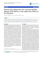
thyrocytes (Fig. 1A). This second population was constituted by small cells, dispersed or aggregated into
small, loosely formed groups; individual cells had a
plasmacytoid-like aspect (Fig. 1A, inset) and occasionally a secretion vacuole. Given the patient’s history,
we decided to use two smears fixed in alcohol to
evaluate oestrogen receptor expression in the second
cell population. Immunocytochemistry revealed positive oestrogen receptor staining (Fig. 1B), which suggested a diagnosis of metastases of the breast cancer
to the thyroid. Positron emission tomography and
total body tomography did not reveal other metastatic
sites, and showed only enlargement of the left thyroid
lobe and an inhomogeneous pattern of colloid and
cystic degeneration and calcifications. Therefore, the
patient underwent left hemithyroidectomy in February
2012. Histology revealed thyroid tissue with a colloid
goitre containing dispersed neoplastic cells constituted
by small atypical cells with eccentric nuclei (Fig. 1C).
Page 2 of 8
Fig. 1 a Cytology of thyroid metastases and plasmacytoid-like aspect
(inset). b Immunocytochemistry with positive estrogen receptor
staining. c Histology of thyroid metastases
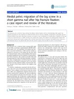
Immunohistochemistry revealed cytokeratin-19 and
oestrogen receptor (Fig. 2 a, b), but not tireoglobulin,
e-cadherin or cytokeratin-7, thereby suggesting metastases from a lobular breast carcinoma. Thirty-two
months after hemithyroidectomy, the patient is alive,
although in May 2014, there was evidence of recurrence in bone. Hormonal treatment with fulvestrant is
ongoing. She died in July 2015.
Pensabene et al. BMC Cancer (2018) 18:146
Page 3 of 8
Fig. 2 Immunohistochemistry showing cytokeratin 19 (a) and estrogen receptor (b)
Discussion and conclusions
Methods of review
We searched PubMed to identify studies about metastases to thyroid from different primary tumours, including
breast cancer. Searches were made using the terms
‘breast cancer’ and ‘metastases to thyroid’, with no limitation of language, publication date, or journal of publication. Eighteen articles were eligible according to our
criteria; these were published between 1962 and 2012.
Given the rarity of metastases to the thyroid and the
limited number of reported cases, we performed only a
descriptive analysis.
Epidemiology
Metastases to the thyroid gland are rare, but not as
rare as previously thought. This is not surprising because the thyroid gland is the second most richly
arterialized organ in the body. The probability of
finding metastases in the thyroid gland depends on
the method of investigation [3]. Large autopsy studies
found that the incidence of thyroid metastases in patients with a history of cancer ranges from 1.9% to 24%
[1, 3, 8, 9]. Two of these studies suggested that thyroid
metastases are more common than primary thyroid cancer [1, 3]. On the other hand, the incidence of thyroid metastases in clinical and surgical series was 3% [4]. Reports
of thyroid metastases have increased in recent years consequent to more sophisticated diagnostic methods, i.e. fine
needle cytology and proton emission tomography with
18F–fluorodeoxyglucose [1, 10].
In autopsy series, breast cancer, lung cancer and melanoma were found to be the most frequent malignancies
to metastasize to the thyroid [11]. Clinical and surgical
series of patients showed that breast carcinoma is the
second most common primary tumour to result in
symptomatic thyroid metastases, the first being clear cell
renal cancer [5, 11, 12].
Clinical and pathological presentation
The characteristics of breast cancer patients with thyroid
metastases are reported in Table 1. We analyzed sex, age
at diagnosis, histology, primary treatment, treatment failure, time between primary diagnosis and thyroid metastases and follow-up in 42 women with thyroid
metastases from breast cancer reported between 1962
and 2012 [1–20]. The development of metastases of the
thyroid gland does not appear to be age-dependent, and
seems to be more frequent in women [1]. The median
age at diagnosis of metastases to the thyroid gland is
51 years (range: 22–83 years) [9, 12]. Time-to-detection
and time from presentation to death differ among reports. The former ranges from 2 months to more than
15 years after the diagnosis of the primary cancer [3, 5],
and the latter from 1 to 34 months [13]. In one case,
thyroid metastases were synchronous to primary breast
cancer [10].
As shown in Table 1, the clinical presentation of
thyroid metastases is very heterogeneous. They are
clinically evident only in a minority of patients and
are frequently found incidentally during postoperative
follow-up by ultrasonography. Thyroid metastases
usually present in the context of widespread metastatic disease, and manifestations in the thyroid are
not clinically significant. On the other hand, when
thyroid metastases are the first presentation of recurrent disease, they usually appear as a palpable neck
mass or, albeit less often, with dysphagia, massive tracheal involvement or dysphonia. Often, patient presented with painless neck mass [21].
In the reports containing histological information,
breast cancer is generically referred to as “adenocarcinoma” [3, 14, 15]. Where indicated, the most prevalent
breast cancer is ductal infiltrating carcinoma [6, 8, 10],
reported in seven cases, while invasive lobular carcinoma
was reported in only our case and in a case described by
Egana et al. [12]. Not all the studies reviewed reported
the site of recurrence. In six cases. The thyroid was the
first and only site of recurrence [2–4, 6, 13]. As shown
in Table 1, other sites of widespread disease were lung,
liver and bone. In eleven cases, breast cancers recurred
in different sites, but the metastatic site other than thyroid was not reported [5, 14, 16].
1985–1994
1995–2000
1982–2002
Nakhjavani MK et
al. 1997 [5]
Chung SY et al.,
2001 [17]
De Ridder M et al.
2003 [13]
1
1985–2002
2005
1997–2003
1989–2004
1984–2003
Wood K et al.,
2004 [3]
Owens CL et al.,
2005 [19]
Kim TY et al. 2005
[10]
Gerges AS et al.
2006 [8]
Cichon A et al.,
2006 [2]
1
1
5
1
1
Loo CK, Burchett
IJ, 2003 [18]
1
6
7
2
1974–1976
3
Pillay SP et al.,
1977 [29]
1907–1962
Wychulis AR et al.,
1964 [14]
1
2
1955
Shimaoka K et al.
1962 [9]
No of pts
Harcourt-Webster
JN, 1965 [15]
Study yrs
Authors
51
32
22
33
F
F
F
F
44
55
45
F
F
F
F
50
45
34
F
36
F
64
72
F
F
F
52
61
F
F
49
F
67
58
F
F
50
58
F
F
51
F
56
51
F
38
F
44
Age
F
F
Sex
ductal
ductal
ductal
ductal
ductal
ductal
NR
adenocarcinoma
neuroendocrine
carcinoma
NR
NR
adenocarcinoma
adenocarcinoma
adenocarcinoma
adenocarcinoma
adenocarcinoma
adenocarcinoma
adenocarcinoma
carcinoma
Histology
S
S + CT + RT
CT
CT
CT
CT
CT
HT
HT
NR
S
S
S + RT
S + RT
S
CT
Primary treatment
Table 1 Characteristics of breast cancer patients with metastases to thyroid in a clinical series
No
No
No
No
No
No
No
No
NR
no
no
NR
Yes
no
no
no
bone
Neck LN, lung, bone
lung, ax LN, neck
Nil
lung, ax LN, scal
Neck LN, lung
shoulder subcutaneous
nodule, liver
no
bone
no
bone, peritoneum
lung
lung, liver
lung, bone, liver
lung
lung, bone
NR
yes
liver, bone
NR
NR
lung, bone
LN, bone, liver, skin, CNS
Treatment failure
Local Distant
10 yrs
63 months
85 months
68 months
37 months
25 months
18 months
5 yrs
15 yrs
8 yrs
0 months
from 2 months to 22 yrs
Synchronous (0 months)
3 yrs
13 yrs
5 yrs
Time between BC diagnosis
and thyroid metastasis
24 months alive
37 months alive
post-thyroidectomy
8 months alive
26 months alive
4 months alive
17 months alive
6 months alive
36 months alive
Died after 2 years for
disseminated disease
3 patients < 3 months
4 pts. 3–23 months
1 yr. lost to FU
5 months lost to FU
4 yrs. died of the disease
4 months
Follow-up
Pensabene et al. BMC Cancer (2018) 18:146
Page 4 of 8
2005
1993–2003
1995–2005
NR
2007
2011
Molina Garrido MJ
et al., 2006 [11]
Papi G et al., 2007
[6]
Calzolari F et al.,
2008 [4]
Saber A et al.
2007 [20]
Egaña N et al.,
2012 [12]
Current report
1
1
1
1
5
1
No of pts
F
F
F
64
NR
F
83
NR
F
ILC
NR
NR
NR
NR
F
F
NR
F
NR
Histology
ductal
43
Age
F
F
Sex
S + CT + RT
mastectomy,
lymphadenectomy
HT
S + CT
mastectomy,
lymphadenectomy
CT + RT
Primary treatment
No
No
NR
No
No
No
bone
bone, liver
and pelvic mass
no
no
no
lung, liver
Treatment failure
Local Distant
6 months
3 yrs
60 (range: 13–135 months)
30 months
Time between BC diagnosis
and thyroid metastasis
18 months alive
1 month
60 months
died of the r disease
5 yrs. 1 alive
1 month
Follow-up
F female, CT chemotherapy, FU follow-up, LN nodes, yrs. years, HT hormonal treatment, S surgery, RT radiotherapy, NR not reported, ILC infiltrating lobular carcinoma, CNS Central Nervous System
Study yrs
Authors
Table 1 Characteristics of breast cancer patients with metastases to thyroid in a clinical series (Continued)
Pensabene et al. BMC Cancer (2018) 18:146
Page 5 of 8
Pensabene et al. BMC Cancer (2018) 18:146
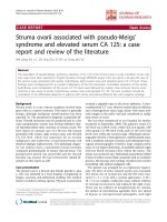
Diagnostic work-up
Figure 3 shows a diagnostic and therapeutic work-flow
of patients with suspected metastases to the thyroid
from breast cancer. Thyroid nodules in a patient with a
history of malignancy should be investigated particularly
if they appear many years after the primary tumour. In
fact, a malignant thyroid nodule in such patients is
much more likely to be metastatic than a new primary
tumour. Thyroid metastases cannot be differentiated
from a primary thyroid cancer based on clinical features, therefore fine needle cytology must be included
in the diagnostic work-up, particularly given its low
morbidity and cost, and its high negative predictive
value [22–24]. Cytology generally shows abundant cellularity and the cells may be typical of the original
site, especially when specific immunohistochemical
stains are used. Negative staining with antithyroglobulin and anti-calcitonin antibodies would
favour a diagnosis of metastatic tumour.
Page 6 of 8
When papillary and follicular carcinomas have a complex
pathological pattern, cytology alone cannot reveal the origin of the metastatic tumour. The diagnosis is particularly
difficult in case of less common primary thyroid cancers
such as small cell, giant cell and spindle cell carcinomas,
anaplastic cancer and the clear cell variant of follicular carcinoma. Therefore, biopsy is needed to reach a definitive
diagnosis. In all the series reported so far, the diagnosis was
confirmed cytologically and histologically [1]. Regarding
the cytological differential diagnosis, a non-cohesive cell
population and a plasmacytoid-like aspect can mimic a
medullary carcinoma of the thyroid. In medullary carcinoma, cytological smears are usually more cellular in a background without colloid, and frequently contain amorphous
material consistent with amyloid. Tumour cells are predominantly isolated, but clusters and rosettes may also be
seen. Cells have a plasmocytoid appearance and are uniform in size and shape with moderate or abundant, finely
granular cytoplasm and eccentrically placed nuclei. Many
Fig. 3 Diagnostic work-up and treatment of patient with thyroid metastases from breast cancer
Pensabene et al. BMC Cancer (2018) 18:146
smears show large cells with nuclear megaly, and binucleated and multinucleated cells. These aspects were not
observed in our patient. Indeed, in our case, the history of
breast cancer, the absence of typical findings of papillary or
follicular carcinoma, positive staining of oestrogen and
progesterone receptors, negative staining of both thyroglobin and calcitonin, and the histological pattern of the primary and metastatic tumour enabled us to establish a
diagnosis of metastases from thyroid. In particular, the following immunocytochemical markers were analyzed: cytokeratin 7, cytokeratin 19, E-cadherin, CD34, besides
estrogen and progesterone receptors.
When thyroid metastases are found, it is important to
re-evaluate the diagnosis of the primary tumour and
search for other metastatic sites. Because breast cancer
has been associated with thyroid disease and because
thyroid nodules are more frequently found in women
[25], it is important to examine the thyroid during breast
ultrasonography. Ultrasonography generally shows lesions with irregular shape and inhomogeneus [21]. At
computer tomography, metastases to thyroid are hypodense; while they look iso-hyperintense in comparison
to the normal thyroid tissue at magnetic resonance imaging [21]. Thyroid examination in the work-up of patients with breast cancer and goitre or nodules can
reveal thyroid metastases in an early phase. The oncologist should consider that thyroid functional tests and
radioiodine uptake are normal in most patients [1].
Management of thyroid metastases
The treatment of thyroid metastases depends on the site
of the primary tumour, presence of other metastases and
symptoms caused by the thyroid mass. Surgery is considered the gold standard treatment for thyroid metastases. Radical treatment of an isolated metastasis to the
thyroid can be curative, and an aggressive surgical approach has been recommended especially in case of slow
growing tumours such as breast or kidney carcinomas
[3]. The extension of surgical resection does not seem to
significantly impact on survival. In fact, no significant
differences in survival were found between total thyroidectomy and conservative surgery [5]. Surgical treatment
of isolated metastasis may prolong survival [5]. However,
more data are necessary regarding the best surgical approach in patients with a single thyroid metastasis [4].
Patients with single metastases to the thyroid should
be treated surgically, whereas patients with multiple metastases in different organs should be treated with a hormonal or chemotherapeutic approach in accordance
with international advanced breast cancer guidelines for
extensive disease [26]. For patients with metastatic sites
other than thyroid, surgery is generally performed to reduce pressure, which causes discomfort, and to avoid
airway obstruction and skin ulceration [4].
Page 7 of 8
Data concerning radiotherapy or chemotherapy for
metastatic disease are fragmentary and limited. Wychulis
et al. [14] reported that radiotherapy relieved symptoms,
and should thus be considered an option, particularly in
patients with high anaesthetic risk and a clinical condition that precludes surgery. Radioactive iodine 131I has
not been found to be effective [27].
Prognosis
Reports of thyroid metastases span over more than four
decades. It is not feasible to make a global evaluation of
the outcome of patients because of the heterogeneity of
treatments and some systemic therapies that have become
obsolete. However, numerous case reports suggest that metastases to the thyroid gland are associated with a poor
prognosis [28]. Multifocal metastases seem to adversely
affect prognosis. Indeed, a significantly worst survival has
been reported in patients with multiple foci. Survival after
surgical treatment is variable with some patients succumbing to metastatic disease within a few months, while others
have a long-term survival. Data on prognosis cannot be extrapolated also in view of the many advances made in systemic treatment and in the identification of distinctive
biological features of breast cancer from the first published
case in 1962 until now. In particular, the introduction of
taxanes as well as targeted therapies, such as trastuzumab
and pertuzumab for HER2-positive tumours and bevacizumab for HER2-negative tumours, which have had an enormous positive effect on outcome. There are no case
reports that describe the type of systemic treatment used,
and patients were often treated with surgery, so that outcomes cannot be extrapolated to define the prognosis of
breast cancer with metastases to the thyroid gland.
Conclusions
In this review and case report we examine aspects of
breast cancer metastases to the thyroid going from the
diagnostic workup to the treatment. Metastases to the thyroid gland can present many years after treatment of a distant primary tumour; however, in a patient with a history
of malignancy, a neoplastic thyroid nodule is more likely
to be a metastasis than a new thyroid malignancy. Fineneedle aspiration biopsy of the thyroid gland should be
performed in patients with breast cancer and a nodular
goitre, even in the absence of clinical signs of metastatic
disease. Biological features are important for treatment
decision-making. Given the availability of targeted biological therapies, i.e. transtuzumab, pertuzumab and bevacizumab, that modify the natural history of metastatic
breast cancer, it is no longer the time to disregard the thyroid metastases from breast cancer and other primary
malignancies.
The history of our patient suggested thyroid goitre and
showed no clinical feature suggestive of metastasis. Our
Pensabene et al. BMC Cancer (2018) 18:146
case report highlights that metastases, also such an unusual site as the thyroid, should be considered in patients diagnosed with breast cancer. Studies dealing with
thyroid metastases are very heterogeneous in terms of
the primary cancer, which makes it difficult to evaluate
the impact of thyroid metastases on prognosis. Most patients with thyroid metastases have widespread metastatic disease but occasionally the thyroid may be the
only site of disease. Although therapy of metastatic malignancies is often considered to be palliative, aggressive
surgical treatment in isolated cases may be curative and
may benefit survival. This highlights the importance of
early recognition and management of thyroid metastases
in prolonging survival in some patients and in preventing the onset of life-threatening complications.
Page 8 of 8
2.
3.
4.
5.
6.
7.
8.
9.
Acknowledgements
The authors thank Jean Ann Gilder (Scientific Communication) for editing
the manuscript.
Funding
No specific funding has been used for data collection, analyses, results
reporting or manuscript writing.
10.
11.
12.
Availability of data and materials
The datasets used and/or analysed during the current study are available
from the corresponding author on reasonable request.
13.
14.
Authors’ contributions
SB, PM, and RL give a relevant contribution to conception of the review and
interpretation of published data; SB, IC, RR and PM have involved in drafting
the manuscript. FV, GC and IC were involved in diagnostic flow. CC, MG have
been involved in patient follow-up. DPS, AG, GDL and LR have been involved
in revising the manuscript critically for important content. All the authors
read and gave their final approval of the version to be published.
Ethics approval and consent to participate
Not applicable.
Consent for publication
Written informed consent was obtained from the patient for the publication
of this case report and any accompanying images. A copy of the written
consent is available for review by the Editor-in-Chief of this journal.
Competing interests
The authors declare that they have no competing interests.
Publisher’s Note
Springer Nature remains neutral with regard to jurisdictional claims in
published maps and institutional affiliations.
Author details
1
Department of Clinical Medicine and Surgery, University of Naples “Federico
II”, via Sergio Pansini, 80131 Naples, Italy. 2Department of Medical Oncology,
National Cancer Institute, Aviano, PN, Italy. 3Department of Advanced
Biomedical Sciences, Pathology Unit, University of Naples “Federico II”, via
Sergio Pansini, 80131 Naples, Italy.
Received: 3 November 2016 Accepted: 25 January 2018
References
1. Chung AY, Tran TB, Brumund KT, Weisman RA, Bouvet M. Metastases to the
thyroid: a review of the literature from the last decade. Thyroid. 2012;22(3):
258–68.
15.
16.
17.
18.
19.
20.
21.
22.
23.
24.
25.
26.
27.
28.
29.
Cichon S, Anielski R, Konturek A, Barczynski M, Cichon W. Metastases to the
thyroid grand: seventeen cases operated on in an single clinical center.
Langenbeck's Arch Surg. 2006;391(6):581–7.
Wood K, Vini L, Harmer C. Metastases to the thyroid gland: the Royal
Marsden experience. Eur J SurgOncol. 2004;30(6):583–8.
Calzolari F, Sartori PV, Talarico C, Parmeggiani D, Beretta E, Pezzullo L, Bovo
G, Sperlongano P, Monacelli M, Lucchini R, Misso C, Gurrado A, D'Ajello M,
Uggeri F, Puxeddu E, Nasi P, Testini M, Rosato L, Barbarisio A, Avenia N.
Surgical treatment of intrathyroid metastases: preliminary results of a
multicentric study. Anticancer Res 2008 Sep-Oct;28(5B):2885–8.
Nakhjavani M, Gharib H, Coellner JR, van Heerden J. Metastases to the
thyroid gland, a report of 43 cases. Cancer. 1997;79:574–8.
Papi G, Fadda G, Corsello SM, Corrado S, Rossi ED, Radighieri E, Miraglia A,
Carani C, Pontecorvi A. Metastases to the thyroid gland: prevalence,
clinicopathological aspects and prognosis: a 10-year experience. Clin
Endocrinol. 2007 Apr;66(4):565–71.
Diaconescu MR, Costea I, Glod M, Grigorovici M, Diaconescu S. Unusual
malignant tumors of the thyroid gland. Chirurg. 2013;108:482–9.
Gerges AS, Shehata SR, Gouda IA. Metastasis to the thyroid gland: unusual
site of metastasis. J Egypt Natl Canc Inst. 2006;18(1):67–72.
Shimaoka K, Sokal JE, Pickren JW. Metastatic neoplasms in the thyroid gland.
Pathological and clinical findings. Cancer. 1962 May-Jun;15:557–65.
Kim TY, Kim WB, Gong G, Hong SJ, Shong YK. Metastasis to the thyroid
diagnosed by fine-needle aspiration biopsy. Clin Endocrinol. 2005, Feb;62(2):
236–41.
Molina Garrido MJ, Guillén Ponce C, Maciá Escalante S, Martínez Y Sevila C,
Carrato Mena A. Dysphagia and dysphonia in a woman with a previous
breast cancer. Clin Transl Oncol. 2006, Jul;8(7):533–5.
Egaña N, Socias C, Matteucci T, Bilbao I, Alvarez-Coca M. Thyroid metastasis
of lobular breast carcinoma. Endocrinol Nutr. 2012, Mar;59(3):219–20.
De Ridder M, Sermeus AB, Urbain D, Storme GA. Metastases to the thyroid
gland-a report of six cases. Eur J Intern Med. 2003;14(6):377–9.
Wychulis AR, Beahrs OH, Woolner LB. Metastasis of carcinoma of the thyroid
gland. Ann Surg. 1964 Aug;160:169–77.
Secondary H-WJN. Neoplasm of the thyroid presenting as a goiter. J Clin
Pathol. 1965;18:282–7.
Angorn IB, Baker LW. Tumour metastasis to the thyroid gland. S Afr Med J.
1977 Apr 9;51(15):509–12.
Chung SY, Kim EK, Kim JH, Oh KK, Kim DJ, Lee YH, An HJ, Kim JS.
Sonographic findings of metastatic disease to the thyroid. Yonsei Med J.
2001 Aug;42(4):411–7.
Loo CK, Burchett IJ. Fine needle aspiration biopsy of neuroendocrine breast
carcinoma metastatic to the thyroid. A case report. Acta Cytol. 2003
Jan-Feb;47(1):83–7.
Owens CL, Basaria S, Nicol TL. Metastatic breast carcinoma involving the
thyroid gland diagnosed by fine-needle aspiration: a case report. Diagn
Cytopathol. 2005 Aug;33(2):110–5.
Saber A, Ramzy S, Gouda I. Metastasis to the thyroid gland; unusual site of
metastasis. Gulf J Oncolog. 2007 Jan;1(1):51–7.
Surov A, Machens A, Holzhausen HJ, Spielmann RP, Dralle H. Radiological
features of metastases to the thyroid. Acta Radiol. 2016 Apr;57(4):444–50.
Chaco MS, Greenebaum E, Moussouris HF, Schreiber K, Koss LG. Value of
aspiration cytology of the thyroid in metastatic disease. Acta Cytol. 1987;31:
705–12.
Ménégaux F, Chigot JP. Les metastases thyroïdiennes. Ann Chir. 2001;126:
981–4.
Michelow PM, Leiman GM. Metastases to the thyroid gland: diagnosis by
aspiration cytology. Diagn Cytopathol. 1995;13:209–13.
Hardefeldt PJ, Eslick GD, Edirimanne S. Benign thyroid disease is associated
with breast cancer: a meta-analysis. Breast Cancer Res Treat. 2012 Jun;133(3):
1169–77.
/>(last accessed 31/01/2017).
Vini L, Harmer C. Radioiodine treatment for differentiated thyroid cancer.
Clin Oncol. 2000;12(6):365–72.
Lam KY, Lo CY. Metastatic tumours of the thyroid gland: a study of 79 cases
in Chinese patients. Arch Pathol Lab Med. 1998;122(1):37–41.
Pillay SP, Angorn IB, Baker LW. Tumour metastasis to the thyroid gland. S
Afr Med J. 1977;Apr;51(15):509–12.