Uterine rupture and its management in a queen cat
Bạn đang xem bản rút gọn của tài liệu. Xem và tải ngay bản đầy đủ của tài liệu tại đây (197.9 KB, 3 trang )
Int.J.Curr.Microbiol.App.Sci (2020) 9(5): 442-444
International Journal of Current Microbiology and Applied Sciences
ISSN: 2319-7706 Volume 9 Number 5 (2020)
Journal homepage:
Case Study
/>
Uterine Rupture and its Management in a Queen Cat
A. Sabarinathan*, S. Rangasamy, J. Umamageswari, U. S. Kalyaan and Pradeep Nag
Department of Veterinary Gynaecology and Obstetrics
Madras Veterinary College, Chennai-7, Tamil Nadu, India
*Corresponding author
ABSTRACT
Keywords
Cat, uterine rupture,
caesarean section
Article Info
Successful management of life threatening case of uterine rupture with
emphysematous fetus by caesarean section in a queen cat was reported.
Accepted:
05 April 2020
Available Online:
10 May 2020
Introduction
Case history and observations
Uterine rupture is a major emergency which
occur during late pregnancy (Roberts, 1986).
The most common causes of uterine rupture is
external trauma during pregnancy, severe
alterations of the uterine wall, improper
obstetrical procedures and indiscriminate use
of drugs such as oxytocin or prostaglandin F2
alpha (Jackson, 1995). This paper describes
about emergency caesarean section in a queen
cat following diagnosis of uterine rupture and
presence of two emphysematous fetuses in the
abdominal cavity with the help of
ultrasonographic and radiographic aids.
A Three years old, domestic short hair queen
cat in lateral recumbency was presented to
Small Animal Obstetrics and Gynecology
ward of Madras Veterinary College Teaching
Hospital with the history of having queened
three live kittens on the previous day morning
and anorexia for past two days.
Clinical examination revealed distended and
tensed abdomen. Vaginal examination
showed edematous vulval lips with oozing of
blood and no foetus palpable in the vaginal
passage.
442
Int.J.Curr.Microbiol.App.Sci (2020) 9(5): 442-444
Further the case was subjected to diagnostic
aids such as ultrasound scanning and
radiographic examination of abdomen which
were revealed the presence of dead and
emphysematous fetuses (Fig.1) in the
abdominal
cavity
respectively.
The
radiographic findings of fetus, in terms of
radio opacity and shape, were in accordance
with fetal death or ectopic pregnancy (Carrig
et al., 1972) and fetal viability assessed
through ultrasonography. Caesarean section
was carried out through a midline laparotomy.
removed followed by flushing of the
abdominal cavity with isotonic saline at the
rate of 200 ml/kg (Seim, 1995). The uterus
was exteriorized and ovariohysterectomy was
done as per standard surgical procedure. The
Surgical site was closed with Polyglycolic
acid (PGA) 2-0 and skin incision was closed
with silk. Post-operative treatment with
Ringer’s lactate solution at the rate of 10 ml
per kg, antibiotic Inj. Intacef-Tazo 20mg /Kg
B. Wt and analgesic Inj. Tramadol (2mg/Kg
B.Wt) was continued for seven days.
Treatment and Discussion
The case of uterine rupture can be
successfully treated with fluid replacement,
antibiotic
therapy,
ovariohysterectomy,
removal of the fetuses as recommended by
Linde Forsberg (2010). The queen cat had an
uneventful
recovery
without
any
complication. Uterine rupture in the cat is
generally asymptomatic, with only abdominal
distension occurring (Ristic and Raijmakers,
1997). Feline fetuses when they are expelled
in to the abdomen after uterine rupture mostly
get mummified and are essentially an
incidental finding in surgical exploration or
routine ovariohysterectomy (Johnston et al.,
2001).
The Queen cat was premedicated with Inj.
Atropine sulphate (0.04 mg/Kg B.Wt, S/C),
and sedated with Inj. Xylazine (1 mg/ Kg
B.Wt.I/M). Anaesthesia was induced and
maintained with Inj. Ketamine + Inj.
Diazepam in the ratio of 4:1 at the dose rate
of 5 mg/Kg B.Wt. I/V were administered. A
Midline
laparotomy
was
performed.
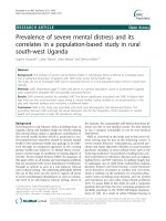
Immediately after opening the linea alba, two
emphysematous fetus (Fig.2) was found in the
abdominal cavity. Further detailed inspection
of the uterus revealed rupture of left uterine
horn. The emphysematous fetuses were
Fig.1 Radiography with extra uterine emphysematous fetus
443
Int.J.Curr.Microbiol.App.Sci (2020) 9(5): 442-444
Fig.2 Ruptured uterus along with emphysematous fetus
W.B.Saunders., Pp. 414-430.
Linde-Forsberg, C. (2010). Abnormalities in
pregnancy,
parturition
and
the
periparturient period. In: Ettinger, S.J.;
Feldman, E.C. (Ed.), Textbook of
Veterinary Internal Medicine. seventh ed.
St.Louis, Missouri: Elsevier Saunders.,
Pp. 1890-1901.
Palmer, N.E. 1989. Ectopic pregnancy in the
cat. Veterinary Record. 125, 24.
Ristic, J and Raijmakers, H. (1997). Abdominal
distension in a cat. Veterinary Record
140: 664.
Roberts, S. J. (1986). Veterinary Obstetrics and
Genital
Diseases.
In:
Veterinary
Obstetrics,
second ed. CBS Publishers and
Distributors Pvt.Ltd.
Seim, H.B. (1995). Management of peritonitis.
In: Bonagura JD, Kirk RW, eds. Current
Veterinary Therapy XII: Small Animal
Practice. Philadelphia: WB Saunders., Pp.
933-937.
There are few reports where these cases
referred to as ectopic pregnancies, implying
extra uterine fetal growth (Palmer, 1989).
The prompt diagnosis and subjecting the
animal to caesarean section at the earliest is
the key to good prognosis to safeguard the
animal. Feline uterine rupture with
emphysematous fetus can be successfully
treated by caesarean section.
References
Carrig, C.B., Gourley, L.M. and Philbrick, A.L.
(1972). Primary abdominal pregnancy in
a cat subsequent to ovariohysterectomy. J.
Am. Vet. Med. Assoc. 160, 308-310.
Jackson,
P.G.G.
(2004).
Postparturient
problems in the dog and cat. Handbook of
Veterinary
Obstetrics. 2nd edition.
W.B. Saunders Company, Philadelphia.,
Pp. 233–237.
Johnston, S.D., Root Kustritz, M.V. and Olson,
P. 2001. Feline pregnancy. In Canine and
Feline Theriogenology. Philadelphia,
How to cite this article:
Sabarinathan. A., S. Rangasamy, J. Umamageswari, U. S. Kalyaan and Pradeep Nag. 2020.
Uterine Rupture and its Management in a Queen Cat. Int.J.Curr.Microbiol.App.Sci. 9(05): 442444. doi: />
444