Isolation and characterization of three new anti-proliferative Sesquiterpenes from Polygonum barbatum and their mechanism via apoptotic pathway
Bạn đang xem bản rút gọn của tài liệu. Xem và tải ngay bản đầy đủ của tài liệu tại đây (1.73 MB, 13 trang )
Farooq et al. BMC Cancer (2017) 17:694
DOI 10.1186/s12885-017-3667-9
RESEARCH ARTICLE
Open Access
Isolation and characterization of three new
anti-proliferative Sesquiterpenes from
Polygonum barbatum and their mechanism
via apoptotic pathway
Umar Farooq1*, Sadia Naz1, Binte Zehra2, Ajmal Khan1*, Syed Abid Ali3, Ayaz Ahmed2*, Rizwana Sarwar1,
Syed Majid Bukhari1, Abdur Rauf4, Izhar Ahmad5 and Yahia Nasser Mabkhot6
Abstract
Background: The emergence of chemoresistant cancers and toxicity related to existing chemotherapeutic agents,
demand the search for new pharmacophore with enhanced anti-cancer activity and least toxicity. For this purpose,
three new sesquiterpenes were isolated from ethyl acetate fraction of the aerial parts of the plant Polygonum
barbatum and evaluated for their anti-cancer potential.
Methods: The structural elucidation and characterization of the isolated compounds 1–3 were performed using
various spectroscopic techniques such as mass, UV, IR, and extensive 1D/2D–NMR spectroscopy. Furthermore, the
compounds 1–3 were subjected to screening of anti-cancer activity against different cell lines followed by brief
analysis of apoptotic and anti-angiogenic potentials of the potent hit against non-small cell lung carcinoma cell
line.
Results: All the compounds 1–3 were subjected to anti-proliferative potential against non-small cell lung carcinoma
(NCI-H460), breast cancer (MCF-7), cervical cancer (HeLa) and normal mouse fibroblast (NIH-3 T3) cell lines. Among
these, compound 3 was found to be more cytotoxic against NCI-H460 and MCF-7 cells (IC50 = 17.86 ± 0.72 and 11.
86 ± 0.46 μM respectively). When compared with the standard drug cisplatin compound 3 was found to have more
potent activity against NCI-H460 (IC50 = 19 ± 1.24 μM) as compared to MCF-7 cell lines (IC50 = 9.62 ± 0.5 μM). Compound
3 induced apoptosis in NCI-H460 cells in a dose dependent manner. It significantly downregulated, the expression of antiapoptotic (BCL-2 L1 and p53) and increased the expression of pro-apoptotic (BAK and BAX) genes. Besides apoptosis, it
also significantly reduced the cell migration and downregulated the angiogenic genes (i.e. VEGF and COX-2), thereby,
inhibiting angiogenesis in NCI-H460 cells.
Conclusion: Compound 3 possesses potent anti-proliferative potential as well as induced apoptosis and inhibited the cell
migration of the cancerous cells by altering the gene expression, responsible for it.
Keywords: Polygonum Barbatum, Sesquiterpenes, Non-small cell lung carcinoma, Angiogenesis, VEGF, Cox-2, Apoptosis
* Correspondence: ; ;
1
Department of Chemistry, COMSATS Institute of Information Technology,
Abbottabad, KPK 22060, Pakistan
2
Dr. Panjwani Center for Molecular Medicine and Drug Research,
International Center for Chemical and Biological Sciences, University of
Karachi, Karachi 75270, Pakistan
Full list of author information is available at the end of the article
© The Author(s). 2017 Open Access This article is distributed under the terms of the Creative Commons Attribution 4.0
International License ( which permits unrestricted use, distribution, and
reproduction in any medium, provided you give appropriate credit to the original author(s) and the source, provide a link to
the Creative Commons license, and indicate if changes were made. The Creative Commons Public Domain Dedication waiver
( applies to the data made available in this article, unless otherwise stated.
Farooq et al. BMC Cancer (2017) 17:694
Background
Polygonum barbatum is a herbaceous perennial weed
commonly known as “Knot weed” belonging to Polygonaceae, which grows mostly in shady and moist areas, along
the sides of rivers and ponds [1]. The family Polygonaceae
comprises of 50 genera and ~1200 species widely distributed in Asia and America, and is represented in Pakistan
by 19 genera and 103 species [2]. Polygonum barbatum is
used as traditional medicine; the leaf extract of Polygonum
barbatum has been used for the treatment of ulcers, while
its roots are used as an astringent traditionally by local
practitioners [3]. According to literature survey, Polygonum barbatum possesses cholinergic, antinociceptive,
anti-tumor, anti-inflammatory, antivenom and diuretic
activities [4]. In addition, brine shrimp toxicity and spasmolypic activity of its dichloromethane extract has also
been reported previously [5].
Various secondary metabolites, such as flavonoids,
anthraquinones, phenylpropanoids and proanthocyanidins have been reported from various species of the
genus Polygonum [6–8]. The bioactive constituents of
Polygonum barbatum, including sitosterone, viscozulenic
acid and acetophenone, have also been reported previously [1]. Natural products, because of their excellent
bioavailability and abundance are getting much attention
in cancer therapy for the last few decades as sources to
find new and novel drug alternates.
Globally, cancers are the leading cause of death after cardiovascular diseases. Among various cancers, non-small cell
lung carcinoma (NSCLC) is the most common type of lung
cancer occurring worldwide with high rates of morbidity
and mortality. Lung cancer alone accounts for one-third of
the cancer related mortality across the world. Excessive
tobacco use makes the lung cancer as the third most prevalent disease in Pakistan. The advancement of therapeutic
regimen increased the survival rate to about 65%, however,
cancer progression, metastatic potential and chemoresistance still are the major concerns associated with high
mortality rates [9]. Modern lifestyle, smoking habits and
occupational exposures to the chemicals such as asbestos,
uranium and coke are the contributing factors responsible
for the initiation and progression of cancer.
Tumor formation is a complex process, which involves
sustained growth signals, inhibition of growth suppressors, preventing cellular death, angiogenesis, invasion
and spread to the secondary sites [10]. Tumor mass is
known to emerge from normal cells after mutation,
which then deviates from their normal cycle of homeostasis, survival and death [11]. It starts as a single tumor
cell, which after interaction with nearby vascular components, develops and results in neoplasm with altered
gene expression [12]. Lack of apoptosis, migration and
invasion are the hallmarks of cancer. Apoptosis is an
orderly cell death in response to any damage caused to
Page 2 of 13
cell’s basic functionality. It begins as a cascade of molecular events, eventually resulting in the cellular death and
phagocytosis to prevent inflammation [13]. A number of
apoptotic markers including tumor suppressor proteins,
caspases, BCL-2 proteins and several others are known to
be crucial for the process [14]. Studies have shown that
apoptosis could occur through any of the two pathways
(intrinsic or extrinsic) depending upon the initiating signals. Both pathways converge to the activation of effector
caspases, which leads to apoptosis [15].
Angiogenesis plays an important role in the advancement of cancer. Many pro-angiogenic factors work in
association with each other to provide vasculature to the
newly formed mass [16]. Vascular epithelial growth factor
(VEGF) is the major angiogenic protein that stimulates
the growth of vascular endothelial cells. Many cancers
including breast, prostate and lung cancers secrete high
levels of this mitogen [17, 18]. While cyclooxygenase-2
(COX-2) is an enzyme, which converts arachidonic acid to
prostaglandin H2 (PGH2). The PGH2 are acted upon by
prostaglandin synthase E to produce prostaglandin E,
which results in tumor invasion [19, 20].
The emergence of chemoresistant cancer and toxic
effects of existing pharmacophore, demand the search for
the alternates with potent anti-cancer activity and less toxicity. In the present study, we are reporting the isolation,
structure elucidation and the anticancer activities of three
new sesquiterpenes 1–3 from aerial parts of the Polygonum barbatum against non-small cell lung carcinoma
(NCI-H640), breast cancer (MCF-7), cervical cancer
(HeLa) and normal mouse fibroblast (NIH-3 T3) cell lines.
These cell lines were selected on the basis of its availability
as well as its prevalence in Pakistani population.
Moreover, in depth analysis of the active compounds were
further evaluated against the respective cell lines.
Results
The ethyl acetate fraction of Polygonum barbatum was
subjected to column chromatography on silica gel packed
columns. The repeated column chromatography resulted
in the isolation of three new sesquiterpenes derivatives
(1–3); their characterization was done by using various
spectroscopic techniques as well as through literature
comparison.
Compound 1
Compound 1 was isolated as a brownish gum having
molecular formula C16H18O4 based on molecular ion
peak at m/z 274.1211 in HR-EI-MS, while the fragmentations were found at m/z (%)256 (50), 242 (75), 238
(65), 227 (60), 192 (70), 188 (30), 151 (100), 126 (80) and
93 (40). The UV spectrum showed absorption bands at
λmax 220 (2.8), 272 (3.6), 314 (4.3) and 336 cm−1 (2.9),
while the IR spectrum showed absorptions at 3398,
Farooq et al. BMC Cancer (2017) 17:694
Page 3 of 13
3075, 2982, 1718, 1630 indicating the presence of hydroxyl group, ketone and aromatic moiety, respectively.
The 1H NMR spectrum showed the presence of two
aromatic protons at δH 7.22 (1H, d, J = 9.0 Hz) and δH
7.46 (1H, d, J = 9.0 Hz) ortho coupled to each other
along with one methylene singlet at δH 4.99 (2H, s), a
multiplet for one methine proton at δH 3.20 (1H, m) and
a singlet for three methoxy protons at δH 3.90 (3H, s).
Similarly, two olefinic protons at chemical shift values of
δH 6.80 (1H, s) and δH 6.51 (1H, m) and a methyl doublet integrating for six protons, appeared at δH 1.26 (6H,
d, J = 7.4 Hz) in 1H NMR spectrum (Table 1).
The 13C NMR and DEPT spectra revealed the presence
of 16 carbon atoms including three methyls, five methines,
one methylene, and seven quaternary carbons. Two methyl
carbons being identical resonated at δC 27.6, while the
methoxy group reappeared at δC 58.7 and methylene carbon having hydroxyl group as substituent centered at δC
62.4 (Table 2). The 13C NMR spectrum showed presence of
five methine carbons at δC 32.9, 143.2, 132.3, 119.7,
133.4 ppm. Similarly, one carbonyl carbon at δC 179.8 along
with other quaternary carbons resonating at δC 138.1,
139.6, 123.9, 158.5, 129.7, and 140.1 were also observed in
Table 2 13C NMR (125 MHz, CDCl3), δC in ppm
Compound 1
Compound 2
Compound 3
13
C–NMR
(δC ppm)
13
C–NMR
(δC ppm)
13
C–NMR
(δC ppm)
1
123.9
123.2
125.3
2
138.1
140.4
140.7
3
132.3
130.7
122.6
4
140.1
138.8
134.7
5
139.6
137.7
138.4
6
119.7
120.1
120.1
7
133.4
125.4
128.1
8
129.7
144.7
146.7
9
158.5
160.1
159.6
10
143.2
141.4
142.1
11
32.9
32.4
32.1
12
27.6
28.3
25.4
13
27.6
28.3
25.4
14
179.8
180.7
175.1
15
62.4
174.1
172.6
Carbon No
16
–
23.4
22.7
9-OCH3
58.7
60.4
59.6
14-OCH3
–
–
57.6
Table 1 1H NMR (500 MHz, CDCl3), δH in ppm
Compound 1
Compound 2
Compound 3
Carbon
No
1
H–NMR
(δH ppm)
1
H–NMR
(δH ppm)
1
1
–
–
–
13
9
–
–
–
10
6.51, (d,
J = 11.1 Hz)
6.48, (d,
J = 11.0 Hz)
6.50, (d, J = 10.8 Hz)
m
11
3.20, m
3.20, m
3.18, m
12
1.26, (d,
J = 7.4 Hz)
1.28, (d,
J = 7.1 Hz)
1.29, (d, J = 7.8 Hz)
C NMR spectrum. The HMBC spectrum showed correlation of H-3 with C-1, C-2, C-4, C-5, while proton present
at position H-6 showed strong HMBC correlation with C1, C-5, C-7, C-8 and C-9 indicating the presence of indene
ring in compound 1 [21]. The placement of substituents on
this indene ring were also established from HMBC
spectrum like methylpropylidine substituent was placed at
position 2 of indene ring on the basis of strong HMBC correlation of olefinic proton at position H-10 with C-2, C-1
and C-3 along with HMBC correlation of other olefinic
proton at position H-3 with C-2, C-4 and C-10. The position of other substituents like carboxylic group, methoxy
group and methyl alcohol moiety were also confirmed on
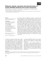
the basis of HMBC spectrum as depicted in Fig. 1. The
structure of compound 1 was suggested to be (E)-6-(hydroxymethyl)-7-methoxy-1-(2-methylpropylidene)-1H–indene-3-carboxylic acid on the basis of above mentioned
spectral data as well as comparison with literature.
13
1.26, (d,
J = 7.4 Hz)
1.28, (d,
J = 7.1 Hz)
1.29, (d, J = 7.8 Hz)
Compound 2
14
–
–
–
H–NMR
(δH ppm)
2
–
–
–
3
6.80, s
6.83, s
6.75, s
4
–
–
–
5
–
–
–
6
7.22 (d,
J = 9.0 Hz)
7.20 (d,
J = 8.3 Hz)
7.22 (d, J = 9.0 Hz)
7
7.46 (d,
J = 9.0 Hz)
7.28 (d,
J = 8.3 Hz)
7.48 (d, J = 9.0 Hz)
8
–
–
–
15
4.99, s
–
–
16
–
2.40, s
2.43, s
9-OCH3
3.90, s
3.88, s
3.80, s
14-OCH3
–
–
3.87, s
Compound 2 was isolated as brownish gummy solid from
ethyl acetate fraction of Polygonum barbatum. The HREI-MS showed molecular ion peak at m/z 302.1160 suggesting a molecular formula C17H18O5 for Compound 2
and m/z (%) other fragments ion peaks were observed at
284 (70), 252 (65), 238 (40), 194 (57), 166 (100), 151 (90),
124 (75) and 93 (20). The UV spectrum revealed
Farooq et al. BMC Cancer (2017) 17:694
Page 4 of 13
CH3
O
H
O
H
O
H
HC
CH3
O
3
OCH3
H
CH3
Fig. 1 Important HMBC (→) correlations of compound 1–3
absorptions bands at λmax 218 (3.6), 274 (4.2), 316 (3.7)
and 328 (2.9), while the IR spectrum showed peaks at
3460, 3009, 1752, 1705, 1640 cm−1 were quite similar
to that of compound 1 suggesting the presence of hydroxyl moiety, alkene, ketone and aromatic groups in
compound 2.
All the spectroscopic data like; 1H NMR and 13C
NMR of compound 2 is quite identical to compound 1
having two aromatic proton along their 13C NMR appeared at δH 7.20 (1H, d, J = 8.3 Hz, δC 120.1) and δH
7.28 (1H, d, J = 8.3 Hz, δH 125.4) showed ortho coupling
with each other, while the two olefinic protons at δH
6.82 (1H, s, δC 141.4) and δH 6.48 (1H, m, δC 130.7) presenting two pair of double bonds (C-2―C-4, C-10). Two
identical methyl group appeared at δH 1.28 (6H, d,
J = 7.1, δC 28.3) and protons of methoxy group gave
singlet at δH 3.88 (3H, s, δC 60.4) suggesting presence of
indene ring similar to compound 1 [21]. The only difference observed was the presence of ester group at position 8 of indene ring protons of compound 2, where
methyl of ester moiety appeared as singlet at δH 2.40
(3H, s, δC 23.4).
The position of substituents at indene ring of compound 2 was established through HMBC correlations
almost similar to compound 1, the only difference
observed was presence of ester group at position 8.
Finally, the structure of compound 2 on the basis of all
the above spectroscopic data and comparison with
literature was as (E)-6-acetoxy-7-methoxy-1-(2-methylpropylidene)-1H–indene-3-carboxylic acid.
at m/z (%) 285 (40), 256 (55), 225 (63), 196 (75), 168
(70), 154 (100), 128 (80) and 94 (35). The UV spectrum
showed absorption bands at λmax 222 (4.3), 270 (3.8),
318 (3.1) and 332 (3.6) while IR spectrum showed absorption bands at 3390, 2960, 1746, 1676 br and
1560 cm−1 similar to compound 1 and 2.
The spectroscopic data obtained from both 1H NMR
and 13C NMR were similar to compound 1 and 2 with
two olefinic protons resonated at δH 6.75 (1H, s, δC
122.6) and δH 6.50 (1H, m, δC 142.1), while two aromatic protons were also observed at δH 7.22 (1H, d,
J = 9.0 Hz, δC 120.1) and δH 7.48 (1H, d, J = 9.0 Hz, δC
128.1). Protons of two methoxy groups appeared as singlet at δH 3.80 (3H, s, δC 59.6) and 3.87 (3H, s, δC 57.6),
while two methyl group resonated at δH 1.29 (6H, d,
J = 7.8 Hz, δC 25.4) and another methyl group directly
connected to carbonyl carbon appeared at δH 2.43 (3H,
s, δC 22.7). The 13C NMR spectra revealed the presence
of 18 carbon atoms, while HMBC correlations were
quite helpful for placement of substituents on basic
skeleton (indene ring). The appearance of one methoxy
group 3.87 (3H, s, δC 57.6) signals and the disappearance
of one hydroxyl group were observed in compound 3.
Further analysis of its HMBC correlations suggested the
methoxy group located at C-14. All spectral data of
compound 3 was found similar to compound 2 the only
difference observed was extra methoxy group attached
to carbonyl carbon at position 14, further supported by
mass fragmentation pattern and structure assigned to
compound 3 was (E)-methyl 6-acetoxy-7-methoxy-1-(2methylpropylidene)-1H–indene-3-carboxylate.
Characterization of compound 1
A brownish gum; UV (MeOH) λmax 220 (2.8), 272 (3.6),
314 (4.3), 336 (2.9) nm; IR (KBr) max 3398, 3075, 2982,
1718, 1630, 1540, 1160, 880, 793 cm−1; EI-MS m/z (%):
256 (50), 242 (75), 238 (65), 227 (60), 192 (70), 188 (30),
151 (100), 126 (80), 93 (40); HR-EI-MS: m/z [M]+ Calcd.
274.1205 for C16H18O4; found 274.1211, 1H NMR
(500 MHz, CDCl3) δ (ppm): 6.80 (H-3, s), 7.22 (H-6, d,
J = 9.0 Hz), 7.46 (H-7, d, J = 9.0 Hz), 6.51 (H-10, (d,
J = 11.1 Hz), 3.20 (H-11, m), 1.26 (H-12/13, d, J = 7.4 Hz),
4.99 (H-15, s), 3.90 (9-OMe, s); 13C NMR (125 MHz,
CDCl3) δ (ppm): 123.9 (C-1), 138.1 (C-2), 132.3 (C-3),
140.1 (C-4), 139.6 (C-5), 119.7 (C-6), 133.4 (C-7), 129.7
(C-8), 158.5 (C-9), 143.2 (C-10), 32.9 (C-11), 27.6 (C-12/
13), 179.8 (C-14), 62.4 (C-15), 58.7 (9-OMe).
Characterization of compound 2
Compound 3
Compound 3 was isolated as light brown gummy solid
with molecular formula C18H20O5 as suggested by molecular ion peak at m/z 316.1320 in HR-EI-MS (calcd.
316.1311). The other fragmentation peaks were observed
A brownish gum; UV (MeOH) λmax 218 (3.6), 274 (4.2),
316 (3.7), 328 (2.9) nm; IR (KBr) max 3460, 3009, 2982,
1752, 1705, 1640, 1570, 1480, 1176, 910 cm−1; EI-MS m/z
(%): 284 (70), 252 (65), 238 (40), 194 (57), 166 (100), 151
(90), 124 (75), 93 (20); HR-EI-MS: m/z [M]+ Calcd.
Farooq et al. BMC Cancer (2017) 17:694
Page 5 of 13
302.1154 for C17H18O5; found 302.1160, 1H NMR
(500 MHz, CDCl3) δH (ppm): 6.82 (H-3, s), 7.20 (H-6, d,
J = 8.3 Hz), 7.28 (H-7, d, J = 8.3 Hz), 6.48 (H-10, (d,
J = 11.0 Hz), 3.20 (H-11, m), 1.28 (H-12/13, d, J = 7.1 Hz),
2.40 (H-16, s), 3.88 (9-OMe, s); 13C NMR (125 MHz,
CDCl3) δC (ppm): 123.2 (C-1), 140.4 (C-2), 130.7 (C-3),
138.8 (C-4), 137.7 (C-5), 120.1 (C-6), 125.4 (C-7), 144.7
(C-8), 160.1 (C-9), 141.1 (C-10), 32.4 (C-11), 28.3 (C-12/
13), 180.7 (C-14), 174.1 (C-15), 23.4 (C-16), 60.4 (9-OMe).
Characterization of compound 3
A light brown gummy solid; UV (MeOH) λmax 222 (4.3),
270 (3.8), 318 (3.1), 332 (3.6) nm; IR (KBr) max 3390,
2960, 1746, 1676 br, 1560, 1330 cm−1; EI-MS m/z (%): 285
(40), 256 (55), 225 (63), 196 (75), 168 (70), 154 (100), 128
(80), 94 (35); HR-EI-MS: m/z [M]+ Calcd. 316.1311 for
C18H20O5; found 316.1320, 1H NMR (500 MHz, CDCl3)
δH (ppm): 6.75 (H-3, s), 7.22 (H-6, d, J = 9.0 Hz), 7.48 (H-7,
d, J = 9.0 Hz), 6.50 (H-10, (d, J = 10.8 Hz), 3.18 (H-11, m),
1.29 (H-12/13, d, J = 7.8 Hz), 2.43 (H-16, s), 3.80 (9-OMe,
s), 57.6 (14-OMe); 13C NMR (125 MHz, CDCl3) δC (ppm):
125.3 (C-1), 140.7 (C-2), 122.6 (C-3), 134.7 (C-4), 138.4 (C5), 120.1 (C-6), 128.1 (C-7), 146.7 (C-8), 159.6 (C-9), 142.1
(C-10), 32.1 (C-11), 25.4 (C-12/13), 175.1 (C-14), 57.6 (14OMe), 172.6 (C-15), 22.7 (C-16), 59.6 (9-OMe).
Effects of compound 1–3 on proliferation and survival of
cancer cell lines
Reduction of viable cells after the treatment with compounds 1–3 was determined by MTT assay. Cytotoxicity
of these compounds was evaluated against human nonsmall cell lung carcinoma (NCI-H460), MCF-7 (breast
cancer), HeLa (cervical cancer) and normal mouse fibroblast cells (NIH-3 T3). Among all cell lines, the three sesquiterpenes showed anti-cancer activity against cancer
cells and was less active against normal 3 T3 cells. However, compound 3 was found to be potentially antiproliferative in a dose dependent manner against NSCLC
cells when compared to compounds 1–2 and the standard
drug cisplatin (Table 3). Compound 3 was also found to
be less cytotoxic against normal cells as compared to lung
cancer cells. By considering its higher anti-cancer and
selective toxicity towards cancer cells at a lower concentration as compared to normal cells, it was selected for
further detailed studies against NCI-H460 cells.
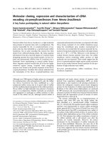
The anti-proliferative potential of the compound 3 on
H460 and 3 T3 cells was further confirmed by phase
contrast microscopy. Images of cells were captured after
treatment with compound 3 at 20 and 40 μM concentrations at 0, 24 and 48 h. After 24 h, H460 cells started to
change their normal morphology and got detached from
the monolayer, while after 48 h, majority of the cells
died, formed clumps with other dead cells in the media
as compared to vehicle treated cells (Fig. 2). Whereas,
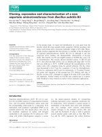
the same doses were applied to 3 T3 cells; inhibition was
observed at both doses after 24 h of treatment but growing cells with normal morphology appeared after 48 h of
the treatment: depicting compound’s selective toxicity
against cancer cells (Fig. 3).
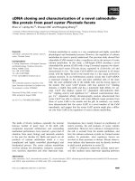
Compound 3 induces apoptosis in NCI-H460 cells
Phase contrast microscopy results showed the presence of
necrotic or dead cells, after treating with compound 3. The
extent of apoptosis and percent population of the cells in
early, late apoptotic phases and necrosis were also analyzed.
The population of non-treated cells was concentrated in
lower left quadrant indicating the normal cells. On the
other hand, treated cells showed significant apoptotic
change with major cell density undergoing late phases of
apoptosis (upper right quadrant) and necrosis (upper left
quadrant) at 20 μM concentration, while at 40 μM, more
cells were found to be in late apoptotic phase (Fig. 4). The
overall percentage of apoptotic cells after treating with
compound 3 at 20 and 40 μM was found to be around 7%
and 8.5% respectively.
The apoptotic ability of compound 3 was further confirmed by gene expression analysis of anti-apoptotic
genes (BCL-2 L1 and p53), pro-apoptotic genes (BAK
and BAX). As observed, the expression of anti-apoptotic
genes BCL-2 L1 and p53 was significantly down regulated and expression of pro-apoptotic genes BAK and
BAX was upregulated as compared to vehicle control
after 48 h treatment (Fig. 5). GAPDH was used as constitutive gene and its expression remained constant in
the treated cells, when compared to control.
Anti-angiogenic potential of compound 3 on NCI-H460
cells
Angiogenesis is the most important processes during
wound healing or cancer cell migration [22, 23]. Therefore,
Table 3 50% inhibitory concentration of compound 1–3 against various cell lines. The compound 3 showed potent anti-cancer activity against NSCLC cells as compared to cisplatin (standard drug)
NCI-H460
MCF-7
HeLa
NIH-3 T3
Compound 1
0.23 mM ± 0.85
Inactive
Inactive
0.42 mM ± 0.13
Compound 2
0.13 mM ± 0.38
Inactive
Inactive
0.28 mM ± 0.5
Compound 3
17.86 μM ± 0.72
11.86 μM ± 0.46
32.13 μM ± 0.6
30.32 μM ± 0.93
Cisplatin
19 μM ± 1.24
09.62 μM ± 0.5
16.19 μM ± 0.7
–
Farooq et al. BMC Cancer (2017) 17:694
Page 6 of 13
20 µM Comp-3
40 µM Comp-3
48 Hours
24 Hours
0 Hours
Control
Fig. 2 Phase contrast microscopic images of NCI-H460 cells after treatment with compound 3 at 20 and 40 μM concentrations. In control well,
the cells were in their normal morphology while treated cells died and escaped the monolayer after 24 h of the treatment. The images were
taken at 10X magnification
20 µM Comp-3
40 µM Comp-3
48 Hours
24 Hours 0 Hours
Control
Fig. 3 Phase contrast microscopic images of normal NIH-3 T3 cells after treatment with compound 3 at 20 and 40 μM concentrations at different
time intervals. Control well showed cells in their normal morphology while both live and dead populations were found in treated wells after
48 h. The images were taken at 10X magnification
Farooq et al. BMC Cancer (2017) 17:694
Page 7 of 13
Fig. 4 a The apoptotic potential of compound 3 in lung cancer NCI-H460 cells. FACS images showed percent apoptotic cells after treatment with
compound 3. Cells were significantly undergoing apoptosis as compared to the vehicle control. b Graphical representation of the cell population
in each phase of apoptosis and expressed as the mean of three independent experiments. *** p < 0.001 as compared to control
the compound 3 was also evaluated for its anti-migratory
potential. Results showed that it significantly delayed the
rate of wound healing at 20 and 40 μM concentrations as
compared to untreated cells (Fig. 6). After 48 h, only 17%
and 24% scratch were closed at 20 and 40 μM concentrations, respectively, as compared to vehicle treated control
(52%). Gene expression of two important angiogenic
markers i.e., VEGF and COX-2, responsible for potentiating
the migration and invasion were also established (Fig. 7).
Significant downregulation of both genes further links
with the anti-angiogenic potential of compound 3.
These compounds also alter the expression of matrix
metalloproteinases in a dose dependent manner (data
not shown). Interestingly, at 40 μM concentrations,
there was a slight increase in the expression of genes
as compared to 20 μM clearly suggesting a dose
dependent phenomenon.
Discussion
Cancer, being one of the emerging concerns of mortality
worldwide, demands the alternative approaches to deal
with it. Due to the toxicity of available drugs and emerging chemo-resistance, the medicinal plants can provide
better alternates that could be developed into new pharmacophores with enhanced anticancer activity and less
toxicity. In fact, 60% of the anti-cancer drugs (e.g.
Farooq et al. BMC Cancer (2017) 17:694
A
p53
BCL-2L1
BAK
BAX
GAPDH
B
Fig. 5 a Gene expression analysis of pro-apoptotic (BAK and BAX)
and anti-apoptotic genes (BCL-2 L1 and p53) after 48 h treatment of
NCI-H460 cells with compound 3. GAPDH was used as a control
housekeeping gene. b Quantitative analysis of the expression by
calculating fold change in integrated density treated versus control
genes. *** p < 0.001 and ** p < 0.01 when compared to the control
vinblastine, paclitaxel, etoposide, doxorubicin etc.) are
derived from the natural origin [24, 25]. Thus, this study
was designed to screen the anti-cancer potential of three
novel sesquiterpenes isolated from the aerial parts of
Polygonum barbatum on different cancer cell lines
(Table 3). Compound 1 and 2 were found to be inactive
against the tested cell lines. Whereas, compound 3 was
found to be anti-proliferative against all tested cancer
cell lines. In comparison to its activity against normal
cells (3 T3), higher anti-cancer activity was observed in
NCI-H460 and MCF-7 cells than HeLa cells. A possible
explanation to this observation could be interference of
tumor suppressor protein p53. All cell lines are positive
for TP53 but it gets inactivated in HeLa cells due to
human papilloma virus; HeLa cells contain HPV-18
sequences. While when compared to IC50 values of compound 3 and the standard drug, it was most potent
against NCI-H460 cells. Hence, in-depth study of mechanism was evaluated against non-small cell lung carcinoma (NSCLC) cells.
Page 8 of 13
Interestingly, compound 3 showed prominent antiproliferative effects after 24 h of treatment as observed
by phase contrast microscopy in H460 cells (Fig. 2).
However, slight inhibition was also observed in treated
wells of 3 T3 cells but the effect reverted after 48 h
resulting in re-emergence of actively growing cells (Fig.
3). Compound 3 also induced apoptosis and inhibit cell
migration in NCI-H460 cells after 24 and 48 h of treatment, and altered the genes responsible for apoptosis
and migration of cancerous cells.
Apoptosis is a very vital maintenance system, which
keeps a check on the unhealthy, malignant, dead or infected cells of the body. It functions in a regulatory manner and can be triggered by a variety of responses,
including physiological or pathological or both [11]. The
early phases of apoptosis correspond to the early events
of activation of intrinsic or extrinsic pathways whereas
events downstream the cleavage of caspases-3 encompasses the late phase of apoptosis [26]. Cancer cells take
control of apoptotic machinery and promote tumour
progression. Literature revealed that most of the existing
drugs target apoptosis, which is crucial in cancer advancement [14, 27]. Our present results demonstrate
that compound 3 also induced apoptosis in the treated
cells. The FACS analysis by using dual staining with YOPRO-1 and PI dyes revealed that after 48 h treatment,
cells were found to be in late phases of apoptosis and
underwent necrosis (Fig. 4).
Usually, a cell undergoes either intrinsic mitochondrial
pathway or extrinsic receptor pathways to initiate the
process of apoptosis. Absence of growth factors or cytokines, radiation, hypoxia, ROS and infections, results in
initiation of mitochondrial pathway. Stimuli lead to a
change in mitochondrial membrane permeability releasing cytochrome c and thereby activating caspases 9 and
formation of the apoptosome. While extrinsic pathway
involved, the ligand binding to death receptor triggering
intracellular signals to activate caspases 8. Once executioner caspases are activated, the effector caspases come
into play and form apoptotic bodies [28, 29]. Likewise,
several proteins are responsible for inducing intrinsic
pathway of apoptosis. The BCL-2 family and tumour
suppressor proteins are of prime importance [30]. It
consists of both anti-apoptotic (BCL-2 L1, BCL-XL etc.)
and pro-apoptotic (BAK, BAX etc.) proteins that work
in association with one another [31]. The BAK and BAX
induce mitochondrial outer membrane permeabilization
leading to mitochondrial dysfunction, hence marking it
for apoptosis [32]. Our results also revealed the significant increase of pro-apoptotic and down regulation of
anti-apoptotic genes after treated with compounds 3
(Fig. 5), suggesting the involvement of intrinsic mitochondrial pathway. The expression of p53, a tumour
suppressor gene, also significantly down regulated after
Farooq et al. BMC Cancer (2017) 17:694
Page 9 of 13
Fig. 6 a Anti-migration potential of compound 3 at 20 and 40 μM concentrations against lung cancer NCI-H460 cells. The control cells healed
33% and 52% scratch after 24 h and 48 h whereas 17% and 24% healing was observed at 40 μM of compound 3. b Graphical representation of
the rates of migration in control and treated wells. ** p < 0.01 and * p < 0.05 control vs treated
treatment, which gets mutated in most of the cancers
and responsible for inhibiting apoptosis [33].
Angiogenesis is an important physiological process for
growth and is also known to be involved in the pathogenesis of many inflammatory diseases including cancer [34]. It
further strengthens and develops tumor mass by providing
important nutrients and promotes migration and invasion
to the secondary sites of the body [35]. Our results showed
that compound 3 also inhibited the bidirectional migration
to almost two folds of the NSCLC cells in a dose dependent
manner by using wound healing assay. It delayed the healing process of the scratch as compared to control. It has
been reported that VEGF-A (VEGF) is the key stimulator
in the angiogenesis [17, 36]. VEGF binds to its receptor and
triggers the intracellular signals, which lead to the initiation
of a cascade of the events involved in angiogenesis. The
VEGF levels increase drastically during tumour growth and
contributes to enhanced stroma [37]. While COX-2, is another major enzyme responsible for converting arachidonic
acid into prostaglandin H2. It is also involved in supplementing the process of angiogenesis in association with
VEGF [38–40]. Thus the compounds, which can act and
downregulates their expression might be an attractive target
for cancer treatment [41]. Our results also showed that
compound 3 significantly downregulates both the genes responsible for angiogenesis of tumour cells. They also effect
the expression of matrix metalloproteinases genes (data not
shown). Studies also confirmed that inhibiting COX-2
enzyme in preclinical models not only prevented angiogenesis, but also reduced the migratory and metastatic potential of tumor cells [42, 43].
Conclusions
This current study resulted in isolation of three new sesquiterpenes namely (E)-6-(hydroxymethyl)-7-methoxy-1(2-methylpropylidene)-1H–indene-3-carboxylic acid (1),
(E)-6-acetoxy-7-methoxy-1-(2-methylpropylidene)-1H–indene-3-carboxylic acid (2) and (E)-methyl 6-acetoxy-7methoxy-1-(2-methylpropylidene)-1H–indene-3-carboxylate (3) from ethyl acetate fraction of the aerial parts of the
plant Polygonum barbatum. The structure elucidation and
characterization of the isolated compounds 1–3 were
done by using various spectroscopic techniques such as
Farooq et al. BMC Cancer (2017) 17:694
Page 10 of 13
Hitachi UV-3200 spectrophotometer was employed to
record UV spectra. The precoated silica gel plates were
used to carry out TLC and ceric sulphate in 10% H2SO4
solution was used for detection of UV active compounds.
Similarly, silica gel (E. Merck, 230–400 mesh and 70–230
mesh) was used for column chromatography.
A
COX-2
Extraction and isolation
VEGF
GAPDH
B
Fig. 7 a Gene expression of angiogenic VEGF and COX-2 genes after the
treatment of NCI-H460 cells. b Graphical representation of quantitative
analysis of the gene expression. *** p < 0.001 as compared to the control
mass spectrometry, UV, IR, 1H NMR, 13C NMR and Heteronuclear multiple bond correlation spectroscopy.
Among all three sesquiterpenes, compound 3 possesses
the most potent anti-proliferative potential against non-small cell lung carcinoma cells (NCI-H460). It induced
apoptosis and inhibited the cell migration of the cancerous cells and alters the gene expression, which is responsible for the apoptosis and angiogenesis. Thus, compound
3 can be further evaluated for its effects on the proteome
by means of 2D PAGE and 2D DiGE proteomics
approaches to further confirm the gene expression
analysis to make it potential drug candidate.
The whole plant of Polygonum barbatum (5.4 kg) was collected from Northern areas (Mansehra), Khyber Pakhtunkhwa Pakistan in October 2015. The plant was
identified by Dr. Manzoor Ahmed (Taxonomist), at the
Department of Botany, Government Postgraduate College,
Abbottabad, Pakistan. A voucher specimen (No. 66130)
has been submitted in herbarium of the same department.
The aerial part of the plant material was shade dried,
ground into fine powder and extracted thrice with
methanol (3 × 10 L) at room temperature and filtered.
The filtrate was subjected to vacuum rotary evaporator
to get crude extract (245 g). The whole extract was further partitioned into three fractions, namely n-hexane
(85 g), ethyl acetate (48 g) and n-butanol (94 g).
The ethyl acetate fraction was chromatographed on
silica gel (E. Merck, 230–400 mesh and 70–230 mesh)
using the solvents with increasing polarity, n-hexane was
used with gradient of ethyl acetate up to 100% followed
by methanol, which resulted in sub-fractions depending
on the polarity of compounds. Sub-fractions number
4―10 out of the total 12, were further, subjected to column chromatography to get compound 1 (8.6 mg) at
EtOAc: n-hexane (40:60), while compound 2 (9 mg) and
compound 3 (7.5 mg) were purified at EtOAc: n-hexane
(36:64), and (27:73), respectively (Fig. 8).
Cell culture
Cell lines (i.e., NCI-H460, MCF-7, HeLa and NIH-3 T3)
were purchased from American type culture collection
(ATCC, USA). Cell lines were grown and maintained
using RPMI-1640 (GIBCO, Auckland, NZ) supplemented with 2 mM Lglutamine, 2 g/L D-glucose, and
1.5 g/L sodium bicarbonate, 10% heat inactivated-FBS
(Hyclone, USA) and 1% antibiotic solution in a humidified (95% air: 5% CO2) incubator at 37 °C. The cells were
regularly passaged on reaching 80% confluency using
trypsin-EDTA in a t75 flask.
Methods
General
The double focusing Varian MAT-312 spectrometer was
used for EI-MS and HR-EI-MS analysis and 1H NMR and
13
C NMR spectra were recorded through Bruker AMX500 MHz Spectrometer with tetramethyl silane (TMS) as
internal standard. The chemical shift values were reported
as ppm and scalar coupling as Hertz (Hz). The IR spectra
were recorded by using Hitachi JASCO-320-A, while
MTT (3-(4,5-Dimethylthiazol-2-yl)-2,5dipheynyltetrazolium bromide) assay
In order to evaluate the effects of the compounds on cell
viability, 10,000 cells per well were seeded in a 96-well
micro titer plate. After 24 h, cells were treated with various concentrations (5–250 μM) of the test compounds
(1–3). After 48 h of incubation, 10 μL MTT dye (Biobasic, Canada) was added to the wells followed by 4 h of
Farooq et al. BMC Cancer (2017) 17:694
Page 11 of 13
O
O
14
6
5
7
1
HO
8
15
14
OH
6
4
3
1
2
13
15
9
8
O
16
11
10
OCH3
5
7
O
4
3
2
13
9
OCH3
2
12
1
OH
10
11
12
O
14
6
5
7
O
1
15
16
O
8
O
4
3
2
13
9
OCH3
10
3
11
12
Fig. 8 Structures of compound 1–3
incubation. Later, the dye was removed and formazan
crystals were solubilized in DMSO and absorbance was
noted at 590 nm using a SpectraMax spectrophotometer
(Molecular Devices, USA). The cytotoxicity at different
concentrations and IC50 of the compounds against both
cell lines were also calculated.
and PI stock solution (Component B) were added and incubated on ice for 30 min. FACSCalibur (Becton Dickinson, USA) was used to analyze the samples whereas,
CellquestPro software was used to calculate the cells in
their respective phase of apoptosis. Total of 10,000 events
of each sample were recorded as one reading and all
experiments were performed in triplicate.
Phase contrast microscopy
The effect of compound 3 on non-small cell lung carcinoma and normal fibroblast cells was evaluated under the
Phase Contrast microscope (Nikon, Tokyo, Japan). Cells
were grown in a 6-well plate to 60% confluency. Later,
the cells were treated with compound 3 at two different
concentrations (20 and 40 μM). The wells were photographed at 0, 24 and 48 h by using the microscope at
10X magnification. The images were taken immediately
after the addition of compounds for 0 h as a control.
YO-PRO-1 assay
The apoptotic potential of compound 3 was analyzed by
Vybrant Apoptosis Assay Kit # 4 (Invitrogen, USA)
according to manufacturer protocol. Briefly, 1 × 10⁶ NCIH460 cells were seeded in a 6-well plate (Corning, USA).
After 24 h, cells were treated with compound 3 and
further incubated for 48 h in a humidified CO2 incubator.
After incubation, the cells were washed with PBS and
dissociated using trypsin-EDTA. Cell pellet was collected
by centrifugation and suspended in 1 mL PBS. One microliter (1 μL) of YO-PRO-1 stock solution (Component A)
Wound healing assay
To observe the anti-migratory effects of compound 3, cells
were grown to 80% confluency in a 6-well plate (Corning,
USA). A scratch was gently made using 100 μL micropipette tip. The well was thoroughly washed with 1X PBS to
remove all the scratched cells and compound 3 was added.
Image taken immediately was marked as 0 h reading.
Scratch images were taken at 24 and 48 h by using a Phase
Contrast microscope (Nikon, Tokyo, Japan). The images
were processed and area of the wound was determined by
using Image J software.
RT-PCR
For the total RNA extraction, 1.5 × 10⁶ NCI-H460 cells
were grown in the presence or absence of compound 3
in a 6-well plate. After 48 h of incubation, cells were
washed with PBS and total RNA was extracted using
TRIzol reagent (Life technologies, USA), according to
the standard protocol. Extracted RNA was quantified
and 1 μg RNA was used to synthesize cDNA through
RevertAid First Strand cDNA Synthesis Kit (Thermo
Farooq et al. BMC Cancer (2017) 17:694
Page 12 of 13
Table 4 Primer sequences and annealing temperatures of
genes used in this study
Gene
Sequence
Annealing
References
Temperature
(°C)
BCL2 L1
R: ATGGTCAGTGTCTGGTCATT
F: TTGTGGAACTCTATGGGAAC
57
[44]
p53
R: CTCTCGGAACATCTCGAAGCG
F: GCTCTGACTGTACCACCATCC
57
[45]
BAX
R: GGCCCCAGTTGAAGTTGC
F: AAGAAGCTGAGCGAGTGTC
54
[46]
BAK
R: CCTGAGAGTCCAACTGCAAA
F: GGTCCTGCTCAACTCTACCC
60
[47]
VEGF
R: ACCGCCTCGGCTTGTCAC
F: GTGTGCCCCTGATGCGATGCG
54
[48]
COX-2
R:
56
CGCTCAGCCATACAGCAAATCCTT
F: GTGCACTGTGTTTGGAGTGGGTTT
[49]
GAPDH
R: GGTCTACATGGCAACTGTGA
F: ACGACCACTTTGTCAAGCTC
[50]
59
Scientific, USA), according to the user guide. This cDNA
was used as template and genes related to apoptosis and
angiogenesis were amplified at their respective annealing
temperatures (Table 4). The final volume of PCR reaction mix was kept at 25 μL. The amplified products were
resolved on 1% agarose gel. The gel was analyzed and
quantified using Image J software.
Statistical analysis
All the results are presented as the mean ± standard
deviation of triplicate experiments. Student’s t-test was
used to compare the treated and control groups whereas
p < 0.05 was reported as significant.
Abbreviations
BCL-2: B-cell lymphoma 2; COX-2: Cyclooxygenase-2; DIGE: Difference gel
electrophoresis; EDTA: Ethylenediaminetetraacetic acid; FACS: Fluorescenceactivated cell sorting; FBS: Fetal bovine serum; GAPDH: Glyceraldehyde 3phosphate dehydrogenase; HR-EI-MS: High Resolution Electroionization Mass
Spectrometry; NSCLC: Non-small cell lung cancer; NSCLC: Non-small cell lung
carcinoma; PAGE: Polyacrylamide gel electrophoresis; PGH2: Prostaglandin
H2; PI: Propidium iodide; ROS: Reactive oxygen species; RPMI: Roswell Park
Memorial Institute; RT-PCR: Reverse transcription polymerase chain reaction;
TMS: Tetramethyl silane; VEGF: Vascular endothelial growth factor
Acknowledgements
We acknowledge Higher Education Commission (HEC) of Pakistan for
financial support under NRPU programme No. 20-2798/NRPU. The authors
extend their sincere appreciation to the Deanship of Scientific Research at
king Saud University for its funding this Prolific Research group (PRG-1437-2).
Availability of data and materials
All datasets on which the conclusions of the manuscript rely are presented
in the paper.
Authors’ contributions
UF and AK supervised and designed the study. SN and RS performed
isolation and identification of compounds. AR helped in the structure
elucidation. BZ, AA and SAA carried out the anticancer activities and their
mechanism. SMB, IA and YNB were involved in writing, editing of
manuscript. All authors have read and approved the final version of the
manuscript.
Ethics approval and consent to participate
Not applicable.
Consent for publication
Not applicable.
Competing interests
The authors declare that they have no competing interests.
Publisher’s Note
Springer Nature remains neutral with regard to jurisdictional claims in
published maps and institutional affiliations.
Author details
1
Department of Chemistry, COMSATS Institute of Information Technology,
Abbottabad, KPK 22060, Pakistan. 2Dr. Panjwani Center for Molecular
Medicine and Drug Research, International Center for Chemical and
Biological Sciences, University of Karachi, Karachi 75270, Pakistan. 3H.E.J.
Research Institute of Chemistry, International Center for Chemical and
Biological Sciences, University of Karachi, Karachi 75270, Pakistan.
4
Department of Chemistry, University of Swabi, Anbar, Khyber Pakhtunkhwa
23561, Pakistan. 5Department of Botany, Islamia College Peshawar, Peshawar,
Pakistan. 6Department of Chemistry, College of Science, King Saud University,
P. O. Box 2455, Riyadh 11451, Saudi Arabia.
Received: 11 April 2017 Accepted: 28 September 2017
References
1. Mazid MA, Datta BK, Nahar L, Bashar SK, Bachar SC, Sarker SD.
Phytochemical Studies on Polygonum barbatum (L.) Hara var. barbata
(Polygonaceae). Records of Natural Product. 2011;5:143–5.
2. Sanchez A, Kron KA. Phylogenetics of Polygonaceae with an emphasis on
the evolution of Eriogonoideae. Syst Bot. 2008;33(1):87–96.
3. Ramani A. In-vitro antioxidant activity of Polygonum barbatum leaf extract.
Asian Journal of Pharmaceutical and Clinical Research. 2011;4(1):113–5.
4. Mazid MA, Datta BK, Nahar L, Bashar S, Bachar SC, Sarker SD.
Antinociceptive, anti-inflammatory and diuretic properties of Polygonum
barbatum (L.) Hara var. barbata. Rev Bras. 2009;19(3):749–54.
5. Chaudhry BA, Syad MY, Janbaz K, Dasti A, Loothar BA. Biological activities of
Polygonum barbatum. Journal of Research (Science). 2003;14:169–75.
6. Isobe T, Kanazawa K, Fujimura M, Noda Y. Flavonoids of Polygonum sieboldi
and P. filiforme. Bull Chem Soc Jpn. 1981;54(10):3239.
7. Brown LL, Larson SR, Sneden AT. Vanicosides CF, new phenylpropanoid
glycosides from Polygonum pensylvanicum. J Nat Prod. 1998;61(6):762–6.
8. Kim H-M. Antiallergy drugs from Oriental medicines. Orient Pharm Exp Med.
2000;1(1):01–7.
9. Song Y, Xue L, Du S, Sun M, Hu J, Hao L, Shao S. Caveolin-1 knockdown is
associated with the metastasis and proliferation of human lung cancer cell
line NCI-H460. Biomed Pharmacother. 2012;66(6):439–47.
10. Hanahan D, Weinberg RA. Hallmarks of cancer: the next generation. Cell.
2011;144(5):646–74.
11. Fernald K, Kurokawa M. Evading apoptosis in cancer. Trends Cell Biol.
2013;23(12):620–33.
12. Kerbel RS. Tumor angiogenesis. N Engl J Med. 2008;358(19):2039–49.
13. Elmore S. Apoptosis: A Review of Programmed Cell Death. Toxicol Pathol.
2007;35(4):495–516.
14. Herr I, Debatin K-M. Cellular stress response and apoptosis in cancer
therapy. Blood. 2001;98(9):2603–14.
15. Wong RS. Apoptosis in cancer: from pathogenesis to treatment. J Exp Clin
Cancer Res. 2011;30(87):01–14.
16. Kubota Y. Tumor angiogenesis and anti-angiogenic therapy. The Keio
Journal of Medicine. 2012;61(2):47–56.
17. Hoeben A, Landuyt B, Highley MS, Wildiers H, Oosterom ATV, Bruijn EAD.
Vascular endothelial growth factor and angiogenesis. Pharmacol Rev.
2004;56(4):549–80.
Farooq et al. BMC Cancer (2017) 17:694
18. Jang Y, Kim DS, Jeon O, Kim D. Saxatilin suppresses tumor-induced
angiogenesis by regulating VEGF expression in NCI-H460 human lung
cancer cells. J Biochem Mol Biol. 2007;30(3):439–43.
19. Clària J. Cyclooxygenase-2 biology. Curr Pharm Des. 2003;9(27):2177–90.
20. Gately S, Li WW. Multiple roles of COX-2 in tumor angiogenesis: a target for
antiangiogenic therapy. Semin Oncol. 2004;31(7):02–11.
21. Shen Q, Xu X, Liu C, Zhao W, Xiang N, Chen Y, Miao M, Liu Z, Yang G. Two
new sesquiterpenes from the leaves of Nicotiana tabacum and their antitobacco mosaic virus activities. Nat Prod Res. 2016;0(0):01–6.
22. Eming SA, Brachvogel B, Odorisio T, Koch M. Regulation of angiogenesis:
wound healing as a model. Prog Histochem Cytochem. 2007;42(3):115–70.
23. Tonnesen MG, Feng X, Clark RA. Angiogenesis in wound healing. J Investig
Dermatol Symp Proc. 2000;5(1):40–6.
24. Newman DJ, Cragg GM. Natural products as sources of new drugs over the
30 years from 1981 to 2010. J Nat Prod. 2012;75(3):311–35.
25. Bhanot A, Sharma R, Noolvi MN. Natural sources as potential anti-cancer
agents: A review. International Journal of Phytomedicine. 2011;2(1):09–26.
26. Sankari SL, Masthan K, Babu NA, Bhattacharjee T, Elumalai M. Apoptosis in
cancer-an update. Asian Pac J Cancer Prev. 2012;13(10):4873–8.
27. Kaufmann SH, Earnshaw WC. Induction of apoptosis by cancer
chemotherapy. Exp Cell Res. 2000;256(1):42–9.
28. Indran IR, Tufo G, Pervaiz S, Brenner C. Recent advances in apoptosis,
mitochondria and drug resistance in cancer cells. Biochimica et Biophysica
Acta (BBA)-Bioenergetics. 2011;1807(6):735–45.
29. Ashkenazi A. Targeting the extrinsic apoptotic pathway in cancer: lessons
learned and future directions. J Clin Invest. 2015;125(2):487–9.
30. Shamas-Din A, Kale J, Leber B, Andrews DW. Mechanisms of action of Bcl-2
family proteins. Cold Spring Harb Perspect Biol. 2013;5(4):01–22.
31. Hardwick JM, Soane L. Multiple functions of BCL-2 family proteins. Cold
Spring Harb Perspect Biol. 2013;5(2):01–23.
32. Volkmann N, Marassi F, Newmeyer D, Hanein D. The rheostat in the
membrane: BCL-2 family proteins and apoptosis. Cell Death Differ.
2014;21(2):206–15.
33. Muller PA, Vousden KH. Mutant p53 in cancer: new functions and
therapeutic opportunities. Cancer Cell. 2014;25(3):304–17.
34. Otrock ZK, Mahfouz RA, Makarem JA, Shamseddine AI. Understanding the
biology of angiogenesis: review of the most important molecular
mechanisms. Blood Cell Mol Dis. 2007;39(2):212–20.
35. Shahi PK, Pineda IF. Tumoral angiogenesis: review of the literature. Cancer
Investig. 2008;26(1):104–8.
36. Wang S, Tu J, Zhou C, Li J, Huang L, Tao L, Zhao L. The effect of Lfcin-B on
non-small cell lung cancer H460 cells is mediated by inhibiting VEGF
expression and inducing apoptosis. Arch Pharm Res. 2015;38(2):261–71.
37. Ferrara N. Vascular endothelial growth factor: basic science and clinical
progress. Endocr Rev. 2004;25(4):581–611.
38. Yoshida S, Amano H, Hayashi I, Kitasato H, Kamata M, Inukai M, Majima M.
COX-2/VEGF-dependent facilitation of tumor-associated angiogenesis and
tumor growth in vivo. Lab Investig. 2003;83(10):1385–94.
39. Toomey D, Murphy J, Conlon K. COX-2, VEGF and tumour angiogenesis.
Surgeon. 2009;7(3):174–80.
40. Ghosh N, Chaki R, Mandal V, Mandal SC. COX-2 as a target for cancer
chemotherapy. Pharmacol Rep. 2010;62(2):233–44.
41. Das M, Wakelee H. Targeting VEGF in lung cancer. Expert Opin Ther Targets.
2012;16(4):395–406.
42. Xu L, Stevens J, Hilton MB, Seaman S, Conrads TP, Veenstra TD, Brad SC.
COX-2 inhibition potentiates antiangiogenic cancer therapy and prevents
metastasis in preclinical models. Sci Transl Med. 2014;6(242):242–84.
43. Chesney JA, Mitchell RA. 25 Years On: A Retrospective on Migration
Inhibitory Factor in Tumor Angiogenesis. Mol Med. 2015;21(1):S19–24.
44. Choi Y-C, Yoon S, Byun Y, Lee G, Kee H, Jeong Y, Yoon J, Baek K. MicroRNA
library screening identifies growth-suppressive microRNAs that regulate
genes involved in cell cycle progression and apoptosis. Exp Cell Res.
2015;339(2):320–32.
45. Suo H, Song J-L, Zhou Y, Liu Z, Yi R, Zhu K, Xie J, Zhao X. Induction of
apoptosis in HCT-116 colon cancer cells by polysaccharide of Larimichthys
crocea swim bladder. Oncol Lett. 2015;9(2):972–8.
46. Hanif F, Perveen K, Jawed H, Ahmed A, Malhi SM, Jamall S, Simjee SU. N-(2hydroxyphenyl) acetamide (NA-2) and Temozolomide synergistically induce
apoptosis in human glioblastoma cell line U87. Cancer Cell Int. 2014;14(1):1.
Page 13 of 13
47. Guan H, Xie L, Leithäuser F, Flossbach L, Möller P, Wirth T, Ushmorov A.
KLF4 is a tumor suppressor in B-cell non-Hodgkin lymphoma and in classic
Hodgkin lymphoma. Blood. 2010;116(9):1469–78.
48. Jang Y, Kim DS, Jeon O, Kim D. Saxatilin suppresses tumor-induced
angiogenesis by regulating VEGF expression in NCI-H460 human lung
cancer cells. J Biochem Mol Biol. 2007;40(3):439.
49. Chen W, Bai L, Wang X, Xu S, Belinsky SA, Lin Y. Acquired activation of the
Akt/cyclooxygenase-2/Mcl-1 pathway renders lung cancer cells resistant to
apoptosis. Mol Pharmacol. 2010;77(3):416–23.
50. Shi Y, Fu X, Hua Y, Han Y, Lu Y, Wang J. The side population in human lung
cancer cell line NCI-H460 is enriched in stem-like cancer cells. PLoS One.
2012;7(3):e33358.
Submit your next manuscript to BioMed Central
and we will help you at every step:
• We accept pre-submission inquiries
• Our selector tool helps you to find the most relevant journal
• We provide round the clock customer support
• Convenient online submission
• Thorough peer review
• Inclusion in PubMed and all major indexing services
• Maximum visibility for your research
Submit your manuscript at
www.biomedcentral.com/submit