Diagnostic and prognostic value of CEA, CA19–9, AFP and CA125 for early gastric cancer
Bạn đang xem bản rút gọn của tài liệu. Xem và tải ngay bản đầy đủ của tài liệu tại đây (458.3 KB, 6 trang )
Feng et al. BMC Cancer (2017) 17:737
DOI 10.1186/s12885-017-3738-y
RESEARCH ARTICLE
Open Access
Diagnostic and prognostic value of CEA,
CA19–9, AFP and CA125 for early gastric
cancer
Fan Feng1†, Yangzi Tian2†, Guanghui Xu1†, Zhen Liu1, Shushang Liu1, Gaozan Zheng1, Man Guo1, Xiao Lian1,
Daiming Fan1 and Hongwei Zhang1*
Abstract
Background: The diagnostic and prognostic significance of carcinoembryonic antigen (CEA), carbohydrate associated
antigen 19–9 (CA19–9), alpha-fetoprotein (AFP) and cancer antigen 125 (CA125) in early gastric cancer have not been
investigated yet. Thus, the present study aimed to explore the diagnostic and prognostic significance of the four tumor
markers for early gastric cancer.
Methods: From September 2008 to March 2015, 587 early gastric cancer patients were given radical gastrectomy in
our center. The clinicopathological characteristics were recorded. The association between levels of CEA and CA19–9
and clinicopathological characteristics and prognosis of patients were analyzed.
Results: There were 444 men (75.6%) and 143 women (24.4%). The median age was 57 years (ranged 21–85). The 1-, 3and 5-year overall survival rate was 99.1%, 96.8% and 93.1%, respectively. The positive rate of CEA, CA19–9, AFP and
CA125 was 4.3%, 4.8%, 1.5% and 1.9%, respectively. The positive rate of all markers combined was 10.4%. The
associations between the clinicopathological features and levels of CEA and CA19–9 were analyzed. No significant
association was found between CEA level and clinicopathological features. However, elevated CA19–9 level was
correlated with female gender and presence of lymph node metastasis. Age > 60 years old, presence of lymph node
metastasis and elevation of CEA level were independent risk factors for poor prognosis of early gastric cancer.
Conclusions: The positive rates of CEA, CA19–9, APF and CA125 were relatively low for early gastric cancer. Elevation
of CA19–9 level was associated with female gender and presence of lymph node metastasis. Elevation of CEA level was
an independent risk factor for the poor prognosis of early gastric cancer.
Keywords: Early gastric cancer, Diagnosis, Prognosis, Tumor marker
Background
Gastric cancer is the fourth commonest malignancy and
the second leading cause of tumor related death all over
the world [1]. Early gastric cancer is a lesion only invading mucosa or submucosa, with or without lymph node
metastasis (LNM) [2]. Early diagnosis of gastric cancer is
critical for optimal treatment. The ratio of early gastric
cancer at diagnosis is increasing with advanced techniques and screening programs [3]. As detection of
* Correspondence:
†
Equal contributors
1
Division of Digestive Surgery, Xijing Hospital of Digestive Diseases, the
Fourth Military Medical University, 127 West Changle Road, 710032, , Xian,
Shaanxi, China
Full list of author information is available at the end of the article
serum tumor markers are more convenient than other
approaches, they are widely applied in early diagnosis of
gastric cancer [4]. Unfortunately, the optimal serum
biomarker for the detection of early gastric cancer is still
under investigation [5].
The prognosis of early gastric cancer is favorable after
radical gastrectomy, with a 5-year overall survival rate
exceed 97% [6]. A variety of factors have been recognized
as prognostic factors for early gastric cancer, including
tumor size, differentiation status, tumor depth, LNM and
vessel involvement [7]. In addition, tumor markers including CEA [8], CA19–9 [9], and AFP [10] were demonstrated
to be prognostic factors for gastric cancer. However,
© The Author(s). 2017 Open Access This article is distributed under the terms of the Creative Commons Attribution 4.0
International License ( which permits unrestricted use, distribution, and
reproduction in any medium, provided you give appropriate credit to the original author(s) and the source, provide a link to
the Creative Commons license, and indicate if changes were made. The Creative Commons Public Domain Dedication waiver
( applies to the data made available in this article, unless otherwise stated.
Feng et al. BMC Cancer (2017) 17:737
prognostic significance of these markers for early gastric
cancer have not been investigated yet.
Given this situation, the present study aims to explore
the diagnostic and prognostic significance of CEA,
CA19–9, AFP and CA125 for early gastric cancer.
Methods
This study was carried out in the Xijing Hospital of Digestive Diseases, the Fourth Military Medical University.
From September 2008 to March 2015, 587 early gastric
cancer patients with radical gastrectomy were enrolled
in our present study. This study was approved by the
Ethics Committee of Xijing Hospital, and written informed consent was obtained from all patients before
surgery.
All patients were treated with proximal, distal or total
D2 gastrectomy. The procedure was based on the Japanese Gastric Cancer Treatment Guidelines [11]. Tumor
depth and LNM were defined by pathologists in the department of pathology according to the TNM
classification.
Preoperative data including gender, age, tumor location, serum CEA, CA19–9, AFP and CA125 levels were
recorded. Tumor size, differentiation status, tumor
depth and LNM were collected based on pathology reports. Patients were followed up till November 2016
every 3 months.
The tumor markers were detected within 7 days before
surgery. The cut off value of CEA, CA19–9, AFP and
CA125 levels were 5 ng/ml, 27 U/ml, 8.1 ng/ml, 35 U/
ml. The positive rates of tumor markers were defined as
number of cases with elevated markers divided by total
number of cases. The positive rates of combined
markers were defined as number of cases with elevation
in any of the markers divided by total number of cases.
Data were analyzed using SPSS 22.0 for Windows
(SPSS Inc., Chicago, IL, USA). Discrete variables were
analyzed by Fisher’s exact test or Chi-square test. Significant prognostic factors for early gastric cancer patients
identified by univariate analysis were further assessed
with multivariate analysis using the Cox’s proportional
hazards regression model. Survival curves for overall
survival were obtained using the Kaplan-Meier method.
The P value less than 0.05 was considered to be statistically significant.
Results
The features of the entire cohort were summarized in
Table 1. There were 444 men (75.6%) and 143 women
(24.4%). The median age was 57 years (21–85 years).
The median follow up time was 39 months (5–
75 months). The total number of death during follow up
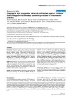
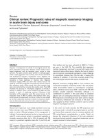
was 25. The 1-, 3- and 5-year overall survival rate was
99.1%, 96.8% and 93.1%, respectively (Fig. 1).
Page 2 of 6
Table 1 Clinicopathological characteristics of early gastric
cancer patients
Characteristics
No. of patients
Percent
Male
444
75.6
Female
143
24.4
Gender
Age
≤ 60
368
62.7
> 60
219
37.3
Upper third
102
17.4
Middle third
100
17.0
Lower third
385
65.6
≤2
365
62.2
>2
222
37.8
Well differentiated
186
31.7
Moderately differentiated
163
27.8
Poorly differentiated
220
37.5
Signet ring cell or Mucinous
18
3.0
T1a
255
43.4
T1b
332
56.6
N0
495
84.3
N1
55
9.4
N2
29
4.9
N3
8
1.4
Tumor location
Tumor size (cm)
Pathological type
Tumor depth
Lymph node metastasis
The positive rates of the four markers were summarized in Table 2. The positive rate of CEA, CA19–9, AFP
and CA125 level were 4.3%, 4.8%, 1.5% and 1.9%,
respectively. The highest positive rate was 8.2% for
combination of two markers (CA19–9 and CEA), 9.4%
for combination of three markers (CA19–9, CEA and
AFP or CA19–9, CEA and CA125), and 10.4% for combination of all four markers.
Considering the extremely low positive rates of AFP
and CA125, we only analyzed the correlation between
level of CEA and CA19–9 and clinicopathological features. No association was found between CEA level and
clinicopathological features (Table 3). However, elevation
of CA19–9 level was correlated with female gender and
presence of LNM (Table 4).
Prognostic factors for early gastric cancer patients
were analyzed using univariate analysis (Table 5). The
results showed that age, LNM and CEA level were
Feng et al. BMC Cancer (2017) 17:737
Page 3 of 6
Fig. 1 Overall survival of early gastric cancer patients
prognostic factors for early gastric cancer. The variables used for adjustment in the multivariate analyses
were age, LNM and CEA level. The results showed
that age, LNM and CEA level were independent prognostic factors according to multivariate analysis
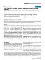
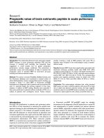
(Table 6). The overall survival of early gastric cancer
patients according to the levels of CEA and CA19–9
were shown in Figs. 2 and 3.
Discussion
Serum tumor markers are widely applied in the diagnosis, treatment effect assessment and disease monitoring [12]. Up to date, a series of studies have
explored the diagnostic and prognostic value of various serum tumor markers for gastric cancer [5].
However, no study has explored the diagnostic and
prognostic value of serum tumor markers for early
gastric cancer. Our present study found that the positive rates of serum CEA, CA19–9, APF and CA125
were relatively low for early gastric cancer. Elevation
Table 3 Comparison of clinicopathological characteristics
between two groups stratified by CEA level
Characteristics
CEA(−)
CEA(+)
P
Male
422
22
0.161
Female
140
3
Gender
Age
≤ 60
351
17
> 60
211
8
99
3
Tumor location
Upper third
Middle third
95
5
Lower third
368
17
≤2
346
19
>2
216
6
Well differentiated
180
6
Table 2 Positive rates of single and combined tumor markers in
early gastric cancer patients
Moderately differentiated
153
10
Poorly differentiated
212
8
Tumor marker
Signet ring cell or Mucinous
17
1
T1a
245
9
T1b
317
16
CEA
25(4.3%)
CA19–9
28(4.8%)
AFP
CA125
CA19–9
AFP
CA125
48(8.2%)
31(5.3%)
35(6.0%)
37(6.3%)
33(5.6%)
9(1.5%)
20(3.4%)
0.205
Pathological type
0.537
Tumor depth
0.539
Lymph node metastasis
11(1.9%)
55(9.4%)
N0
474
21
N1
53
2
CA19–9 + AFP
44(7.5%)
N2
28
1
CEA + CA19–9 + AFP
61(10.4%)
N3
7
1
CEA + AFP
0.744
Tumor size (cm)
41(7.0%)
CEA + CA19–9
0.675
55(9.4%)
0.698
Feng et al. BMC Cancer (2017) 17:737
Page 4 of 6
Table 4 Comparison of clinicopathological characteristics
between two groups stratified by CA 19–9 level
Characteristics
Table 6 Multivariate analysis of prognostic factors for early
gastric cancer
CA19–9(−)
CA19–9(+)
P
Male
428
16
0.025
Female
131
12
Gender
Prognostic factors
β
Hazard ratio (95% CI)
P value
Age
1.379
3.971(1.671–9.435)
0.002
Lymph node metastasis
0.682
1.978(1.248–3.136)
0.004
CEA
1.284
3.611(1.065–12.245)
0.039
Age
≤ 60
351
17
> 60
208
11
95
7
0.843
Tumor location
Upper third
Middle third
96
4
Lower third
368
17
≤2
345
20
>2
214
8
Well differentiated
178
8
Moderately differentiated
156
7
0.543
Tumor size (cm)
0.327
Pathological type
Poorly differentiated
208
12
Signet ring cell or Mucinous
17
1
T1a
243
12
T1b
316
16
N0
475
20
N1
52
3
N2
26
3
N3
6
2
0.936
Tumor depth
1.000
Lymph node metastasis
0.020
Table 5 Univariate analysis of prognostic factors for early gastric
cancer
Prognostic factors
β
Hazard ratio (95% CI)
P value
Gender
0.105
1.110(0.443–2.783)
0.824
Age
1.195
3.304(1.425–7.661)
0.005
Tumor location
−0.283
0.754(0.478–1.189)
0.224
Tumor size
−0.687
0.503(0.201–1.260)
0.142
Pathological type
−0.388
0.679(0.431–1.067)
0.093
Tumor depth
0.736
2.088(0.831–5.241)
0.117
Lymph node metastasis
0.577
1.781(1.124–2.821)
0.014
CEA
1.404
4.070(1.208–13.713)
0.024
CA19–9
0.576
1.779(0.419–7.546)
0.435
AFP
−3.019
0.049(0.000–590,647.114)
0.717
CA125
0.740
2.095(0.283–15.490)
0.469
of CA19–9 level was correlated with female gender
and presence of LNM. Elevation of CEA level was an
independent risk factor for the poor prognosis of
early gastric cancer.
The positive rates of the four markers for early
gastric cancer varied widely. It was reported that the
positive rate was 4.4%–15.4% for CEA [13–15],
11.7% for CA19–9 [15], 2.5%–3.3% for AFP [16, 17]
and 6.7% for CA125 [17]. In the present study, the
positive rates of all four tumor markers were lower
than previous reports. Even with the combination of
four tumor markers, the positive rate was only
10.4%. This indicated that the diagnostic value of the
four tumor markers was extremely low for early gastric cancer.
A strong correlation between elevated tumor
markers and clinicopathological features has been reported previously. It was reported that serum CEA
level was correlated with tumor depth, LNM [13] and
liver metastasis [18]. Other studies have reported that
CA19–9 level was correlated with tumor depth, LNM
and tumor stage [19, 20]. However, the association
between tumor markers and the clinicopathological
features of early gastric cancer has not been investigated yet. In our present study, no association was
found between CEA level and clinicopathological features. However, elevation of CA19–9 level was correlated with female gender and presence of LNM.
Early gastric cancer has a favorable outcome after
radical gastrectomy. The preoperative tumor markers
have been reported as valuable predictors for the
prognosis of gastric cancer. A meta-analysis containing 14,651 gastric cancer patients demonstrated that
serum CEA level was an independent prognostic factor for gastric cancer [8]. Another meta-analysis revealed that CEA protein and mRNA levels in
peritoneal lavage were associated with peritoneal recurrence after radical gastrectomy [21]. A metaanalysis containing 11,408 gastric cancer patients
showed that elevated serum CA19–9 level was correlated with poor prognosis [22]. Elevated AFP level
was reported to be associated with liver metastasis
and poor prognosis of gastric cancer [10, 23, 24]. Elevation of peritoneal lavage CA125 level was correlated
with peritoneal dissemination and poor outcomes of
Feng et al. BMC Cancer (2017) 17:737
Page 5 of 6
Fig. 2 Overall survival of early gastric cancer patients stratified by CEA level
gastric cancer [25]. However, the prognostic value of
these tumor markers for early gastric cancer was unclear. In our study, considering the extremely low
positive rate of AFP and CA125 level, only the prognostic significance of CEA and CA19–9 level were
analyzed. The results showed that serum CEA level
was an independent prognostic factor for early gastric
cancer. However, serum CA19–9 level had no prognostic significance.
There are some limitations in our study. Firstly, we
did not evaluate the predictive value of postoperative
levels of serum tumor markers for recurrence patterns
and prognosis of early gastric cancer. Secondly, the
sample size was not large enough, and the positive
rate of tumor markers was relatively low, which may
result in bias during analysis. Thirdly, mortality was
extremely low in early gastric cancer, which will influence the prognostic significance analysis of tumor
markers.
Conclusions
The positive rates of CEA, CA19–9, APF and CA125
were relatively low for early gastric cancer. Elevation of
CA19–9 level was associated with female gender and
presence of lymph node metastasis. Elevation of CEA
level was an independent risk factor for the poor prognosis of early gastric cancer.
Fig. 3 Overall survival of early gastric cancer patients stratified by CA19–9 level
Feng et al. BMC Cancer (2017) 17:737
Page 6 of 6
Abbreviations
AFP: alpha-fetoprotein; CA125: cancer antigen 125; CA19–9: carbohydrate
associated antigen 19–9; CEA: carcinoembryonic antigen; LNM: lymph node
metastasis
4.
Acknowledgments
We wish to thank Xingbin Hu for his help with the revision of manuscript.
6.
Funding
This study was supported in part by grants from the National Natural
Scientific Foundation of China [NO. 31100643, 31,570,907, 81,572,306,
81,502,403, XJZT12Z03]. The funding body had no role in the design of the
study and collection, analysis, and interpretation of data and in writing of
this manuscript.
7.
Availability of data and materials
The datasets used and/or analysed during the current study are available
from the corresponding author on reasonable request.
5.
8.
9.
10.
11.
Authors’ contributions
FF, TYZ and XGH conceived the study and drafted the manuscript. LZ, LSS
and ZGZ collected the data and participated in drafting the manuscript. GM
and LX performed statistical analysis. FDM designed the study and revised
the manuscript. ZHW designed and supervised the study. All authors read
and approved the final manuscript. All authors contributed to the writing of
the manuscript and provided final approval of the manuscript. All authors
have read and approved the final version of this manuscript. All authors
agreed to be accountable for all aspects of the work in ensuring that
questions related to the accuracy or integrity of any part of the work are
appropriately investigated and resolved.
12.
Authors’ information
Not further applicable.
16.
Ethics approval and consent to participate
This study was approved by the Ethics Committee of Xijing Hospital, and
written informed consent was obtained from the patients in our center.
17.
13.
14.
15.
18.
Consent for publication
Not applicable.
Competing interests
There are no financial or other relations that could lead to a conflict of
interest. Prof. Daiming Fan, one of co-authors in the present study, is a member of the editorial board of this journal.
19.
20.
21.
Publisher’s Note
Springer Nature remains neutral with regard to jurisdictional claims in
published maps and institutional affiliations.
Author details
1
Division of Digestive Surgery, Xijing Hospital of Digestive Diseases, the
Fourth Military Medical University, 127 West Changle Road, 710032, , Xian,
Shaanxi, China. 2Department of Dermatology, Xijing Hospital, the Fourth
Military Medical University, 127 West Changle Road, 710032, , Xian, Shaanxi,
China.
Received: 1 August 2016 Accepted: 30 October 2017
References
1. Jemal A, Bray F, Center MM, Ferlay J, Ward E, Forman D. Global cancer
statistics. CA Cancer J Clin. 2011;61(2):69–90.
2. Feng F, Sun L, Xu G, Cai L, Hong L, Yang J, et al. Is it reasonable to treat
early gastric cancer with mucosal infiltration and well differentiation by
endoscopic submucosal resection? J Gastrointest Surg. 2015;19(12):2111–9.
3. Zhu L, Qin J, Wang J, Guo T, Wang Z, Yang J. Early gastric cancer. Current
Advances of Endoscopic Diagnosis and Treatment Gastroenterol Res Pract.
2016;2016:9638041.
22.
23.
24.
25.
Tian SB, JC Y, Kang WM, Ma ZQ, Ye X, Cao ZJ, et al. Combined detection of
CEA, CA 19-9, CA 242 and CA 50 in the diagnosis and prognosis of
resectable gastric cancer. Asian Pac J Cancer Prev. 2014;15(15):6295–300.
Jin Z, Jiang W, Wang L. Biomarkers for gastric cancer. Progression in early
diagnosis and prognosis (review). Oncol Lett. 2015;9(4):1502–8.
Pyo JH, Lee H, Min BH, Lee JH, Choi MG, Lee JH, et al. Long-term outcome
of endoscopic resection vs. surgery for early gastric cancer: a non-inferioritymatched cohort study. Am J Gastroenterol. 2016;111(2):240–9.
Huang B, Wang Z, Xing C, Sun Z, Zhao B, Long-term XH. Survival results and
prognostic factors of early gastric cancer. EXP THER MED. 2011;2(6):1059–64.
Deng K, Yang L, Hu B, Wu H, Zhu H, Tang C. The prognostic significance of
pretreatment serum CEA levels in gastric cancer: a meta-analysis including
14651 patients. PLoS One. 2015;10(4):e124151.
Xiao J, He X, Wang Z, Hu J, Sun F, Qi F, et al. Serum carbohydrate antigen 19-9
and prognosis of patients with gastric cancer. Tumour Biol. 2014;35(2):1331–4.
Liu X, Cheng Y, Sheng W, Lu H, Xu Y, Long Z, et al. Clinicopathologic
features and prognostic factors in alpha-fetoprotein-producing gastric
cancers: analysis of 104 cases. J Surg Oncol. 2010;102(3):249–55.
Japanese gastric cancer treatment guidelines 2010 (ver. 3). Gastric Cancer.
2011;14(2):113–23.
Rodriguez-Enriquez S, Pacheco-Velazquez SC, Gallardo-Perez JC, MarinHernandez A, Aguilar-Ponce JL, Ruiz-Garcia E, et al. Multi-biomarker pattern for
tumor identification and prognosis. J Cell Biochem. 2011;112(10):2703–15.
Park SH, Ku KB, Chung HY, Yu W. Prognostic significance of serum and
tissue carcinoembryonic antigen in patients with gastric adenocarcinomas.
Cancer Res Treat. 2008;40(1):16–21.
Wang W, Chen XL, Zhao SY, YH X, Zhang WH, Liu K, et al. Prognostic
significance of preoperative serum CA125, CA19-9 and CEA in gastric
carcinoma. Oncotarget. 2016;7(23):35423–36.
Liang Y, Wang W, Fang C, Raj SS, Hu WM, Li QW, et al. Clinical significance
and diagnostic value of serum CEA, CA19-9 and CA72-4 in patients with
gastric cancer. Oncotarget. 2016;7(31):49565–73.
Wang D, Li C, Xu Y, Xing Y, Qu L, Guo Y, et al. Clinicopathological
characteristics and prognosis of alpha-fetoprotein positive gastric cancer in
Chinese patients. Int J Clin Exp Pathol. 2015;8(6):6345–55.
He CZ, Zhang KH, Li Q, Liu XH, Hong Y, Lv NH. Combined use of AFP, CEA,
CA125 and CAl9-9 improves the sensitivity for the diagnosis of gastric
cancer. BMC Gastroenterol. 2013;13:87.
Ucar E, Semerci E, Ustun H, Yetim T, Huzmeli C, Gullu M. Prognostic value of
preoperative CEA, CA 19-9, CA 72-4, and AFP levels in gastric cancer. Adv
Ther. 2008;25(10):1075–84.
Sisik A, Kaya M, Bas G, Basak F, Alimoglu OCEA. CA 19-9 are still valuable
markers for the prognosis of colorectal and gastric cancer patients. Asian
Pac J Cancer Prev. 2013;14(7):4289–94.
Kochi M, Fujii M, Kanamori N, Kaiga T, Kawakami T, Aizaki K, et al. Evaluation
of serum CEA and CA19-9 levels as prognostic factors in patients with
gastric cancer. Gastric Cancer. 2000;3(4):177–86.
Xiao Y, Zhang J, He X, Ji J, Wang G. Diagnostic values of carcinoembryonic
antigen in predicting peritoneal recurrence after curative resection of
gastric cancer: a meta-analysis. Ir J Med Sci. 2014;183(4):557–64.
Song YX, Huang XZ, Gao P, Sun JX, Chen XW, Yang YC, et al.
Clinicopathologic and prognostic value of serum carbohydrate antigen 19-9
in gastric cancer: a meta-analysis. Dis Markers 2015, 2015:549843.
Chen Y, Qu H, Jian M, Sun G, He Q. High level of serum AFP is an
independent negative prognostic factor in gastric cancer. Int J Biol Markers.
2015;30(4):e387–93.
Zuo C, An JQ. Analysis on clinical characteristics and prognosis of patients
with serum alpha-fetoprotein-positive gastric cancer. Minerva Med. 2015;
106(4):185–91.
Yamamoto M, Baba H, Toh Y, Okamura T, Maehara Y. Peritoneal lavage CEA/
CA125 is a prognostic factor for gastric cancer patients. J Cancer Res Clin
Oncol. 2007;133(7):471–6.