LINE-1 hypermethylation in white blood cell DNA is associated with high-grade cervical intraepithelial neoplasia
Bạn đang xem bản rút gọn của tài liệu. Xem và tải ngay bản đầy đủ của tài liệu tại đây (515.75 KB, 10 trang )
Barchitta et al. BMC Cancer (2017) 17:601
DOI 10.1186/s12885-017-3582-0
RESEARCH ARTICLE
Open Access
LINE-1 hypermethylation in white blood
cell DNA is associated with high-grade
cervical intraepithelial neoplasia
Martina Barchitta1, Annalisa Quattrocchi1, Andrea Maugeri1, Carolina Canto2, Nadia La Rosa3,
Maria Antonietta Cantarella3, Giuseppa Spampinato3, Aurora Scalisi3 and Antonella Agodi1*
Abstract
Background: Long Interspersed Nuclear Elements-1 (LINEs-1) methylation from white blood cells (WBCs) DNA has
been proposed as biomarker associated with different types of cancer. The aim of the present study was to investigate
the degree of WBCs LINE-1 methylation, according to high-risk Human Papilloma Virus (hrHPV) status in a healthy
population, and the association with high-grade Cervical Intraepithelial Neoplasia (CIN2+) in hrHPV positive women.
Methods: Women with abnormal cervical cells were enrolled and classified by histological diagnosis and hrHPV
infection. A structured questionnaire was used to obtain information on socio-demographic variables and lifestyle
factors. LINE-1 methylation level in WBCs was measured by pyrosequencing-based methylation analysis after bisulfite
conversion.
Results: Among 252 women diagnosed with normal cervical epithelium, with regard to LINE-1 methylation level no
significant difference was observed between hrHPV positive and hrHPV negative women, also adjusting for known risk
factors of infection. The association between WBCs LINE-1 methylation and CIN2+ status was analyzed in hrHPV positive
women. The median value of LINE-1 methylation levels was higher in cases (CIN2+) than in controls (75.00% versus 73.17%;
p = 0.002). For a one-unit increase in LINE-1 methylation level, the odds of being diagnosed with CIN2+ increased by 10%,
adjusting for known factors related to LINE-1 methylation (adjOR: 1.10; 95% CI:1.01–1.20; p = 0.032). The Receiver-Operating
Characteristic (ROC) curve analysis identified the cut-off value of 73.8% as the best threshold to separate cases from controls
(sensitivity: 63.4% and specificity: 61.8%).
Conclusions: LINE-1 methylation status in WBCs DNA may represent a cost-effective and tissue-accessible biomarker for
high-grade CIN in hrHPV positive women. However, LINE-1 hypermethylation cannot be considered specific for cervical
cancer (CC) and a model based solely on LINE-1 methylation levels has limited performance. Further investigations are
necessary to propose and validate a novel methylation biomarker panel, based on LINE-1 methylation and other
differentially methylated regions, for the screening of women at risk of CC.
Keywords: LINE-1 methylation, Global DNA methylation, Hypermethylation, Cervical cancer, Cervical Intraepitelial
Neoplasia, ROC curve analysis, Pyrosequencing-based methylation analysis, Prevention
* Correspondence:
1
Department of Medical and Surgical Sciences and Advanced Technologies
“GF Ingrassia”, University of Catania, via S. Sofia, 87, 95121 Catania, Italy
Full list of author information is available at the end of the article
© The Author(s). 2017 Open Access This article is distributed under the terms of the Creative Commons Attribution 4.0
International License ( which permits unrestricted use, distribution, and
reproduction in any medium, provided you give appropriate credit to the original author(s) and the source, provide a link to
the Creative Commons license, and indicate if changes were made. The Creative Commons Public Domain Dedication waiver
( applies to the data made available in this article, unless otherwise stated.
Barchitta et al. BMC Cancer (2017) 17:601
Background
Cervical cancer (CC) is the fourth most common cancer
and an important cause of death worldwide [1]. CC
arises through a multistage process of carcinogenesis,
and persistence of high risk Human Papilloma Virus
(hrHPV) infection represents the major etiological factor
for neoplasia development [2–4], through the progression of precursor lesions (i.e. Cervical Intraepithelial
Neoplasia, CIN) to invasive cancer [5, 6]. Among the
putative molecular alterations leading to morphological
modifications, aberrant DNA methylation might be an
important event in cervical carcinogenesis [7, 8]. DNA
methylation at specific CpG sites in hrHPV or in human genes has shown the potential for the detection of
CIN2+ and some biomarkers have been proposed [8–15].
Methylation in repetitive elements has been shown to
correlate with global genomic DNA methylation, as a
result of the high occurrence of these sequences
throughout the genome [16]. Methylation of Long Interspersed Nuclear Elements - 1 (LINEs-1) has been
proposed as a surrogate marker for estimating the global
DNA methylation levels in cancer tissues [17] and in
peripheral blood samples [18]. Furthermore, a systematic
review and meta-analysis reported that LINE-1 methylation levels were significantly lower in cancer patients
compared to healthy controls in tissue samples but not
in blood [19]. However, several studies have shown that
LINE-1 hypo- and hyper-methylation from white blood
cells (WBCs) DNA are associated with different types of
cancer [20–32]. Particularly, evidence from women
recruited in the “Prognostic Significance of DNA &
Histone Methylation” project showed that a higher degree of LINE-1 methylation in peripheral blood mononuclear cells (PBMCs) was associated with lower risk of
CIN2+ [8]. Although susceptibility to hrHPV related
carcinogenesis may also be an epigenetically modified
process, further studies are needed to clarify the association between HPV status and LINE methylation [33].
The aims of the present study were to investigate the
degree of WBCs LINE-1 methylation, by bisulfite pyrosequencing, in a population of women referring to a cervical cancer screening program and to evaluate the
association with their hrHPV status, and with high-grade
CIN in the hrHPV positive women subgroup.
Methods
Study design
During a three-years period (from 2013 to 2015), all
women diagnosed with abnormal PAP test, referring to
the cervical cancer screening unit (Unità Operativa di
Screening Ginecologico) at the Azienda Sanitaria Provinciale
(ASP 3) in Catania (Italy), for further examination by
colposcopy and biopsy, were invited to participate in a
cross-sectional study.
Page 2 of 10
The study protocol was approved by the ethics committee of the involved Institution (CE Catania 2; Prot. N.
227/BE and 275/BE) and performed according to the
Declaration of Helsinki. Participants were fully informed
of the purpose and procedures of the study, and a signed
written consent was obtained.
Women were classified by histological diagnosis and
tested for hrHPV (hrHPV16, 18, 31, 33, 35, 39, 45, 51,
52, 56, 58, 59, and 68) using digene HC2 HPV DNA Test
(Qiagen, Italy). Thus, women were classified as hrHPV
positive if they were infected with any of the thirteen
hrHPV types, otherwise women were classified as
hrHPV negative. Notably, the specific HPV genotype is
not provided by the test.
Women who tested positive for hrHPV were further
classified as cases (CIN2+: CIN2, CIN3 or carcinoma in
situ - CIS) or controls (≤CIN1: CIN1 or normal cervical
epithelium), according to the histological result.
A structured questionnaire was used by trained epidemiologists to obtain information on socio-demographic
variables and lifestyle factors. Women were classified
into two categories of educational level: low-medium
(primary school, i.e., ≤8 years of school) and high
education level (high school education or greater, i.e.,
>8 years of school). Body mass index (BMI) was calculated
based on criteria from the World Health Organization [34].
DNA extraction and methylation analysis
Genomic DNA was extracted from whole blood using
the Illustra blood genomic Prep Mini Spin Kit (GE
Healthcare, Italy) according to the manufacturer’s protocol. LINE-1 methylation level in WBCs was measured by
pyrosequencing-based methylation analysis, using the
PyroMark Q24 instrument (Qiagen, Italy), as previously
reported [35]. Briefly, bisulfite conversion and clean-up of
DNA for methylation analysis of 30–40 ng of WBCs
DNA were completed using the EpiTect Bisulfite Kit
(Qiagen, Italy) and the converted DNA was eluted in
20 μl of Eluition Buffer.
PCR was conducted in a reaction volume of 25 μl,
using the PyroMark PCR Kit (Qiagen, Italy). According
to the manufacturer’s instructions, each reaction mixture
contained 1.5 μl of bisulfite-converted DNA, 12.5 μl of
PyroMark PCR Master Mix 2X, containing HotStartTaq
DNA Polymerase, 2.5 μl of Coral Load Concentrate 10X,
2 μl of the forward primer (5′-TTTTGAGTTA
GGTGTGGGATATA-3′) and the reverse-biotinylated
primer (5′-biotin-AAAATCAAAAAATTCCCTTTC-3′)
(0.2 μM for each) [36]. HotStart PCR cycling conditions
were 1 cycle at 95 °C for 15 min, 40 cycles at 94 °C for
30 s, 50 °C for 30 s, and 72 °C for 30s, and a final
extension at 72 °C for 10 min. Then, the PCR product
underwent pyrosequencing using 0.3 mM of the sequencing primer (5′-AGTTAGGTGTGGGATATAGT-3′).
Barchitta et al. BMC Cancer (2017) 17:601
All runs included 0% and 100% methylated human DNA
as positive controls and a nontemplate control. Any
failed LINE-1 methylation assays were excluded from
the statistical analysis.
The degree of methylation was expressed for each DNA
locus as percentage of methylated cytosines over the sum
of methylated and unmethylated cytosines. The degree of
LINE-1 methylation was reported for each locus as well as
the average percentage of methylation of the three evaluated CpG sites (GenBank Accession No. X58075).
Statistical analyses
Statistical analyses were performed using the SPSS software (version 22.0, SPSS, Chicago, IL). Descriptive statistics were used to characterize the population using
frequencies, means ± standard deviations (SDs), median
values and interquartile ranges (IQRs). The two-tailed
Chi-squared test was used for the statistical comparison
of proportions, whereas continuous variables were tested
using Student’s t test.
The Kolmogorov-Smirnov test was performed to determine whether LINE-1 methylation levels were normally
distributed. Accordingly, median LINE-1 methylation
levels were compared, between case and control groups,
using the Mann–Whitney U test. Correlation between
LINE-1 methylation level and continuous variables was
also evaluated using Pearson correlation coefficient.
In order to measure the strength of the association between categorical variables, the crude odds ratios (ORs)
and the corresponding 95% confidence intervals (95% CIs)
were computed. Unconditional multivariable logistic
regression analyses were used to evaluate the association
between the degree of LINE-1 methylation, hrHPV infection and CIN status. The analyses were adjusted for age
(continuous), BMI (continuous), smoking status (current
smokers vs non-smokers/former smokers), and parity (< 1
live births vs ≥ 1 live births). The adjusted ORs with the
respective 95% CIs were reported. A p value <0.05 was
considered statistically significant in all performed analyses.
The Receiver-Operating Characteristic (ROC) curve
analysis was performed in order to separate cases from
controls, according to mean LINE-1 methylation percentage. Area Under the Curve (AUC) and 95% CIs were
calculated to assess the performance (sensitivity and specificity) of the test for each methylation value. To determine the optimal threshold of LINE-1 methylation level,
suitable to distinguish cases from controls, the point on
the ROC curve with the shortest distance value from the
top left corner (point: 0,1) was calculated using the
formula [(1 – sensitivity)2 + (1 – specificity)2] [37].
Results
Overall, 539 women with abnormal PAP test were classified by histological diagnosis and tested for hrHPV.
Page 3 of 10
Among these, 252 were diagnosed with normal cervical
epithelium (46.7%), 160 CIN1 (29.7%), 57 CIN2 (10.6%),
67 (12.4%) CIN3 and 3 (0.6%) CIS. With regard to
hrHPV status, women were classified as hrHPV positive
(hrHPV+; N = 302; 56%) and hrHPV negative (hrHPV-;
N = 237; 44%). The analysis of WBC LINE-1 methylation level was performed on women who provided blood
sample for DNA analysis and the following results refer
to this subgroup of women (N = 260). Notably, comparing women who provided blood samples with those who
did not, no significant differences for socio-demographic
and life-style factors were observed (data not shown).
Differences in WBC LINE-1 methylation levels,
according to hrHPV status, were analyzed among
women with normal cervical epithelium. Among the 252
women diagnosed with normal cervical epithelium, 96
had provided the blood sample for methylation analyses
and were further classified as hrHPV- (N = 64) and
hrHPV+ (N = 32). Table 1 displays the characteristics of
women diagnosed with normal cervical epithelium according to hrHPV status. Particularly, the odds of being
diagnosed with hrHPV infection increased among
younger women (≤median age) (OR = 2.4; 95% CI = 1.0–
5.8; p = 0.043), smokers (OR = 2.6; 95% CI = 1.1–6.2;
p = 0.035), underweight-normal weight (OR = 3.2; 95%
CI = 1.1–9.5; p = 0.028) and nulliparous women
(OR = 4.9; 95% CI = 1.7–14.1; p = 0.002).
Mean LINE-1 methylation level was 73.57% (median = 74.00%) and no significant difference was observed
between hrHPV- and hrHPV+ women (Table 2). Results
by multivariable logistic regression analysis showed that
changes in LINE-1 methylation level were not associated
with hrHPV status, adjusting for age, BMI, smoking status
and parity (Table 3).
Table 1 Characteristics of healthy women according to hrHPV
status
Characteristics
hrHPV+
(n = 32)
hrHPV(n = 64)
p-valuea
Age (mean ± SD)
38.50 ± 9.54
42.39 ± 9.47
0.061
Smoking status (current)
50.0%
28.1%
0.035
BMI (mean ± SD)
22.26 ± 3.93
24.73 ± 5.31
0.022
0.030
Nutritional status
Underweight
15.6%
3.1%
Normal weight
68.8%
59.4%
Overweight
12.5%
20.3%
Obese
3.1%
17.2%
Parity (≥1 live births)
62.5%
89.1%
0.002
Education level (low)
37.5%
35.9%
0.881
Oral contraceptive use (yes)
12.5%
7.8%
0.458
Abbreviations: SD standard deviation, BMI Body Mass Index
a
Statistically significant p values (p < 0.05) are indicated in bold font
Barchitta et al. BMC Cancer (2017) 17:601
Page 4 of 10
Table 2 Differences in LINE-1 methylation levels between
hrHPV+ and hrHPV- women
LINE-1 methylation
levels
hrHPV+ (n = 32)
hrHPV- (n = 64)
Median
IQR
Median
IQR
Site 1
67.00
17.00
71.00
18.00
Table 4 Characteristics of hrHPV positive women according to
cases/controls classification
p-value
0.498
Site 2
75.00
5.00
75.00
5.00
0.640
Site 3
77.00
6.00
76.00
3.00
0.913
Mean (all three sites)
73.50
3.83
74.33
4.25
0.407
Abbreviations: LINE-1 Long Interspersed Nucleotide Element- 1, IQR
Interquartile range
Among the 302 hrHPV positive women, 139 have provided the blood sample for methylation analyses and
were further classified as cases (n = 71; 51.1%), diagnosed as CIN 2 [n = 28], CIN 3 [n = 42] or CIS [n = 1],
and controls (n = 68; 48.9%) including CIN 1 [n = 36] or
normal cervical epithelium [n = 32].
Table 4 shows the characteristics of hrHPV+ women
according to cases/controls classification. Taking into account socio-demographic variables and lifestyle factors,
no statistically significant differences were observed between cases and controls. Mean LINE-1 methylation
levels were 71.83 ± 10.20 (site 1), 74.28 ± 5.30 (site 2)
and 76.91 ± 3.91 (site 3), respectively. No significant differences in LINE-1 methylation levels were observed according to age, BMI, smoking status, parity and oral
contraceptive use (data not shown).
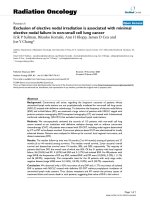
Table 5 and Fig. 1 show differences in LINE-1 methylation levels between cases and controls. Particularly,
overall mean LINE-1 methylation level, and site 3,
were higher in cases compared with controls (p = 0.002
and p = 0.032, respectively). Accordingly, logistic regression analysis showed a 1.1-fold increased odds of CIN2+
diagnosis associated with 1 unit increase in LINE-1
methylation level, adjusting for known factors related to
LINE-1 methylation, such as age, BMI and smoking status
(adjOR: 1.10; 95% CI:1.01–1.20; p = 0.032) (Table 6).
Table 3 Association between hrHPV status and LINE-1 methylation
levels (logistic regression analysis adjusting for age, BMI, smoking
status and parity)
β (SE)
p-valuea adjOR 95% CI
Lower Upper
Characteristics
Cases
(n = 71)
Controls
(n = 68)
p-value
Age (mean ± SD)
36.10 ± 7.88
37.84 ± 9.28
0.235
Smoking status (current)
49.3%
50.0%
0.934
BMI (mean ± SD)
22.89 ± 3.74
22.44 ± 3.63
0.470
0.928
Nutritional status
Underweight
11.3%
11.8%
Normal weight
62.0%
66.2%
Overweight
22.5%
19.1%
Obese
4.2%
2.9%
Parity (≥1 live births)
64.8%
54.4%
0.212
Education level (low)
46.5%
35.3%
0.180
Oral contraceptive use (yes)
14.1%
11.8%
0.684
Abbreviations: SD standard deviation, BMI Body Mass Index
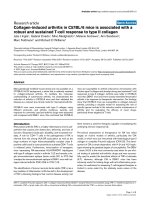
To evaluate the performance of a model, based on
LINE-1 methylation status, to distinguish cases from
controls, an ROC curve analysis was performed. Figure
2 shows the ROC curve for detecting CIN2+ based on
LINE-1 methylation level (AUC = 0.652, 95% CI = 0.560–
0.744; p = 0.002). According to the definition of the
minimum distance on the ROC curve from the (0,1)
point (distance: 0.280), the cut-off value of 73.83% was
the best threshold to separate cases from controls (sensitivity: 63.4% and specificity: 61.8%).
Discussion
Identification of high-grade CIN lesions (CIN2+) by organized screening programs has shown high efficacy in
reducing CC incidence and mortality worldwide [38, 39].
Since evidence from large randomized controlled trials
demonstrated that hrHPV testing is more sensitive than
cytology testing [40–44], the Italian Ministry of Health has
recommended that regions shift toward HPV-based
screening and has provided guidelines for its application
[45, 46]. The identification of hrHPV+ women who are
at risk of CIN2+ and CC and the validation of new
Table 5 Differences in LINE-1 methylation levels between cases
and controls
0.011 (0.047)
0.809
1.01
0.92
1.11
LINE-1 methylation
levels
Cases (n = 71)
Median
IQR
Median
IQR
Age (continuous)
−0.014 (0.28)
0.633
0.97
0.93
1.04
Site 1
70.00
21.00
66.00
17.00
BMI (continuous)
−0.076 (0.062) 0.220
0.93
0.82
1.05
Site 2
76.00
4.00
75.00
5.00
0.090
Smoking status
(current)
0.997 (0.493)
0.043
2.71
1.03
7.12
Site 3
78.00
3.00
77.00
5.00
0.032
Mean (all three sites)
75.00
6.00
73.17
2.92
0.002
Parity (<1 live births) 1.302 (0.629)
0.038
3.68
1.07
12.61
Abbreviations: SE standard error, adjOR adjusted Odds Ratio, CI Confidence
Interval, LINE-1 Long Interspersed Nuclear Element- 1
a
Statistically significant p values (p < 0.05) are indicated in bold font
Controls (n = 68)
p-valuea
LINE-1 methylation
level (continuous)
0.103
Abbreviations: LINE-1 Long Interspersed Nuclear Element- 1, IQR
Interquartile range
a
Statistically significant p values (p < 0.05), based on the Mann-Whitney U test,
are indicated in bold font
Barchitta et al. BMC Cancer (2017) 17:601
Page 5 of 10
Fig. 1 Methylation levels of LINE-1 in cases (CIN2+) and controls (≤CIN1). Mean methylation levels of LINE-1 sequences (mean percentage of
methylation of the three evaluated CpG sites) in CIN2+ patients (cases) and in CIN 1 or normal cervical epithelium patients (controls) obtained
using pyrosequencing of bisulfite converted DNA from WBCs (p-value = 0.002, based on the Mann-Whitney U test)
biomarkers of disease progression are big challenges for
the management of cervical abnormalities [46]. Particularly, the validation of blood-based methylation biomarkers is of great interest because they are easier to
obtain and adaptable to population screening for the
identification of cancer-affected individuals or those who
are at higher risk of cancer. Among cancer patients and
healthy controls, recent systematic reviews and metaanalyses have shown significantly different LINE-1
methylation levels in tissue samples [19], but not in
blood leukocytes [19, 47]. We investigated whether
LINE-1 methylation level in WBCs may represent a
Table 6 Association between LINE-1 methylation level and case
status (logistic regression analysis adjusting for age, BMI and
smoking status)
β (SE)
p-valuea
adjOR
Lower
Upper
LINE-1 methylation
level (continuous)
0.096 (0.045)
0.032
1.10
1.01
1.20
Age (continuous)
−0.030 (0.22)
0.178
0.97
0.93
1.01
BMI (continuous)
0.049 (0.051)
0.339
1.05
0.95
1.16
Smoking status
(current)
−0.044 (0.351)
0.900
0.96
0.48
1.90
95% CI
Abbreviations: SE standard error, adjOR adjusted Odds Ratio, CI Confidence
Interval, LINE-1 Long Interspersed Nuclear Element- 1
a
Statistically significant p values (p < 0.05) are indicated in bold font
biomarker of cervical precursor lesions and cancer in
hrHPV+ women. However, LINE-1 methylation has been
investigated in several types of cancer and cannot be
considered specific for CC. Furthermore, although the
mechanisms leading to LINE-1 methylation changes in
WBCs of cancer patients are currently uncertain, both
LINE-1 hypomethylation and hypermethylation have
been previously reported [21, 22, 32, 48–50].
Hypomethylation of repetitive elements which causes
chromosomal instability is considered a molecular biomarker of cancer cells. Several studies have shown reduced LINE-1 methylation levels in cancer tissues and
WBCs, especially in patients with head and neck, bladder and gastric cancer [27–32]. In contrast, other studies
on bladder, renal, colorectal, ovarian, pancreatic cancers
and cutaneous melanoma have reported higher LINE-1
methylation levels in WBCs of cancer patients [20–26].
A plausible explanation for this relationship is that
LINE-1 sequences with double strand DNA breaks had
higher methylation levels around the area of the break,
compared to DNA without double strand breaks [51].
Thus, the DNA damage and the increased frequency of
double strand DNA breaks in non-healthy individuals
could explain the hypermethylation in WBCs DNA.
At the best of our knowledge, only the study by
Piyathilake et al. [8] has currently evaluated the
association between LINE-1 methylation and CIN2+
Barchitta et al. BMC Cancer (2017) 17:601
Fig. 2 ROC curve analysis of LINE-1 methylation and CIN2+ detection.
ROC (Receiver Operator Characteristics) curve of LINE-1 methylation
levels for the detection of CIN2+. LINE-1 methylation level was suitable
for detecting CIN2+ with an AUC of 0.652 (95% CI = 0.560–0.744). The
cut-off value of 73.83% is the best threshold to separate cases
from controls
status, in blood samples. The degree of LINE-1 methylation
was lower in high grade CIN patients (mean = 63% ± 7%)
than in controls (mean = 64% ± 7%), albeit difference was
small. Particularly, the risk to be diagnosed with CIN2+
was lower among women in the highest tertile of LINE-1
methylation level (≥70%), compared to women in the lower
tertiles [8]. To support this association, the authors
assumed that higher LINE-1 methylation levels could mediate a positive effect on immune response against HPV
infection [8]. However, an in vitro study on squamous cell
carcinoma cell lines revealed higher LINE-1 methylation
level in HPV+ compared to HPV- cells [52]. This result
partially confirmed the positive correlation between the
maintenance of normal LINE methylation and HPVpositivity, observed by Richards et al. in head and neck cancer tissues and cell lines [33].
Accordingly, in order to investigate the potential association between WBC LINE-1 methylation level and
hrHPV status, we analyzed women with normal cervical
epithelium, to avoid the possibility of reverse causation
mediated by the carcinogenic process (i.e. the degree of
LINE-1 methylation could be influenced by the carcinogenic process). Results of our study showed that LINE-1
methylation levels were not different between hrHPV+
and hrHPV- women. Besides, the degree of LINE-1
methylation was not associated with hrHPV status, also
taking into account hrHPV related variables such as age,
Page 6 of 10
BMI, smoking status and parity. However, additional
studies are required to assess the role of LINE-1 methylation in cell-mediated response to HPV infection.
Among hrHPV+ women, we were able to show that
WBC LINE-1 methylation level was higher in subjects
diagnosed with CIN2+ (median = 75.00%; IQR = 73.00%–
79.00%), compared to healthy women and those with low
grade cervical lesions (median = 73.17%; IQR = 72.00%–
75.33%).
This small, but statistically significant, difference in
LINE-1 methylation levels could be due to factors that
influence the association between DNA methylation and
cancer risk [53]. For example, previous studies have
shown that global hypomethylation can occur with
increasing age [54, 55].
Since, in the present study, cases were younger than
controls, we analysed whether LINE-1 methylation levels
were different according to age. Consistently with results
from previous studies [28, 56–59], we did not observe
association between age and LINE-1 methylation levels
in WBCs DNA. Moreover, on the basis of a multivariable model, the association between LINE-1 methylation
and CIN2+ did not depend on age, BMI, and smoking
status. Particularly, for a one-unit increase in LINE-1
methylation level, the odds of being diagnosed with
CIN2+ increased by 10% (adjOR = 1.10; 95% CI:1.01–
1.20), adjusting for age, BMI, and smoking status. Thus,
the odds of being diagnosed with CIN2+ looked to be
slightly associated with LINE-1 methylation status.
However, the retrospective nature of our study did not
make it possible to establish whether the increase in
LINE-1 methylation level was a cause or a consequence
of tumor progression. Moreover, although the present
study did not show evidence of association between
LINE-1 methylation and other socio-demographic and
life-style factors, the contribution of other unmeasured
variables cannot be excluded. Particularly, previous
studies have reported the influence on LINE-1 methylation levels of MTHFR polymorphisms [60], diet, nutrient
intakes, folate deficiency [35] and amount of physical activity [61]. Thus, future studies should consider other influential factors to confirm the present findings.
In order to evaluate the potential use of LINE-1
methylation as a biomarker for CC risk, the optimal cutoff value, suitable to distinguish cases from controls, has
been assessed through an ROC curve analysis. Our results demonstrate that a model based on LINE-1 methylation level had limited performance for the diagnosis of
CIN2+ lesions, with moderate sensitivity (63.4%) and
specificity (61.8%). Moreover, the cut-off value (73.8%),
obtained from the ROC curve analysis, is very close to
median value of LINE-1 methylation in hrHPV+ healthy
controls (73.4%). Thus, results from ROC curve analysis
do not encourage the use of LINE-1 methylation as a
Barchitta et al. BMC Cancer (2017) 17:601
stand-alone blood-based biomarker for CC risk. Its
potential clinical value for the screening of women at risk
of CC needs to be evaluated by large prospective studies
and randomized controlled trials, which take into account
tumor progression through pre-neoplastic lesions.
However, a potential goal for the future would be that
a novel methylation biomarker panel, using LINE-1
methylation status and other differentially methylated
regions [62–64], could be proposed and validated for the
screening of women at risk of CC.
Strengths of this study consist in the use of protocols
and methodologies for blood collection, DNA extraction
and DNA methylation analysis consistent between cases
and controls. Moreover, to investigate difference and variability in LINE-1 methylation levels within histological
groups, data were analysed with a robust statistical
approach. The potential effect of hrHPV infection on
WBC LINE-1 methylation level was investigated in
women with normal cervical epithelium, also taking into
account hrHPV related risk factors, through a multivariable logistic regression model. The degree of LINE-1
methylation was not associated with hrHPV status, even
though we were not able to stratify the effect for specific
hrHPV types (i.e. HPV16, HPV18 and others).
As reported by the previous contrasting study [8],
difference in LINE-1 methylation levels between cases
and controls was modestly different. A multivariable
logistic regression model was applied to adjust our result
for factors that are commonly known to affect methylation biomarkers. Conversely to previously published
results [8], independent variables (i.e. LINE-1 methylation level, age and BMI) were entered in the regression
model as continuous variables, to avoid considerable loss
of statistical power and residual confounding caused by
dichotomization of continuous variables [65]. This
makes more accurate the interpretation of the coefficient
of LINE-1 methylation level in the regression model,
being able to partially explain controversial findings.
With regard to molecular analysis, precision and
reproducibility of the DNA methylation assay are very
important characteristics to assess the utility of LINE-1
methylation as a biomarker in clinical practice. High reliability and flexibility have made pyrosequencing of
bisulfite-treated DNA the “gold standard” [66, 67], and a
high-throughput and replicable methodology to evaluate
LINE-1 methylation as a surrogate marker for global
DNA methylation [66–70]. Furthermore, several studies
have reported that pyrosequencing has good precision at
higher methylation levels, and can provide a reliable
measure of LINE-1 methylation in WBC DNA [71–76].
Particularly, results by Iwagami et al. [77] indicate that
run-to-run variation of LINE-1 methylation degrees is
not large, and a single run of PCR pyrosequencing can
provide reasonably precise measures.
Page 7 of 10
Additional important issues should be considered
when interpreting results of the present study. Firstly,
LINE-1 methylation levels can vary depending on the
target CpG site and on the tissue type [68, 69]. The
distinctiveness of LINE-1 methylation levels discourages
the comparison between results from studies which
evaluate LINE-1 methylation status at different CpG
sites [29]. Since CpG sites analysed in the present study
differ from those analysed in others, this could partially
explain both the discrepancies with findings reported by
Piyathilake et al. [8] and also the high variability in
LINE-1 methylation levels among our population, when
compared to previously published studies [20, 22].
Recent results report the variability of methylation
degree of LINE-1 sequences. It has been reported that
repetitive elements, including LINE-1 and Alu, are
strongly hypomethylated in epithelial ovarian cancer tissue as compared to the normal tissue of control subjects.
Conversely, WBCs DNA of cancer patients was hypermethylated compared to controls, suggesting that the
mechanisms controlling global methylation in cancer
and in normal tissues are distinct [24]. Secondly, previous studies have reported that differences in blood cell
composition could lead to variation in methylation levels
[70]. In our study, DNA was extracted from whole blood
and differences in the proportion of blood cell subtypes
could represent a limitation of this study, reinforcing the
importance of accounting for cellular heterogeneity in
clinical practice and research [26].
Finally, to detect methylation changes and variability,
an exhaustive investigation of the relationship between
LINE-1 DNA methylation and CC risk would require
the study of a large cohort of prospectively collected
blood samples.
Conclusions
Although several previous studies have investigated the
association between WBCs DNA methylation levels and
cancer, to the best of our knowledge, our study is the
first to identify an association between LINE-1 hypermethylation and CIN2+. LINE-1 methylation status in WBCs
may represent a cost-effective and tissue-accessible
biomarker for high-grade CIN in hrHPV positive women.
However, a model based solely on LINE-1 methylation
levels has limited performance and other investigations
are necessary to further elicit the role of WBCs DNA
methylation in CC. As a result, LINE-1 methylation in
WBCs could be proposed as a target in a novel methylation biomarker panel, based on differentially methylated
regions, for non-invasive early diagnosis in women at risk
of CC. However, genome-wide analyses to identify
differentially methylated regions and further validation of
potential markers through a systematic approach should
be encouraged.
Barchitta et al. BMC Cancer (2017) 17:601
Abbreviations
95% CIs: 95% confidence intervals; AUC: Area Under the Curve; BMI: Body
mass index; CC: Cervical cancer; CIN: Cervical Intraepithelial Neoplasia;
CIS: carcinoma in situ; hrHPV: high risk Human Papilloma Virus; IQRs: interquartile
ranges; LINEs-1: Long Interspersed Nuclear Elements - 1; ORs: odds ratios;
ROC: Receiver-Operating Characteristic; SDs: standard deviations; WBCs: white
blood cells
Acknowledgments
We are grateful to Fabrizio Italia (Oncopath.r.l, Floridia, SR, Italy) for his
technical support.
Funding
The Authors would like to thank Bench Srl, University of Catania, Italy for
partial financial support and assistance in data analysis.
Page 8 of 10
5.
6.
7.
8.
9.
10.
Availability of data and materials
The original version of the questionnaire used and the datasets generated
during and/or analysed during the current study are available from the
corresponding author on reasonable request. The accession number of the
Human LINE-1 transposon (L1Hs) DNA is: GenBank Accession No. X58075.
Authors’ contributions
AA conceived and designed the study, reviewed the data quality, interpreted the
data and drafted the manuscript and provided the final editing. MB, AM and AQ
performed the experiments, conducted the statistical analyses, interpreted the
data and drafted the manuscript. CC performed the experiments, interpreted the
data and drafted the manuscript. MAC, GS, NLR and AS were responsible for
cohort enrollment, sample collection, histological diagnosis and hrHPV
identification and provided the final editing of the manuscript. All authors
read, edited, and approved the final manuscript.
Ethics approval and consent to participate
The study protocol was approved by the ethics committee of the involved
Institution (CE Catania 2; Prot. N. 227/BE and 275/BE) and performed according
to the Declaration of Helsinki. Participants were fully informed of the purpose
and procedures of the study, and a signed written consent was obtained.
11.
12.
13.
14.
15.
16.
Consent for publication
Not Applicable.
Competing interests
Carolina Canto is an employee of Oncopath s.r.l.; the other authors declare
that they have no competing interests.
17.
18.
19.
Publisher’s Note
Springer Nature remains neutral with regard to jurisdictional claims in
published maps and institutional affiliations.
20.
Author details
1
Department of Medical and Surgical Sciences and Advanced Technologies
“GF Ingrassia”, University of Catania, via S. Sofia, 87, 95121 Catania, Italy.
2
Oncopath s.r.l, Floridia, SR, Italy. 3Unità Operativa di Screening Ginecologico,
Azienda Sanitaria Provinciale 3, Catania, Italy.
21.
Received: 1 April 2017 Accepted: 22 August 2017
22.
References
1. Ferlay J, Soerjomataram I, Dikshit R, Eser S, Mathers C, Rebelo M, et al.
Cancer incidence and mortality worldwide: sources, methods and major
patterns in GLOBOCAN 2012. Int J Cancer. 2015;136(5):E359–86.
2. Cannistra SA, Niloff JM. Cancer of the uterine cervix. N Engl J Med.
1996;334:1030–8.
3. Pornthanakasem W, Shotelersuk K, Termrungruanglert W, Voravud N,
Niruthisard S, Mutirangura A. Human papillomavirus DNA in plasma of
patients with cervical cancer. BMC Cancer. 2001;1:2.
4. Tavassoli FA, Devilee P. World Health Organization Classification of Tumours.
Pathology and Genetics of Tumours of the Breast and Female Genital
Organs. Lyon: IARC Press; 2003.
23.
24.
25.
26.
Rositch AF, Koshiol J, Hudgens MG, Razzaghi H, Backes DM, Pimenta JM,
et al. Patterns of persistent genital human papillomavirus infection among
women worldwide: a literature review and meta-analysis. Int J Cancer.
2013;133:1271–85.
Robertson JH, Woodend B. Negative cytology preceding cervical cancer:
causes and prevention. J ClinPathol. 1993;46:700–2.
Tost J. DNA methylation: an introduction to the biology and the disease
associated changes of a promising biomarker. MolBiotechnol. 2010;44:71–81.
Piyathilake CJ, Macaluso M, Alvarez RD, Chen M, Badiga S, Siddiqui NR, et al.
A higher degree of LINE-1 methylation in peripheral blood mononuclear
cells, a one-carbon nutrient related epigenetic alteration, is associated with
a lower risk of developing cervical intraepithelial neoplasia. Nutrition.
2011;27(5):513–9.
Lorincz AT. Cancer diagnostic classifiers based on quantitative DNA
methylation. Expert Rev MolDiagn. 2014;14:293–305.
Wentzensen N, Sun C, Ghosh A, Kinney W, Mirabello L, Wacholder S, et al.
Methylation of HPV18, HPV31, and HPV45 genomes is associated with cervical
intraepithelial neoplasia grade 3. J Natl Cancer Inst. 2012;104:1738–49.
Mirabello L, Schiffman M, Ghosh A, Rodriguez AC, Vasiljevic N, Wentzensen
N, et al. Elevated methylation of HPV16 DNA is associated with the
development of high grade cervical intraepithelial neoplasia. Int J Cancer.
2012;132:1412–22.
Vasiljevic N, Scibior-Bentkowska D, Brentnall A, Cuzick J, Lorincz A. A
comparison of methylation levels in HPV18, HPV31 and HPV33 genomes
reveals similar associations with cervical precancers. J ClinVirol. 2014;59:161–6.
Vasiljevic N, Scibior-Bentkowska D, Brentnall AR, Cuzick J, Lorincz AT.
Credentialing of DNA methylation assays for human genes as diagnostic
biomarkers of cervical intraepithelial neoplasia in high-risk HPV positive
women. GynecolOncol. 2014;132:709–14.
Verhoef VM, Bosgraaf RP, van Kemenade FJ, Rozendaal L, Heideman DA,
Hesselink AT, et al. Triage by methylation-marker testing versus cytology in
women who test HPV-positive on self-collected cervicovaginal specimens
(PROHTECT-3):a randomised controlled non-inferiority trial. Lancet Oncol.
2014;15:315–22.
Lorincz AT, Brentnall AR, Scibior-Bentkowska D, Reuter C, Banwait R,
Cadman L, et al. Validation of a DNA methylation HPV triage classifier in a
screening sample. Int J Cancer. 2016;138(11):2745–51.
Jordan IK, Rogozin IB, Glazko GV, Koonin EV. Origin of a substantial fraction
of human regulatory sequences from transposable elements. Trends Genet.
2003;19:68–72.
Fabris S, Ronchetti D, Agnelli L, Baldini L, Morabito F, Bicciato S, et al.
Transcriptional features of multiple myeloma patients with chromosome 1q
gain. Leukemia. 2007;21:1113–6.
Woo HD, Kim J. Global DNA Hypomethylation in peripheral blood leukocytes
as a biomarker for cancer risk: a meta-analysis. PLoS One. 2012;7:e34615.
Barchitta M, Quattrocchi A, Maugeri A, Vinciguerra M, Agodi A. LINE-1
hypomethylation in blood and tissue samples as an epigenetic marker for cancer
risk: a systematic review and meta-analysis. PLoS One. 2014;9(10):e109478.
Andreotti G, Karami S, Pfeiffer RM, Hurwitz L, Liao LM, Weinstein SJ, et al.
LINE1 methylation levels associated with increased bladder cancer risk in
pre-diagnostic blood DNA among US (PLCO) and European (ATBC) cohort
study participants. Epigenetics. 2014;9(3):404–15.
Walters RJ, Williamson EJ, English DR, Young JP, Rosty C, Clendenning M,
et al. Association between hypermethylation of DNA repetitive elements in
white blood cell DNA and early-onset colorectal cancer. Epigenetics.
2013;8(7):748–55.
Liao LM, Brennan P, van Bemmel DM, Zaridze D, Matveev V, Janout V, et al.
LINE-1 methylation levels in leukocyte DNA and risk of renal cell cancer.
PLoS One. 2011;6:e27361.
Karami S, Andreotti G, Liao LM, Pfeiffer RM, Weinstein SJ, Purdue MP, et al.
LINE1 methylation levels in pre-diagnostic leukocyte DNA and future renal
cell carcinoma risk. Epigenetics. 2015;10(4):282–92.
Akers SN, Moysich K, Zhang W, Collamat Lai G, Miller A, Lele S, et al. LINE1 and Alu
repetitive element DNA methylation in tumors and white blood cells from
epithelial ovarian cancer patients. Gynecol Oncol. 2014;132(2):462–7.
Neale RE, Clark PJ, Fawcett J, Fritschi L, Nagler BN, Risch HA, et al.
Association between hypermethylation of DNA repetitive elements in white
blood cell DNA and pancreatic cancer. Cancer Epidemiol. 2014;38(5):576–82.
De Araújo ÉS, Kashiwabara AY, Achatz MI, Moredo LF, De Sá BC, Duprat JP,
et al. LINE-1 hypermethylation in peripheral blood of cutaneous melanoma
patients is associated with metastasis. Melanoma Res. 2015;25(2):173–7.
Barchitta et al. BMC Cancer (2017) 17:601
27. Kitkumthorn N, Mutirangura A. Long interspersed nuclear element-1
hypomethylation in cancer: biology and clinical application. Clin Epigenet.
2011;2:315–30.
28. Hsiung DT, Marsit CJ, Houseman EA, Eddy K, Furniss CS, McClean MD, et al.
Global DNA methylation level in whole blood as a biomarker in head and neck
squamous cell carcinoma. Cancer Epidemiol Biomark Prev. 2007;16:108–14.
29. Pobsook T, Subbalekha K, Sannikorn P, Mutirangura A. Improved
measurement of LINE-1 sequence methylation for cancer detection.
Clin Chim Acta. 2011;412:314–21.
30. Wilhelm CS, Kelsey KT, Butler R, Plaza S, Gagne L, Zens MS, et al.
Implications of LINE1 methylation for bladder cancer risk in women. Clin
Cancer Res. 2010;16:1682–9.
31. Moore LE, Pfeiffer RM, Poscablo C, Real FX, Kogevinas M, Silverman D, et al.
Genomic DNA hypomethylation as a biomarker for bladder cancer
susceptibility in the Spanish bladder cancer study: a case–control study.
Lancet Oncol. 2008;9:359–66.
32. Hou L, Wang H, Sartori S, Gawron A, Lissowska J, Bollati V, et al. Blood
leukocyte DNA hypomethylation and gastric cancer risk in a high-risk polish
population. Int J Cancer. 2010;127:1866–74.
33. Richards KL, Zhang B, Baggerly KA, Colella S, Lang JC, Schuller DE, et al.
Genome-wide hypomethylation in head and neck cancer is more
pronounced in HPV-negative tumors and is associated with genomic
instability. PLoS One. 2009;4(3):e4941.
34. World Health Organization. Physical status: the use and interpretation of
anthropometry. Report of a WHO expert committee. World Health Organ
Tech Rep Ser. 1995;854:1–452.
35. Agodi A, Barchitta M, Quattrocchi A, Maugeri A, Canto C, Marchese AE, et al.
Low fruit consumption and folate deficiency are associated with LINE-1
hypomethylation in women of a cancer-free population. Genes Nutr.
2015;10(5):480.
36. Bollati V, Schwartz J, Wright R, Litonjua A, Tarantini L, Suh H, et al. Decline in
genomic DNA methylation through aging in a cohort of elderly subjects.
Mech Ageing Dev. 2009;130(4):234–9.
37. Pepe MS. The Statistical Evaluation of Medical Tests for Classification and
Prediction. New York: Oxford University Press; 2003.
38. Serraino D, Gini A, Taborelli M, Ronco G, Giorgi-Rossi P, Zappa M, et al.
Changes in cervical cancer incidence following the introduction of
organized screening in Italy. Prev Med. 2015;75:56–63.
39. No PB. 157: cervical cancer screening and prevention. Obstet Gynecol.
2016;127(1):e1–e20.
40. Mayrand MH, Duarte-Franco E, Rodrigues I, Walter SD, Hanley J, Ferenczy A,
et al. Human papillomavirus DNA versus Papanicolaou screening tests for
cervical cancer. N Engl J Med. 2007;357:1579–88.
41. Ronco G, Dillner J, Elfstrom KM, Tunesi S, Snijders PJ, Arbyn M, et al. Efficacy
of HPV-based screening for prevention of invasive cervical cancer: follow-up
of four European randomised controlled trials. Lancet. 2014;383:524–32.
42. Ronco G, Giorgi-Rossi P, Carozzi F, Confortini M, Dalla Palma P, Del Mistro A,
et al. Efficacy of human papillomavirus testing for the detection of invasive
cervical cancers and cervical intraepithelial neoplasia: a randomised
controlled trial. Lancet Oncol. 2010;11:249–57.
43. Bulkmans NW, Berkhof J, Rozendaal L, van Kemenade FJ, Boeke AJ, Bulk S,
et al. Human papillomavirus DNA testing for the detection of cervical
intraepithelial neoplasia grade 3 and cancer: 5-year follow-up of a
randomised controlled implementation trial. Lancet. 2007;370:1764–72.
44. Rijkaart DC, Berkhof J, Rozendaal L, van Kemenade FJ, Bulkmans NW,
Heideman DA, et al. Human papillomavirus testing for the detection of
high-grade cervical intraepithelial neoplasia and cancer: final results of the
POBASCAM randomised controlled trial. Lancet Oncol. 2012;13:78–88.
45. Ronco G, Giorgi Rossi P, Giubilato P, Del Mistro A, Zappa M, Carozzi F. HPV
screening surveygroup. A first survey of HPV-based screening in routine
cervical cancer screening in Italy. Epidemiol Prev. 2015;39(Suppl 1):77–83.
46. Carozzi FM, Iossa A, Scalisi A, Sideri M, Andersson KL, Confortini M, et al. HrHPV testing in the management of women with ASC-US+ and in the
follow-up of women with cytological abnormalities and negative
colposcopy. Recommendations of the Italian group for cervical cancer
screening (GISCi). Epidemiol Prev. 2015;39(Suppl 1):84–90.
47. Brennan K, Flanagan JM. Is there a link between genome-wide hypomethylation
in blood and cancer risk? Cancer Prev Res (Phila). 2012;5(12):1345–57.
48. Cash HL, Tao L, Yuan JM, Marsit CJ, Houseman EA, Xiang YB, et al. LINE-1
hypomethylation is associated with bladder cancer risk among nonsmoking Chinese. Int J Cancer. 2012;130:1151–9.
Page 9 of 10
49. Di JZ, Han XD, Gu WY, Wang Y, Zheng Q, Zhang P, et al. Association of
hypomethylation of LINE-1 repetitive element in blood leukocyte DNA with an
increased risk of hepatocellular carcinoma. J Zhejiang UnivSci B. 2011;12:805–11.
50. Gainetdinov IV, Kapitskaya KY, Rykova EY, Ponomaryova AA, Cherdyntseva
NV, Vlassov VV, et al. Hypomethylation of human-specific family of LINE-1
retrotransposons in circulating DNA of lung cancer patients. Lung Cancer.
2016;99:127–30.
51. Pornthanakasem W, Kongruttanachok N, Phuangphairoj C, Suyarnsestakorn
C, Sanghangthum T, Oonsiri S, et al. LINE-1 methylation status of
endogenous DNA double-strand breaks. Nucleic Acids Res. 2008;36:3667–75.
52. Sartor MA, Dolinoy DC, Jones TR, Colacino JA, Prince ME, Carey TE, et al.
Genome-wide methylation and expression differences in HPV(+) and HPV(−)
squamous cell carcinoma cell lines are consistent with divergent
mechanisms of carcinogenesis. Epigenetics. 2011;6(6):777–87.
53. Terry MB, Delgado-Cruzata L, Vin-Raviv N, Wu HC, Santella RM. DNA
methylation in white blood cells: association with risk factors in
epidemiologic studies. Epigenetics. 2011;6:828–37.
54. Suzuki K, Suzuki I, Leodolter A, Alonso S, Horiuchi S, Yamashita K, et al.
Global DNA demethylation in gastrointestinal cancer is age dependent and
precedes genomic damage. Cancer Cell. 2006;9:199–207.
55. Fuke C, Shimabukuro M, Petronis A, Sugimoto J, Oda T, Miura K, et al. Age
related changes in 5-methylcytosine content in human peripheral leukocytes
and placentas: an HPLC-based study. Ann Hum Genet. 2004;68:196–204.
56. Chalitchagorn K, Shuangshoti S, Hourpai N, Kongruttanachok N, Tangkijvanich P,
Thong-ngam D, et al. Distinctive pattern of LINE-1 methylation level in normal
tissues and the association with carcinogenesis. Oncogene. 2004;23:8841–6.
57. Rusiecki JA, Baccarelli A, Bollati V, Tarantini L, Moore LE, Bonefeld-Jorgensen EC.
Global DNA hypomethylation is associated with high serum-persistent
organic pollutants in Greenlandic Inuit. Environ Health Perspect.
2008;116:1547–52.
58. Figueiredo JC, Grau MV, Wallace K, Levine AJ, Shen L, Hamdan R, et al.
Global DNA hypomethylation (LINE-1) in the normal colon and lifestyle
characteristics and dietary and genetic factors. Cancer Epidemiol Biomark
Prev. 2009;18:1041–9.
59. Jintaridth P, Mutirangura A. Distinctive patterns of age-dependent
hypomethylation in interspersed repetitive sequences. Physiol Genomics.
2010;41:194–200.
60. Stern LL, Mason JB, Selhub J, Choi SW. Genomic DNA hypomethylation, a
characteristic of most cancers, is present in peripheral leukocytes of
individuals who are homozygous for the C677T polymorphism in the
methylenetetrahydrofolate reductase gene. Cancer Epidemiol Biomark Prev.
2000;9:849–53.
61. Zhang FF, Cardarelli R, Carroll J, Zhang S, Fulda KG, Gonzalez K, et al.
Physical activity and global genomic DNA methylation in a cancer-free
population. Epigenetics. 2011;6:293–9.
62. Mersakova S, Visnovsky J, Holubekova V, Nachajova M, Kudela E, Danko J, et al.
Detection of methylation of the promoter region of the MAL and CADM1 genes
by pyrosequencing in cervical carcinoma. Neuro Endocrinol Lett. 2014;35(7):619–23.
63. Holubeková V, Mendelová A, Grendár M, Meršaková S, Kapustová I, Jašek K,
et al. Methylation pattern of CDH1 promoter and its association with
CDH1 gene expression in cytological cervical specimens. Oncol
Lett. 2016;12(4):2613–21.
64. Verlaat W, Snijders PJ, Novianti PW, Wilting SM, De Strooper LM,
Trooskens G, et al. Genome-wide DNA methylation profiling reveals
methylation markers associated with 3q gain for detection of cervical precancer and cancer. Clin Cancer Res. 2017; 10.1158/1078-0432.CCR-16-2641.
65. Royston P, Altman DG, Sauerbrei W. Dichotomizing continuous predictors in
multiple regression: a bad idea. Stat Med. 2006;25(1):127–41.
66. Rakyan VK, Down TA, Balding DJ, Beck S. Epigenome-wide association
studies for common human diseases. Nat Rev Genet. 2011;12:529–41.
67. Beck S, Rakyan VK. The methylome: approaches for global DNA methylation
profiling. Trends Genet. 2008;24:231–7.
68. Nelson H, Marsit C, Kelsey K. Global Methylation in exposure biology and
translational medical science. Environ Health Perspect. 2011;119:1528–33.
69. Nüsgen N, Goering W, Dauksa A, Biswas A, Jamil MA, Dimitriou I, et al. Interlocus as well as intra-locus heterogeneity in LINE-1 promoter methylation in
common human cancers suggests selective demethylation pressure at
specific CpGs. Clin Epigenetics. 2015;7(1):17.
70. Reinius LE, Acevedo N, Joerink M, Pershagen G, Dahlén SE, Greco D, et al.
Differential DNA methylation in purified human blood cells: implications for
cell lineage and studies on disease susceptibility. PLoS One. 2012;7:e41361.
Barchitta et al. BMC Cancer (2017) 17:601
Page 10 of 10
71. Yang AS, Estecio MR, Doshi K, Kondo Y, Tajara EH, Issa JP. A simple method
for estimating global DNA methylation using bisulfite PCR of repetitive DNA
elements. Nucleic Acids Res. 2004;32:38.
72. Weisenberger DJ, Campan M, Long TI, Kim M, Woods C, Fiala E, et al.
Analysis of repetitive element DNA methylation by MethyLight. Nucleic
Acids Res. 2005;33:6823–36.
73. Ogino S, Kawasaki T, Nosho K, Ohnishi M, Suemoto Y, Kirkner GJ, et al. LINE1 hypomethylation is inversely associated with microsatellite instability and
CpG island methylator phenotype in colorectal cancer. Int J Cancer.
2008;122:2767–73.
74. Estecio MR, Gharibyan V, Shen L, Ibrahim AE, Doshi K, He R, et al. LINE-1
hypomethylation in cancer is highly variable and inversely correlated with
microsatellite instability. PLoS One. 2007;2:399.
75. Choi SH, Worswick S, Byun HM, Shear T, Soussa JC, Wolff EM, et al. Changes
in DNA methylation of tandem DNA repeats are different from interspersed
repeats in cancer. Int J Cancer. 2009;125:723–9.
76. Irahara N, Nosho K, Baba Y, Shima K, Lindeman NI, Hazra A, et al. Precision of
pyrosequencing assay to measure LINE-1 methylation in colon cancer, normal
colonic mucosa, and peripheral blood cells. J Mol Diagn. 2010;12:177–83.
77. Iwagami S, Baba Y, Watanabe M, Shigaki H, Miyake K, Ida S, et al.
Pyrosequencing assay to measure LINE-1 methylation level in esophageal
squamous cell carcinoma. Ann Surg Oncol. 2012;19:2726–32.
Submit your next manuscript to BioMed Central
and we will help you at every step:
• We accept pre-submission inquiries
• Our selector tool helps you to find the most relevant journal
• We provide round the clock customer support
• Convenient online submission
• Thorough peer review
• Inclusion in PubMed and all major indexing services
• Maximum visibility for your research
Submit your manuscript at
www.biomedcentral.com/submit