Starvation stress attenuates the miRNAtarget interaction in suppressing breast cancer cell proliferation
Bạn đang xem bản rút gọn của tài liệu. Xem và tải ngay bản đầy đủ của tài liệu tại đây (2.01 MB, 10 trang )
Lü et al. BMC Cancer
(2020) 20:627
/>
RESEARCH ARTICLE
Open Access
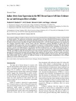
Starvation stress attenuates the miRNAtarget interaction in suppressing breast
cancer cell proliferation
Jinhui Lü1†, Chuyi Zhang1†, Junyi Han2†, Zhen Xu1, Yuan Li1, Lixiao Zhen1, Qian Zhao1, Yuefan Guo1,
Zhaohui Wang1,3, Evelyne Bischof4,5* and Zuoren Yu1*
Abstract
Background: Emerging evidence has demonstrated the limited access to metabolic substrates as an effective
approach to block cancer cell growth. The mechanisms remain unclear. Our previous work has revealed that miR221/222 plays important role in regulating breast cancer development and progression through interaction with
target gene p27.
Results: Herein, we determined the miRNA-mRNA interaction in breast cancer cells under induced stress status of
starvation. Starvation stimulation attenuated the miR-221/222-p27 interaction in MDA-MB-231 cells, thereby
increased p27 expression and suppressed cell proliferation. Through overexpression or knockdown of miR-221/222,
we found that starvation-induced stress attenuated the negative regulation of p27 expression by miR-221/222.
Similar patterns for miRNA-target mRNA interaction were observed between miR-17-5p and CyclinD1, and between
mR-155 and Socs1. Expression of Ago2, one of the key components of RNA-induced silencing complex (RISC), was
decreased under starvation-induced stress status, which took responsibility for the impaired miRNA-target
interaction since addition of exogenous Ago2 into MDA-MB-231 cells restored the miR-221/222-p27 interaction in
starvation condition.
Conclusions: We demonstrated the attenuated interaction between miR-221/222 and p27 by starvation-induced
stress in MDA-MB-231 breast cancer cells. The findings add a new page to the general knowledge of negative
regulation of gene expression by miRNAs, also demonstrate a novel mechanism through which limited access to
nutrients suppresses cancer cell proliferation. These insights provide a basis for development of novel therapeutic
options for breast cancer.
Keywords: Breast cancer, Starvation, miRNA, Target interaction, Proliferation
* Correspondence: ;
†
Jinhui Lü, Chuyi Zhang and Junyi Han contributed equally to this work.
4
Shanghai University of Medicine and Health Sciences Clinical Medicine
Division, Shanghai, China
1
Research Center for Translational Medicine, Tongji University School of
Medicine, 150 Jimo Road, Shanghai 200120, China
Full list of author information is available at the end of the article
© The Author(s). 2020 Open Access This article is licensed under a Creative Commons Attribution 4.0 International License,
which permits use, sharing, adaptation, distribution and reproduction in any medium or format, as long as you give
appropriate credit to the original author(s) and the source, provide a link to the Creative Commons licence, and indicate if
changes were made. The images or other third party material in this article are included in the article's Creative Commons
licence, unless indicated otherwise in a credit line to the material. If material is not included in the article's Creative Commons
licence and your intended use is not permitted by statutory regulation or exceeds the permitted use, you will need to obtain
permission directly from the copyright holder. To view a copy of this licence, visit />The Creative Commons Public Domain Dedication waiver ( applies to the
data made available in this article, unless otherwise stated in a credit line to the data.
Lü et al. BMC Cancer
(2020) 20:627
Background
Breast cancer is still one of the most prevalent neoplasms globally, with a persistently high prevalence despite numerous diagnostic progresses and prevention
measures. The mortality is still significant, despite a
slight decline in the last two decades. Old age is the
most important risk factor for breast cancer, which explains the persistently increasing incidence, especially in
developed countries [1–3].
Breast cancer is one of the most heterogenous diseases. Albeit there is a number of established predictive
and prognostic factors, most of which are also therapeutic targets (e.g. estrogen, progesterone receptor (ER,
PR) or HER-2), the management options are still primarily based on the cancer’s basic clinicopathological features, such as tumor size, lymph node stage, histological
grade, type, and lymphovascular invasion [4–7]. Especially, the oldest old cancer patients are being excluded
from various therapeutic options due to their frailty and
vulnerability towards anticancer-therapy-related side effects [8–10]. Increasingly, discoveries in breast cancer
genomics, proteomics and lipidomics are opening our
understanding and categorizations [11–13]. However,
these analyses are relatively expensive and remain mostly
in the theoretical frame. Therefore, there is an urgent
need for new indicators and approaches in personalized
therapies, offering targeted care with minimal toxicities.
While caloric restrictions in humans showed positive
clinical influence on various neurocognitive disorders
(NCD) and aging, fasting in cancer patients is controversially debated, especially due to potential increase of
tumor growth due to reactive increase of growth factors
after fasting cessation, hyperglycemia and hyperinsulinemia, but no valid and reliable trials were conducted so
far. Emerging evidence showed starving tumor cells of
nutrients are capable to stop cancer cell growing [14].
The constitutive anabolism of cancer cells not only supports proliferation but also addicts tumor cells to a
steady influx of exogenous nutrients. Limiting access to
metabolic substrates is effective to block cancer growth
[15]. Recent reports from animal models illustrated positive relations between starvation and cancer, such as better outcomes in mice after 2–3 fasting days prior to
chemotherapy [16–18]. Related hypotheses suggest that
fasting renders tumor cells susceptible towards chemotherapy due to a differential stress resistance: since
tumor cells are supposedly less adaptable to acute environmental changes, they become hyperactive and hypermitotic under starvation [19]. Such processes lead to
chromosomal damage, DNA- and cellular instability,
thus a higher sensitivity to chemo- or radio- therapy,
despite the fact that paradoxically, these pathophysiologic processes are also causative of tumor growth and
progression [20, 21].
Page 2 of 10
Therefore, our study aimed to elaborate on the underlying
molecular mechanisms of how fasting links to shrinkage of
solid tumors and whether these findings can be translated to
humans, using miRNAs (microRNAs) as crucial players in
tumorigenesis and progression. miRNAs become severely
deregulated during cancerous processes, which impairs their
main function on targeted genes by degradation of mRNA
or inhibition of mRNA translation. Recently, miRNA-mRNA
interactions have been described in the context of signature
interactions within well-established cancer pathways, e.g. in
breast and pancreatic cancer [22–24]. Nevertheless, some
studies revealed that miRNAs have the capability of activating gene expression directly or indirectly in respond to different cell types and cell conditions [24, 25]. However, it is thus
far unclear whether miRNA-mRNA interactions depend on
the cell status, such as stress induced by external conditions
(e.g. starvation). Establishing such correlation would allow
for new therapeutic options.
In our previous research, we showed that miR-221/222
is a potential oncogenic gene in breast cancer, and validated target genes p27 downstream of miR-221/222
[26]. The expression levels of miR-221/222 are upregulated in breast cancer, especially in basal-like breast cancer subtype. For example, miR-221/222 showed ~ 20–40
times higher in expression in MDA-MB-231 cell, compared to luminal breast cancer cell types [26]. Targeted
knockdown of miR-221 and/or miR-222 suppressed cell
proliferation and promoted the cell sensitivity to chemotherapy in human breast cancer [26]. Herein we found
that the interaction between miR-221/222 and p27 is
dependent on the cell status in human breast cancer
cells. Starvation-induced stress showed influence of
miRNA-mRNA interaction.
Methods
Cell lines and culture
Human breast cancer cell lines MCF-7 and MDA-MB-231
were originally obtained from American Type Culture
Collection (ATCC), and maintained in our laboratory.
The two cell lines were not included in the list of contaminated cell lines ( Cells are free of mycoplasma
contamination according to a recent test in our lab. Cells
were regularly cultured in DMEM containing glucose (4.5
g/L), penicillin and streptomycin (100 mg of each/L), supplemented with 10% fetal bovine serum (FBS). Starvation
cell culture condition included DMEM containing glucose
(1.0 g/L), penicillin and streptomycin (100 mg of each/L)
and supplementation with only 0.1% FBS.
Oligos, plasmid and transfection
Anti-miR-221 (5′ gaaacccagcagacaauguagcu 3′), antimiR-222 (5′ acccaguagccagauguagcu 3′), anti-miR-NC
scramble (5′ guguaacacgucuauacgccca 3′), miR-17-5p
Lü et al. BMC Cancer
(2020) 20:627
Page 3 of 10
(5′ caaagugcuuacagugcagguag 3′), miR-155 (5′ uuaaugcuaaucgugauagggguu 3′) and miR-NC scramble (5′
agucgcauaccucgacaauaau 3′) oligos were designed following LNA Oligo Tools and Design Guidelines of Exiqon (Vedbaek, Denmark), and synthesized per GenScript
(Nanjing, China). siRNA to Ago2 and negative control
were purchased from Guangzhou RiboBio Co., Ltd.
(Guangzhou, China). The HiPerFect transfection reagent
from Qiagen (Venlo, The Netherland) was used for RNA
oligos cell transfection according to the manufacturer’s
instructions. Final RNA oligo concentrations of 50 nM
were used for all in vitro assays. miR-221 and miR-222
expression vectors, as previously described in the literature [27], were provided by Moffitt Cancer Center & Research Institute. pcDNA3.1-Ago2 plasmid was presented
by Dr. Yandong Li from Tongji University. Lipofectamine 2000 (Invitrogen, USA) was used for plasmid
transfections, following the manufacturer’s instructions.
Sequences for all primers are available upon request.
luciferase activities were measured using Dual-Luciferase
Reporter Assay System (Promega) by AutoLumat.
Cell proliferation assays
Starvation-induced stress attenuated the miR-221/222p27 interaction in MDA-MB-231 breast cancer cells
For the 3-(4,5-dimethylthiazol-2-yl)-2,5 -diphenyltetrazolium (MTT) assay, 4 × 10³ cells/well were seeded into
96-well plate in triplicates. After culturing for 24 and 48
h, cells were stained with MTT solution for 3 h under
cell-culturing conditions, followed by dissolving with
DMSO. The cell growth was determined by measuring
OD value at 570 nm wavelength.
Western blot
Cell lysates (50 μg) were separated by 10% SDS/PAGE.
The proteins were transferred to nitrocellulose membrane.
After being blocked in 5% milk (w/v) at room temperature
for 1 h, the membranes were incubated at 4 °C overnight
with primary antibodies (1:1000). Following 1 × PBST
washing, the membranes were incubated with secondary
antibodies (1:3000) at room temperature for 1 h followed
by ECL staining. The following antibodies were applied:
anti-p27 (sc-776, Santa Cruz), anti-Cyclin D1 (sc-20,044,
Santa Cruz), anti-E2F1 (sc-251, Santa Cruz), anti-Ago2
(04–642, Millipore), anti-Socs1 (ab62584, Abcam) and
anti-β-actin (sc-47778, Santa Cruz).
Luciferase reporter assay
pGL3-p27 3’UTR vector, carrying the whole 3’UTR sequence of human p27 mRNA, was prepared as described
in the literature [26]. 293 T cells were seeded on 12-well
plates at a density of 1 × 10⁵ cells/well in an antibioticfree medium for 24 h under regular or starvation conditions. 24 h later, 1.0 μg/well of pGL3-p27 3’UTR, 0.2 μg/
well of Renilla plasmid and anti-miRNA (50 nM) were
co-transfected using HiPerFect transfection reagent from
Qiagen (Venlo, The Netherland). For the following 18 h,
Statistical analysis
The standard two-tailed student’s t-test was used for
statistical analysis, in which p < 0.05 was considered statistically significant. Data are presented as mean ± SEM.
Results
Starvation condition suppressed cell proliferation in MDAMB-231 breast cancer cells
In order to determine the influence of the cell proliferation and gene expression by starvation stimulation,
MTT assays were conducted in MDA-MB-231 cells
under culturing condition of starvation. We observed a
significant inhibition of cell proliferation (Fig. 1a). Western blot analysis demonstrated increase of p27 (Fig. 1b)
and decrease of Cyclin D1 (Fig. 1c) in expression under
starvation condition.
miRNA has been well defined to interact with target
mRNA through seed sequence complementarity. miR221/222 has two binding sites within the 3’UTR sequence of target gene p27, which is highly conserved between human and rodents (Fig. 2a-c). Adding miRNA
inhibitors to MDA-MB-231 led to a suppression of cell
proliferation by both anti-miR-221 and anti-miR-222
(Fig. 2d), which were attenuated by starvation stimulation (Supplemental Fig. S1). miR-221/222 were knocked
down or overexpressed in MDA-MB-231 cells, followed
by western blot analysis and quantitative real-time PCR
to detect the p27 expression at both protein and mRNA
levels. As shown in Fig. 2e and Supplemental Fig. S2,
p27 expression was significantly upregulated after inhibition of miR-221 and/or miR-222 under regular culture
condition, while this upregulation was not seen when
the cells were cultured under starvation condition. In
consistence, overexpression of miR-221 and/or miR-222
significantly downregulated the expression of p27 under
regular condition, but not under starvation condition
(Fig. 2f and Supplemental Fig. S3).
In order to further analyze the miRNA-mRNA interaction
in cells at starvation-induced stress, a luciferase reporter vector carrying 3’UTR of p27 containing two binding sites to
miR-221/222 (Fig. 3a) was transfected into cells. As shown in
Fig. 3b, knockdowns of miR-221 and/or miR-222 upregulated the luciferase activity to over 3-fold through interaction
with p27 3’UTR under normal cell culture condition. However, the luciferase activity did not show influence by antimiR221/222 under starvation condition (Fig. 3c), which is
consistent with the results in Fig. 2. The expression status of
the immediate downstream and associating genes of p27,
Lü et al. BMC Cancer
(2020) 20:627
Page 4 of 10
Fig. 1 Starvation stimulation suppressed cell proliferation in human breast cancer. a: MTT assays showing decreased cell proliferation of
MDA-MB-231 cells by starvation stimulation, compared to regular cell culture condition. b: Western blot analysis demonstrating the increased p27
expression by starvation stimulation in MDA-MB-231 cells. c: Western blot analysis demonstrating the decreased Cyclin D1 expression under
starvation in MDA-MB-231 cells. β-actin served as loading control. Original gels for western blots were shown in Supplemental Fig. S6. Data are
derived from three independent analyses, and presented as mean ± SEM (n = 3). *p < 0.05, **p < 0.01, ***p < 0.0001
such as E2F1 and Cyclin D1, was further analyzed under
normal and starvation status with or without the presence of
miR-221/222. As shown in Fig. 3d, downregulation of E2F1
and Cyclin D1 was associated with upregulation of p27 by
knockdown of miR-221/222 under normal cell culture condition. However, this kikd of regulation was not seen under
starvation condition (Fig. 3e).
Other miRNA-target interaction under regular and
starvation conditions
In addition to miR-221/222, two more miRNAs with welldefined target gene and regulatory function in human
breast cancer were selected to further confirm the influence of the miRNA-mRNA interaction by starvation. As
shown in Fig. 4a and b, Cyclin D1 is a demonstrated target
gene of miR-17-5p in MCF-7 breast cancer cells [28]. Cyclin D1 expression was significantly lower after transfection
with miR-17-5p mimic in MCF-7 cells under normal condition (Fig. 4b). This effect was not observed in cells at
starvation-induced stress status (Fig. 4b). Similar analysis
was applied to confirmed the suppression of Socs1 by
miR-155 in MDA-MB-231 cells under regular culture
condition [29], while starvation-induced stress condition
completely attenuated this regulation (Fig. 4c-d).
Lü et al. BMC Cancer
(2020) 20:627
Page 5 of 10
Fig. 2 Starvation stimulation attenuated the miR-221/222-p27 target interaction in regulating cell proliferation in MDA-MB-231. a: Two
binding sites of miR-221/222 were identified in the highly conserved 3’UTR region of p27 mRNAs. b, c: Sequence alignment between conserved
3’UTR of p27 mRNA and miR-221/222. The two binding sites were highlighted and boxed. d: MTT assays showing decrease of cell proliferation in
MDA-MB-231 cells after the knockdown of miR-221 and/or miR-222, compared to negative control (anti-NC). e: Western blot analysis showing
increased p27 protein level after addition of anti-miR-221 and/or anti-miR-222 into MDA-MB-231 cells under regular cell culture condition, but not
under starvation condition. f: Western blot showing suppression of p27 at the protein level by overexpression of miR-221 and/or miR-222 in
MDA-MB-231 cells under regular cell culture condition, but not under starvation condition. β-actin served as loading control. Original gels for
western blots were shown in Supplemental Fig. S6. Data are derived from three independent analyses, and presented as mean ± SEM (n = 3).
*p < 0.05, **p < 0.01, ***p < 0.0001
Decreased Ago2 expression was associated with
attenuated miRNA-mRNA interaction under starvation
condition
In order to determine the mechanisms through which
miRNA-mRNA interaction is attenuated by starvation
stimulation, Ago2 and Dicer1, two key components in
the RNA-induced silencing complex (RISC) and regulating the target interaction between miRNAs and mRNAs,
as well as Exportin 5 and Drosha, two factors regulating
miRNA biogenesis, were detected in MDA-MB-231 cells
under regular and starvation conditions. As shown in
Supplemental Fig. S4, Ago2 showed downregulation by
starvation stimulation, while Dicer1, Drosha and Exportin did not. Western blot analyses were applied to further conform the downregulation of Ago2 by starvation
stress (Fig. 5a). The attenuated regulation of p27 expression by anti-miR-221 and/or anti-miR-222 under starvation stimulation was rescued by addition of exogenous
Lü et al. BMC Cancer
(2020) 20:627
Page 6 of 10
Fig. 3 Luciferase reporter assays demonstrated the attenuated interaction between miR-221/222 and p27 by starvation stimulation. a:
Schematic representation of the luciferase reporter structure carrying human p27 3’UTR. b: Luciferase reporter assay demonstrating the direct
interaction between p27 3’UTR and miR-221/222 under regular cell culture condition. c: Luciferase reporter assay demonstrating the attenuated
interaction between p27 3’UTR and miR-221/222 under starvation conditions. d: Western blot showing downregulated E2F1 and cyclin D1, two
downstream genes of p27, by knockdown of miR-221/222 under normal cell culture condition. e: Western blot showing the expression of E2F1
and Cyclin D1 was not affected by miR-221/222 under starvation condition. Original gels for western blots were shown in Supplemental Fig. S6.
Data are presented as mean ± SEM (n = 4), ***p < 0.0001
Ago2 into MDA-MB-231 cells (Supplemental Fig. S5,
Fig. 5b). Targeted knockdown of Ago2 by siRNA treatment in MDA-MB-231 cells led to decreased cell proliferation (Fig. 5c). As such we hypothesized it is the
downregulation of Ago2 by starvation that takes responsibility for the attenuated miR221/222-p27 interaction,
leading to upregulation of p27 and the cell cycle arrest
in MDA-MB-231 cells (Fig. 5d).
Discussion
In view of the lack of therapeutic target and poor 5-year
survival for triple-negative breast cancer, novel forms of
therapy are urgently needed. Limited access to nutritious
supply is becoming a promising approach for suppressing
tumor growth [14, 15]. In addition, miRNA-mRNAinteraction-targeted strategy could enhance the efficacy of
chemo- and radio- therapy [30]. While beneficial reactions
to fasting have been reported in animals, the underlying
pathways have not been fully understood. Herein, we report that starvation suppresses cancer cell proliferation via
attenuating miRNA-mRNA target interaction.
In the studied culture of MDA-MB-231 (triple-negative breast cancer cells), the content of miR-221/222 was
high [26]. Under normal conditions, it inhibited the
Lü et al. BMC Cancer
(2020) 20:627
Page 7 of 10
Fig. 4 Other miRNA-target interaction under regular and starvation conditions in breast cancer cells. a: Sequence binding between 3’UTR
of Cyclin D1 mRNA and miR-17-5p. b: Western blot analysis showing suppression of the Cyclin D1 expression by miR-17-5p in MCF-7 cells under
regular cell culture condition, but not under starvation condition. β-actin served as protein loading control. c: Sequence binding between 3’UTR
of human Socs1 mRNA and miR-155. d: Western blot analysis confirming the miR-155-Socs1 interaction in MDA-MB-231 cells under regular
culture condition, but not under starvation condition. Original gels for western blots were shown in Supplemental Fig. S6. Data are derived from
three independent analyses, and presented as mean ± SEM (n = 3). *p < 0.05, **p < 0.01
expression of p27 and promoted the proliferation and
viability of cancer cells. Under starvation, miR-221/222
lost its inhibitory effect on p27 expression, which led to
increase of the p27 level and inhibition of the cell cycle.
The current study is the first to demonstrate the interruption of miRNA-mRNA interaction by starvation stimulation, which mediates suppression of cell proliferation in
breast cancer. Furthermore, Ago2 was decreased under
starvation condition, suggesting that RISC structure and
function are inhibited in this specific cell status. As such,
we propose that the starvation-induced downregulation of
Ago2 attenuates the miR-221/222-p27 interaction, leading
to a cell cycle arrest in MDA-MB-231 cells. These findings
not only demonstrate a novel mechanism for starving cancer cells to inhibit tumor growth, but also add a new page
to the general knowledge of the miRNA-mRNA target
interaction. It suggests novel therapeutic targets: inhibition of mRNA-miRNA interaction via induced stress,
could bring benefits to many breast cancer patients, especially those who lack any therapeutic target or cannot
undergo full dose chemo- or radio- therapy.
In addition to miR-221/222-p27 interaction, other
miRNA-mRNA interactions, such as miR-155-Socs1 and
miR-17-5p-Cyclin D1, were also analyzed, and showed
attenuation by starvation stimulation. Socs1, as a member of the suppressor of cytokine signaling family, inhibits Jak/Stat pathway and suppresses cytokine signal
transduction. Socs1 is downregulated in breast cancer,
functioning as a tumor suppressor [26]. Differing from
Socs1, Cyclin D1 is overexpressed in more than 50% of
breast cancer, encoding the regulatory subunit of a holoenzyme that phosphorylates the gene retinoblastoma
(RB) and promotes G1/S cell cycle progression and
oncogenesis [28]. The interrupted miRNA effects on the
expression of target genes, such as oncogenes and/or
tumor suppressors, should be a mechanism mediating
the fasting-induced growth suppression of cancer cells.
In the current study, we demonstrated downregulation of Ago2, a key component of RISC, by starvation
stimulation. In view of the similar mechanisms for
miRNAs and siRNAs in regulating target gene expression through RISC, siRNA-mediated RNA interference
Lü et al. BMC Cancer
(2020) 20:627
Page 8 of 10
Fig. 5 Suppression of Ago2 expression by starvation stimulation. a: Western blot analysis demonstrating the inhibition of Ago2 expression
by starvation stimulation in MDA-MB-231 cells. β-actin served as loading control. b: Western blot analysis demonstrating the attenuated
regulation of p27 expression by anti-miR-221 and/or anti-miR-222 under starvation stimulation, which was rescued by addition of Ago2 using
pcDNA-3.1-Ago2 plasmid. β-actin served as loading control. Original gels for western blots were shown in Supplemental Fig. S6. b: Cell
proliferation assay showing decrease of cell proliferation in MDA-MB-231 cells after knockdown of Ago2 by siRNA. d: Schematic representation of
mechanisms for regulating starvation-induced cell cycle arrest in human breast cancer cells. Downregulation of Ago2 by starvation attenuated
the miR221/222-p27 interaction, leading to upregulation of p27 and cell cycle arrest in MDA-MB-231 cells. Data are derived from three
independent analyses, and presented as mean ± SEM (n = 3). *p < 0.05, **p < 0.01
(RNAi) effects on gene expression are very likely influenced in a starvation state. Although further experimental validation is required for the RNAi
disruption by starvation stimulation, it remains unclear whether the RNAi strategy can be combined
with the fasting strategy for cancer treatment. The
implications of disturbed RNAi on the transcriptome
in a starvation state have to be taken into account.
Conclusion
The current study demonstrated the attenuated interaction
between miR-221/222 and p27 and decreased Ago2 expression by starvation-induced stress in MDA-MB-231 breast
cancer cells. It adds a new page to the general knowledge of
negative regulation of gene expression by miRNAs, also
demonstrate a novel mechanism through which limited access to nutrients suppresses cancer cell proliferation.
Lü et al. BMC Cancer
(2020) 20:627
Supplementary information
Page 9 of 10
Received: 17 October 2019 Accepted: 26 June 2020
Supplementary information accompanies this paper at />1186/s12885-020-07118-3.
Additional file 1 Figure S1: MTT assays showing uninfluenced cell
proliferation in MDA-MB-231 cells by knockdown of miR-221 and/or miR222 under starvation condition. Figure S2: Quantitative real-time PCR
analysis showing increase of p27 expression at mRNA level by anti-miR221 and/or anti-miR-222 under regular cell culture condition, but not
under starvation culture condition. Data are derived from three independent analyses, and presented as mean ± SEM (n = 3). **p < 0.01. Figure S3: Quantitative real-time PCR analysis showing suppressed
expression of p27 at the mRNA levels in MDA-MB-231 cells by miR-221
and/or miR-222 overexpression under regular cell culture condition, but
not under starvation condition. Data are derived from three independent
analyses, and presented as mean ± SEM (n = 3). **p < 0.01. Figure S4:
Quantitative real-time PCR analysis of the key factors regulating miRNA
biogenesis and function, including Exportin 5, Dicer 1, Ago2 and Drosha
in MDA-MB-231 cells under regular and starvation culture conditions.
Data are derived from three independent analyses, and presented as
mean ± SEM (n = 3). **p < 0.01. Figure S5: Western blot analysis demonstrating the overexpression of Ago2 in MDA-MB-231 cells transfected with
pcDNA 3.1-Ago2 plasmid. Empty vector was used as negative control. βactin served as loading control. Figure S6: Original gels for all western
blots in Figures.
Abbreviations
miRNA: microRNA; RISC: RNA-induced silencing complex; ER: Estrogen
receptor; PR: Progesterone receptor; NCD: Neurocognitive disorders;
FBS: Fetal bovine serum; RNAi: RNA interference; RB: Retinoblastoma
Acknowledgements
Not applicable.
Authors’ contributions
ZY and EB designed the research and wrote the paper. JL, CZ, ZX, JH, YL, LZ,
YG and ZW performed experiments. QZ did data analysis. All authors have
read and approved the manuscript.
Funding
This work was supported by grants 81772810 and 81972476 from the
National Natural Science Foundation of China (materials purchase and
manuscript preparation), and grant from the Top-level Clinical Discipline Project of Shanghai Pudong PWYgf 2018–05 (materials sharing). Evelyne Bischof
(Ewelina Biskup) was supported by Krebsliga Schweiz, BIL KFS 4261-08-2017.
Availability of data and materials
Data sharing is not applicable to this article as no datasets were generated
or analyzed during the current study.
Ethics approval and consent to participate
Neither human specimens nor animals were used in this study. The two cell
lines used in the article do not require ethics approval.
Consent for publication
Not applicable.
Competing interests
There are no conflicts of interest associated with this manuscript.
Author details
1
Research Center for Translational Medicine, Tongji University School of
Medicine, 150 Jimo Road, Shanghai 200120, China. 2Department of Surgery,
Shanghai East Hospital, Tongji University School of Medicine, 150 Jimo Road,
Shanghai 200120, China. 3Jinzhou Medical University, Liaoning, China.
4
Shanghai University of Medicine and Health Sciences Clinical Medicine
Division, Shanghai, China. 5Division of Internal Medicine, University Hospital
of Basel, Petersgraben 4, 4051 Basel l, Switzerland.
References
1. Le Saux O, Ripamonti B, Bruyas A, Bonin O, Freyer G, Bonnefoy M, et al.
Optimal management of breast cancer in the elderly patient: current
perspectives. Clin Interv Aging. 2015;10:157–74.
2. Gusterson BA, Stein T. Human breast development. Semin Cell Dev Biol.
2012;23(5):567–73.
3. Ferlay J, Colombet M, Soerjomataram I, Dyba T, Randi G, Bettio M, et al.
Cancer incidence and mortality patterns in Europe: estimates for 40
countries and 25 major cancers in 2018. Eur J Cancer. 2018;103:356–87.
4. Payne SJ, Bowen RL, Jones JL, Wells CA. Predictive markers in breast
cancer--the present. Histopathology. 2008;52(1):82–90.
5. Millar EK, Graham PH, McNeil CM, Browne L, O'Toole SA, Boulghourjian A,
et al. Prediction of outcome of early ER+ breast cancer is improved using a
biomarker panel, which includes Ki-67 and p53. Br J Cancer. 2011;105(2):
272–80.
6. Cai FF, Xu C, Pan X, Cai L, Lin XY, Chen S, et al. Prognostic value of plasma
levels of HIF-1a and PGC-1a in breast cancer. Oncotarget. 2016;7(47):77793–
806.
7. Lin XY, Cai FF, Wang MH, Pan X, Wang F, Cai L, et al. Mammalian sterile 20like kinase 1 expression and its prognostic significance in patients with
breast cancer. Oncol Lett. 2017;14(5):5457–63.
8. Parks RM, Cheung KL. Personalising Care in the Older Woman with primary
breast Cancer. Ann Acad Med Singap. 2019;48(11):370–5.
9. Andersen SL, Terry DF, Wilcox MA, Babineau T, Malek K, Perls TT. Cancer in
the oldest old. Mech Ageing Dev. 2005;126(2):263–7.
10. Lichtman SM. Guidelines for the treatment of elderly cancer patients.
Cancer Control. 2003;10(6):445–53.
11. Baird RD, Caldas C. Genetic heterogeneity in breast cancer: the road to
personalized medicine? BMC Med. 2013;11:151.
12. Sabatier R, Goncalves A, Bertucci F. Personalized medicine: present and
future of breast cancer management. Crit Rev Oncol Hematol. 2014;91(3):
223–33.
13. Dawson SJ, Rueda OM, Aparicio S, Caldas C. A new genome-driven
integrated classification of breast cancer and its implications. EMBO J. 2013;
32(5):617–28.
14. Wokoun U, Hellriegel M, Emons G, Grundker C. Co-treatment of breast
cancer cells with pharmacologic doses of 2-deoxy-D-glucose and
metformin: starving tumors. Oncol Rep. 2017;37(4):2418–24.
15. Selwan EM, Finicle BT, Kim SM, Edinger AL. Attacking the supply wagons to
starve cancer cells to death. FEBS Lett. 2016;590(7):885–907.
16. Mukherjee P, El-Abbadi MM, Kasperzyk JL, Ranes MK, Seyfried TN. Dietary
restriction reduces angiogenesis and growth in an orthotopic mouse brain
tumour model. Br J Cancer. 2002;86(10):1615–21.
17. Maddocks ODK, Athineos D, Cheung EC, Lee P, Zhang T, van den Broek NJF,
et al. Modulating the therapeutic response of tumours to dietary serine and
glycine starvation. Nature. 2017;544(7650):372–6.
18. Lee C, Raffaghello L, Brandhorst S, Safdie FM, Bianchi G, Martin-Montalvo A,
et al. Fasting cycles retard growth of tumors and sensitize a range of cancer
cell types to chemotherapy. Sci Transl Med. 2012;4(124):124ra127.
19. Raffaghello L, Lee C, Safdie FM, Wei M, Madia F, Bianchi G, et al. Starvationdependent differential stress resistance protects normal but not cancer cells
against high-dose chemotherapy. Proc Natl Acad Sci U S A. 2008;105(24):
8215–20.
20. Swanton C, Burrell RA, Futreal PA. Breast cancer genome heterogeneity: a
challenge to personalised medicine? Breast Cancer Res. 2011;13(1):104.
21. Burrell RA, McClelland SE, Endesfelder D, Groth P, Weller MC, Shaikh N, et al.
Replication stress links structural and numerical cancer chromosomal
instability. Nature. 2013;494(7438):492–6.
22. Li Z, Peng Z, Gu S, Zheng J, Feng D, Qin Q, et al. Global analysis of miRNAmRNA interaction network in breast Cancer with brain metastasis.
Anticancer Res. 2017;37(8):4455–68.
23. Lee S, Jiang X. Modeling miRNA-mRNA interactions that cause phenotypic
abnormality in breast cancer patients. PLoS One. 2017;12(8):e0182666.
24. Vasudevan S, Tong Y, Steitz JA. Switching from repression to activation:
microRNAs can up-regulate translation. Science. 2007;318(5858):1931–4.
25. Valinezhad Orang A, Safaralizadeh R, Kazemzadeh-Bavili M. Mechanisms of
miRNA-mediated gene regulation from common Downregulation to
mRNA-specific Upregulation. Int J Genomics. 2014;2014:970607.
Lü et al. BMC Cancer
(2020) 20:627
26. Li Y, Liang C, Ma H, Zhao Q, Lu Y, Xiang Z, et al. miR-221/222 promotes Sphase entry and cellular migration in control of basal-like breast cancer.
Molecules. 2014;19(6):7122–37.
27. Zhao JJ, Lin J, Yang H, Kong W, He L, Ma X, et al. MicroRNA-221/222
negatively regulates estrogen receptor alpha and is associated with
tamoxifen resistance in breast cancer. J Biol Chem. 2008;283(45):31079–86.
28. Yu Z, Wang C, Wang M, Li Z, Casimiro MC, Liu M, et al. A cyclin D1/
microRNA 17/20 regulatory feedback loop in control of breast cancer cell
proliferation. J Cell Biol. 2008;182(3):509–17.
29. Jiang S, Zhang HW, Lu MH, He XH, Li Y, Gu H, et al. MicroRNA-155 functions
as an OncomiR in breast cancer by targeting the suppressor of cytokine
signaling 1 gene. Cancer Res. 2010;70(8):3119–27.
30. Miller TE, Ghoshal K, Ramaswamy B, Roy S, Datta J, Shapiro CL, et al.
MicroRNA-221/222 confers tamoxifen resistance in breast cancer by
targeting p27Kip1. J Biol Chem. 2008;283(44):29897–903.
Publisher’s Note
Springer Nature remains neutral with regard to jurisdictional claims in
published maps and institutional affiliations.
Page 10 of 10