Adjuvant Transarterial chemoembolization does not influence recurrence-free or overall survival in patients with combined hepatocellular carcinoma and Cholangiocarcinoma after curative
Bạn đang xem bản rút gọn của tài liệu. Xem và tải ngay bản đầy đủ của tài liệu tại đây (769.09 KB, 11 trang )
Liu et al. BMC Cancer
(2020) 20:642
/>
RESEARCH ARTICLE
Open Access
Adjuvant Transarterial chemoembolization
does not influence recurrence-free or
overall survival in patients with combined
hepatocellular carcinoma and
Cholangiocarcinoma after curative
resection: a propensity score matching
analysis
Wei-Ren Liu1†, Meng-Xin Tian1†, Chen-Yang Tao1†, Zheng Tang1, Yu-Fu Zhou1,2, Shu-Shu Song1,2, Xi-Fei Jiang1,2,
Han Wang1,2, Pei-Yun Zhou1,2, Wei-Feng Qu1,2, Yuan Fang1,2, Zhen-Bin Ding1,2, Jian Zhou1,2,3,4,5, Jia Fan1,2,3,4,5 and
Ying-Hong Shi1,2,3,4*
Abstract
Background: The prognosis of patients with combined hepatocellular carcinoma and intrahepatic
cholangiocarcinoma (CHC) is usually poor, and effective adjuvant therapy is missing making it important to
investigate whether these patients may benefit from adjuvant transarterial chemoembolization (TACE). We aimed to
evaluate the efficiency of adjuvant TACE for long-term recurrence and survival after curative resection before and
after propensity score matching (PSM) analysis.
Methods: In this retrospective study, of 230 patients who underwent resection for CHC between January 1994 and
December 2014, 46 (18.0%) patients received adjuvant TACE. Univariate and multivariate regression analyses were
used to identify the independent predictive factors of survival. Cox regression analyses and log-rank tests were used
to compare overall survival (OS) and disease-free survival (DFS) between patients who did or did not receive
adjuvant TACE.
(Continued on next page)
* Correspondence:
†
Wei-Ren Liu, Meng-Xin Tian and Chen-Yang Tao contributed equally to this
work.
1
Department of Liver Surgery and Transplantation, Liver Cancer Institute,
Zhongshan Hospital, Fudan University, 180 FengLin Road, Shanghai 200032,
China
2
Key Laboratory of Carcinogenesis and Cancer Invasion of Ministry of
Education, Shanghai, China
Full list of author information is available at the end of the article
© The Author(s). 2020 Open Access This article is licensed under a Creative Commons Attribution 4.0 International License,
which permits use, sharing, adaptation, distribution and reproduction in any medium or format, as long as you give
appropriate credit to the original author(s) and the source, provide a link to the Creative Commons licence, and indicate if
changes were made. The images or other third party material in this article are included in the article's Creative Commons
licence, unless indicated otherwise in a credit line to the material. If material is not included in the article's Creative Commons
licence and your intended use is not permitted by statutory regulation or exceeds the permitted use, you will need to obtain
permission directly from the copyright holder. To view a copy of this licence, visit />The Creative Commons Public Domain Dedication waiver ( applies to the
data made available in this article, unless otherwise stated in a credit line to the data.
Liu et al. BMC Cancer
(2020) 20:642
Page 2 of 11
(Continued from previous page)
Results: A total of 230 patients (mean age 52.2 ± 11.9 years; 172 men) were enrolled, and 46 (mean age 52.7 ± 11.1
years; 38 men) patients received TACE. Before PSM, in multivariate regression analysis, γ-glutamyl transpeptidase (γGT), tumour nodularity, macrovascular invasion (MVI), lymphoid metastasis, and extrahepatic metastasis were
associated with OS. Alanine aminotransferase (ALT), MVI, lymphoid metastasis, and preventive TACE (HR: 2.763, 95%
CI: 1.769–4.314, p < 0.001) were independent prognostic factors for DFS. PSM created 46 pairs of patients. Before
PSM, adjuvant preventive TACE was not associated with an increased risk of OS (HR: 0.911, 95% CI: 0.545–1.520, p =
0.720) or DFS (HR: 3.345, 95% CI: 1.686–6.638, p = 0.001). After PSM, the 5-year OS and DFS rates were comparable in
the TACE group and the non-TACE group (OS: 22.7% vs 14.9%, respectively, p = 0.75; DFS: 11.2% vs 14.4%,
respectively, p = 0.06).
Conclusions: The present study identified that adjuvant preventive TACE did not influence DFS or OS after curative
resection of CHC.
Keywords: Combined hepatocellular carcinoma and intrahepatic cholangiocarcinoma, Transarterial
chemoembolization, Overall survival, Disease-free survival, Propensity score matching analysis
Background
Primary liver cancer (PLC) is a heavy global health burden; it ranks as the second leading cause of mortality in
men in less-developed countries, especially in China,
which accounts for more than 50% of PLC patients in
the world [1, 2]. PLC is composed of several biologically
distinct subtypes: hepatocellular carcinoma (HCC),
intrahepatic cholangiocarcinoma (ICC), and combined
hepatocellular-cholangiocarcinoma (CHC). As a distinct
and rare subtype of PLC, CHC accounts for less than 5%
of PLC cases, with histological evidence of both hepatocellular and biliary epithelial differentiation [3, 4]. Due
to the stem cell features of CHC, this disease is
associated with an aggressive course and a poor
prognosis, with 5-year overall survival (OS) ranging from
9.2–40% [5, 6].
Effective treatments for CHC are deficient. In our previous study, we found that radical surgical resection provided a better outcome that was intermediate between
HCC and ICC [7, 8]. Aggressive surgical treatment, including lymph node dissection, may improve survival in
patients diagnosed with CHC [9]. Regardless of Allen
and Lisa class or the predominance of ICC cells within
the tumour, the 5-year OS rate is 24% after hepatectomy
[10]. Liver transplantation is not an appropriate therapeutic choice for CHC due to the disappointing results,
with a mean OS of 11.7 months and a mean disease-free
survival (DFS) of 7.97 months [11]. However, a group reported that very early CHC resulted in favourable posttransplant prognosis [12]. However, these studies had
relatively small sample sizes and were retrospective in
nature.
Similar to HCC and ICC, for CHC, recurrence is the
most adverse factor influencing OS and DFS; vascular
and lymph node invasion as well as the presence of satellite metastasis have been suggested as significant predictors of poor outcome after curative resection [13–15].
Transarterial chemoembolization (TACE), percutaneous
ethanol injection (PEI) and radiofrequency ablation
(RFA) are the most widely used treatments for HCC and
post-resection recurrence [16–18]. For CHC, TACE
shows an advantageous response and prognosis in recurrent patients after resection [19]. TACE is effective for
prolonging the survival of patients with nonresectable
CHC. Nonetheless, the effect of adjuvant TACE in CHC
patients after curative resection is still unknown.
To address this issue, we conducted a retrospective cohort study to elucidate the relationship between adjuvant
TACE and long-term recurrence and survival after curative resection of CHC using propensity score matching
(PSM) and multivariate Cox regression analyses.
Methods
Participants and criteria
This was a retrospective study that used data collected
at a single medical centre. The study was approved by
the institutional review board and was in accordance
with the standards of the Declaration of Helsinki and
current ethical guidelines. Written informed consent
was obtained for each patient. The inclusion and
exclusion criteria are presented in the supplemental
information.
Between January 1994 and December 2014, a total of
255 patients who underwent curative hepatic resection
and were diagnosed with CHC in the Department of
Liver Surgery were retrospectively enrolled in this study.
Among them, 25 patients who received preoperative
surgery and anticancer treatments were excluded: 16 patients with a previous history of surgery, 2 patients who
received preoperative TACE, and 7 patients with missing
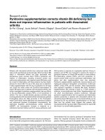
data. Thus, 230 patients were enrolled in the final analyses (Fig. 1). The detailed criteria for curative resection
are shown in the supplemental information [20].
Liu et al. BMC Cancer
(2020) 20:642
Page 3 of 11
Fig. 1 Patients selection flowchart
TACE
PSM
The risk of recurrence after resection was assessed by
tumour characteristics, which were established by the
pathology report, and the patients with intermediate or
high risks of recurrence were advised to undergo TACE
therapy. A high risk of recurrence was defined as a single
tumour with microvascular invasion or two or three
tumours, and an intermediate risk of recurrence was
defined as a solitary tumour larger than 5 cm without
microvascular invasion [16, 21]. Using the Seldinger
technique, a vascular catheter was inserted through a
femoral artery to the hepatic artery, and hepatic angiography was then carried out. A microcatheter was used to
inject Adriamycin (20–30 mg/m2) and lipiodol (3–5 mL)
unselectively into the left and right hepatic arteries. The
unselective embolization of the arterial tumor feeders
was carried out by using 1-mm-diameter absorber gelatin sponge particles (Gelfoam; Upjohn, Kalamazoo, MI,
USA) until arterial flow stasis was achieved.
Patients in the TACE and non-TACE groups were
matched using the PSM method [22], which was carried
out using R software version 2.10.0 (R Project for Statistical Computing, Austria).
First, a propensity score (from 0 to 1) that contained the
information of variates that was selected during matching was generated by logistic regression in PSM. Then,
to create a reliable propensity score model, the variables
that were chosen for matching included all the potential
confounders [23, 24]. Thus, the variables contained all
the independent prognostic factors of CHC. The Cox
proportional hazards model was used to identify the independent prognostic factors, and the variables with
statistical significance (p < 0.25) in univariate analysis
were entered into multivariate analysis. The variables entered into the final propensity model were sex, ALT,
perioperative blood transfusion, and lymphoid metastasis. Then, the model used one-to-one matching without
replacement between TACE and non-TACE patients by
using the nearest-neighbour matching algorithm. The
calliper value was selected as 0.01, and the balance between the two groups after matching was evaluated by
the standardized mean difference (p < 0.1).
Follow-up
Patients were followed in our centre every 3 months
until death or dropout (two patients) from the follow-up
program. The median follow-up time was 15.1 months.
The detailed follow-up procedures are shown in the
supplemental information.
Variables and outcomes
The data were prospectively collected and retrospectively
reviewed. The detailed information from the database is
shown in the supplemental information. The main outcomes of this study were OS and DFS. OS was measured
from the date of the resection to either the date of death
or the date of the last follow-up. DFS was defined from
the date of the resection to the date of first recurrence
or the date of death or the last follow-up visit.
Statistical analysis
Statistical analyses were carried out using IBM SPSS
22.0 (SPSS Inc., Armonk, NY, USA) and SAS 9.1
(SAS Institute Inc., Cary, NC, USA). The demographic, clinical, and tumour characteristics were documented as summary statistics that were obtained
using established methods. In both the TACE and
non-TACE groups, continuous data were presented as
the mean with a 25th–75th percentile range and analysed using Student’s t test or the Mann-Whitney U
test. The categorical variables were presented as absolute and relative frequencies and compared by Pearson’s χ2 analysis or Fisher’s exact test. OS and DFS
Liu et al. BMC Cancer
(2020) 20:642
Page 4 of 11
Table 1 Preoperative clinicopathologic Data of Patients with CHC Who received or not postoperative TACE
Variable
Before Propensity Matching
Without TACE
(n = 184)
Postoperative
TACE (n = 46)
Sex
After Propensity Matching
P
Without TACE
(n = 46)
Postoperative
TACE (n = 46)
0.172
> 0.99
Men
134
38
38
38
Women
50
8
8
8
52.3 ± 12.1
52 ± 10.7
53.4 ± 11.6
52 ± 10.7
Mean age (y)
HBsAg
0.326
> 0.99
136
34
35
34
Negative
48
12
11
12
Positive
153
9
42
9
Negative
31
37
4
37
0.666
HCV antibody
0.834
0.810
Positive
HBcAb
P
0.231
> 0.99
> 0.99
Positive
4
1
1
1
Negative
180
45
45
45
Median AFP, ng/mL
24.7 (1–80,000)
96 (1.8–46,897)
0.006
21.3 (1–30,728)
96 (1.8–46,897)
0.002
Median CEA, μg/mL
2.5 (0–274)
2.1 (0.5–70.5)
0.364
2.7 (0.1–112.4)
2.1 (0.5–70.5)
0.423
Median CA19–9, U/ml
28.1 (0–4370)
19.4 (0.2–300.1)
0.029
22 (0.5–4062.5)
19.4 (0.2–300.1)
0.023
Median bilirubin, μmol/L
11.8 (1.7–314.8)
12.9 (5.7–156.5)
< 0.001
13.7 (2.4–169.3)
12.9 (5.7–156.5)
0.664
Median albumin, g/L
41 (26–55)
42 (35–66)
0.397
41 (30–48)
42 (35–66)
0.556
Median ALT, U/L
28 (5–484)
31 (5–104)
0.094
26 (11–484)
31 (5–104)
0.109
Median ALP, IU/L
89.5 (22–1413)
88.5 (46–184)
0.477
92 (25–331)
88.5 (46–184)
0.599
Median GGT, U/L
59 (3.6–1632)
80 (18–490)
0.923
75.5 (10–658)
80 (18–490)
0.273
Median platelets, 10 /μL
13.7 (2.2–47.6)
16 (3.9–46.1)
0.319
15.3 (5.3–24.7)
16 (3.9–46.1)
0.171
Median prothrombin time, s
11.8 (9–17.6)
12 (10.2–13.8)
0.941
12 (10.2–14.6)
12 (10.2–13.8)
0.903
Median INR
1 (0.5–1.5)
1 (0.8–1.2)
0.227
1 (0.5–1.2)
1 (0.8–1.2)
0.065
Median tumour size, cm
5 (1–24)
7.3 (1.5–17)
0.626
6 (1.5–22)
7.3 (1.5–17)
0.384
Median tumour nodularities
1 (1–10)
1 (1–5)
0.140
1 (1–6)
1 (1–5)
0.648
Median blood loss, ml
200 (30–3500)
200 (10–2500)
0.182
200 (50–1800)
200 (10–2500)
0.480
Mean occlusion, min
6.8 ± 8.6
10 ± 1.6
0.044
5.4 ± 1.1
10 ± 1.6
3
Macrovascular invasion
0.041
Positive
11
7
7
7
Negative
173
39
39
39
Positive
39
11
8
11
Negative
145
35
38
35
Microvascular invasion
0.689
Lymphoid metastasis
0.607
0.840
> 0.99
Positive
22
6
6
6
Negative
162
40
40
40
Positive
6
2
3
2
Negative
178
44
43
44
Extrahepatic metastasis
0.719
Postrecurrent therapy
Resection
0.646
0.451
2
1
0.090
> 0.99
0.583
1
1
Liu et al. BMC Cancer
(2020) 20:642
Page 5 of 11
Table 1 Preoperative clinicopathologic Data of Patients with CHC Who received or not postoperative TACE (Continued)
Variable
Before Propensity Matching
After Propensity Matching
Without TACE
(n = 184)
Postoperative
TACE (n = 46)
27
Regional therapy
Chemothearpy
Selective internal radiation therapy
Stereotactic body radiation
Best supportive care
58
TACE
Without TACE
(n = 46)
Postoperative
TACE (n = 46)
6
10
6
4
1
1
1
66
14
11
14
5
2
1
2
12
5
3
5
17
19
17
P
P
Data are numbers of patients. Data in parentheses are range. Mean data are±standard deviation. Regional therapy: Radiofrequency ablation and percutaneous
ethanol injection
HBsAg hepatitis B surface antigen, HBcAb hepatitis B core antibody, HCV hepatitis C virus, AFP α-fetoprotein, CEA carcino-embryonic antigen, CA19–9 carbohydrate
19–9, INR International normalized ratio, ALT alanine aminotransferase, GGT γ-glutamyl transpeptidase, ALP alkaline phosphatase, MVI microvascular
vascular invasion
were compared using the Kaplan-Meier method, and
survival differences between the two groups were analysed using the log-rank test. Multivariate Cox proportional hazard regression analyses were then carried
out to adjust for other prognostic factors that were
associated with OS and DFS. Moreover, to strengthen
the accuracy of the model, a robust sandwich variance
estimator was used in all the cohorts for estimating
the hazard ratios and their 95% confidence intervals
(CIs). All tests using two-tailed p < 0.05 were considered to be statistically significant.
Results
Demographic and clinicopathological characteristics
Table 1 summarizes the baseline characteristics of
patients with CHC who underwent TACE (n = 46)
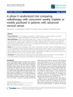
Fig. 2 Kaplan-Meier curves of survival outcomes of adjuvant TACE in patients with CHC before and after PSM analysis. Kaplan-Meier curves of (a)
overall survival (OS) and (b) disease-free survival (DFS) for patients with CHC before propensity score matching analysis; Kaplan-Meier curves of (c)
overall survival (OS) and (d) disease-free survival (DFS) for patients with CHC after PSM analysis
Liu et al. BMC Cancer
(2020) 20:642
Page 6 of 11
and those who did not (n = 184) before PSM. The
mean age of patients in the TACE group (52 ± 10.7
years) was similar to that of patients in the nonTACE group (52.3 ± 12.1 years), and the sex distribution was similar in both groups (38 and 134 male patients in the TACE group and non-TACE group,
respectively). The median AFP (p = 0.006), median
bilirubin (p < 0.001), occlusion time (p = 0.044), and
macrovascular invasion (p = 0.041) were higher in the
TACE group than in the non-TACE group, and the
median CA19–9 was higher in the non-TACE group
than in the TACE group (p = 0.029). After PSM, the
mean age of patients in the TACE group (52 ± 10.7
years) was similar to that of patients in the nonTACE group (53.4 ± 11.6 years), and the sex distribution was similar in both groups. Except for the higher
median AFP (p = 0.006), lower median CA19–9 (p =
0.023), lower median bilirubin (< 0.001), lower mean
occlusion time (p = 0.044), and macrovascular invasion
(p = 0.041) in the TACE group, there were no significant differences between the TACE group and the
non-TACE group in terms of the baseline characteristics (p > 0.05).
OS and DFS before PSM
The median survival of the whole cohort was 22.6
months, and the overall cumulative OS rates at 1, 3, 5,
and 10 years were 48.5, 33.3, 25.8, and 15.3%,
respectively. The median OS of the TACE group and
non-TACE group was 22.0 months and 23.5 months, respectively. The cumulative OS rates were comparable
between the two groups; the 1-, 3-, 5-, and 10-year OS
rates in the TACE group were 46.6, 31.7, 22.7, and
12.6%, respectively, whereas those in the non-TACE
group were 49.0, 33.7, 26.6, and 16.1%, respectively (p =
0.34) (Fig. 2a). The median DFS of the whole cohort was
14.0 months, and the cumulative DFS rates at 1, 3, 5,
and 10 years were 20.9, 10.4, 0.7, and 0.3%, respectively.
Stratified by TACE, the median DFS in the TACE group
was less than that in the non-TACE group (9.3 months
vs. 17.2 months) (p = 0.001) (Fig. 2b).
Table 2 Univariable and multivariable cox analysis of OS before propensity matched analysis
Variable
Univariable
Multivariable
HR
95% CI
P
HR
95% CI
P
Age (≥60/< 60, year)
1.279
0.857–1.908
0.229
–
–
–
Sex (Men/Women)
1.443
0.95–2.193
0.085
–
–
–
HBsAg (yes/no)
1.044
0.719–1.517
0.821
–
–
–
HCV antibody (yes/no)
2.293
0.722–7.283
0.159
–
–
–
AFP (≥20/< 20, ng/mL)
2.819
0.68–11.682
0.153
–
–
–
CEA (≥5/<5, ng/mL)
1.844
0.643–5.29
0.255
–
–
–
CA19–9 (≥37/<37, U/mL)
2.069
0.639–6.702
0.225
–
–
–
Liver cirrhosis, yes (%)
1.252
0.857–1.83
0.245
–
–
–
TB (≥17/< 17, μmol/L)
0.950
0.626–1.443
0.810
–
–
–
ALB (≥40/<40, g/mL)
0.759
0.530–1.086
0.132
–
–
–
ALT (≥35/<35, U/L)
1.327
0.941–1.870
0.106
–
–
–
γ-GT (≥40/<40, U/L)
2.662
1.703–4.163
< 0.001
2.152
1.354–3.421
0.001
PLT (≥10/< 10 103/μL)
1.005
0.665–1.518
0.982
–
–
–
Prothrombin time, median (range), s
1.199
0.781–1.841
0.406
–
–
–
Tumour size, cm
1.769
1.235–2.534
0.002
1.274
0.867–1.872
0.218
Tumour nodularities
1.167
1.055–1.292
0.003
1.130
1.011–1.262
0.031
Occlusion, min (< 20/≥20)
0.290
0.740–2.250
0.369
–
–
–
Macrovascular invasion (yes/no)
1.927
1.442–2.576
< 0.001
1.869
1.375–2.540
< 0.001
Microvascular invasion (yes/no)
1.365
0.921–2.204
0.122
–
–
–
Lymphoid metastasis (yes/no)
2.801
1.745–4.495
< 0.001
2.031
1.201–3.435
0.008
Extrahepatic metastasis (yes/no)
11.435
5.262–24.849
< 0.001
6.392
2.731–14.961
< 0.001
Preventive TACE (yes/no)
1.212
0.807–1.821
0.354
–
–
–
HBsAg hepatitis B surface antigen, HCV hepatitis C virus, AFP α-fetoprotein, CEA carcino-embryonic antigen, CA19–9 carbohydrate 19–9, TB total bilirubin, ALB
albumin, ALT alanine aminotransferase, γ-GT γ-glutamyl transpeptidase, PLT platelet, ALP alkaline phosphatase
Liu et al. BMC Cancer
(2020) 20:642
Page 7 of 11
The prognostic factors of CHC before PSM
PSM for TACE and non-TACE patients
To identify potential confounders, we used the Cox
proportional hazards model to analyse the risk factors for
CHC. For OS, in univariate analysis, the following six variants were enrolled in the multivariate analysis: γ-GT (p <
0.001), tumour size (p = 0.002), tumour nodularities (p =
0.003), macrovascular invasion (p < 0.001), lymphoid metastasis (p < 0.001), and extrahepatic metastasis (p < 0.001).
In multivariate analysis, γ-GT (p = 0.001), tumour nodularities (p = 0.031), macrovascular invasion (p < 0.001),
lymphoid metastasis (p = 0.008), and extrahepatic metastasis (p < 0.001) were independent factors of OS (Table 2).
For DFS, in univariate analysis, the following five variants were enrolled in the multivariate analysis: male sex
(p = 0.034), ALT (p = 0.008), γ-GT (p = 0.016), occlusion
time (p = 0.002), macrovascular invasion (p = 0.001),
lymphoid metastasis (p = 0.005), and preventive TACE
(p < 0.001). In multivariate analysis, we found that ALT
(p = 0.031), macrovascular invasion (p = 0.001), lymphoid
metastasis (p = 0.001), and preventive TACE (HR: 2.763,
95% CI: 1.769–4.314, p < 0.001) were independent prognostic factors of DFS (Table 3).
The distribution of the risk factors and demographic
characteristics differed between the TACE and nonTACE groups. To reduce confounding factors and to reflect the true effect of TACE, we established a PSM
model based on the analysis of the risk factors described
above. Considering OS and DFS, four variates were involved in the model: AFP, CA19–9, total bilirubin, and
macrovascular invasion. Finally, we matched 46 pairs of
TACE and non-TACE patients. Apart from AFP and
CA19–9, all other variables were balanced between the
two groups (all p > 0.2). The balances between the two
groups are shown in Table 1.
OS and DFS after PSM
After PSM, the median OS of the TACE group and nonTACE group was 22.0 months and 16.3 months, respectively. The cumulative survival rates in the TACE group
at 1, 3, 5, and 10 years were 46.6, 31.7, 22.7, and 12.6%,
respectively, whereas those in the non-TACE group were
36.4, 22.4, 14.9, and 14.9%, respectively. However, the
OS between the TACE and non-TACE groups was still
Table 3 Univariable and multivariable cox analysis of DFS before propensity matched analysis
Variable
Univariable
Multivariable
HR
95% CI
P
HR
95% CI
P
Age (≥60/< 60, year)
1.240
0.765–2.010
0.382
–
–
–
Sex (Men/Women)
1.751
1.042–2.941
0.034
1.919
1.097–3.357
0.022
HBsAg (yes/no)
0.672
0.405–1.114
0.123
–
–
–
HCV antibody (yes/no)
0.782
0.108–5.636
0.807
–
–
–
AFP (≥20/< 20, ng/mL)
1.245
0.824–1.881
0.299
–
–
–
CEA (≥5/<5, ng/mL)
1.169
0.672–2.035
0.581
–
–
–
CA19–9 (≥37/<37, U/mL)
1.136
0.727–1.775
0.575
–
–
–
Liver cirrhosis, yes (%)
1.291
0.815–2.044
0.277
–
–
–
TB (≥17/< 17, μmol/L)
0.998
0.607–1.641
0.995
–
–
–
ALB (≥40/<40, g/mL)
0.771
0.499–1.191
0.241
–
–
–
ALT (≥35/<35, U/L)
1.741
1.154–2.267
0.008
1.676
1.050–2.677
0.031
γ-GT (≥40/<40, U/L)
1.811
1.116–2.938
0.016
1.105
0.653–1.870
0.711
PLT (≥10/< 10 103/μL)
0.856
0.529–1.382
0.524
–
–
–
Prothrombin time, median (range), s
1.417
0.845–2.375
0.186
–
–
–
Tumour size, cm
1.226
0.809–1.857
0.338
–
–
–
Tumour nodularities
1.056
0.918–1.215
0.442
–
–
–
Occlusion, min (< 20/≥20)
2.363
1.356–4.119
0.002
1.790
0.974–3.289
0.061
Macrovascular invasion (yes/no)
1.878
1.300–2.713
0.001
2.026
1.342–3.058
0.001
Microvascular invasion (yes/no)
1.084
0.654–1.797
0.754
–
–
–
Lymphoid metastasis (yes/no)
2.300
1.287–4.112
0.005
2.835
1.517–5.297
0.001
Extrahepatic metastasis (yes/no)
2.248
0.538–9.395
0.267
–
–
–
Preventive TACE (yes/no)
2.799
1.815–4.317
< 0.001
2.763
1.769–4.314
< 0.001
HBsAg hepatitis B surface antigen, HCV hepatitis C virus, AFP α-fetoprotein, CEA carcino-embryonic antigen, CA19–9 carbohydrate 19–9, TB total bilirubin, ALB
albumin, ALT alanine aminotransferase, γ-GT γ-glutamyl transpeptidase, PLT platelet, ALP alkaline phosphatase
Liu et al. BMC Cancer
(2020) 20:642
Page 8 of 11
comparable after PSM (p = 0.75) (Fig. 2c). The median
DFS of the TACE group and non-TACE group was 7.3
months and 10.0 months, respectively. The cumulative
DFS rates in the TACE group at 1, 3, 5, and 10 years
were 20.8, 14.9, 11.2, and 5.6%, respectively, whereas
those in the non-TACE group were 28.7, 14.4, 14.4, and
14.4%, respectively. However, the DFS between the
TACE and non-TACE groups was comparable after
PSM (p = 0.06) (Fig. 2d).
The prognostic factors of CHC after PSM
After PSM, for OS, in univariate analysis, the following
three variants were enrolled in the multivariate analysis:
HCV antibody (p = 0.013), macrovascular invasion (p <
0.001), and extrahepatic metastasis (p < 0.001). In multivariate analysis, HCV antibody (p = 0.004), macrovascular invasion (p = 0.001), and extrahepatic metastasis (p <
0.001) were independent factors of OS (Table 4).
For DFS, in univariate analysis, the following four
variants were enrolled in the multivariate analysis: ALT
(p = 0.02), occlusion time (p = 0.005), macrovascular invasion (p = 0.002), and preventive TACE (p = 0.001). In
multivariate analysis, macrovascular invasion (p = 0.006)
and preventive TACE (HR: 3.345, 95% CI: 1.686–6.638,
p = 0.001) were independent factors of DFS (Table 5).
Discussion
CHC is a rare and complex disease with limited treatment options. In our previous study, we constructed a
convenient and reliable prediction model for identifying
individuals with CHC. In this model, 2.73% of the patients diagnosed with liver cancer were definitely diagnosed with CHC [6]. However, even with curative
resection, the prognosis of CHC is dismal. Due to its
more malignant behaviour than HCC, CHC tends to
recur after curative resection [13]. Herein, we answered
this difficult question: can we prolong the survival of
CHC patients after curative resection? We found that
postoperative adjuvant TACE could not prolong DFS in
CHC patients after curative resection.
Regarding HCC recurrence, many postoperative adjuvant therapies, including targeted therapy, have reported
limited success [20, 25, 26]. In our previous retrospective
study, postoperative adjuvant TACE prolonged the
Table 4 Univariable and multivariable cox analysis of OS after propensity matched analysis
Variable
Univariable
Univariable
HR
95% CI
P
HR
95% CI
P
Age (≥60/< 60, year)
0.922
0.463–1.837
0.818
–
–
–
Sex (Men/Women)
1.458
0.689–3.087
0.324
–
–
–
HBsAg (yes/no)
1.711
0.887–3.300
0.109
–
–
–
HCV antibody (yes/no)
6.405
1.491–27.524
0.013
9.142
2.028–41.225
0.004
AFP (≥20/< 20, ng/mL)
1.288
0.761–2.181
0.346
–
–
–
CEA (≥5/<5, ng/mL)
1.643
0.924–2.923
0.091
–
–
–
CA19–9 (≥37/<37, U/mL)
1.591
0.932–2.715
0.089
–
–
–
Liver cirrhosis, yes (%)
1.952
1.091–3.493
1.379
6.264
0.734–2.590
0.318
TB (≥17/< 17, μmol/L)
0.739
0.383–1.427
0.368
–
–
–
ALB (≥40/<40, g/mL)
0.814
0.476–1.391
0.451
–
–
–
ALT (≥35/<35, U/L)
1.459
0.869–2.452
0.153
–
–
–
γ-GT (≥40/<40, U/L)
1.811
0.933–3.515
0.079
–
–
–
PLT (≥10/< 10 103/μL)
1.353
0.683–2.682
0.386
–
–
–
Prothrombin time, median (range), s
1.014
0.547–1.880
0.964
–
–
–
Tumour size, cm
1.466
0.814–2.639
0.203
–
–
–
Tumour nodularities
1.017
0.785–1.318
0.898
–
–
–
Occlusion, min (< 20/≥20)
1.560
0.735–3.310
0.247
–
–
–
Macrovascular invasion (yes/no)
3.343
1.770–6.315
< 0.001
3.035
1.543–5.972
0.001
Microvascular invasion (yes/no)
1.359
0.725–2.546
0.338
–
–
–
Lymphoid metastasis (yes/no)
1.487
0.667–3.315
0.332
–
–
–
Extrahepatic metastasis (yes/no)
6.805
2.549–18.166
< 0.001
6.264
2.277–17.235
< 0.001
Preventive TACE (yes/no)
0.911
0.545–1.520
0.720
–
–
–
HBsAg hepatitis B surface antigen, HCV hepatitis C virus, AFP α-fetoprotein, CEA carcino-embryonic antigen, CA19–9 carbohydrate 19–9, TB total bilirubin, ALB
albumin, ALT alanine aminotransferase, γ-GT γ-glutamyl transpeptidase, PLT platelet, ALP alkaline phosphatase
Liu et al. BMC Cancer
(2020) 20:642
Page 9 of 11
Table 5 Univariable and multivariable cox analysis of DFS after propensity matched analysis
Variable
Univariable
Multivariable
HR
95% CI
P
HR
95% CI
P
Age (≥60/< 60, year)
1.198
0.587–2.443
0.620
–
–
–
Sex (Men/Women)
1.827
0.713–4.685
0.209
–
–
–
HBsAg (yes/no)
1.478
0.706–3.096
0.300
–
–
–
HCV antibody (yes/no)
0.048
0.526–4.934
0.665
–
–
–
AFP (≥20/< 20, ng/mL)
1.075
0.585–1.976
0.815
–
–
–
CEA (≥5/<5, ng/mL)
0.820
0.380–1.771
0.614
–
–
–
CA19–9 (≥37/<37, U/mL)
1.019
0.520–1.997
0.957
–
–
–
Liver cirrhosis, yes (%)
1.436
0.752–2.744
0.273
–
–
–
TB (≥17/< 17, μmol/L)
0.941
0.449–1.973
0.873
–
–
–
ALB (≥40/<40, g/mL)
0.580
0.315–1.068
0.080
–
–
–
ALT (≥35/<35, U/L)
2.083
1.120–3.873
0.020
1.989
0.980–4.037
0.057
γ-GT (≥40/<40, U/L)
1.265
0.616–2.597
0.521
–
–
–
PLT (≥10/< 10 103/μL)
0.975
0.466–2.043
0.947
–
–
–
Prothrombin time, median (range), s
1.841
0.942–3.598
0.074
–
–
–
Tumour size, cm
1.077
0.560–2.071
0.823
–
–
–
Tumour nodularities
0.992
0.731–1.346
0.957
–
–
–
Occlusion, min (< 20/≥20)
3.308
1.388–6.647
0.005
1.565
0.639–3.833
0.327
Macrovascular invasion (yes/no)
3.703
1.607–8.535
0.002
3.361
1.416–7.977
0.006
Microvascular invasion (yes/no)
1.705
0.854–3.407
0.131
–
–
–
Lymphoid metastasis (yes/no)
1.423
0.553–3.663
0.464
–
–
–
Extrahepatic metastasis (yes/no)
2.246
0.520–9.712
0.279
–
–
–
Preventive TACE (yes/no)
3.144
1.610–6.137
0.001
3.345
1.686–6.638
0.001
HBsAg hepatitis B surface antigen, HCV hepatitis C virus, AFP α-fetoprotein, CEA carcino-embryonic antigen, CA19–9 carbohydrate 19–9, TB total bilirubin, ALB
albumin, ALT alanine aminotransferase, γ-GT γ-glutamyl transpeptidase, PLT platelet, ALP alkaline phosphatase, NS non-sense
survival of patients with risk factors [27, 28]. In our prospective study, we found that adjuvant TACE significantly reduced tumour recurrence and improved RFS
and OS in patients with HBV-related HCC who had an
intermediate or high risk for recurrence [16]. Regarding
ICC recurrence, ICC patients with high nomogram
scores benefited from adjuvant TACE following liver resection [29].
In CHC management, TACE is considered to be inefficient, as CHC has less vasculature and is much more fibrotic than HCC [30]. However, one study showed that
TACE was effective for prolonging the survival of patients with nonresectable CHC, and the survival period
after TACE was dependent on tumour size, tumour vascularity, liver function, and the presence or absence of
portal vein invasion [31]. According to the enhanced
pattern, the globally enhancing type showed a better response and prognosis after TACE than the peripherally
enhancing type [19]. In our view, as CHC is less vascular
and much more fibrotic than HCC, thus CHC is less
likely to respond to TACE [30], which may contribute to
the inefficiency of postoperative adjuvant TACE in CHC
patients.
This study has several limitations. First, this is a retrospective cohort study but not a randomized controlled
trial. The initial surgical approach in patients with CHC
has changed over the last 20 years, as especially lymphadenectomy was not performed regularly in the early
years, and approaches to CHC might have changed due
to the CCC component as well. Thus, a randomized trial
is warranted to reduce the bias of patients’ selection and
so on. As was done in the present study, it is the bestsuited study design to apply PSM and multivariate Cox
regression analyses. Second, our study is based on a single institution, and external confirmation is urgently
needed in our future work. Third, the HBV rate was
higher than the rates published from Western countries,
which may cause bias in clinical decision-making.
Finally, we found that adjuvant TACE shortened DFS
and did not affect OS in CHC patients, as OS and DFS
were influenced by tumour characteristics and treatment
modalities. Further, the individual decision on
Liu et al. BMC Cancer
(2020) 20:642
postrecurrence treatment would affect the prognosis of
each patient. Thus, whether adjuvant TACE affects OS
and DFS also needs further investigation.
Conclusions
To summarize, with the use of propensity score analyses
and multivariate Cox regression analyses, our present
study showed that adjuvant TACE shortened DFS and
did not affect OS in CHC patients. Our study showed
that more specific criteria, such as tumour enhancement
type, should be warranted for select patients who will
benefit from postoperative adjuvant TACE.
Supplementary information
Supplementary information accompanies this paper at />1186/s12885-020-07138-z.
Additional file 1.
Abbreviations
AFP: α-fetoprotein; ALP: Alkaline phosphatase; ALT: Alanine aminotransferase;
CA19–9: Carbohydrate 19–9; CEA: Carcino-embryonic antigen;
CHC: Combined hepatocellular carcinoma and intrahepatic
cholangiocarcinoma; CI: Confidence interval; DFS: Disease-free survival; γGT: γ-glutamyl transpeptidase; HBcAb: Hepatitis B core antibody;
HBsAg: Hepatitis B surface antigen; HCV: Hepatitis C virus;
HCC: Hepatocellular carcinoma; ICC: Intrahepatic cholangiocarcinoma;
INR: International normalized ratio; MVI: Vascular invasion; OS: Overall survival;
PEI: Percutaneous ethanol injection; PLC: Primary liver cancer;
PSM: Propensity score matching; RFA: Radiofrequency ablation;
TACE: Transarterial chemoembolization
Acknowledgements
We would thank Professor Li Yan in collecting the clinical information of
each patients, and thanks for all the members of the Department of Hepatic
Oncology who performed TACE.
Authors’ contributions
Conception and design: JZ, JF&YHS; Administrative support: JZ, JF&YHS;
Provision of study materials or patients: All authors; Collection and assembly
of data: WRL, MXT, CYT, ZT, YF, YFZ, SSS, XFJ, HW, PYZ, WFQ, ZBD, JZ, JF&YHS;
Data analysis and interpretation: WRL, MXT, JZ, JF&YHS; Manuscript writing:
WRL, JF&YHS; Final approval of manuscript: All authors.
Funding
This work was supported by the grants from National Natural Science
Foundation of China (No. 81773067, 81800790 and 81902963). Shanghai
Municipal Science and Technology Major Project (Grant No. 2018SHZDZX05).
Shanghai Sailing Program (19YF1407800). Shanghai Municipal Key Clinical
Specialty. CAMS Innovation Fund for Medical Sciences (CIFMS) (2019-I2M-5058).
Availability of data and materials
The datasets used and analyzed during the current study are available from
the corresponding author on reasonable request.
Ethics approval and consent to participate
This study was approved by the Institutional Ethics Committee of the
Zhongshan Hospital, Fudan University. Written informed consents were
obtained from each patient.
Consent for publication
Not applicable.
Competing interests
The authors declare that they have no competing interests.
Page 10 of 11
Author details
Department of Liver Surgery and Transplantation, Liver Cancer Institute,
Zhongshan Hospital, Fudan University, 180 FengLin Road, Shanghai 200032,
China. 2Key Laboratory of Carcinogenesis and Cancer Invasion of Ministry of
Education, Shanghai, China. 3Institutes of Biomedical Sciences, Fudan
University, Shanghai, China. 4Shanghai Key Laboratory of Organ
Transplantation, Shanghai, China. 5State Key Laboratory of Genetic
Engineering and Collaborative Innovation Center for Genetics and
Development, School of Life Sciences, Fudan University, Shanghai, China.
1
Received: 23 February 2020 Accepted: 2 July 2020
References
1. Torre LA, Bray F, Siegel RL, Ferlay J, Lortet-Tieulent J, Jemal A. Global cancer
statistics, 2012. CA Cancer J Clin. 2015;65(2):87–108.
2. Allemani C, Matsuda T, Di Carlo V, Harewood R, Matz M, Niksic M,
Bonaventure A, Valkov M, Johnson CJ, Esteve J, et al. Global
surveillance of trends in cancer survival 2000-14 (CONCORD-3): analysis
of individual records for 37 513 025 patients diagnosed with one of 18
cancers from 322 population-based registries in 71 countries. Lancet.
2018;391(10125):1023–75.
3. Garancini M, Goffredo P, Pagni F, Romano F, Roman S, Sosa JA, Giardini V.
Combined hepatocellular-cholangiocarcinoma: a population-level analysis of
an uncommon primary liver tumor. Liver Transpl. 2014;20(8):952–9.
4. Brunt E, Aishima S, Clavien PA, Fowler K, Goodman Z, Gores G, Gouw A,
Kagen A, Klimstra D, Komuta M, et al. cHCC-CCA: consensus terminology for
primary liver carcinomas with both hepatocytic and cholangiocytic
differentation. Hepatology. 2018;68(1):113–26.
5. Gera S, Ettel M, Acosta-Gonzalez G, Xu R. Clinical features, histology, and
histogenesis of combined hepatocellular-cholangiocarcinoma. World J
Hepatol. 2017;9(6):300–9.
6. Tian MX, He WJ, Liu WR, Yin JC, Jin L, Tang Z, Jiang XF, Wang H, Zhou PY,
Tao CY, et al. A novel risk prediction model for patients with combined
hepatocellular-Cholangiocarcinoma. J Cancer. 2018;9(6):1025–32.
7. Yin X, Zhang BH, Qiu SJ, Ren ZG, Zhou J, Chen XH, Zhou Y, Fan J.
Combined hepatocellular carcinoma and cholangiocarcinoma: clinical
features, treatment modalities, and prognosis. Ann Surg Oncol. 2012;19(9):
2869–76.
8. Tao CY, Liu WR, Jin L, Tang Z, Tian MX, Jiang XF, Wang H, Zhou PY, Fang Y,
Ding ZB, et al. Surgical treatment of combined hepatocellularCholangiocarcinoma is as effective in elderly patients as it is in younger
patients: a propensity score matching analysis. J Cancer. 2018;9(6):1106–12.
9. Kim KH, Lee SG, Park EH, Hwang S, Ahn CS, Moon DB, Ha TY, Song GW,
Jung DH, Kim KM, et al. Surgical treatments and prognoses of patients with
combined hepatocellular carcinoma and cholangiocarcinoma. Ann Surg
Oncol. 2009;16(3):623–9.
10. Ariizumi S, Kotera Y, Katagiri S, Nakano M, Yamamoto M. Combined
hepatocellular-cholangiocarcinoma had poor outcomes after hepatectomy
regardless of Allen and Lisa class or the predominance of intrahepatic
cholangiocarcinoma cells within the tumor. Ann Surg Oncol. 2012;19(5):
1628–36.
11. Magistri P, Tarantino G, Serra V, Guidetti C, Ballarin R, Di Benedetto F. Liver
transplantation and combined hepatocellular-cholangiocarcinoma: feasibility
and outcomes. Digest Liver Dis. 2017;49(5):467–70.
12. Jung DH, Hwang S, Song GW, Ahn CS, Moon DB, Kim KH, Ha TY, Park GC,
Hong SM, Kim WJ, et al. Longterm prognosis of combined hepatocellular
carcinoma-cholangiocarcinoma following liver transplantation and
resection. Liver Transpl. 2017;23(3):330–41.
13. Koh KC, Lee H, Choi MS, Lee JH, Paik SW, Yoo BC, Rhee JC, Cho JW, Park CK,
Kim HJ. Clinicopathologic features and prognosis of combined
hepatocellular cholangiocarcinoma. Am J Surg. 2005;189(1):120–5.
14. Tang D, Nagano H, Nakamura M, Wada H, Marubashi S, Miyamoto A, Takeda
Y, Umeshita K, Dono K, Monden M. Clinical and pathological features of
Allen's type C classification of resected combined hepatocellular and
cholangiocarcinoma: a comparative study with hepatocellular carcinoma
and cholangiocellular carcinoma. J Gastrointest Surg. 2006;10(7):987–98.
15. Sanada Y, Shiozaki S, Aoki H, Takakura N, Yoshida K, Yamaguchi Y. A clinical
study of 11 cases of combined hepatocellular-cholangiocarcinoma
assessment of enhancement patterns on dynamics computed tomography
before resection. Hepatol Res. 2005;32(3):185–95.
Liu et al. BMC Cancer
(2020) 20:642
16. Wang Z, Ren Z, Chen Y, Hu J, Yang G, Yu L, Yang X, Huang A, Zhang X,
Zhou S, et al. Adjuvant Transarterial chemoembolization for HBV-related
hepatocellular carcinoma after resection: a randomized controlled study.
Clin Cancer Res. 2018;24(9):2074–81.
17. Llovet JM, Real MI, Montana X, Planas R, Coll S, Aponte J, Ayuso C, Sala M,
Muchart J, Sola R, et al. Arterial embolisation or chemoembolisation versus
symptomatic treatment in patients with unresectable hepatocellular
carcinoma: a randomised controlled trial. Lancet. 2002;359(9319):1734–9.
18. Peng ZW, Zhang YJ, Chen MS, Xu L, Liang HH, Lin XJ, Guo RP, Zhang YQ,
Lau WY. Radiofrequency ablation with or without transcatheter arterial
chemoembolization in the treatment of hepatocellular carcinoma: a
prospective randomized trial. J Clin Oncol. 2013;31(4):426–32.
19. Na SK, Choi GH, Lee HC, Shin YM, An J, Lee D, Shim JH, Kim KM, Lim YS,
Chung YH, et al. The effectiveness of transarterial chemoembolization in
recurrent hepatocellular-cholangiocarcinoma after resection. PLoS One.
2018;13(6):e0198138.
20. Sun HC, Tang ZY, Wang L, Qin LX, Ma ZC, Ye QH, Zhang BH, Qian YB, Wu
ZQ, Fan J, et al. Postoperative interferon alpha treatment postponed
recurrence and improved overall survival in patients after curative resection
of HBV-related hepatocellular carcinoma: a randomized clinical trial. J
Cancer Res Clin Oncol. 2006;132(7):458–65.
21. European Association for the Study of the Liver. Electronic address eee,
European Association for the Study of the L: EASL clinical practice
guidelines: management of hepatocellular carcinoma. J Hepatol. 2018;69(1):
182–236.
22. Rubin DB, Thomas N. Matching using estimated propensity scores: relating
theory to practice. Biometrics. 1996;52(1):249–64.
23. Kim DH, Pieper CF, Ahmed A, Colon-Emeric CS. Use and interpretation of
propensity scores in aging research: a guide for clinical researchers. J Am
Geriatr Soc. 2016;64(10):2065–73.
24. Garrido MM, Kelley AS, Paris J, Roza K, Meier DE, Morrison RS, Aldridge MD.
Methods for constructing and assessing propensity scores. Health Serv Res.
2014;49(5):1701–20.
25. Bruix J, Takayama T, Mazzaferro V, Chau GY, Yang J, Kudo M, Cai J, Poon RT,
Han KH, Tak WY, et al. Adjuvant sorafenib for hepatocellular carcinoma after
resection or ablation (STORM): a phase 3, randomised, double-blind,
placebo-controlled trial. Lancet Oncol. 2015;16(13):1344–54.
26. Lee JH, Lee JH, Lim YS, Yeon JE, Song TJ, Yu SJ, Gwak GY, Kim KM, Kim YJ,
Lee JW, et al. Adjuvant immunotherapy with autologous cytokine-induced
killer cells for hepatocellular carcinoma. Gastroenterology. 2015;148(7):1383–
91 e1386.
27. Ren ZG, Lin ZY, Xia JL, Ye SL, Ma ZC, Ye QH, Qin LX, Wu ZQ, Fan J, Tang ZY.
Postoperative adjuvant arterial chemoembolization improves survival of
hepatocellular carcinoma patients with risk factors for residual tumor: a
retrospective control study. World J Gastroenterol. 2004;10(19):2791–4.
28. Chen X, Zhang B, Yin X, Ren Z, Qiu S, Zhou J. Lipiodolized transarterial
chemoembolization in hepatocellular carcinoma patients after curative
resection. J Cancer Res Clin Oncol. 2013;139(5):773–81.
29. Li J, Wang Q, Lei Z, Wu D, Si A, Wang K, Wan X, Wang Y, Yan Z, Xia Y, et al.
Adjuvant Transarterial chemoembolization following liver resection for
intrahepatic Cholangiocarcinoma based on survival risk stratification.
Oncologist. 2015;20(6):640–7.
30. Kassahun WT, Hauss J. Management of combined hepatocellular and
cholangiocarcinoma. Int J Clin Pract. 2008;62(8):1271–8.
31. Kim JH, Yoon HK, Ko GY, Gwon DI, Jang CS, Song HY, Shin JH, Sung KB.
Nonresectable combined hepatocellular carcinoma and
cholangiocarcinoma: analysis of the response and prognostic factors after
transcatheter arterial chemoembolization. Radiology. 2010;255(1):270–7.
Publisher’s Note
Springer Nature remains neutral with regard to jurisdictional claims in
published maps and institutional affiliations.
Page 11 of 11