Suppressing Dazl modulates tumorigenicity and stemness in human glioblastoma cells
Bạn đang xem bản rút gọn của tài liệu. Xem và tải ngay bản đầy đủ của tài liệu tại đây (3.85 MB, 13 trang )
Zhang et al. BMC Cancer
(2020) 20:673
/>
RESEARCH ARTICLE
Open Access
Suppressing Dazl modulates tumorigenicity
and stemness in human glioblastoma cells
Fengyu Zhang1,2†, Ruilai Liu1†, Haishi Zhang3†, Cheng Liu1, Chunfang Liu1* and Yuan Lu1*
Abstract
Background: Glioblastoma is devastating cancer with a high frequency of occurrence and poor survival rate and it
is urgent to discover novel glioblastoma-specific antigens for the therapy. Cancer-germline genes are known to be
related to the formation and progression of several cancer types by promoting tumor transformation. Dazl is one
such germline gene and is up-regulated in a few germ cell cancers. In this study, we analyzed the expression of
Dazl in human glioblastoma tissues and cells, and investigated its significance in proliferation, migration, invasion
and chemoresistance of the glioblastoma cell lines.
Methods: We evaluated the expression of Dazl in different pathologic grades of glioblastoma tissues by
immunohistochemistry. We assessed the expression of Dazl in glioblastoma cells and normal human astrocytes (NHA) cells
by western blotting and RT-qPCR. Then we generated Dazl knockout glioblastoma cell lines using the CRISPR/Cas9 geneediting technology to explore the cellular function of Dazl. We detected the proliferation and germline traits via CCK-8
assays and alkaline phosphatase staining, respectively. Boyden chamber assays were performed to measure glioblastoma
cell migration and invasion. Crystal violet staining was used to determine the number of viable cells after the treatment of
Doxorubicin and Temozolomide. Finally, we used subcutaneous xenograft studies to measure the growth of tumors in vivo.
Results: We found that Dazl was upregulated in glioblastoma tissues and glioblastoma cell lines. Dazl knockdown
glioblastoma cells showed decreased cellular proliferation, migration, invasion, and resistance in vitro, and inhibited the
initiation of glioblastoma in vivo. The glioblastoma cell lines A172, U251, and LN229 were found to express stem cell
markers CD133, Oct4, Nanog, and Sox2. The expression of these markers was downregulated in Dazl-deficient cells.
Conclusions: Our results indicated that Dazl contributes to the tumorigenicity of glioblastoma via reducing cell
stemness. Therefore, cancer-germline genes might represent a new paradigm of glioblastoma-initiating cells in the
treatment of malignant tumors.
Keywords: Glioblastoma, Dazl, Cancer-germline, Tumorigenicity, Stemness
Background
Glioblastoma is among the most prevalent primary brain
tumor, accounting for 15–20% of all intracranial tumors.
The median survival time is only 15 months. Among these,
* Correspondence: ;
†
Fengyu Zhang, Ruilai Liu and Haishi Zhang contributed equally to this work.
1
Department of Laboratory Medicine, Huashan Hospital, Shanghai Medical
College, Fudan University, 12 Wulumuqi Road, Jing-an District, Shanghai
200040, China
Full list of author information is available at the end of the article
glioblastoma is characterized by excessive proliferation,
high invasion and high resistance to clinical treatment
[1–3]. The current standard treatment for glioblastoma
patients involves radical surgical resection followed by
adjuvant radiation and chemotherapy, numerous antineoplastic drugs such as Doxorubicin (Dox) and Temozolomide (TMZ), are widely used as in clinical treatment of
glioblastoma [4, 5]. However, glioblastoma is notorious for
its chemoresistance to treatment, and despite many efforts
© The Author(s). 2020 Open Access This article is licensed under a Creative Commons Attribution 4.0 International License,
which permits use, sharing, adaptation, distribution and reproduction in any medium or format, as long as you give
appropriate credit to the original author(s) and the source, provide a link to the Creative Commons licence, and indicate if
changes were made. The images or other third party material in this article are included in the article's Creative Commons
licence, unless indicated otherwise in a credit line to the material. If material is not included in the article's Creative Commons
licence and your intended use is not permitted by statutory regulation or exceeds the permitted use, you will need to obtain
permission directly from the copyright holder. To view a copy of this licence, visit />The Creative Commons Public Domain Dedication waiver ( applies to the
data made available in this article, unless otherwise stated in a credit line to the data.
Zhang et al. BMC Cancer
(2020) 20:673
have been made, the addition of Dox and TMZ against
glioblastoma have largely failed. Recurrence after chemoand radiotherapy is inevitable and eventually leads to high
mortality in patients with glioblastoma [6]. Tumor initiation, therapeutic resistance, and recurrence originate
from cancer-initiating cells (CICs) [7–9]. CICs display
some stem cell markers and exhibit sustained selfrenewal. Glioblastoma cells with stem characteristics
have been isolated from glioblastoma tissues or established glioblastoma cell lines, based on the expression
of stem cell markers and the ability to survive in certain
stem cell circumstances. Glioblastoma-initiating cells
have been found to exhibit resistance to chemotherapy
and radiotherapy, tumor-initiating potential, migration,
and proliferative capacity [10].
Generally, the concepts of how CICs gain their ability to
self-renew and proliferate are hardly understood. In the
past decade, Takahashi [11] found that cancer cells could
gain the embryonic characteristics enabling self-renew,
which might be comparable to the reprogramming of differentiated somatic cells to induced pluripotent stem cells
(iPSCs) by introducing embryonic stem cell transcription
factors. Meanwhile, cancers acquire characteristic properties by reactivating genes normally expressed in embryonic
and fetal life. The description of cancer-embryonic genes
like CEA, the anomalous production of human chorionic
gonadotrophin by a range of histologically distinct cancers,
and the finding that germline genes are involved in the
process of invasion and metastases [12, 13]. Previous work
focusing on germline traits in cancers led to the discovery
of cancer-germline (CG) genes, also called cancer-testis
(CT) genes, which are mainly expressed in germline cells
and are barely expressed in somatic adult tissues; however,
they are abnormally activated in a wide variety of tumors
[14]. Some of these human CG genes are suspected to be
involved in the germline traits of oncogenesis, such as invasiveness, metastasis, immortality, angiogenesis, and hypomethylation, so they are being studied as biomarkers for
cancers [14]. Dazl (Deleted in azoospermia-like), a member
of the DAZ (Deleted in Azoospermia) gene family, which is
also identified as a marker for germ cell identification [15].
Dazl is conserved in all vertebrates and acts as a meiosispromoting factor in developing germ cells [16]. It is also a
“licensing factor” that is required for primordial germ cells
(PGCs) sexual differentiation [17]. Dazl can directly regulate apoptosis in PGCs by suppressing the translation of
Caspase RNAs, loss of Dazl expression results in apoptosis
of the postmigratory germ cells and infertility in both sexes
in mice, with germ cell loss during development and a final
block at meiosis [18, 19]. During the transition of PGCs
into germ cells, Dazl acts as a translational regulator and
regulates the transcription of the stemness genes Sox2,
Sall4, and Suz12 [15, 20]. Sox2 regulates proliferation, migration, invasion, and colony formation of glioblastoma
Page 2 of 13
cells [21, 22]. CD133, Oct4, and Nanog are identified as
stem/progenitor cell markers of glioblastoma [10] and
participate in the tumorigenesis of astrocytic glioblastoma
[22–25]. Moreover, Dazl identified as a novel cancer
germline gene and could promote the proliferation and
resistance to chemical drugs of lung cancer cells by enhancing the translation of RRM2 [26]. However, whether
Dazl is involved in the formation of glioblastoma has not
been reported. Herein, to explore the correlation of Dazl
expression and the tumorigenesis of glioblastoma, we
generated glioblastoma Dazl+/− GBM cell lines using the
CRISPR/Cas9 gene editing system, and we evaluated that
the Dazl knockdown attenuated cell proliferation, reduced
cell migration, invasion, and chemo-resistance. These
results support the concept that Dazl may be a cancergermline gene involved in the development of human glioblastoma cells.
Methods
Cell culture
Experimental analyses were carried out in vitro using
the following cell lines: Normal human astrocytes
(NHA) (KG578, KeyGEN, Nanjing, China), A172 and
U251 cells (HNC241, HNC1088, FDCC, Shanghai,
China), and LN229 cell (the First Affiliated Hospital,
Army Medical University). NHA, A172, U251, and
LN229 cells were cultured in Dulbecco’s modified Eagle
medium (DMEM, HyClone) supplemented with 10% (v/
v) fetal bovine serum (FBS, 10270, Life Technologies), 4
mM glutamine, 100 IU/mL penicillin, 100 μg/mL
streptomycin and 1% nonessential amino acids (Thermo,
Carlsbad, CA, USA). All cell lines were cultured in a
37 °C, 5% CO2 incubator and passaged for less than 2
months after thawing.
CRISPR/Cas9-mediated Dazl knockdown
According to the protocol of Ran et al [27], CRISPR/
Cas9 gene-editing technology was used to mediate Dazl
knockdown in GBM cells. To generate Dazl-silenced
cells using CRISPR-Cas9 gene-editing technology, two
different short guide RNAs (sgRNAs) against DAZL
were bought from Sigma (Clone ID: HS5000028071 and
HS5000028072). The Dazl-sgRNAs sequences are:
GCTGATGAGGACTGGGTGCTGG; GAAGCTTCTT
TGCTAGATATGG. The Dazl sgRNAs were cloned into
a CRISPR/Cas 9-Puro vector: hU6-gRNA-PGK-PuroT2A-BFP. GBM cells were transfected with CRISPR
plasmids and the lenti-cas9 pSpCas9(BB)-2A-GFP
(PX458) plasmid (Addgene plasmid #48138) using XtremeGENE 9 DNA Transfection Reagent (6,365,787,
001, Sigma-Aldrich, USA). Lenti-Cas9 and Dazl sgRNA
plasmids were transfected at a ratio of 150 ng to 50 ng
per well. Puromycin (60210ES25, Yeasen Biotech, China)
and blasticidin (15,205, Sigma-Aldrich, USA) selection
Zhang et al. BMC Cancer
(2020) 20:673
Page 3 of 13
were performed followed by the transfection. Positive
clones were isolated by a medium gradient dilution
method, finally confirmed by sequencing. Then Dazl
deletion was further verified by Western blotting using
anti-Dazl (ab34139, Abcam, USA).
RT 30 min, then all slides were incubated with HRP secondary antibodies and stained with a DAB kit (ab64238,
Abcam, USA) and with hematoxylin solution (MHS1,
Sigma, USA). Finally, dehydration was performed in 85,
95, and 100% ethanol and distilled water sequentially.
Western blotting
Cell proliferation assay
GBM cells and tissues were harvested and lysed in RIPA
lysis buffer (P0013B, Beyotime, China) supplemented with
phenylmethanesulfonyl fluoride (PMSF, 1 mM, ST506,
Beyotime, China) cocktails. Proteins (25 μg / well) were
separated by 10% sodium dodecyl sulfate-polyacrylamide
gel electrophoresis and electro-transferred to a polyvinylidene fluoride membrane (Millipore, Bedford, UK). The
membrane was blocked with 5% nonfat milk, blotted with
primary and secondary antibodies. The immune reaction
was detected with an enhanced chemiluminescence substrate (Thermo, USA) using a chemiluminescence imaging
system (Clinx, Shanghai, China). Band density was statistically analyzed with ImageJ software. The antibodies used
to detect protein expression are shown above.
According to the manufacturer’s instructions, GBM cells
were all planted with a density of 1 × 10³ cells per well
in 96-well plates. Following the 7 consecutive days culture, each well was replaced with 100 μl fresh DMEM
containing 10 μl CCK-8 solution (CK04, DOJINDO,
Japan), and incubated at 37 °C for 2 h. The optical density was measured at 450 nm on a microplate reader (Biotek, USA). Background signal was subtracted, all values
were repeated 4 times.
RNA isolation and RT-PCR
Total RNA from GBM cells was collected using the Trizol
reagent (15,596,018, Thermo, USA) and RNA quantification was done using a NanoDrop2000 spectrophotometer
(Thermo, USA) by detecting absorbance at 260 and 280
nm. Subsequently, reverse- transcription of total RNA
(500 ng) was performed using a PrimeScript™ RT reagent
kit (RR036, Takara, Japan). Quantitative RT-PCR was performed using SYBR premix (RR820, Takara, Japan) and
performed on the ABI 7500 system (Life, USA). mRNA
expression was normalized to the average of human
GAPDH. All reactions were performed in triplicate, and
the RNA level was analyzed via the 2−ΔΔCt method. The
primers used for detecting gene expression were human
Dazl-F: GGTGTCGGGCGCATGTAAT; human Dazl-R:
CTTTGGACACACCAGTTCGAT; human GAPDH-F:
TGCACCACCAACTGCTTAGC; human GAPDH-R:
GGCATGGACTGTGGTCATGAG.
Immunohistochemistry
Immunohistochemistry for Dazl was done on paraffin
tissue array sections. Slices were deparaffinized by incubating in xylene and rehydrated in an ethanol gradient
with decreasing amounts of ethanol until the final wash,
which was water. After antigen retrieval in sodium
citrate-hydrochloric acid buffer (pH 6.0, C8532, Sigma,
USA), subsequent steps were to quench endogenous
peroxidase activity with a 3% H2O2 solution. After
blocking the sections with 10% goat serum (ab7481,
Abcam, USA) for 1 h, the slides were incubated with
monoclonal rabbit anti-Dazl antibodies at 4 °C overnight.
Next day remove the slices from 4 °C and rewarming at
Alkaline phosphatase staining
GBM WT cells and Dazl-knockdown cells were washed
with 100 mM Tris-HCl buffer (pH 8.2). For phosphatase
activity reaction, cells were treated with a Vector® Blue
Alkaline Phosphatase (Blue AP) Substrate kit (SK5300,
Vector Laboratories, USA) according to the manufacturer’s instruction. After staining, randomly selected 10
microscopic fields (200 × magnification) for each treatment and counted stain-positive colonies.
Cell migration and invasion assay
For cell migration assay, GBM cells (5.0 × 10⁴ cells /
well) were seeded into the upper chambers of wells in
24-well plates that had 6.5 mm polycarbonate membranes with an 8 um pore size (3422, Corning, USA).
For the cell invasion experiment, Matrigel matrix (354,
234, Coring, USA) in DMEM (1:3) was coated into the
upper chambers. The DMEM was removed carefully
when the Matrigel matrix was solidified 12 h later. A
total of 5.0 × 10⁴ cells suspended in serum-free DMEM
were seeded into the upper chambers. DMEM with 10%
FBS was added to the lower chambers. Twenty-four
hours later, cells remaining on the upper surfaces of the
membranes were removed, with the others that invaded
through the membrane filters being fixed with methanol
for 30 min, stained with crystal violet (C1021, Beyotime,
China) for 30 min, and photographed.
In vivo experiments: xenograft model
All animal experiments complied with the “Guide for
the Care and Use of Laboratory Animals” of the
National Institutes of Health and all animal experiments
adhered to the ARRIVE guidelines. To explore whether
Dazl is involved in the tumorigenicity of glioblastoma
in vivo, Dazl knocked-down cells (1.5 × 10⁵) and GBM
WT cells (1.5 × 10⁵) were subcutaneously injected into
4-week-old female BALB/c nude mice (n = 6 per group,
Zhang et al. BMC Cancer
(2020) 20:673
Shanghai Lab. Animal Research Center, China) in their
back. Vernier calipers were used to measure the tumor
diameter of nude mice every 6 days to assess tumor
growth. Tumor volumes were calculated according to
the formula: V (mm3) = L × W2 / 2 (where V is the tumor
volume, L is the length, and W is the width). The survival of the remaining mice was assessed via KaplanMeier analysis. The mice were euthanized via CO2 at the
end of the experiments. Tumors from each mouse were
removed, photographed, measured, and weighed, then
were used for biochemical (frozen tissue) and histological (paraffin fixed tissue) analyses.
Statistical analysis
Statistical analysis was carried out by using GraphPad
Prism version 6.0 (San Diego, CA, USA). Each figure
shows an accurate representation of the error bars. Unless otherwise specified, all experiments were performed
at least in triplicate. P < 0.05 were considered as statistically significant.
Results
Upregulation of Dazl expression in both glioblastoma cell
lines and glioblastoma tissues
To determine the clinical significance of the cancer
germline gene in glioblastoma, the expression of Dazl
was examined by Immunohistochemical (IHC) analysis.
Dazl expression was mainly localized in the cytoplasm
and detected in the glioblastoma tissue samples with
strong staining compared with that in normal brain
tissues (P < 0.05, Fig. 1a). Furthermore, Dazl expression
was increased with the malignant grade of brain glioblastoma based on data from the Chinese Glioblastoma
Genome Atlas (CGGA) (P < 0.05, Fig. 1b). Dazl was
negatively associated with overall patient survival based
on the CGGA data (P < 0.05, Fig. 1c). We then analyzed
the mRNA expression of Dazl in three glioblastoma cell
lines and the normal NHA cell lines. High expression of
Dazl was evident in A172, U251, and LN229 cells
compared to that in NHA cells (P < 0.05, Fig.1d and e).
Consistent with the mRNA expression, western blotting
demonstrated that the protein expression of Dazl in glioblastoma cell lines was significantly increased compared
with that in NHA cells (P < 0.05, Fig. 1f and g). These results indicated that Dazl is expressed in the glioblastoma
cell lines, in line with the observations in glioblastoma
tissues.
Dazl knockdown inhibits the proliferation and germline
traits in glioblastoma cells in vitro
To assess the biological functions of Dazl in human glioblastoma, we used the CRISPR/Cas9 system to build
Dazl knockdown cell lines. Lenti-Dazl-sgRNA and lentiCas9 were co-transfected into A172, U251 and LN229
Page 4 of 13
cell lines, separately, the single colonies from the transfected cells were isolated and analyzed by western blotting. The results showed that glioblastoma cells were
successfully transfected with Cas9, and the expression of
Dazl protein was inhibited in Dazl knockdown cell lines
(P < 0.05, Fig. 2a). Since Dazl−/− could completely inhibit
the proliferation of glioblastoma cells, all the deletion
cell lines we acquired were heterozygous (Dazl+/−). Next,
we examined whether Dazl is a critical regulator of glioblastoma cell proliferation and detected the effect of
Dazl knockdown on glioblastoma cell growth. By knocking down Dazl in A172, U251, and LN229 cell lines, we
found that they all displayed decreased proliferation
rates compared to that in the Dazl WT cells (P < 0.05,
Fig. 2b), and the population of cells in sub G1 phase increased significantly, in addition, the cell populations in
G2 phase in Dazl KD cells were decreased (Supplement
Figure S1). Reduction of Dazl protein levels in A172,
U251, and LN229 cell lines reduced colony formation in
a soft agar anchorage-independent colony-forming assay
(Suppl Figure S2). Furthermore, AP stain showed that
Dazl knockdown also reduced the germline characteristics in glioblastoma cells, and germline characteristics
might be related to the tumorigenicity of GBM cells
(P < 0.05, Fig. 2c). These findings demonstrated that Dazl
knockdown inhibit the proliferation and germline traits
of glioblastoma cell in vitro.
Knockdown of Dazl inhibits glioblastoma cell migration
and invasion in vitro
To estimate whether Dazl knockdown affects the migration
and invasion of glioblastoma cells. Firstly, we examined cell
migration by performing the transwell migration assay. The
assay showed that the number of migrated Dazl+/− cells
were decreased compared to the Dazl WT cells in migration experiments (P < 0.05, Fig. 3a and b). The finding
indicated that Dazl deficiency significantly inhibited the migration ability of A172, U251, and LN229 cell lines. Next,
we examined the invasion activity by using a Matrigel invasion assay. Cell invasion assays indicated that Dazl knockdown resulted in a significantly lower proportion of cell
migration through the Matrigel-coated chamber in contrast
to the glioblastoma WT cells (P < 0.05, Fig. 3c and d).
These results revealed that knockdown of Dazl remarkably
inhibited the migration and invasion of glioblastoma cell
in vitro.
Knockdown of Dazl increases the chemosensitivity of
glioblastoma cells to DOX and TMZ in vitro
The role of the Dazl gene in the sensitivity of GBM
cells to TMZ and DOX was explored by incubating the
GBM cells in which Dazl was knocked down, with
TMZ and DOX for 48 h. Under a light microscope, in
the presence of TMZ and DOX, the number of A172,
Zhang et al. BMC Cancer
(2020) 20:673
Fig. 1 (See legend on next page.)
Page 5 of 13
Zhang et al. BMC Cancer
(2020) 20:673
Page 6 of 13
(See figure on previous page.)
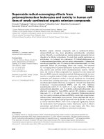
Fig. 1 The expression levels of Dazl in glioblastoma tissues and cell lines. a Dazl expression was examined by immunohistochemical analysis in
human glioblastoma tissues and adjacent normal tissues. Strong cytoplasmic expression of Dazl (brown staining) was detected in stage III/IV
glioblastoma cells, and the nucleus was stained blue with hematoxylin. Nor: Normal. Images were taken from the inverted microscope (bars =
50 μm, magnification × 200). b The correlation of Dazl expression and glioblastoma grade was analyzed from the Chinese Glioblastoma Genome
Atlas (CGGA) data (**P < 0.01, ****P < 0.001). c The correlation of Dazl expression and overall survival of glioblastoma patients was determined from
the CGGA data (**P < 0.01). d The lysates of glioblastoma cells from A172, U251, and LN229 cell lines were harvested and examined for Dazl
expression, the cell lysates of NHA cell line were used as the negative control (**P < 0.01, ****P < 0.001). e Detection of gene expression by agarose
gel electrophoresis after RT-PCR. f Dazl expression in glioblastoma cell lines was detected by western blotting. g Relative Dazl expression was
quantified by Image J software using Gapdh as an internal control. (*P < 0.05)
U251, and LN229 cells per field of vision showed lower
in the Dazl KD group, in contrast to the glioblastoma
WT cells (P < 0.05, Fig. 4a and b). These results revealed the involvement of Dazl in the sensitivity of glioblastoma cells to TMZ and DOX.
Dazl inhibits the initiation of glioblastoma via blocking
the stemness of glioblastoma cells
To validate the contribution of Dazl knockdown on glioblastoma tumorigenesis in vivo, we subcutaneously
injected GBM WT cells and GBM Dazl+/− cells into the
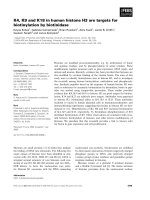
Fig. 2 Dazl knockdown inhibited the proliferation and germline traits of glioblastoma cells in vitro. a Western blot analysis detected whether
Cas9 protein was transfected into GBM cells successfully and whether Dazl protein was deleted. b A CCK-8 cell proliferation assay was performed
after Dazl deletion in A172, U251, and LN229 cells. c An alkaline phosphatase stain assay was performed between the WT GBM cell lines and the
Dazl deletion cells. Images were taken from the inverted microscope (magnification × 200). All experiments were carried out in triplicate. Data are
shown as the mean ± SE (*P < 0.05, **P < 0.01)
Zhang et al. BMC Cancer
(2020) 20:673
Fig. 3 (See legend on next page.)
Page 7 of 13
Zhang et al. BMC Cancer
(2020) 20:673
Page 8 of 13
(See figure on previous page.)
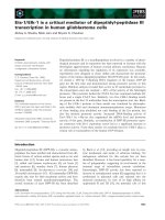
Fig. 3 Knockdown of Dazl inhibited glioblastoma cell migration and invasion in vitro. a Cell migration assays were performed after Dazl deletion in
A172, U251, and LN229 cells. A172, U251, and LN229 cells with Dazl knockdown exhibited decreased ability to migrate through the Boyden chamber
compared with the WT GMB cell lines. Five pictures were collected for each group, and the representative images were shown here. Images were
taken with an inverted microscope (bars = 50 μm, magnification × 200). b The statistical analysis of the ability of the glioblastoma cells’ migration (**P <
0.01, ***P < 0.001). c The invasion of A172, U251, and LN229 cells with Dazl knockdown was measured by transwell assay. Cells migrated through
Matrigel-coated transwell inserts and relative invasion proportion of cells were shown. Five pictures were collected for each group, and the
representative images were shown here. Images were taken with an inverted microscope (bars = 50 μm, magnification × 200). d The statistical analysis
of the ability of the glioblastoma cells’ invasion, the numbers of invasion cells with Dazl knockdown were significantly less than those with untreated
cells. All experiments were carried out in triplicate. Data are shown as the mean ± SE (*P < 0.05, ***P < 0.001)
backs of nude mice to build a xenograft model. The
growth curve of xenografted tumors displayed that U251
and LN229 cells showed rapid tumor growth in vivo
(P < 0.05, Fig. 5a and b), whereas U251 Dazl+/− and
LN229 Dazl+/− cells markedly inhibited tumor growth.
These results suggested that Dazl knocked-down GBM
cells were unable to initiate tumorigenesis in 6 months,
and recipient mice remained survival after 6 months.
The high post-surgical recurrence rate of glioblastoma
is mainly attributed to the existence of cancer stem cells
(CSCs) which can promote tumor initiation, invasion,
metastasis, and increase both differentiation and proliferation [28, 29]. To support this hypothesis, we then
investigated the correlation between Dazl expression and
cell stemness. We explored whether the stem transcriptional core formed by Oct4, Nanog, and Sox2 was
altered by Dazl levels. By qRT-PCR (P < 0.05, Fig. 5c), we
found that Dazl knockdown could significantly reduce
Oct4, Nanog, and Sox2 mRNA expression. Furthermore,
western blot experiments (P < 0.05, Fig. 5d) were utilized
to detect the protein expression, and Dazl knockeddown glioblastoma cell lines showed significantly reduced expression of stemness markers CD133, Oct4,
Nanog, and Sox2, and no changes in the protein expression of beta-Catenin. These results showed that Dazl
induces the tumorigenesis in glioblastoma mainly by
increasing the stemness but not via the WNT signaling
pathway (Fig. 5c and d). Therefore, our reports discovered that germline characteristics of glioblastoma cells
were markedly reduced in Dazl knockdown cells, and
the germline characteristics might be related to the
oncogenicity of glioblastoma.
Discussion
CRISPR/Cas9 technology is a powerful method for targeting desired genomic sites for gene editing or activity
modulation via specific single-guide RNAs (sgRNAs)
[30]. In the experiment, Dazl-sgRNAs were designed,
synthesized and cloned into a lentiviral vector, which
was subsequently transduced into glioma cells at a low
multiplicity of infection to ensure that only one sgRNA
copy was integrated per cell; then, the Cas9 enzyme was
guided to the Dazl gene location, where Cas9 induced a
double-strand break [31] The repair of such a break by
glioma cells led to a knockout of the targeted Dazl gene,
and the Dazl+/− GBM cell lines grew stably for generations.
CRISPR knockout technology has been highly effective in
identifying genes that have functions in tumorigenesis.
CRISPR/Cas9 is the most commonly applied method for
generating clinical trials of human cancer [32], and it is far
superior to the previously reported RNA interference technology because it ensures the functional stability of the Dazl
gene in the cell inheritance.
Glioblastoma is one of the most malignant primary
brain tumors associated with poor prognosis and low
median survival [33, 34]. Glioblastoma is not a surgically
curable disease because the glioblastoma cells invade the
surrounding brain tissue and are among the most resistant to chemotherapy [35, 36]. Therefore, new targets in
molecular knowledge, prognosis factor, and treatment
are urgent. The similarity of the biological characteristics
of cancer cells and germ cells prompted Lloyd J. Old to
discover cancer/testis (CT) antigens [37]. The discovery
elaborated a theory that aberrant expression of germline
genes in cancers reflects the activation of the silenced
gametogenic programme in somatic cells, and this programmatic acquisition is one of the driving forces of
tumorigenesis. Extensive data have been assembled concerning the ectopic activation of germline genes in the
progression of several human cancer types [38]. Dazl is
responsible for germline traits and plays a central role in
controlling pluripotency, differentiation, and apoptosis
[15]. In this study, we demonstrated that ectopic expression of the germline gene Dazl in human glioblastoma
and its association with tumorgenicity. We found that
Dazl promotes cell proliferation in GBM since A172,
U251, and LN229 GBM cells with Dazl knockdown
exhibited a reduced cell proliferation rate (Fig. 2). We
also showed that Dazl increases the ability of migration
and invasion through the transwell assays (Fig. 3). Also,
TMZ and Dox treated cell lines showed increased apoptosis in A172, U251, and LN229 GBM cells with Dazl
knockdown (Fig. 4), which suggested the anti-apoptosis
function of Dazl in GBM cells. Lastly, a screening of
stem cell markers found that their expression decreased
significantly in Dazl-knockdown cells (Fig. 5), suggesting
the involvement of Dazl in the maintenance of the
glioblastoma stem cell population.
Zhang et al. BMC Cancer
(2020) 20:673
Page 9 of 13
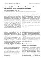
Fig. 4 Knockdown of Dazl inhibited the resistance of glioblastoma cells in vitro. a Dazl KD cells were significantly more sensitive to TMZ and DOX than
GBM WT cells (A172, U251, and LN229). Images were taken with an inverted microscope (bars = 50 μm, magnification × 100). b The statistical analysis of
the glioblastoma cell resistance. All experiments were carried out in triplicate. Data are shown as the mean ± SE (*P < 0.05, **P < 0.01, ***P < 0.001)
Zhang et al. BMC Cancer
(2020) 20:673
Fig. 5 (See legend on next page.)
Page 10 of 13
Zhang et al. BMC Cancer
(2020) 20:673
Page 11 of 13
(See figure on previous page.)
Fig. 5 Dazl inhibited the formation of glioblastoma via inhibiting the stemness of glioblastoma cells. a Tumor growth was observed from GBM cells
with Dazl alterations that were implanted subcutaneously in nude mice, n = 5; tumors were excised, photographed, and measured; b The tumor
growth sizes were recorded every 6 days between Dazl WT and Dazl +/− GBM cell lines in xenograft tumor models, n = 5; data are shown as the
mean ± SE. (***P < 0.001). c Q-RT-PCR analysis of stem cell gene expression in Dazl knockdown GBM cell lines. (*P < 0.05, **P < 0.01). d Western blot
analysis of the relative protein levels of CD133, Oct4, Nanog, and Sox2 in Dazl+/− and WT GBM cells. Quantitative analysis of the relative protein levels
of CD133, Oct4, Nanog, and Sox2 in GBM cells was carried out in triplicate. Data are shown as the mean ± SE. (*P < 0.05, **P < 0.01, ***P < 0.001)
Our work successfully discovered the relationship between Dazl and the proliferation of GBM cells. Dazl has
been known for its involvement in cell proliferation in
the integrity of PGCs in many vertebrates [39], Dazl is
involved in the early proliferation of the germ cells [40],
and also have essential roles in controlling a network of
cell-cycle regulatory genes such as sox3 and Atm [41].
Dazl enhances postnatal germ cell survival via poly Aproximal interactions that promote the cell-cycle regulation and germ cell survival [41]. Dazl can also improve
the spermatogonia proliferation via increasing steadystate levels of inherently unstable mRNA to ensure the
high concentrations of regulatory factors in the germ cell
development [42]. In this work, we found that Dazl was
upregulated in GBM cells and glioblastoma tissues, especially in late-stage. These findings support the oncogenic
function of Dazl in tumor formation and proliferation.
Besides regulating cell proliferation, Dazl was also
found to be responsible for anti-apoptosis in GBM cells
in this work. We found that multiple the number of
GBM cells with Dazl knockdown experienced apoptosis
compared to normal GBM cells under drug treatment
(Fig. 4). Dazl regulates the expression of the key
caspases, reveals a meaningful fail-safe mechanism that
prevents stray PGCs from forming teratomas by sensitizing them to apoptotic cell death [15, 39, 43]. A previous
study also demonstrated that the loss of PGCs in the
Dazl−/− embryo is due to increased apoptosis [43] and
Dazl knockdown in PGCs causes increased apoptosis.
Therefore, the silencing of Dazl induced drug susceptibility in glioblastoma cells by increasing apoptosis. Stemness
is thought to be the main reason for chemoresistance,
then we detected whether Dazl could regulate the stemness marker in glioblastoma.
Finally, we found that GBM cell lines A172, U251, and
LN229 all expressed stemness markers CD133, Nanog,
Oct4, and Sox2. Dazl-knocked down cell lines showed
significantly decreased expression of these markers.
Interestingly, at the transition of PGCs, Dazl-mediated
silencing of both pluripotency factors and the polycomb
complex allows PGCs to reduce the risk of teratoma formation by inhibition of the pluripotent program while
simultaneously preventing somatic differentiation [15].
Dazl likely plays different roles in different developmental stages, and its role in a specific tissue remains the
same in both normal and tumor cells. This findings confirm the theory that CG genes could exist in the GBM
cells, and mainly present in reproductive tissues, such as
testes, fetal ovaries, and trophoblasts, and are aberrantly
expressed in a range of human cancers, but have limited
expression on other normal tissues in adults [44]. Our
results further demonstrated the cancer-promoting role
of Dazl in glioblastoma cells and helped expand the
knowledge that the germline gene could involve in the
formation of glioblastomas. The tumor-suppressive
effect of Dazl was exerted through inhibiting the transcriptional activity of Oct4, Sox2, and Nanog gene to
attenuate the stemness and resistance of glioblastoma
cells. However, our results do not discover the detailed
mechanism of the Dazl regulates the tumorigenicity and
stemness in glioblastoma cells. Further studies on Dazl
expression and its function on these stemness markers
should prove beneficial. Moreover, the relationship
between germline genes and the tumorigenicity merits
further investigation as they are involved in several important cellular signaling pathways. However, our study
demonstrated that Dazl promotes the expression of stem
cell markers and apoptosis of the GBM cells not through
the WNT/beta-Catenin pathway.
Conclusion
In conclusion, Dazl functions as a novel cancer germline
gene to initiate the stemness of glioblastoma cells by
regulating the CD133/Oct4/Nanog/Sox2 regulatory axis
and increasing the resistance of glioblastoma cells to
Dox and TMZ. Additionally, Dazl knockdown not only
promotes the glioblastoma cells proliferation, migration,
and invasion in vitro, but also inhibits the initiation of
glioblastoma in vivo. Therefore, understanding the
underlying mechanisms of the cancer-germline gene in
glioblastoma has new implications in future therapies to
inhibit glioblastoma progression and recurrence.
Supplementary information
Supplementary information accompanies this paper at />1186/s12885-020-07155-y.
Additional file 1.
Additional file 2.
Additional file 3.
Zhang et al. BMC Cancer
(2020) 20:673
Abbreviations
AP: Alkaline phosphatase; BFP: Blue fluorescent protein; BSA: bovine serum
albumin; CCK-8: Cell counting kit-8; CG: Cancer-germline; CICs: Cancerinitiating cells; CRISPR: Clustered regularly interspaced short palindromic
repeats; CSCs: Cancer stem cells; Dazl: Deleted in azoospermia-like;
DMEM: Dulbecco’s modified eagle medium; DOX: Doxorubicin;
GBM: Glioblastoma multiforme; NHA: Normal human astrocytes;
iPSCs: Induced pluripotent stem cells; KD: Knockdown; PBS: Phosphatebuffered saline; PGCs: Primordial germ cells; RT: Room temperature;
sgRNAs: Single guide RNAs; TMZ: Temozolomide; WT: wild type
Acknowledgements
The authors thank the platform provided by the Experiment Animal Research
Center of Shanghai Medical College, Fudan University.
Authors’ contributions
FYZ, CL performed the experiments, generated and analyzed the data,
search literature, originated Figures, HSZ and RLL collected the clinical
tissues, FYZ wrote the manuscript. CFL and YL helped to design the
experiments. All authors had final approval of the submitted and published
version.
Funding
This work was supported by Grant No. 81372141, No. 81372351, and No.
81600202 from Natural Science Foundation of China, grant shslczdzk03303
from Shanghai Municipal Key Clinical Specialty of China. The funding bodies
had no role in the design of the study and collection, analysis and
interpretation of data and in writing the manuscript.
Availability of data and materials
All data generated or analyzed during this study are included in this
published article.
Page 12 of 13
5.
6.
7.
8.
9.
10.
11.
12.
13.
14.
Ethics approval and consent to participate
The study protocol was approved by the Medical Ethics Committee of
Huashan Hospital, Fudan University, and informed consent was obtained
from each patient. All animal studies according to protocols approved by
the Laboratory Animal Committee of Fudan University and handled with
care and euthanized humanely during the experiment.
15.
16.
Consent for publication
Not applicable.
17.
Competing interests
The authors declare that there are no conflicts of interest.
18.
Author details
1
Department of Laboratory Medicine, Huashan Hospital, Shanghai Medical
College, Fudan University, 12 Wulumuqi Road, Jing-an District, Shanghai
200040, China. 2Department of Laboratory Medicine, Shanghai General
Hospital, Shanghai Jiao Tong University, 85 Wujin Road, Hongkou District,
Shanghai 200080, China. 3Department of Neurosurgery, Huashan Hospital,
Fudan University, 12 Wulumuqi Road, Jing-an District, Shanghai 200040,
China.
19.
20.
21.
Received: 1 March 2020 Accepted: 8 July 2020
22.
References
1. Huang W, Zhong Z, Luo C, Xiao Y, Li L, Zhang X, Yang L, Xiao K, Ning Y,
Chen L, et al. The miR-26a/AP-2alpha/Nanog signaling axis mediates stem
cell self-renewal and temozolomide resistance in glioma. Theranostics. 2019;
9(19):5497–516.
2. Xi G, Best B, Mania-Farnell B, James CD, Tomita T. Therapeutic potential for
bone morphogenetic protein 4 in human malignant Glioma. Neoplasia.
2017;19(4):261–70.
3. Wang B, Wang M, Zhang W, Xiao T, Chen C-H, Wu A, Wu F, Traugh N, Wang
X, Li Z, et al. Integrative analysis of pooled CRISPR genetic screens using
MAGeCKFlute. Nat Protoc. 2019;14(3):756–80.
4. López-Valero I, Saiz-Ladera C, Torres S, Hernández-Tiedra S, GarcíaTaboada E, Rodríguez-Fornés F, Barba M, Dávila D, Salvador-Tormo N,
23.
24.
25.
26.
Guzmán M, et al. Targeting Glioma initiating cells with a combined
therapy of cannabinoids and temozolomide. Biochem Pharmacol. 2018;
157:266–74.
Kuo Y-C, Chang Y-H, Rajesh R. Targeted delivery of etoposide,
carmustine and doxorubicin to human glioblastoma cells using
methoxy poly (ethylene glycol)-poly(ε-caprolactone) nanoparticles
conjugated with wheat germ agglutinin and folic acid. Mater Sci Eng C
Mater Biol Appl. 2019;96:114–28.
Chattergoon NN, D'Souza FM, Deng W, Chen H, Hyman AL, Kadowitz PJ,
Jeter JR Jr. Antiproliferative effects of calcitonin gene-related peptide in
aortic and pulmonary artery smooth muscle cells. Am J Physiol Lung Cell
Mol Physiol. 2005;288(1):L202–11.
Auffinger B, Spencer D, Pytel P, Ahmed AU, Lesniak MS. The role of glioma
stem cells in chemotherapy resistance and glioblastoma multiforme
recurrence. Expert Rev Neurother. 2015;15(7):741–52.
Lucena-Cacace A, Otero-Albiol D, Jiménez-García MP, Peinado-Serrano
J. Carnero a.NAMPT overexpression induces cancer stemness and
defines a novel tumor signature for glioma prognosis. Oncotarget.
2017;8(59):99514–30.
Virolle T. Cancer stem cells in glioblastoma. Bull Cancer. 2017;104(12):1075–9.
Hattermann K, Flüh C, Engel D, Mehdorn HM, Synowitz M, Mentlein R, HeldFeindt J. Stem cell markers in glioma progression and recurrence. Int J
Oncol. 2016;49(5):1899–910.
Takahashi K, Yamanaka S. Induction of pluripotent stem cells from mouse
embryonic and adult fibroblast cultures by defined factors. Cell. 2006;126(4):
663–76.
Li X, Hughes SC, Wevrick R. Evaluation of melanoma antigen (MAGE) gene
expression in human cancers using the Cancer genome atlas. Cancer Genet.
2015;208(1):25–34.
Van Tongelen A, Loriot A, De Smet C. Oncogenic roles of DNA
hypomethylation through the activation of cancer-germline genes. Cancer
Lett. 2017;396:130–7.
Janic A, Mendizabal L, Llamazares S, Rossell D, Gonzalez C. Ectopic
expression of germline genes drives malignant brain tumor growth in
drosophila. Science. 2010;330(6012):1824–7.
Chen H-H, Welling M, Bloch DB, Muñoz J, Mientjes E, Chen X, Tramp C, Wu
J, Yabuuchi A, Chou Y-F, et al. DAZL limits pluripotency, differentiation, and
apoptosis in developing primordial germ cells. Stem Cell Reports. 2014;3(5):
892–904.
Lin Y, Gill ME, Koubova J, Page DC. Germ cell-intrinsic and -extrinsic factors
govern meiotic initiation in mouse embryos. Science. 2008;322(5908):1685–7.
Gill ME, Hu Y-C, Lin Y, Page DC. Licensing of gametogenesis, dependent on
RNA binding protein DAZL, as a gateway to sexual differentiation of fetal
germ cells. Proc Natl Acad Sci U S A. 2011;108(18):7443–8.
Ruggiu M, Speed R, Taggart M, McKay SJ, Kilanowski F, Saunders P, Dorin J,
Cooke HJ. The mouse Dazla gene encodes a cytoplasmic protein essential
for gametogenesis. Nature. 1997;389(6646):73–7.
Schrans-Stassen BH, Saunders PT, Cooke HJ, de Rooij DG. Nature of the
spermatogenic arrest in Dazl −/− mice. Biol Reprod. 2001;65(3):771–6.
Zhang J, Tam W-L, Tong GQ, Wu Q, Chan H-Y, Soh B-S, Lou Y, Yang J, Ma Y,
Chai L, et al. Sall4 modulates embryonic stem cell pluripotency and early
embryonic development by the transcriptional regulation of Pou5f1. Nat
Cell Biol. 2006;8(10):1114–23.
Luo G, Luo W, Sun X, Lin J, Wang M, Zhang Y, Luo W, Zhang Y. MicroRNA21 promotes migration and invasion of glioma cells via activation of Sox2
and β-catenin signaling. Mol Med Rep. 2017;15(1):187–93.
Abdelrahman AE, Ibrahim HM, Elsebai EA, Ismail EI, Elmesallamy W. The
clinicopathological significance of CD133 and Sox2 in astrocytic glioma.
Cancer Biomark. 2018;23(3):391–403.
Guo Y, Liu S, Wang P, Zhao S, Wang F, Bing L, Zhang Y, Ling EA, Gao
J, Hao A. Expression profile of embryonic stem cell-associated genes
Oct4, Sox2 and Nanog in human gliomas. Histopathology. 2011;59(4):
763–75.
Sedaghat S, Gheytanchi E, Asgari M, Roudi R, Keymoosi H, Madjd Z.
Expression of Cancer stem cell markers OCT4 and CD133 in transitional cell
carcinomas. Appl Immunohistochem Mol Morphol. 2017;25(3):196–202.
Niwa H, Miyazaki J, Smith AG. Quantitative expression of Oct-3/4 defines
differentiation, dedifferentiation or self-renewal of ES cells. Nat Genet. 2000;
24(4):372–6.
Du Y. The role and molecular mechanism of DAZL gene in lung cancer.
Hunan Normal University. 2019;16–36. [in Chinese].
Zhang et al. BMC Cancer
(2020) 20:673
27. Ran FA, Hsu PD, Wright J, Agarwala V, Scott DA, Zhang F. Genome
engineering using the CRISPR-Cas9 system. Nat Protoc. 2013;8(11):2281–308.
28. Li XT, Zhou YX, Du ZW. Novel lncRNA-ZNF281 regulates cell growth,
stemness and invasion of glioma stem-like U251s cells. Neoplasma. 2019;
66(1):118–27.
29. Clarke MF, Fuller M. Stem cells and cancer: two faces of eve. Cell. 2006;
124(6):0–1115.
30. Cong L, Ran FA, Cox D, Lin S, Barretto R, Habib N, et al. Multiplex genome
engineering using CRISPR/Cas systems. Science. 2013;339(6121):819.
31. Wang B, Wang M, Zhang W, Xiao T, Chen C-H, Wu A, et al. Integrative
analysis of pooled CRISPR genetic screens using MAGeCKFlute. Nat Protoc.
2019;14(3):756–80.
32. Huang K, Yang C, Wang Q-X, Li Y-S, Fang C, Tan Y-L, et al. the CRISPR/Cas9
system targeting EGFR exon 17 abrogates NF-κB activation via epigenetic
modulation of UBXN1 in EGFRwt/vIII glioma cells. Cancer Lett. 2017;388:
269–80.
33. Masui K, Cloughesy TF, Mischel PS. Review: molecular pathology in adult
high-grade gliomas: from molecular diagnostics to target therapies.
Neuropathol Appl Neurobiol. 2012;38(3):271–91.
34. Batash R, Asna N, Schaffer P, Francis N, Schaffer M. Glioblastoma Multiforme,
diagnosis and treatment; recent literature review. Curr Med Chem. 2017;
24(27):3002–9.
35. Gzell C, Back M, Wheeler H, Bailey D, Foote M. Radiotherapy in
Glioblastoma: the past, the present and the future. Clin Oncol (R Coll
Radiol). 2017;29(1):15–25.
36. Sahebjam S, Sharabi A, Lim M, Kesarwani P, Chinnaiyan P. Immunotherapy
and radiation in glioblastoma. J Neuro-Oncol. 2017;134(3):531–9.
37. Old LJ. Cancer vaccines: an overview. Cancer Immun. 2008;8(Suppl 1):1.
38. Wang J, Rousseaux S, Khochbin S. Sustaining cancer through addictive
ectopic gene activation. Curr Opin Oncol. 2014;26(1):73–7.
39. Lee HC, Choi HJ, Lee HG, Lim JM, Ono T. Han JY.DAZL expression explains
origin and central formation of primordial germ cells in chickens. Stem Cells
Dev. 2016;25(1):68–79.
40. Stefanidis K, Pergialiotis V, Christakis D, Patta J, Stefanidi D. Loutradis D.OCT4 and DAZL expression in precancerous lesions of the human uterine cervix.
J Obstet Gynaecol Res. 2015;41(5):763–7.
41. Zagore LL, Sweet TJ, Hannigan MM, Weyn-Vanhentenryck SM, Jobava R,
Hatzoglou M, Zhang C. Licatalosi DD.DAZL regulates germ cell survival
through a network of PolyA-Proximal mRNA interactions. Cell Rep. 2018;
25(5):1225–1240.e1226.
42. Li M, Zhu F, Li Z, Hong N, Hong Y. Dazl is a critical player for primordial
germ cell formation in medaka. Sci Rep. 2016;6:28317.
43. Lin Y, Page DC. Dazl deficiency leads to embryonic arrest of germ cell
development in XY C57BL/6 mice. Dev Biol. 2005;288(2):309–16.
44. Simpson AJG, Caballero OL, Jungbluth A, Chen Y-T, Old LJ. Cancer/testis
antigens, gametogenesis and cancer. Nat Rev Cancer. 2005;5(8):615–25.
Publisher’s Note
Springer Nature remains neutral with regard to jurisdictional claims in
published maps and institutional affiliations.
Page 13 of 13