Effect of neoadjuvant therapy on breast cancer biomarker profile
Bạn đang xem bản rút gọn của tài liệu. Xem và tải ngay bản đầy đủ của tài liệu tại đây (599.86 KB, 9 trang )
Rey-Vargas et al. BMC Cancer
(2020) 20:675
/>
RESEARCH ARTICLE
Open Access
Effect of neoadjuvant therapy on breast
cancer biomarker profile
Laura Rey-Vargas1,2, Juan Carlos Mejía-Henao3, María Carolina Sanabria-Salas4 and Silvia J. Serrano-Gomez1*
Abstract
Background: Breast cancer clinical management requires the assessment of hormone receptors (estrogen (ER) and
progesterone receptor (PR)), human epidermal growth factor receptor 2 (HER2) and cellular proliferation index Ki67,
by immunohistochemistry (IHC), in order to choose and guide therapy according to tumor biology. Many studies
have reported contradictory results regarding changes in the biomarker profile after neoadjuvant therapy (NAT).
Given its clinical implications for the disease management, we aimed to analyze changes in ER, PR, HER2, and Ki67
expression in paired core-needle biopsies and surgical samples in breast cancer patients that had either been
treated or not with NAT.
Methods: We included 139 patients with confirmed diagnosis of invasive ductal breast carcinoma from the
Colombian National Cancer Institute. Variation in biomarker profile were assessed according to NAT administration
(NAT and no-NAT treated cases) and NAT scheme (hormonal, cytotoxic, cytotoxic + trastuzumab, combined). Chisquared and Wilcoxon signed-rank test were used to identify changes in biomarker status and percentage
expression, respectively, in the corresponding groups.
Results: We did not find any significant variations in biomarker status or expression values in the no-NAT group. In
cases previously treated with NAT, we did find a statistically significant decrease in Ki67 (p < 0.001) and PR (p =
0.02605) expression. When changes were evaluated according to NAT scheme, we found a significant decrease in
both Ki67 status (p = 0.02977) and its expression values (p < 0.001) in cases that received the cytotoxic treatment.
Conclusions: Our results suggest that PR and Ki67 expression can be altered by NAT administration, whereas cases
not previously treated with NAT do not present IHC biomarker profile variations. The re-evaluation of these two
biomarkers after NAT could provide valuable information regarding treatment response and prognosis for breast
cancer patients.
Keywords: Breast neoplasms, Immunohistochemistry, Neoadjuvant therapy, Biomarkers, Heterogeneity
Background
Breast cancer is the malignancy with the highest incidence (46.3 per 100.000) and mortality rates (13.0 per
100.000) in women worldwide. According to the Surveillance, Epidemiology and End Results Program (SEER), in
2018 breast cancer accounted for 15.3% of all new
* Correspondence:
1
Grupo de investigación en biología del cáncer, Instituto Nacional de
Cancerología, Calle 1a #9-85, Bogotá D. C, Colombia
Full list of author information is available at the end of the article
cancer cases and 6.7% of all cancer deaths in the United
States (US) [1, 2].
Neoadjuvant therapy (NAT) has become an important
strategy to reduce tumor size in locally advanced breast
cancer and facilitate breast conservative surgery, along
with monitoring treatment response and eliminating
possible micrometastasis [3–5]. In order to choose an
appropriate NAT scheme according to tumor biology, a
preoperative evaluation on core-needle biopsies from the
primary tumor is performed, where histological type and
grade are assessed. Additionally, immunohistochemistry
© The Author(s). 2020 Open Access This article is licensed under a Creative Commons Attribution 4.0 International License,
which permits use, sharing, adaptation, distribution and reproduction in any medium or format, as long as you give
appropriate credit to the original author(s) and the source, provide a link to the Creative Commons licence, and indicate if
changes were made. The images or other third party material in this article are included in the article's Creative Commons
licence, unless indicated otherwise in a credit line to the material. If material is not included in the article's Creative Commons
licence and your intended use is not permitted by statutory regulation or exceeds the permitted use, you will need to obtain
permission directly from the copyright holder. To view a copy of this licence, visit />The Creative Commons Public Domain Dedication waiver ( applies to the
data made available in this article, unless otherwise stated in a credit line to the data.
Rey-Vargas et al. BMC Cancer
(2020) 20:675
(IHC) of biomarkers, such as: hormone receptors (estrogen receptor (ER) and progesterone receptor (PR)), human epidermal growth factor receptor 2 (HER2) and the
cellular proliferation index (Ki67), is also analyzed to
guide the therapy and predict survival [4, 6, 7].
These biomarkers have been used as surrogates for
breast cancer classification into four main intrinsic subtypes: luminal A, luminal B, HER2-enriched and triple
negative (TN) [8]. Both, luminal A and luminal B tumors
express ER, therefore these patients are candidates for
hormone therapy with ER modulators or aromatase inhibitors [8–11], whilst HER2-enriched and TN subtypes
lack the expression of hormone receptors, therefore are
mainly treated with biological therapy agents such as
trastuzumab or pertuzumab, and cytotoxic chemotherapy, respectively [5, 11, 12].
Standard clinical recommendations indicate the assessment of ER, PR, HER2 and Ki67 by IHC in biopsy samples [13, 14]. Nevertheless, many retrospective studies
have reported changes in biomarker expression in surgical specimens after NAT administration [15–21]. The
main changes correspond to discordances in hormone
receptors and HER2 status [22], along with decreases in
the percentage of expression, especially for PR and Ki67
[23–26]. Most of these studies end up suggesting the
need to re-evaluate its expression, justifying its importance not only to assess tumor response to treatment but
to adjust therapy according to these changes [3, 27].
However, other studies show that these changes are not
statistically significant [28] and suggest that the reevaluation of biomarker expression after NAT might not
be necessary, especially for health care institutions with
limited resources, as it is the case for many hospitals in
Latin-America, including Colombia [29].
Given the prognostic value of biomarkers expression
and its important role for deciding treatment scheme,
the aim of this study was to compare the IHC expression
of ER, PR, HER2, and Ki67 in core-needle biopsies and
surgical excision specimens in NAT-treated and nontreated breast cancer samples from patients diagnosed in
the Colombian National Cancer Institute (NCI), in order
to evaluate NAT effect on biomarker expression profile.
Methods
Clinical samples and data collection
This is a retrospective study that included 139 breast
cancer patients diagnosed with invasive ductal carcinoma (IDC) at the Colombian NCI between 2013 and
2014. Patients were included if they met the following
eligibility criteria: 1) histologically confirmed diagnosis
of IDC, 2) availability of formalin-fixed paraffinembedded (FFPE) tissue blocks from mastectomies or
breast-conserving surgeries that contained at least 10%
of tumor content, 3) availability of IHC slides from core-
Page 2 of 9
needle biopsies, and 4) paired IHC biomarker information on biopsy and surgical specimens. Patients with in
situ breast carcinoma were excluded. A single pathologist confirmed the histological diagnosis and reevaluated the expression of the IHC markers from each
patient.
This study was approved by the Colombian NCI ethics
committee, and according to the Colombian laws, it was
considered that no informed consent was required.
Pathology reports were reviewed to obtain information
regarding histopathological diagnosis, nodal status, surgical margins, invasion and histological grade. Treatment
information was retrieved from clinical records. NATtreated patients were categorized based on their neoadjuvant scheme in four groups: 1) hormonal, which includes letrozole and/or exemestane, 2) cytotoxic, which
includes AC (doxorubicin and cyclophosphamide), taxanes and/or platinums, 3) cytotoxic + trastuzumab,
which includes the same therapeutic agents from the
cytotoxic scheme plus trastuzumab, and 4) combined,
which includes both hormonal and cytotoxic therapeutic
agents.
Immunohistochemistry
IHC for ER, PR, HER2 and Ki67 expression was performed on 3 μm-thick sections from a single FFPE
with the highest tumor representation. Staining was
carried out using the Roche Benchmark XT automated slide preparation system (Roche Ltd.,
Switzerland). Positive and negative controls were included and DAB (3,3′ diaminobenzidine) was used as
chromogen.
A single pathologist analyzed biomarker expression
from surgery blocks and re-evaluated the IHC slides
from core-needle biopsies. Hormone receptors (ER
and PR) and Ki67 expression values were calculated
as the percentage of positive nuclear staining in the
IHC slide evaluated. Status of hormone receptors
was considered positive when they exceeded 1% of
nuclear staining in tumor cells. HER2 was defined
as: positive (3+) for complete and intense circumferential membrane within > 10% of tumor cells; ambiguous (2+) for incomplete and/or weak/moderate
circumferential membrane staining within > 10% of
tumor cells, or complete membrane staining but
within ≤10% of tumor cells; negative (1+) for incomplete faint membrane staining within > 10% of tumor
cells; and negative (0+) for absence of staining, according to the recommendations of the American
Society of Clinical Oncology (ASCO)/College of
American Pathologists (CAP) guideline [30]. For analysis purposes, Ki67 was categorized as high (≥20%)
or low (< 20%) expression.
Rey-Vargas et al. BMC Cancer
(2020) 20:675
Statistical analysis
We analyzed changes in biomarker status in paired samples from biopsy and surgical specimens, according to
NAT administration, using Chi-squared test for categorical variables. Changes in biomarker expression, as continuous variables, were evaluated using the Wilcoxon
signed-rank test for paired samples. All analyses were
conducted using R software (version 1.2.5033). Differences were considered statistically significant if p < 0.05.
Page 3 of 9
Table 1 Clinical-pathological characteristics of patients at
diagnosis
N (%)
Clinical stage
I (I, Ia, Ib)
11 (7.9)
II (IIa, IIb)
57 (41.0)
III (IIIa, IIIb, IIIc)
69 (49.6)
IV
2 (1.4)
Scarff-Bloom Richardson
Results
I
10 (7.2)
Clinical-pathological characteristics
II
83 (59.7)
III
46 (33.1)
Clinical-pathological characteristics of patients included
in this study are presented in Table 1. All tumors were
classified as IDC, of which the majority presented a clinical stage of III (49.6%) and a Scarff-Bloom-Richardson
score of II (59.7%). Sixty-four patients (46%) had positive
invasion, from which 38 (45.2%) corresponded to
lympho-vascular type. One hundred thirteen (81.3%) of
the patients included had axillary lymph node dissection,
from which 67 (59.3%) had lymph node involvement.
Seventy-eight patients (56.1%) received NAT based
mainly in cytotoxic chemotherapy (71.8%).
Biomarker status in core-needle biopsy and surgical
excisional specimens
Results from biomarker status in core-needle biopsy
showed that most cases were ER positive (80.6%), PR
positive (73.4%), HER2 negative (77.0%) and had a high
Ki67 proliferation index (≥20%) (66.2%). Biomarker status in surgical specimens presented a similar distribution, where most cases were also ER positive (79.1%), PR
positive (71.2%), HER2 negative (71.9%) and had a high
Ki67 proliferation index (53.3%). In order to assess the
impact of NAT treatment in biomarker status and its expression values, we performed an analysis in paired samples stratified by NAT administration.
Changes in biomarker status and expression in the noNAT group
Biomarker status
We compared the biomarker status between core-needle
biopsy and the surgical specimen in sixty-one cases
(43.9%) that did not receive NAT. We neither find statistically significant changes for ER/PR status (p = 1) nor
for Ki67 (p = 0.5796) (Table 2). Even though these are
cases that did not receive any type of treatment before
surgery that could have affected their tumor biology,
interestingly, we observed two cases that changed from
PR positive status in biopsy to negative in the surgical
specimen; other two cases went from negative status in
the biopsy to positive in the surgical specimen for the
same biomarker. On the other hand, although not statistically significant, we also observed variations in Ki67
Invasion
Yes
64 (46.0)
No
55 (39.6)
Unknown
20 (14.4)
Type of invasion
Lympho-vascular
38 (45.2)
Dermal
6 (7.1)
Lympho-vascular and perineural
9 (10.7)
Dermal lymphatic
5 (6.0)
Perineural
4 (4.8)
Dermal lymphatic and perineural
1 (1.2)
Dermal and perineural
1 (1.2)
Unknown
20 (23.8)
Axillary lymph node dissection
Yes
113 (81.3)
No
26 (18.7)
Involvement of lymph nodes
Yes
67 (59.3)
No
46 (40.7)
NAT administration
Yes
78 (56.1)
No
61 (43.9)
Type of NAT scheme
Hormonal
6 (7.7)
Cytotoxic
56 (71.8)
Cytotoxic + trastuzumab
11 (14.1)
Combined (hormonal+ cytotoxic)
5 (6.4)
NAT Neoadjuvant therapy
status. From thirty-seven cases with a high Ki67 expression (≥20%) in the biopsy, thirty-two remained in this
category, while 4 cases changed its status from high to
low Ki67 expression (< 20%) (See Supplementary Table 1,
Additional file 1).
For HER2 status, we did not find statistically significant changes in paired samples (p = 0.416). However, we
Rey-Vargas et al. BMC Cancer
(2020) 20:675
Page 4 of 9
Table 2 Biomarker status in cases that did and did not receive NAT, in biopsy and surgical specimens
Estrogen Receptor
Progesterone Receptor
HER2
Ki67 status
No-NAT group
NAT group
(N = 61)
(N = 78)
Categories
Biopsy
Surgery
p value
Biopsy
Surgery
Positive
46 (75.4)
46 (75.4)
1
66 (84.6)
64 (82.1)
Negative
15 (24.6)
15 (24.6)
12 (15.4)
14 (17.9)
Positive
42 (68.9)
42 (68.9)
60 (76.9)
57 (73.1)
Negative
19 (31.1)
19 (31.1)
18 (23.1)
21 (26.9)
Positive
9 (14.8)
11 (18.0)
10 (12.8)
9 (11.5)
Negative
47 (77.0)
41 (67.2)
60 (76.9)
59 (75.6)
Ambiguous
5 (8.2)
9 (14.8)
7 (9.0)
10 (12.8)
1
0.4165
Low (< 20%)
21 (34.4)
23 (37.7)
19 (24.4)
41 (52.6)
High (≥20%)
37 (60.7)
37 (60.7)
0.5796
55 (70.5)
37 (47.4)
Unknown
3 (4.9)
1 (1.6)
4 (5.1)
0 (0.0)
p value
0.8299
0.7115
0.6616
< 0.001
NAT Neoadjuvant therapy
did observe a modification in the HER2 classification for
some cases not previously treated with NAT. Even
though an ambiguous status does not correspond to an
actual HER2 classification, fluorescence in situ
hybridization (FISH) confirmatory results were not available for all cases within this category. Among cases that
presented a gain of positive HER2 status, two changed
from negative to positive and one changed from ambiguous to positive. Additionally, five cases changed from
negative to ambiguous status; from these cases, FISH
confirmatory results were available for two of them, with
negative result for HER2 amplification in the surgical
specimen. The remaining three cases did not have FISH
confirmatory results at the surgical sample.
On the other hand, among cases that presented loss of
HER2 status, one case changed from positive to ambiguous and another changed from ambiguous to negative
status. Both cases had HER2 negative amplification confirmatory results at the surgical sample (Table 3).
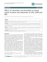
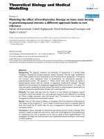
Biomarker expression
We also compared ER, PR and Ki67 expression values
between paired specimens. Median values were 100, 70
and 27.5%, respectively, in core-needle biopsies. After
surgery, expression values did not show statistically significant changes for any of the three biomarkers (100, 70
and 20%, respectively) (Fig. 1).
Changes in biomarker status and expression in the NAT
group
Biomarker status
Seventy-eight cases (56.1%) received NAT. Tumor size
measured before and after therapy were compared in
these patients. The mean tumor size before NAT were
51.9 mm, and after treatment it significantly decreased
to 29.3 mm (p < 0.01).
We did not find any statistically significant changes in
ER nor PR status in paired samples in the NAT group
(Table 2). Nevertheless, as expected, we observed some
cases that did present a modification in their hormone
receptor status after NAT. We observed changes from
positive to negative status for ER in two cases, one of
which also showed loss of PR expression. Four additional
cases also changed their PR status from positive to negative. On the other hand, we observed gain of PR status
in two cases.
Table 3 HER2 classification changes in the NAT and no-NAT group from paired biopsy and surgical specimens
No-NAT group (N = 61)
NAT group (N = 78)
NAT Neoadjuvant therapy
Surgery
Biopsy
Negative
Ambiguous
Positive
p value
No. cases with loss of HER2
No. cases with gain of HER2
Negative
40
5
2
0.4165
2
8
0.6616
4
5
Ambiguous
1
3
1
Positive
0
1
8
Negative
55
4
1
Ambiguous
2
5
0
Positive
2
0
8
Unkown
0
1
0
Rey-Vargas et al. BMC Cancer
(2020) 20:675
Page 5 of 9
Fig. 1 Changes of biomarker expression in biopsy and surgical samples, in the no-NAT group. Points indicate the median value for each measure.
ER: Estrogen receptor; PR: Progesterone receptor
Evaluation of Ki67 status showed statistically significant changes between paired biopsy and surgical specimen (p < 0.001) (Table 2). From fifty-five cases with a
high Ki67 expression (≥20%) in the biopsy, thirty-five
remained in this category, while twenty cases changed
its status from high to low Ki67 expression (< 20%) (See
Supplementary Table 1, Additional file 1).
Regarding HER2 status, no statistically significant
changes between biopsy and surgical sample were observed (p = 0.662), nonetheless, a small number of cases
showed changes in HER2 status. Among positive HER2
tumors at biopsy, two cases changed to negative status,
and similarly, among cases with ambiguous HER2 status
at diagnosis, two cases changed to negative. Lastly, from
60 cases initially defined as HER2 negative in the biopsy,
four turned to ambiguous and one case to positive status
(Table 3).
Changes in biomarkers status according to NAT
scheme was also analyzed. We only found statistically
significant variations in Ki67 status between biopsy and
surgical specimens in cases treated with the cytotoxic
scheme (p = 0.02977) (See Supplementary Table 2, Additional file 1). Other findings, although not statistically
significant, include a trend for hormone receptors and
Ki67 status loss after treatment with the cytotoxic (ER
positive: 83.9% vs. 82.1%; PR positive: 76.8% vs. 73.2%)
and cytotoxic + trastuzumab schemes (ER positive:
72.7% vs. 63.6%; PR positive: 72.7% vs. 54.5%, High Ki67:
81.8% vs. 54.5%). Interestingly, cases previously treated
with cytotoxic therapy presented a trend for a gain of
HER2 positive and ambiguous status (HER2 positive: 0%
vs 1.8%; HER2 ambiguous: 8.9% vs. 12.5%), whereas in
the cytotoxic + trastuzumab scheme group, HER2
showed a tendency for loss of positive status (HER2
positive: 90.9% vs 72.7%).
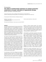
Biomarker expression
The ER, PR and Ki67 median expression values in coreneedle biopsy were 100, 80 and 30%, respectively. After
surgery, PR and Ki67 expression significantly decreased
to 65% (p = 0.01466) and 15% (p < 0.001), respectively,
whilst ER showed no statistically significant variation
(Fig. 2). Additionally, we analyzed changes of biomarker
expression according to the NAT scheme, and only
found a statistically significant decrease for Ki67 expression in cases that received cytotoxic treatment (p <
0.001). Even though not statistically significant, we still
could observe a trend for a lower PR expression values
in surgical samples for all treatment schemes groups
(Hormonal: 100% vs. 80%, Cytotoxic: 80% vs. 70%, Cytotoxic + trastuzumab: 40% vs. 20%, Combined: 90% vs.
20%) (See Supplementary Table 3, Additional file 1).
Discussion
In the current study, we aimed to analyze changes in
IHC biomarker status and expression in paired biopsy
and surgical samples in breast cancer cases treated and
non-treated with NAT, given that changes in biomarker
expression may have several clinical implications for disease outcome in breast cancer patients [31–33]. It may
also affect adjuvant therapy regimen, as variation in
breast cancer intrinsic subtype after NAT could lead to
the addition or discontinuation of therapy schemes [16,
34].
It has been well described that NAT treatment affects
Ki67 index, as it targets mainly cycling cells and major
cell proliferation pathways [35]. We observed a significant decrease in tumor size and in Ki67 expression
values only in the NAT-treated group. Despite the fact
that we did not analyze differences in outcome according to these changes, a decrease in Ki67 expression after
NAT has been associated with a good clinicalpathological response, better disease-free survival and
overall survival [36, 37], whereas no reduction in Ki67
expression after NAT have been associated with a significantly higher risk of breast cancer recurrence and
death [38]. Interesting results reported by Dowsett et al.
[35] show that Ki67 expression values after 2 weeks of
NAT were more useful as prognostic markers for prediction of recurrence-free survival than baseline Ki67
Rey-Vargas et al. BMC Cancer
(2020) 20:675
Page 6 of 9
Fig. 2 Changes of biomarker expression in biopsy and surgical samples in the NAT group. Points indicate the median value in each measure. ER:
Estrogen receptor; PR: Progesterone receptor
expression before NAT. Our results showing a significant decrease in Ki67 expression after NAT, along with
previously reported results, highlight the utility of assessing this biomarker in the surgical specimen after NAT,
for prognosis and patients’ clinical outcomes evaluation.
It has been well reported that chemotherapy may have
several effects on tumor biology, which could potentially
alter biomarker expression [3, 39]. We found a statistically significant decrease in PR expression values between biopsy and surgical specimens after NAT, which
is consistent with other reports where the PR, along with
the Ki67 index, are the most commonly altered biomarkers after NAT administration [7, 15, 16, 31]. Contrary to what has been reported for Ki67, PR expression
loss has been associated with worse tumor characteristics [15] and poor clinical outcomes [23, 32, 40]. It has
also been shown that loss of PR expression may be an
indicator of a decrease of hormone sensitivity in tumor
cells, activation of alternate proliferation pathways such
as PI3K/AKT/mTOR [41, 42], and also, the induction of
a non-functional ER state which could lead to a diminished response to endocrine therapy, specifically in ERpositive/PR-negative tumors [18, 43].
Loss of ER expression has also been associated with
bad clinical outcomes as it affects tumor response to
endocrine therapy [44]. Nevertheless, as we reported
here, variation in ER expression after NAT is much less
frequent than for PR. We did not observe statistically
significant changes in ER status nor its expression in neither of the NAT treated or not-treated cases, although
we did observe two cases with status loss in the NATtreated group. These results suggest that analyzing ER
after NAT administration may not be as useful as a
prognostic marker, as it could be PR and Ki67 status
evaluation.
On the other hand, gain of hormone receptor expression after NAT is associated with significantly better
outcomes, compared with patients with unchanged hormone receptor expression [45, 46]. It has even been
shown that the improvement on survival rates for patients with ER and PR expression gain is dependent on
the magnitude of change [47], however, gain of hormone
receptor expression is much less frequently reported [22,
31, 32]. In our study, no gain in ER status was observed
in neither of the NAT-treated nor non-treated cases,
whilst for PR, we observed gain of status in only two
cases from the NAT-treated group. Overall, these results
suggest that the gain of hormone receptor may not be
frequent enough for its implementation to assess treatment response and disease outcomes.
HER2 status variation between biopsy and surgical
samples are reported to be less frequent than for hormone receptors and Ki67 index [32, 48]. We did not find
statistically significant changes in HER2 biomarker status
neither in the NAT-treated nor non-treated cases. Some
reports have found important changes in HER2 expression, which seem to be driven not just by NAT, but by
specific types of therapeutic agents [33, 40, 49]. Ignatov
et al. [25] reported that trastuzumab administration was
associated with a decrease in HER2 expression in 47.3%
of cases, and interestingly, when pertuzumab was added
to the trastuzumab-NAT scheme, the decrease in HER2
expression rise to 63.2%. In our data, when we assessed
HER2 variations according to type of NAT regimen, no
statistically significant changes were found in neither of
the NAT-schemes groups, including the cytotoxic +
trastuzumab group. Despite our results not being statistically significant, we did observe some cases in the cytotoxic + trastuzumab scheme group with a decrease in
HER2 status. Hypotheses regarding HER2 downregulation after treatment includes the internalization in endosomal compartments and lysosomal degradation of
HER2 receptor induced by anti-HER2 agents (pertuzumab, trastuzumab) [50]. Nonetheless, as have been
shown, ERBB2 amplification when evaluated by FISH remains stable after NAT treatment [51].
Undoubtedly, cancer treatments may alter in some degree tumor gene expression [52], leading to possible
Rey-Vargas et al. BMC Cancer
(2020) 20:675
modification of the IHC biomarker profile. However,
tumor heterogeneity is also an important factor to take
into account when considering changes in biomarker expression between tumor samples, especially when
changes are observed in non-previously NAT treated
cases [53], as we reported here. Tumor heterogeneity in
breast cancer has been observed in multiple studies [17,
54–56]. Rye et al. [56] evaluated tumor heterogeneity of
ER and HER2 expression within individual breast tumors
at different time points, and reported the presence of
tumor cells within the same sample with both HER2+/
ER+ and HER2+/ER- expression profile, reveling a high
rate of cell-to-cell variation. Tumor heterogeneity may
have several clinical implications for patient’s outcome.
For example, a heterogeneous expression of HER2 copy
number in tumors have been reported to be associated
with higher risks of relapse and breast cancer death [56].
Such findings are expected given that this kind of intratumoral heterogeneity is often the result of clonal evolution, which is highly correlated with metastatic events
[57, 58].
External factors different from tumor heterogeneity
may also account for changes in biomarkers expression
between biopsy and surgical samples in cases not previously treated with NAT [17, 51, 54]. Among these are
technical preparation of the IHC stain, fixation times,
and inter- and intra-observer variability [17]. It has also
been reported that this variability could be a result of
the so-called dilution effect, which refers to a decrease
of biomarker expression with increasing number of
available tumor cells to evaluate at the surgical sample
[59]. As have been shown before, larger tumors from
surgical excision procedures are more likely to present
variations of biomarkers expression between biopsy and
surgical samples [15, 55]. This may indicate that at the
initial biopsy only a small portion of a tumor is sampled
for its evaluation, therefore large tumors could end up
being poorly represented and present with IHC profile
variations.
Our study certainly had limitations, mainly regarding
the small sample size, which limited the statistical power
of the analyses, especially when NAT-treated cases were
grouped according to the therapy scheme. Small sample
size in the hormonal, cytotoxic + trastuzumab and combined NAT-scheme groups may not have allowed us to
observe statistically significant expression changes between biopsy and surgical samples. Additionally, all cases
were recruited from a single institution and only pathological, but not clinical information was collected. However, our results regarding changes of biomarker
expression in NAT-treated and non-treated cases are
mostly consistent with what has been reported previously in other studies [17, 51, 54]. On the other hand,
we did not have FISH confirmatory amplification results
Page 7 of 9
for some cases with HER2 ambiguous result by IHC,
which did not allow us to give a more precise classification of these cases. Additionally, it is important to highlight that, unlike most studies evaluating changes of IHC
biomarker expression after NAT treatment, we included
a group of cases not previously treated with NAT in our
analysis, which allowed us to determine if NAT administration could in fact induce changes in the IHC biomarker profile.
Conclusions
Overall, our results confirmed that NAT administration
may cause changes in IHC biomarker profile, mainly in
Ki67 and PR expression, and that patients not previously
treated with NAT do not present significant changes in
biomarker expression. Since it is not cost-effective for
the health care system to reassess the expression of each
biomarker, for every patient after NAT, we suggest that
only PR and Ki67 biomarkers should be reassessed after
NAT treatment, as it has been shown that changes in
these two may have prognosis implications for breast
cancer patients. The implementation of the PR and Ki67
biomarkers as prognosis tools, along with other clinical
variables such as tumor stage and nodal status [60, 61],
could provide enough information about treatment response and it could be used by physicians to readjust
therapy.
Supplementary information
Supplementary information accompanies this paper at />1186/s12885-020-07179-4.
Additional file 1: Table S1. Ki67 classification changes in the NAT and
no-NAT group from paired biopsy and surgical specimens. Table S2. Biomarker status in biopsy and surgical specimens according to NAT
scheme. Table S3. Median biomarker expression in biopsy and surgical
specimens according to NAT scheme.
Abbreviations
NAT: Neoadjuvant therapy; IHC: Immunohistochemistry; ER: Estrogen
receptor; PR: Progesterone receptor; HER2: Human epidermal growth factor
receptor 2; TN: Triple negative; NCI: National Cancer Institute; IDC: Invasive
ductal carcinoma; FFPE: Formalin-fixed paraffin-embedded;
FISH: Fluorescence in situ hybridization.
Acknowledgements
Not applicable.
Authors’ contributions
The concept of the study was conceived by SJSG and LRV. SJSG, MCSS and
LRV have contributed to the background check and design of the study.
JCMH and LRV have contributed to data collection. JCMH performed the
pathology and histology evaluations. LRV has written the manuscript in
collaboration with SJSG and MCSS. SJSG and LRV have contributed to the
statistical analysis. The authors read and approved the final manuscript.
Funding
This study was funded by the Colombian NCI, project number: C19010300–
411.
Rey-Vargas et al. BMC Cancer
(2020) 20:675
Availability of data and materials
The dataset analyzed during the current study are available from the
corresponding author on reasonable request.
Ethics approval and consent to participate
This study was approved by the Colombian NCI research and ethics
committee. In accordance with Colombian resolution 8430 of 1993, this
study was considered without risk, given that no intervention was performed
in any patient, therefore, no informed consent was required. We also
acquired administrative permission from the pathology department to
access patients’ clinical information.
Page 8 of 9
16.
17.
18.
19.
Consent for publication
Not applicable.
Competing interests
The authors declare that they have no competing interests.
20.
Author details
1
Grupo de investigación en biología del cáncer, Instituto Nacional de
Cancerología, Calle 1a #9-85, Bogotá D. C, Colombia. 2Pontificia Universidad
Javeriana, Bogotá, Colombia. 3Grupo de patología oncológica, Instituto
Nacional de Cancerología, Bogotá, Colombia. 4Subdirección de
Investigaciones - Instituto Nacional de Cancerología de Colombia, Bogotá,
Colombia.
21.
22.
23.
Received: 7 April 2020 Accepted: 13 July 2020
24.
References
1. Globocan. Breast: Cancer incidence and mortality statistics worldwide and
by region. 2018 [cited 2018 Oct 29]. Available from: />2. SEER: Surveillance E and ERP. Female Breast Cancer - Cancer Stat Facts. 2018
[cited 2019 Jan 28]. Available from: />breast.html.
3. Penault-Llorca F, Radosevic-Robin N. Biomarkers of residual disease after
neoadjuvant therapy for breast cancer. Nat Rev Clin Oncol. 2016;13(8):487–
503.
4. Untch M, Konecny GE, Paepke S, Von Minckwitz G. Current and future role
of neoadjuvant therapy for breast cancer. Breast. 2014;23:526–37.
5. Liu SV, Melstrom L, Yao K, Russell CA, Sener SF. Neoadjuvant therapy for
breast cancer. J Surg Oncol. 2010;101(4):283–91.
6. National Institute for Health and Care Excellence. Early and locally advanced
breast cancer: diagnosis and management, NICE guideline. 2018.
7. Lee HC, Ko H, Seol H, Noh DY, Han W, Kim TY, et al. Expression of
immunohistochemical markers before and after neoadjuvant chemotherapy
in breast carcinoma, and their use as predictors of response. J Breast
Cancer. 2013;16(4):395–403.
8. Prat A, Perou CM. Deconstructing the molecular portraits of breast cancer.
Mol Oncol. 2011;5(1):5–23.
9. Perou CM, Sørlie T, Eisen MB, van de Rijn M, Jeffrey SS, Rees CA, et al.
Molecular portraits of human breast tumours. Nature. 2000;406(6797):747–
52.
10. Sørlie T, Perou CM, Tibshirani R, Aas T, Geisler S, Johnsen H, et al. Gene
expression patterns of breast carcinomas distinguish tumor subclasses with
clinical implications. Proc Natl Acad Sci U S A. 2001;98(19):10869–74.
11. Eroles P, Bosch A, Alejandro Pérez-Fidalgo J, Lluch A. Molecular biology in
breast cancer: intrinsic subtypes and signaling pathways. Cancer Treat Rev.
2012;38(6):698–707.
12. Dai X, Li T, Bai Z, Yang Y, Liu X, Zhan J, et al. Breast cancer intrinsic subtype
classification , clinical use and future trends. Am J Cancer Res. 2015;5(10):
2929–43.
13. Duffy MJ, Harbeck N, Nap M, Molina R, Nicolini A, Senkus E, et al. Clinical
use of biomarkers in breast cancer: Updated guidelines from the European
Group on Tumor Markers (EGTM). Eur J Cancer Elsevier Ltd. 2017;75:284–98.
14. Provenzano E, Bossuyt V, Viale G, Cameron D, Badve S, Denkert C, et al.
Standardization of pathologic evaluation and reporting of postneoadjuvant
specimens in clinical trials of breast cancer: recommendations from an
international working group. Mod Pathol. 2015;28:1185–201.
15. Zhou X, Zhang J, Yun H, Shi R, Wang Y, Wang W. Alterations of biomarker
profiles after neoadjuvant chemotherapy in breast cancer : tumor
25.
26.
27.
28.
29.
30.
31.
32.
33.
34.
heterogeneity should be taken into consideration. Oncotarget. 2015;6(34):
36894–902.
Xian Z, Quinones AK, Tozbikian G, Zynger DL. Breast cancer biomarkers
before and after neoadjuvant chemotherapy: does repeat testing impact
therapeutic management? Hum Pathol. 2017;62:215–21.
Yang YF, Liao YY, Li LQ, Xie SR, Xie YF, Peng NF. Changes in ER, PR and
HER2 receptors status after neoadjuvant chemotherapy in breast cancer.
Pathol Res Pract. 2013;209(12):797–802.
Neubauer H, Gall C, Vogel U, Hornung R, Wallwiener D, Solomayer E, et al.
Changes in tumour biological markers during primary systemic
chemotherapy (PST). Anticancer Res. 2008;28(3 B):1797–804.
Gahlaut R, Bennett A, Fatayer H, Dall BJ, Sharma N, Velikova G, et al. Effect of
neoadjuvant chemotherapy on breast cancer phenotype, ER/PR and HER2
expression - implications for the practising oncologist. Eur J Cancer. 2016;
60(January 2014):40–8.
Jin G, Han Y, Liu C, Chen L, Ding B, Xuan S, et al. Evaluation of biomarker
changes after administration of various neoadjuvant chemotherapies in
breast cancer. Int J Clin Exp Pathol. 2015;8(1):914–21.
Dede DS, Gumuskaya B, Guler G, Onat D, Altundag K, Ozisik Y. Evaluation of
changes of biologic markers ER, PR, HER 2 and Ki-67 in breast cancer with
administration of neoadjuvant dose- dense doxorubicin, cyclophosphamide
followed by paclitaxel. J BUON. 2013;18(1):57–63.
Peng J, Zhang X, Song J, Ran L, Luo R, Wang Y. Neoadjuvant chemotherapy
reduces the expression rates of ER, PR, HER2, Ki67, and P53 of invasive
ductal carcinoma. Medicine (Baltimore). 2019;98(2):1–8.
Tural D, Karaca M, Zirtiloglu A, M Hacioglu B, Sendur MA, Ozet A. Receptor
discordances after neoadjuvant chemotherapy and their effects on survival.
J BUON. 2019;24(1):20–5.
Jin X, Jiang YZ, Chen S, Da Yu K, Shao ZM, Di GH. Prognostic value of
receptor conversion after neoadjuvant chemotherapy in breast cancer
patients: a prospective observational study. Oncotarget. 2015;6(11):9600–11.
Ignatov T, Gorbunow F, Eggemann H, Ortmann O, Ignatov A. Loss of HER2
after HER2-targeted treatment. Breast Cancer Res Treat. 2019;175(2):401–8.
Wu YT, Li X, Lu LJ, Gan L, Dai W, Shi YL, et al. Effect of neoadjuvant
chemotherapy on the expression of hormone receptors and Ki67 in Chinese
breast cancer patients: a retrospective study of 525 patients. J Biomed Res.
2018;32(3):191–7.
Kang Y-J, Lee H-B, Kim YG, Han J, Kim Y, Yoo T-K, et al. Ki-67 expression is a
significant prognostic factor only when progesterone receptor expression is
low in estrogen receptor-positive and HER2-negative early breast Cancer. J
Oncol. 2019;2019:8.
Kinsella MD, Nassar A, Siddiqui MT, Cohen C. Estrogen receptor (ER),
progesterone receptor (PR), and HER2 expression pre- and postneoadjuvant chemotherapy in primary breast carcinoma: a single
institutional experience. Int J Clin Exp Pathol. 2012;5(6):530–6.
Population NRC (US) C on P, Gribble JN, Preston SH. Health Policy Issues in
Three Latin American Countries: Implications of The Epidemiological Transition.
In: National Academies Press (US), editor. The Epidemiological Transition: Policy
and Planning Implications for Developing Countries: Workshop Proceedings.
Washington (DC): National Academies Press (US); 1993.
Wolff AC, Hammond MEH, Hicks DG, Dowsett M, McShane LM, Allison KH,
et al. Recommendations for human epidermal growth factor receptor 2
testing in breast Cancer: American Society of Clinical Oncology/College of
American Pathologists Clinical Practice Guideline Update. J Clin Oncol. 2013;
31(31):3997–4013.
Yang L, Zhong X, Pu T, Qiu Y, Ye F, Bu H. Clinical significance and
prognostic value of receptor conversion in hormone receptor positive
breast cancers after neoadjuvant chemotherapy. World J Surg Oncol. 2018;
16(1):1–9.
Ahn S, Kim HJ, Kim M, Chung YR, Kang E, Kim EK, et al. Negative conversion
of progesterone receptor status after primary systemic therapy is associated
with poor clinical outcome in patients with breast cancer. Cancer Res Treat.
2018;50(4):1418–32.
Shuai Y, Ma L. Prognostic value of pathologic complete response and the
alteration of breast cancer immunohistochemical biomarkers after
neoadjuvant chemotherapy. Pathol Res Pract. 2019;215:29–33 Elsevier
GmbH.
De La Cruz LM, Harhay MO, Zhang P, Ugras S. Impact of Neoadjuvant
Chemotherapy on Breast Cancer Subtype: Does Subtype Change and, if so,
How?: IHC Profile and Neoadjuvant Chemotherapy. Ann Surg Oncol. 2018;
25(12):3535–40.
Rey-Vargas et al. BMC Cancer
(2020) 20:675
35. Dowsett M, Smith IE, Ebbs SR, Dixon JM, Skene A, A’Hern R, et al. Prognostic
value of Ki67 expression after short-term presurgical endocrine therapy for
primary breast cancer. J Natl Cancer Inst. 2007;99(2):167–70.
36. Enomoto Y, Morimoto T, Nishimukai A, Higuchi T, Yanai A, Miyagawa Y,
et al. Impact of biomarker changes during neoadjuvant chemotherapy for
clinical response in patients with residual breast cancers. Int J Clin Oncol.
2016;21(2):254–61.
37. Penault-Llorca F, Abrial C, Raoelfils I, Chollet P, Cayre A, Mouret-Reynier M, et al.
Changes and predictive and prognostic value of the mitotic index, Ki-67, Cyclin
D1, and Cyclo-oxygenase-2 in 710 operable breast Cancer patients treated
with Neoadjuvant chemotherapy. Oncologist. 2008;13(12):1235–45.
38. Cabrera-Galeana P, Muñoz-Montaño W, Lara-Medina F, Alvarado-Miranda A,
Pérez-Sánchez V, Villarreal-Garza C, et al. Ki67 changes identify worse
outcomes in residual breast Cancer tumors after Neoadjuvant
chemotherapy. Oncologist. 2018;23(6):670–8.
39. Schmitt MW, Loeb LA, Salk JJ. The influence of subclonal resistance
mutations on targeted cancer therapy. Nat Rev Clin Oncol. 2016;13:335–47
Nature Publishing Group.
40. Guarneri V, Dieci MV, Barbieri E, Piacentini F, Omarini C, Ficarra G, et al. Loss
of HER2 positivity and prognosis after neoadjuvant therapy in HER2-positive
breast cancer patients. Ann Oncol. 2013;24(12):2990–4.
41. Brodie A, Sabnis G. Adaptive changes result in activation of alternate
signaling pathways and acquisition of resistance to aromatase inhibitors.
Clin Cancer Res. 2011;17:4208–13 NIH Public Access.
42. Riggio M, Polo L, Blaustein M, Colman-Lerner A, Lü I, Lanari C, et al. PI3K/
AKT pathway regulates phosphorylation of steroid receptors, hormone
independence and tumor differentiation in breast cancer. Carcinogenesis.
2012;33(3):509–18.
43. Cui X, Schiff R, Arpino G, Osborne CK, Lee AV. Biology of progesterone
receptor loss in breast cancer and its implications for endocrine therapy. J
Clin Oncol. 2005;23(30):7721–35.
44. Li C, Fan H, Xiang Q, Xu L, Zhang Z, Liu Q, et al. Prognostic value of
receptor status conversion following neoadjuvant chemotherapy in breast
cancer patients: a systematic review and meta-analysis. Breast Cancer Res
Treatment. 2019;178:497–504 Springer New York LLC.
45. Lim SK, Lee MH, Park IH, You JY, Nam B-H, Kim BN, et al. Impact of
molecular subtype conversion of breast cancers after Neoadjuvant
chemotherapy on clinical outcome. Cancer Res Treat. 2016;48(1):133–41.
46. Tacca O, Penault-Llorca F, Abrial C, Mouret-Reynier M, Raoelfils I, Durando X,
et al. Changes in and prognostic value of hormone receptor status in a
series of operable breast Cancer patients treated with Neoadjuvant
chemotherapy. Oncologist. 2007;12(6):636–43.
47. Parinyanitikul N, Lei X, Chavez-Macgregor M, Liu S, Mittendorf EA, Litton JK,
et al. Receptor status change from primary to residual breast cancer after
neoadjuvant chemotherapy and analysis of survival outcomes. Clin Breast
Cancer. 2015;15(2):153–60.
48. Niikura N, Tomotaki A, Miyata H, Iwamoto T, Kawai M, Anan K, et al.
Changes in tumor expression of HER2 and hormone receptors status after
neoadjuvant chemotherapy in 21 755 patients from the Japanese breast
cancer registry. Ann Oncol. 2016;27(3):480–7.
49. Van de Ven S, Smit VTHBM, Dekker TJA, Nortier JWR, Kroep JR. Discordances
in ER, PR and HER2 receptors after neoadjuvant chemotherapy in breast
cancer. Cancer Treat Rev. 2011;37(6):422–30.
50. Hughes JB, Rødland MS, Hasmann M, Madshus IH, Stang E. Pertuzumab
increases 17-AAG-induced degradation of ErbB2, and this effect is further
increased by combining pertuzumab with trastuzumab. Pharmaceuticals.
2012;5(7):674–89.
51. Li P, Liu T, Wang Y, Shao S, Zhang W, Lv Y, et al. Influence of Neoadjuvant
chemotherapy on HER2/neu status in invasive breast Cancer. Clin Breast
Cancer. 2013;13(1):53–60.
52. Lee SC, Xu X, Lim YW, Lau P, Sukri N, Lim SE, et al. Chemotherapy-induced
tumor gene expression changes in human breast cancers. Pharmacogenet
Genomics. 2009;19(3):181–92.
53. Zardavas D, Irrthum A, Swanton C, Piccart M. Clinical management of breast
cancer heterogeneity. Nat Rev Clin Oncol. 2015;12(7):381–94.
54. Mann GB, Fahey VD, Feleppa F, Buchanan MR. Reliance on hormone
receptor assays of surgical specimens may compromise outcome in
patients with breast cancer. J Clin Oncol. 2005;23(22):5148–54.
55. Cavaliere A, Sidoni A, Scheibel M, Bellezza G, Brachelente G, Vitali R, et al.
Biopathologic profile of breast cancer core biopsy: is it always a valid
method? Cancer Lett. 2005;218(1):117–21.
Page 9 of 9
56. Rye IH, Trinh A, Sætersdal AB, Nebdal D, Lingjærde OC, Almendro V, et al.
Intratumor heterogeneity defines treatment-resistant HER2+ breast tumors.
Mol Oncol. 2018;12(11):1838–55.
57. Krøigård AB, Larsen MJ, Lænkholm AV, Knoop AS, Jensen JD, Bak M, et al.
Clonal expansion and linear genome evolution through breast cancer
progression from pre-invasive stages to asynchronous metastasis.
Oncotarget. 2015;6(8):5634–49.
58. Barry P, Vatsiou A, Spiteri I, Nichol D, Cresswell GD, Acar A, et al. The
spatiotemporal evolution of lymph node spread in early breast cancer. Clin
Cancer Res. 2018;24(19):4763–70.
59. Romero Q, Bendahl P-O, Klintman M, Loman N, Ingvar C, Rydén L, et al. Ki67
proliferation in core biopsies versus surgical samples-a model for neoadjuvant breast cancer studies. BMC Cancer. 2011;11:341.
60. Hayashi N, Takahashi Y, Matsuda N, Tsunoda H, Yoshida A, Suzuki K, et al.
The prognostic effect of changes in tumor stage and nodal status after
Neoadjuvant chemotherapy in each primary breast Cancer subtype. Clin
Breast Cancer. 2018;18(2):e219–29.
61. Knutsvik G, Stefansson IM, Aziz S, Arnes J, Eide J, Collett K, et al. Evaluation
of Ki67 expression across distinct categories of breast Cancer specimens: a
Population-based study of matched surgical specimens, Core needle
biopsies and tissue microarrays. PLoS One. 2014;9(11):e112121.
Publisher’s Note
Springer Nature remains neutral with regard to jurisdictional claims in
published maps and institutional affiliations.