XLF-mediated NHEJ activity in hepatocellular carcinoma therapy resistance
Bạn đang xem bản rút gọn của tài liệu. Xem và tải ngay bản đầy đủ của tài liệu tại đây (2.41 MB, 10 trang )
Yang and Wang BMC Cancer (2017) 17:344
DOI 10.1186/s12885-017-3345-y
RESEARCH ARTICLE
Open Access
XLF-mediated NHEJ activity in
hepatocellular carcinoma therapy
resistance
Sitian Yang1 and Xiao Qi Wang1,2*
Abstract
Background: DNA repair pathways are used by cancer cells to overcome many standard anticancer treatments, causing
therapy resistance. Here, we investigated the role of XRCC4-like factor (XLF), a core member of the non-homologous end
joining (NHEJ) repair pathway, in chemoresistance in hepatocellular carcinoma (HCC).
Methods: qRT-PCR analysis and western blotting were performed to detect expression levels of genes and proteins
related to NHEJ. NHEJ repair capacity was assessed in vitro (cell-free) and in vivo by monitoring the activity of the NHEJ
pathway. Cell viability and IC50 assays were used to measure sensitivity to drug therapy. A xenograft HCC model was
used to develop methods of targeting XLF-induced chemosensitization. Clinicopathological analysis was conducted on
patients with HCC treated with transarterial chemoembolization (TACE).
Results: Many conventional cancer chemotherapeutics induce DNA double-strand breaks (DSBs). HCC cells respond to
these breaks by increasing their NHEJ activity, resulting in resistance. XLF-knockdown cells show an inhibition of NHEJ
activity in both cell-free and live-cell assays as well as a high level of unrepaired cellular DSBs. These results indicate that
XLF facilitates DNA end-joining and therefore promotes NHEJ activity in cancer cells. Consequently, knockdown of XLF
significantly chemosensitized resistant cells both in vitro and in xenograft tumors. A low rate of XLF genomic alteration
was found in patients with primary HCC, but XLF expression was induced after drug treatment. Clinically, a high level of
XLF expression is significantly associated with advanced HCC and shorter overall survival.
Conclusion: Chemotherapy-induced overexpression of XLF and XLF-mediated enhancements in NHEJ activity contribute
to chemoresistance in HCC cells and patients with HCC. Targeting XLF to modulate DSB repair could enhance drug
sensitivity and may be a therapeutically useful addition to conventional therapy.
Keywords: XLF, NHEJ activity, DNA repair, Chemoresistance, HCC
Background
Many chemotherapeutic drugs induce DNA damage. Druginduced DNA lesions are recognized by DNA damage response (DDR) factors, which activate cell cycle checkpoints
and direct DNA repair pathways. These events enable
tumor cells to survive chemotherapy. Therefore, the effectiveness of DNA-damaging drugs largely depends on the
DNA damage repair capacity of a cancer cell. Using a combination of DNA-damaging drugs and drugs targeting DDR
and DNA damage repair pathways is an obvious
* Correspondence:
1
Department of Surgery, The University of Hong Kong, 21 Sassoon Road,
Hong Kong, China
2
State Key Laboratory for Liver Research, The University of Hong Kong, Hong
Kong, China
therapeutic strategy for cancer, not only to enhance therapeutic sensitivity but also for targeted cancer therapy [1–4].
Current DDR-targeting therapies include (a) restoration of
wild type p53 activity, as commonly occurring mutations
can inactivate p53 and consequently abrogate cell cycle
checkpoints; (b) direct inhibition of cell cycle checkpoint
regulators, leading to cell death; and (c) inhibition of DNA
damage repair pathways, causing general disease-associated
deregulation of DNA repair [1, 4].
Cancers often develop defects in genes associated with
DNA repair pathways. Therapeutic interventions targeting
proteins with functions dispensable for normal cells but essential for cancer cell survival provide synthetic sensitivity
or lethality (SSL) [4, 5]. As expected, due to the severe
© The Author(s). 2017 Open Access This article is distributed under the terms of the Creative Commons Attribution 4.0
International License ( which permits unrestricted use, distribution, and
reproduction in any medium, provided you give appropriate credit to the original author(s) and the source, provide a link to
the Creative Commons license, and indicate if changes were made. The Creative Commons Public Domain Dedication waiver
( applies to the data made available in this article, unless otherwise stated.
Yang and Wang BMC Cancer (2017) 17:344
consequences associated with unrepaired DSBs, the homologous recombination (HR) and non-homologous end
joining (NHEJ) pathways display a high level of SSL with
many other pathways. For example, PARP1, a PARP inhibitor, creates synthetic lethality in HR-deficient cancers, such
as breast and ovarian tumors harboring BRCA1 and
BRCA2 gene mutations [2, 4, 6]. However, resistance to
PARP inhibitors has been reported [7], and crosstalk between DNA repair pathways such as the HR and NHEJ
pathways could result in the acquisition of resistance mechanisms in tumors [3]. Thus, the identification of novel DDR
targets is needed and requires further investigation [4].
Direct double-strand breaks (DSBs) are considered the
most lethal type of DNA lesion [8]. DSBs induced by ionizing radiation (IR) and radiomimetic drugs are mainly
repaired by NHEJ [9], whereas replication-associated DSBs
are repaired by HR and related replication repair pathways
[10]. Unlike HR, which is restricted in the S and G2/M
phases of the cell cycle, NHEJ is active throughout the cell
cycle and is the only DSB repair mechanism available in G1,
during which there is no template for HR [1]. Wellcharacterized core members of the NHEJ pathway include
KU70/KU80, DNA-dependent kinase (DNA-PKcs),
Artimes, ligase 4 (LIG4), X-ray-cross-complementation
gene 4 (XRCC4), and XRCC4-like factor (XLF). DNA-PKcs
regulatory subunits KU70 and KU80 bind to DSB ends and
dictate NHEJ pathway choice. In association with XRCC4
and XLF, LIG4 ligates exposed ends of DNA [1, 11]. A recently identified component of the NHEJ pathway in human
cells is PAXX (Paralog of XRCC4 and XLF), which functions in concert with XRCC4 and XLF to mediate DSB repair [12]. XLF (also called Cernunnos or NHEJ1) has
recently been identified as a core NHEJ factor. XLF-null human cells are highly sensitive to IR and have profound DBS
repair defects. The absence of XLF also leads to V(D)J recombination defects (13, 14).
Chemotherapy is a principal treatment for cancer, but resistance to chemotherapy drugs and molecular targeted
therapeutics creates a major obstacle for cancer therapy.
Hepatocellular carcinoma (HCC) is a highly chemoresistant
cancer with limited therapeutic options. Many types of cancers have been shown to possess DDR gene mutations and
DDR pathway gene dis-regulation [1, 4]; however, these features have not been identified in HCC [4]. Thus, for HCC,
whether the DDR pathway is deregulated and how this
pathway is related to therapy resistance are not well defined. In this study, we investigated the roles of XLFmediated NHEJ in drug response and resistance in HCC.
Page 2 of 10
(provided by Liver Cancer Institute of Fudan University,
China) were cultured in DMEM containing 10% FBS.
CD133-PE-labeled Huh7 cells were sorted using magnetic
microbeads conjugated to an anti-PE antibody (Miltenyi
Biotec. Germany). Sorted CD133+ and CD133− cells were
cultured for further experiments. The chemotherapeutic
drugs cisplatin (cis, Mayne Pharma, Melbourne,
Australia), oxaliplatin (oxa, Jiangsu Hengrui Medicine Co.,
China), doxorubicin (dox, Main Luck Pharmaceuticals,
China), and 5-fluorouacial (5FU, Sigma-Aldrich, St. Louis,
MO) were applied to cells, and cell viability was determined by incubation with tetrazolium salt (Cell Counting
Kit 8, Dojindo, Japan) and using colony-forming assays.
Transfection
Small interfering RNAs (siRNAs) against human XLF and
ERCC1 (Santa Cruz Biotechnology, Dallas, TX) were transfected into 97 L cells using Lipofectamine RNAiMAX (Life
Technologies, Carlsbad, CA). Scrambled siRNA was used
as a negative control. shRNA against human NHEJ1 and a
scrambled control were constructed using a pEco-LentiH1-shRNA (GFP) kit (GenTarget Inc., San Diego, CA).
Lentivirus particles were produced in 293 T cells using
ViraPower Lentiviral Packaging Mix (Life Technologies)
and concentrated by ultracentrifugation (20,000 g). 97 L
cells were infected with shXLF or shCon lentiviral particles,
followed by puromycin selection for 7–10 days.
Antibodies, western blotting, and immunofluorescence
A phospho-histone H2AX (Ser139) (γH2AX) antibody
was obtained from Cell Signaling Technology (Beverly,
MA). An antibody against Ligase IV was purchased from
Santa Cruz Biotechnology (Santa Cruz Biotechnology,
Dallas, Texas), and an ERCC1 antibody was purchased
from Thermo Fisher Scientific (Waltham, MA). For western blotting (WB), PVDF membranes containing proteins
electrophoretically separated from cell lysates were probed
with relevant antibodies. The resultant immune complexes were visualized using enhanced chemiluminescence
detection reagents (Bio-Rad, Hercules, CA). For immunofluorescence staining, cells were harvested and cyto-spun,
followed by fixation with 4% paraformaldehyde. After
blocking and permeabilization with 1%BSA/0.3%TritonX100 in PBS for 1 h, the cells were incubated with antiγH2AX antibody overnight at 4 °C. A FITC-conjugated
secondary antibody (Life Technologies) was utilized to
visualize the signal.
Cell-free (in vitro) and living cell (in vivo) NHEJ assays
Methods
Cell culture and drug sensitivity
The HCC cell lines PLC/PRF/5 (ATCC, CRL-8024), Huh7
(provided by Dr. H Nakabayashi, Hokkaido University,
Japan), MHCC97 L (97 L) and MHCC97H (97H)
For the in vitro NHEJ assay, cellular nuclear protein fractions were isolated and applied to repair DNA DSBs in
vitro as previously described [15]. Briefly, cellular nuclear
protein was incubated with linearized plasmid DNA in T4
ligase buffer and 1 mM dNTPs for 2 h at 14 °C. After de-
Yang and Wang BMC Cancer (2017) 17:344
proteinization, the quantity of end-joined DNA products
was measured by quantitative PCR with a pair of primers
flanked primer-joining junction of the plasmid. Relative
NHEJ activity was calculated as the ratio of end-joined
products normalized to the loading control. For the in
vivo NHEJ assay, the engineered construct pEGFP-PEM1Ad2 [16] was transfected using X-tremeGENE HP reagent
(Roche, Hong Kong). The starting construct was GFPnegative, and the successful repair of Hind III-induced
DSBs by cellular NHEJ was identified by restored functionality of the GFP gene [16]. After transfection, the
number of GFP-positive cells was measured by flow cytometry and normalized to the transfection efficiency for
in vivo NHEJ activity.
Xenograft tumor model
97 L cells (1 × 10⁶) infected with lentiviruses harboring
shRNA-XLF or shRNA-scramble were subcutaneously
injected into nude mice to generate an HCC xenograft
model. Two weeks after tumor cell injection, 4 cycles of
oxaliplatin (4–10 mg/kg) were given via weekly intraperitoneal injection. Tumor volumes were measured at the
endpoint. Nuclear proteins from the xenograft tumors
were extracted to perform cell-free NHEJ assays. All animal experiments were approved by the Committee on
the Use of Live Animals of The University of Hong Kong
(CULATR 3091–13).
HCC tumor specimens
Tissue specimens were collected from 31 patients with
HCC who received transarterial chemoembolization
(TACE) as a first treatment followed by a hepatectomy.
All patients were treated at Queen Mary Hospital. Tissue specimens were also collected from patients with
HCC who underwent hepatectomy as a first treatment.
This study was approved by the Institutional Review
Board of the University of Hong Kong/Hospital Authority of Hong Kong (UW05–3597/I022). The need for informed consent was waived because the study was
retrospective in design.
Page 3 of 10
accomplish this, three types of drugs with different DNAdamaging mechanisms were applied. γH2AX nuclear foci
staining was used to detect DSBs: within minutes of the
occurrence of DNA damage, H2A.X becomes phosphorylated at Ser139 (γH2AX) at sites of damage [17]. Cisplatin
and oxaliplatin (DNA crosslinking agents), doxorubicin
(topoisomerase II inhibitor), and 5-FU (antimetabolites)
can all induce DSBs both directly and indirectly (Fig. 1a).
Following treatments with these drugs, 25% to 50% of
97 L and PLC cells showed positive staining for γH2AX in
nuclear foci (Fig. 1b). The γH2AX-positive nuclear foci
peaked at 9 h after drug treatment (Fig. 1c). Interestingly,
at 48 h, the percentage of γH2AX-positive cells in the
97 L cell group was significantly reduced (Fig. 1c, right
panel) compared to that in the PLC cell group, where the
percentage remained high (Fig. 1c, left panel). This result
indicates that these two cell lines harbor differences in
their DSB repair capacity.
We further compared NHEJ activity in vitro and in vivo.
Nuclear proteins were isolated from drug-treated cells to
perform in vitro NHEJ assays using double-stranded plasmid DNA as a substrate. NHEJ activity in 97 L cells was
5- to 10-fold higher in cells submitted to drug treatment
compared to untreated cells. In contrast, NHEJ activity in
PLC cells did not change in response to drug treatment
(Fig. 1d). Similarly, the drugs oxaliplatin and doxorubicin
induced NHEJ activity in 97 L cells in live-cell assays but
not in PLC cells (Fig. 1e). Thus, 97 L cells displayed significantly higher in vitro and in vivo NHEJ activity than
PLC cells. Concordantly, we found that PLC cells were
sensitive to the drugs, whereas 97 L cells were resistant to
them (Fig. 1f). The half maximal inhibitory concentration
(IC50) for oxaliplatin for inducing HCC cell death was
consistently significantly higher in 97 L cells (4.8 μg/ml)
than in PLC cells (2.6 μg/ml) (Fig. 1g). Thus, conventional
chemotherapeutic drugs for HCC treatment can induce
DSBs, and NHEJ responses following treatment with
DNA-damaging drugs differ among different cells. HCC
cells (97 L) with increased NHEJ activity are more resistant to drugs, whereas HCC cells (PLC) with no enhancement of NHEJ activity are more sensitive.
Statistical analysis
Data are presented as the mean ± SD. Paired and independent Student’s t tests were performed using SPSS 21
software (IBM Corp. Armonk, NY). Overall and diseasefree survival rates for the included patients were analyzed using the Kaplan-Meier log-rank method.
Results
Therapy sensitivity is associated with NHEJ activity
To better understand how DNA damage repair contributes to HCC therapy resistance, we first investigated the
correlation between the presence of DNA lesions induced
by chemotherapeutic drugs and cellular sensitivity. To
Liver CSCs possess high NHEJ capacity
Cancer stem cells (CSCs) are highly resistant to chemotherapeutic drugs [18, 19]. We found that a correlation
exists between relative chemotherapeutic response and
NHEJ activity in HCC CD133+ (potential CSC population) and CD133− populations. In studying Huh7CD133+ and Huh7-CD133− populations, we found that
the former was more drug resistant than the latter, with
75–85% of CD133+ cells surviving after oxaliplatin or
doxorubicin treatment (Fig. 2a). When investigating
whether NHEJ mechanisms contribute to liver CSC resistance, we found that gene expression levels of the
Yang and Wang BMC Cancer (2017) 17:344
Page 4 of 10
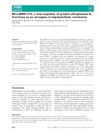
Fig. 1 Chemosensitivity in HCC cells is associated with drug-induced increases in NHEJ activity. a Following conventional therapy regimens with
the drugs cisplatin (cis), oxaliplatin (oxa) doxorubicin (dox), as well as treatment with 5-FU, DSB foci in PLC and 97 L cells were localized using an
anti-γH2AX antibody. b Quantification of the percentage of γH2AX foci-positive cells. Data are reported as the mean ± SD from 2 independent
experiments. c Statistical comparison of the percentage of γH2AX-positive cells between PLC and 97 L groups at 9 h and 48 h after drug
treatment. A total of 1000 cells were randomly selected for counting. The mean ± SD was from 3 independent experiments. p values ≤0.05 and
≤0.01 were denoted as * and **, respectively. d In vitro NHEJ activity was assayed after oxaliplatin and doxorubicin treatment and quantified by
plasmid-based quantitative PCR (see methods). NHEJ activity from 3 independent experiments was statistically analyzed using a paired Student’s t
test. e HCC cells were transfected with the plasmid pEGFP-PEM1-Ad2, followed by drug treatment. In vivo NHEJ activity was calculated based on
the frequency of GFP-positive cells normalized to the transfection efficiency. Drug-induced NHEJ activity was counted as a ratio of drug-induced
activity versus control activity. Representative data are reported as the mean ± SD, n = 3. f PLC and 97 L cells were treated with cisplatin (1 μg/
ml), oxaliplatin (1 μg/ml), or doxorubicin (0.2 μg/ml). Cell viability (%) was determined using a Cell Counting Kit 8 and standardized against cells
without drug treatment. Representative data are reported as the mean ± SD, n = 3. g The therapy drug (oxaliplatin) response curve and the IC50
concentration for the PLC and 97 L cells were analyzed from 2 independent experiments using GraphPad 6.0
NHEJ factors Ku, DNA-PKcs, and XLF were significantly
higher in CD133+ than in CD133− cells (Fig. 2b). We
next transfected pEGFP-PEM1-Ad2 plasmids into both
CD133+ and CD133− populations and then observed
drug-induced NHEJ activity in vivo. The CD133+ cells
contained a significantly higher GFP+ population than
the CD133− cells (Fig. 2c), and this pattern was consistent with all three drug treatments (Fig. 2c). This result
indicates that DSB repair capacity in CD133+ cells is
higher than in CD133− cells. Statistical comparisons of
the in vivo NHEJ assay results are summarized in Fig.
2d. The results indicate that significantly higher NHEJ
activity is found in CD133+ cells. These results suggest
that CSCs possess higher intrinsic NHEJ capacity.
XLF knockdown enhances chemosensitivity in HCC cells
XLF is a core member of the NHEJ protein complex.
XLF interacts with XRCC4 to form an XRCC4/XLF
dimer that bridges DNA ends in vitro; thus, XLF might
stimulate ligation [20–22]. XLF also accumulates at sites
with DSBs [14]. Whether the known functions of XLF
have a role in drug-induced DSB repair and cancer resistance is not known. Resistant 97 L HCC cells displayed high NHEJ activity both in vitro and in vivo (Fig.
1d, e). After knocking down XLF, the drug-induced in
vitro NHEJ activity was significantly inhibited (Fig. 3a).
In vivo, NHEJ activity was still induced by the drugs in
shRNA-control-transfected cells, whereas it was not in
cells transfected with shRNA-XLF (Fig. 3b). These
Yang and Wang BMC Cancer (2017) 17:344
Page 5 of 10
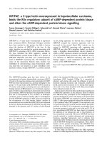
Fig. 2 Liver CD133+ CSCs possess high intrinsic NHEJ capacity. a Statistical comparison of drug sensitivity in CD133+ and CD133− liver cancer cells
measured by cell viability assays. b Expression levels of the NHEJ genes Ku, DNA-PK, and XLF in CD133+ and CD133− HCC cells determined by qRTPCR. Expression levels were normalized to the reference gene 18S rRNA. The data are reported as the mean ± SD obtained from 2 experiments with
duplicates. c Huh7 cells were transfected with pEGFP-PEM1-Ad2 plasmid, followed by cisplatin, oxaliplatin, or doxorubicin treatment. GPF+ gating was
based on CD133-PE-positive (upper part) and CD133-PE-negative (lower part) populations. Live-cell NHEJ activity is presented as the percentage of GFP
+ cells within CD133+ and CD133− cell populations and was normalized to the transfection efficiency. d Statistical comparison of drug-induced NHEJ
activity in vivo of c between CD133+ and CD133− cells from 2 independent experiments
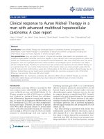
Fig. 3 Knockdown of XLF chemosensitizes resistant 97 L cells by causing inhibition of NHEJ activity. a In vitro NHEJ activity was assayed and quantified
in shRNA-Con and shRNA-XLF lentivirus-infected 97 L cells from 3 independent experiments. b In vivo NHEJ activity was calculated as the percentage
of GFP-positive cells and normalized to transfection efficiency for the shCon and shXLF group, which were transfected with pEGFP-PEM1-Ad2 plasmid
and treated with or without oxaliplatin. The data are reported as the mean ± SD, n = 4. c Quantification of the percentage of cells with <5 and >5
γΗ2ΑX foci per nucleus at 26 h after drug treatment for the siRNA-Con and siRNA-XLF groups (left panel). The data are reported as the mean ± SD,
n = 2. Image of γH2AX speckles with <5 and >5 foci per nucleus (right panel). d Statistical comparison of drug sensitivity based on colony formation
assay results from shCon and shXLF cells. Number of colonies is reported as the mean ± SD from 2 independent experiments. e Gene expression levels
(left panel) and protein levels (right panel) of LIG4 and ERCC1 were determined by qRT-PCR and WB, respectively, in shCon and shXLF HCC cells. f
Cisplatin response curves and IC50 concentrations for siCon, siXLF, siERCC1, and siXLF + siERCC1 cells, respectively. Statistical comparison was between
siXLF, siERCC1, and siXLF + siERCC1 versus siCon, respectively, with treatment by cisplatin 1 or 5 μg/ml. * p < 0.05; ** p < 0.01; *** p < 0.001
Yang and Wang BMC Cancer (2017) 17:344
results imply that down-regulating XLF significantly impairs NHEJ capacity. 97 L cells had higher DSB repair efficiency, and γH2AX foci were reduced after longer-term
drug treatment (Fig. 1c). Following XLF knockdown, the
number of γH2AX foci remained high, with 83.1% ± 6.1
of cells displaying >5 γH2AX foci (Fig. 3c), indicating
the presence of unrepaired DSBs. As a result, 97 L cells
became sensitized to the chemotherapeutic drugs tested
(Fig. 3d).
XLF knockdown results in down-regulation of ERCC1
We next explored the cross-regulation of XLF with other
DNA repair genes, such as LIG4, as XLF stimulates the
XRCC4/LIG4 complex [13]. We also investigated excision
repair cross-complementing 1 (ERCC1) of the nucleotide
excision repair (NER) pathway. Achieving SSL is ideal for
producing therapeutic effects, and the HR and NHEJ
pathways show high levels of SSL with the NER pathway
[4]. Down-regulation of XLF was associated with reduced
LIG4 mRNA (Fig. 3e, left panel) and protein expression
levels (Fig. 3e, right panel), which is consistent with previous findings that XLF regulates LIG4 in cancer. Interestingly, in XLF-knockdown cells, ERCC1 mRNA and
protein expression levels also decreased (Fig. 3e). We then
investigated whether a synergistic effect exists between
the chemotherapeutic drugs after two DNA repair genes
are inhibited. In combination with ligase inhibitor (L189),
the drugs inhibited the growth of shXLF cells more than
the growth of shCon cells (Additional file 1: Fig. S1). Inhibiting XLF enhanced the chemotherapeutic sensitivity in
resistant 97 L HCC cells (Fig. 3d). We then determined a
synergistic effect compared to cisplatin alone, particularly
when both XLF and ERCC1 were knocked down because
ERCC1 positive-tumors predict cisplatin resistance in
non-small-cell lung cancer and squamous cell carcinoma
[23–25]. The IC50 of cisplatin was 5.56 μg/ml for the
siCon group, 3.15 μg/ml for the siXLF group, 2.51 μg/ml
for the siERCC1 group, and 1.43 μg/ml for the siXLF +
siERCC1 group (Fig. 3f), indicating a chemosensitization
effect when there was knockdown of either XLF or
ERCC1, where the most enhanced chemosensitivity was
seen with knockdown of both XLF and ERCC1. Crosstalk
between DNA repair pathways often results in the acquisition of resistance mechanisms in cancer [3]. This result
suggests that targeting independent pathways may produce stronger synergy than targeting the same pathway at
multiple points [2].
XLF knockdown enhances therapeutic efficacy in HCC
xenograft model
To establish a xenograft model, shCon- and shXLFtransfected 97 L cells were subcutaneously injected into
the left and right flanks, respectively, of the same nude
mouse. Using the drug administration protocol depicted
Page 6 of 10
in Fig. 4a, oxaliplatin treatment was started at the third
week after cell injection and was continued for 4 weeks.
At the endpoint, overall tumor volumes were significantly smaller for the shXLF-HCC xenografts than the
shCon-HCC xenografts (Fig. 4b, c), indicating that XLF
knockdown sensitized HCC xenografts to oxaliplatin.
Cell-free NHEJ activity was measured in nuclear proteins
extracted from xenograft tissue. A dramatic enhancement in NHEJ activity was observed in the shCon xenografts, whereas NHEJ activity was consistently inhibited
in the shXLF xenografts (Fig. 4d). Thus, the shXLF-HCC
xenograft tumors were more responsive to oxaliplatin as
a result of shXLF-mediated reductions in NHEJ activity.
Low frequency of XLF gene alterations in patients with
HCC
Cancer cells are frequently dysfunctional in one or more
DDR and DNA damage repair pathways, which can be specifically inhibited to induce SSL. These dysfunctions can
also lead to additional alterations in DDR pathways that
may induce therapy resistance [1, 4]. Given that XLFmediated induction of NHEJ activity in HCC cells is associated with chemotherapeutic drug sensitivity (Figs. 1 and 2),
we investigated the incidence of XLF gene alterations using
bioinformatic analysis. We also investigated genomic alterations of the core factors in the NHEJ pathway in HCC and
other cancers. The NHEJ pathway includes 8 genes: NHEJ1
(XLF), XRCC4, XRCC5, XRCC6, LIG4, DNA-dependent
protein kinase catalytic subunit (PRKDC), TP53BP1, and
DNA cross-link repair 1C (DCLRE1C). Genomic alterations were identified using cBioPortal [] [26, 27] based on 90 studies from 17 to 26 types
of primary tumor and cancer lines assembled by The Cancer Genomics Atlas (TCGA) [28] and Asan Medical Center
(AMC) [29]. The frequency of alterations, including mutation, deletion, amplification, and multiple alterations, for
NHEJ pathway genes in HCC was found to be 17.6% by
TCGA and 10% by AMC (Additional file 1: Fig. S2); in contrast, the frequency of alterations in the XLF gene was extremely low in HCC. Indeed, only one of 231 patients had a
missense mutation (mutation rate: 0.4%)(Fig. 5a) in the
AMC data collection, and no mutations were found in the
371 patients included in the TCGA data collection (Additional file 1: Fig. S3; Additional file 2: Table S1). Moreover,
upregulation of XLF mRNA was found in only 5% of patients with HCC (Additional file 2: Table S1). Given the low
genomic alteration rate of XLF in patients with HCC, we
sought to measure XLF levels in primary HCC tissues from
patients who did or did not receive TACE treatment before
liver tumor resection to determine whether therapy drugs
can impact XLF expression, because TACE delivers chemotherapy in combination with embolization to administer
therapy directly to liver tumors. As shown in Fig. 5b, XLF
expression was significantly increased in patients with HCC
Yang and Wang BMC Cancer (2017) 17:344
Page 7 of 10
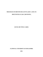
Fig. 4 XLF knockdown restores drug sensitivity in HCC xenograft model. a Experimental setup used to create the xenograft model. shCon and shXLF
97 L cells (1 × 10⁶) were subcutaneously injected into nude mice. Oxaliplatin (4 mg/kg/week or 10 mg/kg/week) was administered to the mice by
intra-peritoneal injection. b Tumor volume was reported in mm3 as the mean ± SD and statistically compared between shCon and shXLF tumors.
c Representative images of xenograft tumor. d In vitro NHEJ activity in nuclear proteins extracted from xenograft tumor tissue. The statistical
comparison was based on the mean ± SD from 3 independent experiments
Fig. 5 Clinical significance of XLF expression. a Genomic alteration frequency for XLF was calculated using cBioPortal [] from the
TCGA (193 patients) and AMC (231 patients) databases. Tumor types are indicated at the bottom and ordered by the frequency of samples harboring
mutation, deletion, amplification and multiple alterations. The XLF mutation rate in HCC is highlighted. b Statistical comparison of XLF gene expression in
tumor tissue between patients with HCC who underwent TACE (n = 31) and those who did not (n = 22). c Kaplan-Meier analysis of overall survival
between patients with HCC with high and low expression of XLF, who all underwent TACE treatment. d Kaplan-Meier analysis of disease-free survival for
patients with HCC as in c
Yang and Wang BMC Cancer (2017) 17:344
Page 8 of 10
who underwent TACE compared to those who did not, indicating that the delivery of drugs by TACE may directly or
indirectly stimulate XLF expression.
Therapy drugs induce XLF expression and affect clinical
outcome
Given that XLF expression (Fig. 5b) and XLF-mediated alterations in NHEJ activity (Fig. 1d, e; Fig. 3a, b) can be induced by therapy drugs, we wanted to assess whether
enhancing XLF expression would affect HCC clinical parameters. The patients in the TACE treatment group who
showed higher levels of XLF expression displayed poor
prognosis features such as larger tumor size, venous infiltration, and advanced-stage HCC disease (Table 1). More
importantly, XLF expression was significantly associated
with overall survival of HCC patients, with patients exhibiting higher levels of XLF showing shorter survival times
(Fig. 5c). High XLF expression was also associated with a
shorter disease-free survival time, although the trend did
not reach statistical significance (Fig. 5d).
Discussion
Cancer cells frequently exhibit dysfunctional DNA repair.
Mutations in DNA repair pathways can also be a predisposing factor for cancer. However, once tumors develop,
DNA repair pathways can be used by cancer cells to overcome many standard anticancer treatments and are often
a cause of chemotherapeutic resistance [30]. DDR pathway deregulation has been analyzed in 15 types of cancer
[4], although there have been no previous reports describing this deregulation in HCC [4]. Based on a large dataset
assembled by TCGA and AMC with information from 17
to 26 types of primary tumor and cancer lines, we showed
here for the first time the genomic alterations that can be
found in core factors of NHEJ pathway, including XLF, in
patients with HCC. Our analysis demonstrated an extremely low mutation rate (0.4%) for XLF in the AMC
data collection and no mutations for XLF in the TCGA
data collection for patients with HCC. Furthermore, XLF
Table 1 Correlation between XLF expression and
clinicopathologic features in TACE-treated HCC
Clinical features
P
XLF expression
Low
High
Venous infiltration
(−)
(+)
11
2
7
11
0.025*
Size of tumor (cm)
≤5
>5
9
4
5
13
0.022*
No. of nodules
Single
Multiple
6
7
8
10
0.925
UICC grade
I, II
IIIa, IIIb
12
1
10
8
0.045*
Recurrence
(−)
(+)
6
7
6
12
0.47
mRNA overexpression was found in only 5% of patients
with HCC. These results suggest that dysregulation of
XLF does not contribute to the phenotypic profile of
HCC. However, significant upregulation of XLF was observed in patients who underwent TACE, indicating that
therapeutic drugs can induce overexpression of XLF. This
overexpression was further predictive of poor prognosis
and reduced survival. These results led us to develop a
new hypothesis: therapy resistance associated with DNA
repair can be induced by standard therapeutic treatment
and might not be related to pre-existing deregulation of
DNA repair pathways. This hypothesis is reinforced by
clinical observations that TACE therapy induces robust
XLF expression and that XLF induces high levels of NHEJ
activity in patients with HCC. These patients might develop DNA repair-mediated therapy resistance, a critical
contributor to treatment failure and reduced survival,
even after hepatectomy. Thus, rationale for including
DNA repair inhibitors as part of the cancer drug armamentarium should be based not only on the presence of
abnormally high levels of intrinsic expression of DNA repair pathway members in tumor tissue but also on high
levels of expression induced by chemotherapeutic drugs.
By comparing two HCC cell lines with different degrees of drug sensitivity, we determined that conventional chemotherapy can induce NHEJ activity in HCC
cells. Increases in NHEJ activity leads to the repair of
drug-induced DSBs and reduces the presence of cellular
γH2AX foci, which significantly decreased the effectiveness of the chemotherapeutic drugs used in this study.
Drug-induced activation of NHEJ activity also contributed to the chemoresistance of liver CSCs. We further
demonstrated that XLF plays an important role in drug
resistance mediated by NHEJ activity. Knocking down
XLF significantly enhanced chemosensitization in vitro
and in vivo by decreasing NHEJ activity. Thus, for the
first time, we have demonstrated that XLF-mediated increases in NHEJ activity are responsible for chemoresistance in HCC cells. The XLF-XRCC4 complex is
essential for NHEJ, although how XLF mechanistically
functions in NHEJ is not well understood [31]. Recent
studies have suggested that XLF-XRCC4 filaments provide both protection to and alignment of DNA ends for
accurate and efficient ligation [31, 32]. Whether this
mechanism is relevant in our current experimental setting is not known. However, downregulation of XLF significantly impaired ligation and thus reduced NHEJ
efficiency, resulting in 80% of γH2AX-positive cells containing >5 γH2AX foci per cell as well as the development of chemosensitization. Moreover, XLF might
directly regulate LIG4, as knocking down XLF downregulated LIG4. Together, the roles of the XLF-XRCC4
complex in promoting ligation [31] and XLF-stimulated
NHEJ activity in chemoresistance suggests that targeting
Yang and Wang BMC Cancer (2017) 17:344
XLF is a potential avenue for the development of a new
DNA repair inhibitor for combination therapy.
DNA-dependent protein kinase catalytic subunit (DNAPKcs), including Ku70 and Ku80, plays key roles in NHEJ
repair for DSB repair and V(D)J recombination. Recent investigations demonstrated that levels of DNA-PKcs were
directly associated with genomic stability, cancer cell proliferation index, and patients’ survival length in HCC, suggesting that DNA-PKcs contributes to liver malignant
transformation and carcinogenesis. Moreover, elevated
DNA-PKcs expression identified HCC patients with
therapy-resistance [33, 34]. In combination with our study,
intrinsic and drug-induced DNA repair genes of the NHEJ
pathway indeed represent prognostic factors and, more
importantly, specific therapeutic targets.
γH2AX phosphorylation occurs several minutes after irradiation and DSB formation. Immunological detection of
γH2AX indicates the presence of unrepaired DNA breaks.
Thus, γH2AX has been utilized as a pharmacodynamic
marker in clinical studies. For example, γH2AX foci were
enumerated to determine irradiation toxicity at different
dosages and to evaluate chemotherapeutic drug responses
[4, 35, 36]. According to our observations, the remaining
levels but not initial level of γH2AX positive cells is a better predictor for drug sensitivity. Many cancer cells possess high DNA repair capacity, and the number of
γH2AX-positive cells and foci after longer-term drug
treatment are predictive of cellular response or resistance.
Conclusion
Overexpression of XLF and increased NHEJ activity mediated by XLF in response to treatment with chemotherapeutic drugs contribute to chemoresistance in HCC
cells and patients with HCC. Inhibition of XLF-mediated
NHEJ activity results in chemosensitization in a HCC
xenograft model, suggesting that XLF is a novel candidate for the development of new DNA repair inhibitors
for combination therapy.
Additional files
Additional file 1: Figure S1. Colony formation in shCon or shXLF cells
treated with DMSO, cisplatin, cisplatin + L189 (LIG4 inhibitor), oxaliplatin,
or oxaliplatin + L189, respectively. Figure S2. Genomic alteration
frequency of core factors of NHEJ pathway (XLF, XRCC4, XRCC5, XRCC6,
LIG4, PRKDC, TP53BP1, and DCLRE1C) was generated using cBioPortal
[] from database of TCGA (193 patients) and
AMC (231 patients). Tumor types are indicated at the bottom and
ordered by the frequency of samples harboring mutation, deletion,
amplification and multiple alterations. Overall alteration frequency in liver
cancer was highlighted. Figure S3. RNA-sequencing data organization of
NHEJ pathway genes (XLF, XRCC4, XRCC5, XRCC6, LIG4, PRKDC, TP53BP1,
and DCLRE1C) by TCGA to show frequency of NHEJ pathway gene amplification, gene expression, missense or truncating mutations. (ZIP 1521 kb)
Additional file 2: Table S1. Alteration of NHEJ pathway genes in HCC
by RNA-seq. (DOCX 56 kb)
Page 9 of 10
Abbreviations
DDR: DNA damage response; DSBs: double strand breaks; ERCC1: excision repair
cross-complementing 1; HR: homologous recombination; IC50: inhibitory
concentration; NER: nucleotide excision repair; NHEJ: non-homologous end
joining; SSL: synthetic sensitivity or lethality; TACE: transarterial
chemoembolization; XLF: XRCC4-like factor
Acknowledgements
We thank Drs. M. Huen and B. Liu at the University of Hong Kong for
providing the suggestions and plasmids.
Funding
This study was supported by the Seed Funding Program for Basic Research,
The University of Hong Kong (59,203 to XQW) and partially supported by
Healthy and Medical Research Fund, Research Council of Hong Kong
(03143396 to XQW). The funding bodies partially support the expenses for
the experiments.
Availability of data and materials
The dataset of this article is available at request from the corresponding
author.
The dataset of bioinformatic analysis of genomic alterations of the NHEJ
pathway, which are assembled by TCGA and AMC, are downloaded from
cBioPortal [].
Authors’ contributions
SY contributed to conception, data collection, and data analysis. XQW
contributed to conception, experimental design, data analysis, and writing
and finalizing the manuscript. All authors have read and approved the
manuscript.
Competing interests
The authors declare that they have no competing interests.
Consent for publication
Not applicable.
Ethics approval and consent to participate
The study has been approved by the Institutional Review Board of the
University of Hong Kong/Hospital Authority of Hong Kong (UW05–3597/
I022), which is the consent for patients donating clinical specimens before
hepatectomy to surgical tissue bank for research purpose at Department of
Surgery, The University of Hong Kong. For this retrospective study, the frozen
tumor tissue samples were obtained from the tissue bank of Department of
Surgery. The mouse experiment was approved by the Committee on the
Use of Live Animals of The University of Hong Kong (CULATR 3091–13).
Publisher’s Note
Springer Nature remains neutral with regard to jurisdictional claims in
published maps and institutional affiliations.
Received: 21 September 2016 Accepted: 11 May 2017
References
1. Bouwman P, Jonkers J. The effects of deregulated DNA damage signalling
on cancer chemotherapy response and resistance. Nat Rev Cancer. 2012;
12(9):587–98.
2. Holohan C, Van Schaeybroeck S, Longley DB, Johnston PG. Cancer drug
resistance: an evolving paradigm. Nat Rev Cancer. 2013;13(10):714–26.
3. Helleday T, Petermann E, Lundin C, Hodgson B, Sharma RA. DNA repair
pathways as targets for cancer therapy. Nat Rev Cancer. 2008;8(3):193–204.
4. Pearl LH, Schierz AC, Ward SE, Al-Lazikani B, Pearl FM. Therapeutic
opportunities within the DNA damage response. Nat Rev Cancer. 2015;15(3):
166–80.
5. Brough R, Frankum JR, Costa-Cabral S, Lord CJ, Ashworth A. Searching for
synthetic lethality in cancer. Curr Opin Genet Dev. 2011;21(1):34–41.
6. Farmer H, McCabe N, Lord CJ, Tutt AN, Johnson DA, Richardson TB, et al.
Targeting the DNA repair defect in BRCA mutant cells as a therapeutic
strategy. Nature. 2005;434(7035):917–21.
Yang and Wang BMC Cancer (2017) 17:344
7.
8.
9.
10.
11.
12.
13.
14.
15.
16.
17.
18.
19.
20.
21.
22.
23.
24.
25.
26.
27.
28.
29.
30.
Edwards SL, Brough R, Lord CJ, Natrajan R, Vatcheva R, Levine DA, Boyd J,
Reis-Filho JS, Ashworth A. Resistance to therapy caused by intragenic
deletion in BRCA2. Nature. 2008;451(7182):1111–5.
Hsiang YH, Lihou MG, Liu LF. Arrest of replication forks by drug-stabilized
topoisomerase I —DNA cleavable complexes as a mechanism of cell killing
by camptothecin. Cancer Res. 1989;49(18):5077–82.
Sargent RG, Brenneman MA, Wilson JH. Repair of site-specific double-strand
breaks in a mammalian chromosome by homologous and illegitimate
recombination. Mol Cell Biol. 1997;17(1):267–77.
Arnaudeau C, Lundin C, Helleday T. DNA double-strand breaks associated
with replication forks are predominantly repaired by homologous
recombination involving an exchange mechanism in mammalian cells. J
Mol Biol. 2001;307(5):1235–45.
Fattah F, Lee EH, Weisensel N, Wang Y, Lichter N, Hendrickson EA. Ku regulates
the non-homologous end joining pathway choice of DNA double-strand
break repair in human somatic cells. PLoS Genet. 2010;6(2):e1000855.
Ochi T, Blackford AN, Coates J, Jhujh S, Mehmood S, Tamura N, et al. DNA
repair. PAXX, a paralog of XRCC4 and XLF, interacts with Ku to promote
DNA double-strand break repair. Science. 2015;347(6218):185–8.
Fattah FJ, Kweon J, Wang Y, Lee EH, Kan Y, Lichter N, et al. A role for XLF in
DNA repair and recombination in human somatic cells. DNA Repair. 2014;
15:39–53.
Koike M, Yutoku Y, Koike M. Dynamic changes in subcellular localization of
cattle XLF during cell cycle, and focus formation of cattle XLF at DNA damage
sites immediately after irradiation. J Vet Med Sci. 2015;77(9):1109–14.
Wang XQ, Chan KK, Ming X, Lui VC, Poon RY, Lo CM, Norbury C, Poon RT.
G1 checkpoint establishment in vivo during embryonic liver development.
BMC Dev Biol. 2014;14:23.
Seluanov A, Mao Z, Gorbunova V. Analysis of DNA double-strand break
(DSB) repair in mammalian cells. J Vis Exp. 2010;43:1–6.
Rogakou EP, Boon C, Redon C, Bonner WM. Megabase chromatin domains
involved in DNA double-strand breaks in vivo. J Cell Biol. 1999;146(5):905–16.
Valent P, Bonnet D, De Maria R, Lapidot T, Copland M, Melo JV, et al. Cancer
stem cell definitions and terminology: the devil is in the details. Nat Rev
Cancer. 2012;12(11):767–75.
Colak S, Medema JP. Cancer stem cells-important players in tumor therapy
resistance. FEBS J. 2014;281(21):4779–91.
Gu J, Lu H, Tsai AG, Schwarz K, Lieber MR. Single-stranded DNA ligation and
XLF-stimulated incompatible DNA end ligation by the XRCC4-DNA ligase IV
complex: influence of terminal DNA sequence. Nucleic Acids Res. 2007;
35(17):5755–62.
Tsai CJ, Kim SA, Chu G. Cernunnos/XLF promotes the ligation of mismatched
and noncohesive DNA ends. Proc Natl Acad Sci U S A. 2007;104(19):7851–6.
Roy S, de Melo AJ, Xu Y, Tadi SK, Négrel A, Hendrickson E, et al. XRCC4/XLF
interaction is variably required for DNA repair and is not required for Ligase
IV stimulation. Mol Cell Biol. 2015;35(17):3017–28.
Olaussen KA, Postel-Vinay S. Predictors of chemotherapy efficacy in non-smallcell lung cancer: a challenging landscape. Ann Oncol. 2016;27(11):2004-16.
Olaussen KA, Dunant A, Fouret P, Brambilla E, André F, Haddad V, et al. DNA
repair by ERCC1 in non-small-cell lung cancer and cisplatin-based adjuvant
chemotherapy. N Engl J Med. 2006;355(10):983–91.
Pierceall WE, Olaussen KA, Rousseau V, Brambilla E, Sprott KM, Andre F, et al.
Cisplatin benefit is predicted by immunohistochemical analysis of DNA
repair proteins in squamous cell carcinoma but not adenocarcinoma:
theranostic modeling by NSCLC constituent histological subclasses. Ann
Oncol. 2012;23(9):2245–52.
Cerami E, Gao J, Dogrusoz U, Gross BE, Sumer SO, Aksoy BA, et al. The cBio
cancer genomics portal: an open platform for exploring multidimensional
cancer genomics data. Cancer Discov. 2012;2(5):401–4.
Gao J, Aksoy BA, Dogrusoz U, Dresdner G, Gross B, Sumer SO, et al.
Integrative analysis of complex cancer genomics and clinical profiles using
the cBioPortal. Sci Signal 2013;6(269):pl1.
Weinstein JN, Collisson EA, Mills GB, Shaw KR, Ozenberger BA, Ellrott K, et al.
The cancer genome atlas pan-cancer analysis project. Nat Genet. 2013;
45(10):1113–20.
Ahn SM, Jang SJ, Shim JH, Kim D, Hong SM, Sung CO, et al. Genomic
portrait of resectable hepatocellular carcinomas: implications of RB1 and
FGF19 aberrations for patient stratification. Hepatology. 2014;60(6):1972–82.
Plummer R. Perspective on the pipeline of drugs being developed with
modulation of DNA damage as a target. Clin Cancer Res. 2010;16(18):
4527–31.
Page 10 of 10
31. Mahaney BL, Hammel M, Meek K, Tainer JA, Lees-Miller SP. XRCC4 and XLF
form long helical protein filaments suitable for DNA end protection and
alignment to facilitate DNA double strand break repair. Biochem Cell Biol.
2013;91(1):31–41.
32. Hammel M, Rey M, Yu Y, Mani RS, Classen S, Liu M, et al. XRCC4 protein
interactions with XRCC4-like factor (XLF) create an extended grooved
scaffold for DNA ligation and double strand break repair. J Biol Chem. 2011;
286(37):32638–50.
33. Evert M, Frau M, Tomasi ML, Latte G, Simile MM, Seddaiu MA, et al.
Deregulation of DNA-dependent protein kinase catalytic subunit contributes
to human hepatocarcinogenesis development and has a putative
prognostic value. Br J Cancer. 2013;109(10):2654–64.
34. Cornell L, Munck JM, Alsinet C, Villanueva A, Ogle L, Willoughby CE, et al. DNAPK-A candidate driver of hepatocarcinogenesis and tissue biomarker that predicts
response to treatment and survival. Clin Cancer Res. 2015;21(4):925–33.
35. Pouliliou S, Koukourakis MI. Gamma histone 2AX (γ-H2AX) as a predictive
tool in radiation oncology. Biomarkers. 2014;19(3):167–80.
36. Ivashkevich A, Redon CE, Nakamura AJ, Martin RF, Martin OA. Use of the γH2AX assay to monitor DNA damage and repair in translational cancer
research. Cancer Lett. 2012;327(1–2):123–33.
Submit your next manuscript to BioMed Central
and we will help you at every step:
• We accept pre-submission inquiries
• Our selector tool helps you to find the most relevant journal
• We provide round the clock customer support
• Convenient online submission
• Thorough peer review
• Inclusion in PubMed and all major indexing services
• Maximum visibility for your research
Submit your manuscript at
www.biomedcentral.com/submit