Switch in KRAS mutational status during an unusual course of disease in a patient with advanced pancreatic adenocarcinoma: Implications for translational research
Bạn đang xem bản rút gọn của tài liệu. Xem và tải ngay bản đầy đủ của tài liệu tại đây (599.81 KB, 5 trang )
Baechmann et al. BMC Cancer (2017) 17:374
DOI 10.1186/s12885-017-3376-4
CASE REPORT
Open Access
Switch in KRAS mutational status during an
unusual course of disease in a patient with
advanced pancreatic adenocarcinoma:
implications for translational research
Sibylle Baechmann1†, Steffen Ormanns1† , Michael Haas2, Stephan Kruger2, Anna Remold1,2,
Dominik Paul Modest2, Thomas Kirchner1,4, Andreas Jung1,4, Jens Werner3, Volker Heinemann2,4† and
Stefan Boeck2*†
Abstract
Background: Despite the introduction of novel effective treatment regimens like gemcitabine plus nab-paclitaxel
and FOLFIRINOX, pancreatic ductal adenocarcinoma (PDAC) remains one of the most aggressive epithelial tumors.
Among the genetic alterations frequently found in PDAC, mutations in the KRAS gene might play a prognostic role
regarding overall survival and may also have the potential to predict the efficacy of anti-EGFR treatment.
Case presentation: We report the clinical case of a 69 year old Caucasian female that was diagnosed with
histologically confirmed locally advanced PDAC with lymph node involvement in August 2010. At the time of first
diagnosis, tumor tissue obtained from an open regional lymph node biopsy showed a poorly differentiated
adenocarcinoma with a wild type sequence within exon 2 (codon 12/13) of the KRAS gene. The patient initially
received single-agent gemcitabine and a subsequent 5-FU-based chemoradiotherapy with a sequential
maintenance chemotherapy with oral capecitabine resulting in a long term disease control. Local disease
progression occurred in May 2014 and the patient underwent pancreaticoduodenectomy in September 2014.
A novel KRAS gene mutation (c.35G > T, p.G12 V) in exon 2 (codon 12) was detected within the surgical
specimen. As of January 2016 the patient is still alive and without evidence of the underlying disease.
Conclusions: Specifically in the context of clinical trials and translational research in PDAC a re-assessment of
molecular biomarkers, i. e. KRAS, at defined time points (e. g. relapse, disease progression, unusual clinical
course) may be indicated in order to detect a potential switch in biomarker status during the course of disease.
Keywords: Pancreatic ductal adenocarcinoma (PDAC), KRAS mutation, Tumor heterogeneity
Background
Pancreatic ductal adenocarcinoma (PDAC) is one of the
most aggressive epithelial tumors worldwide. In most
patients it represents a deadly disease [1] due to an
advanced stage at the time of diagnosis and the difficulties in therapeutic treatment, but also due to genetic
heterogeneity [2]. Surgical resection remains the only
* Correspondence:
†
Equal contributors
2
Department of Internal Medicine III and Comprehensive Cancer Center,
Klinikum Grosshadern, Ludwig-Maximilians University of Munich,
Marchioninistr. 15, 81377 Munich, Germany
Full list of author information is available at the end of the article
curative treatment option for localized PDAC. During
the last decade, systemic treatment with single-agent
gemcitabine has evolved as standard chemotherapy for
the adjuvant and palliative treatment setting [3, 4].
Gemcitabine offers a median survival of about 5 to
7 months in patients with advanced disease and shows
comparatively good tolerability [5]; more recently,
gemcitabine-based combination regimens with the oral
epidermal growth factor receptor (EGFR) inhibitor
erlotinib or together with nab-paclitaxel [6] showed a
statistically significant improvement in overall survival
(OS). The development and progression of PDAC
© The Author(s). 2017 Open Access This article is distributed under the terms of the Creative Commons Attribution 4.0
International License ( which permits unrestricted use, distribution, and
reproduction in any medium, provided you give appropriate credit to the original author(s) and the source, provide a link to
the Creative Commons license, and indicate if changes were made. The Creative Commons Public Domain Dedication waiver
( applies to the data made available in this article, unless otherwise stated.
Baechmann et al. BMC Cancer (2017) 17:374
include different genetic alterations in oncogenic activation, loss of tumor-suppressor gene function and
overexpression of receptor-ligand systems [7, 8].
Among these genetic alterations, mutations in the
KRAS gene, which often are already present in precursor lesions, play an important role in tumor development and progression [8]. Gain of function mutations
in the KRAS gene are detected in about 70 to 90% of
PDAC cases [9], commonly as point mutations in exon
2 (codon 12/13), most frequently as p.G12D (c.35G > A)
or p.G12 V (p.35G > T). Several studies showed that
constitutively activating KRAS mutations are associated
with worse OS, whereas KRAS wildtype status is associated with improved OS in PDAC [7, 10, 11]. Thus, in
PDAC, KRAS mutations may be regarded as prognostic
biomarker. The role of KRAS mutational status as predictive biomarker regarding the use of EGFR-targeting
agents like erlotinib in advanced PDAC still remains a
matter of debate to date [12–14].
Here, we report the case of a PDAC patient with an
unusual clinical course: the tumor of the patient harbored a wildtype KRAS gene at the time of initial PDAC
diagnosis; however, upon disease progression 4 years
later, a mutation within exon 2 of the KRAS gene was
detectable.
Case presentation
A currently 75-year-old woman was diagnosed with
locally advanced PDAC at our comprehensive cancer
center (CCC) in 2010. An explorative laparotomy in
August 2010 showed metastatic disease spread extensively to regional lymph nodes and thus the primary
tumor in the pancreatic head was not resected. By CT
imaging criteria no other distant metastatic disease was
evident. Lymph nodes were sampled surgically from the
right gastric artery, the hepatic artery, the coeliac trunc
and from the interaortocaval region; in all samples,
tumor infiltration by a poorly differentiated adenocarcinoma was confirmed by histology. Immunohistochemical
staining was positive for CK7, CK20 and CA 19–9 (with
CDX-2 being negative). At that time point an additional
analysis for KRAS mutational status and EGFR protein
expression (which were conducted within a translational
research project) detected a wildtype sequence of KRAS
exon 2 by pyrosequencing and a moderately positive
immunohistochemical staining for membranous EGFR
expression in about 80% of the tumor cells.
The patient initially received systemic chemotherapy
with three cycles of standard dose (1000 mg/m2) gemcitabine between September and December 2010. Imaging
studies in January 2011 confirmed stable disease and the
CA 19–9 levels decreased from 3700 U/ml at first diagnosis to 180 U/ml. In February 2011 5-FU-based chemoradiotherapy (30 Gy) was applied at an external hospital.
Page 2 of 5
During re-exploration performed in May 2011 surgical
biopsies from the peritoneum histologically confirmed
metastatic disease of PDAC; thus, no attempt to resect
the primary tumor in the pancreas was performed. We
then decided, also based on the wish of the patient, to
re-start systemic chemotherapy and treatment with oral
capecitabine was initiated in July 2011 and given until
April 2012. During this chemotherapy, a further decline
of CA 19–9 values was observed (nadir: 30 U/ml) and
repeated CT imaging did not show any signs of local disease progression or metastatic disease (as assessed by
imaging criteria). After a treatment rest for two years
(beginning in May 2012), local tumor progression of the
pancreatic primary was observed within a CT scan in
May 2014. Again, no radiographic signs of distant metastasis were observed. Systemic chemotherapy with
single-agent gemcitabine was re-introduced in June 2014
resulting in a CA 19–9 decrease from 690 U/ml at
disease progression to 380 U/ml after three gemcitabine
applications. Due to a progressive duodenal infiltration
with clinical and endoscopic signs of gastrointestinal obstruction, a surgical re-exploration was performed in
September 2014. Intraoperatively, no signs of peritoneal
carcinomatosis were apparent and a liver biopsy showed
no signs of malignancy. Thus, the pancreatic primary
was removed by a pylorus preserving pancreaticoduodenectomy (modified Whipple-Kausch procedure). The
tumor was classified as ypT3 ypN0 (0/15) L0 V0 Pn0,
ductal adenocarcinoma G3, R0 resection (according to
UICC criteria, TNM classification 7th edition, 2010). An
additionally executed KRAS mutational analysis at this
time point revealed a new point mutation p.G12 V
(c.35G > T) in exon 2, codon 12. After surgery, CA 19–9
values decreased to levels of 20 U/ml. The patient was
offered adjuvant chemotherapy with S-1 (tegafur, gimeracil, oteracil) after pancreaticoduodenectomy and started
this treatment December 2014; however, S-1 was tolerated poorly due to gastrointestinal toxicity (diarrhea
grade 4 and accompanying renal insufficiency) and was
therefore terminated in March 2015. As of January 2016
the patient is still alive and without clear evidence of the
underlying disease. An overview of this unusual disease
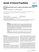
course is shown within Fig. 1.
Discussion
Up to now, no prognostic or predictive tissue biomarker
is available for PDAC [12]. In contrast to other diseases
like breast, lung or colorectal cancer no specific biomarker has been validated for clinical use in pancreatic
cancer and several clinical and translational trials are ongoing in order to better define the molecular basis of
this disease and to search specifically for predictive
markers for treatment efficacy. Thus, only limited data is
available on the clinical role of biomarkers in PDAC
Baechmann et al. BMC Cancer (2017) 17:374
Page 3 of 5
Fig. 1 Therapy, procedures and KRAS mutational status over the time course of the disease (5-FU = 5-fluouracile, Gy = Gray, PPPD = pylorus
preserving pancreaticoduodenectomy)
[12]; specifically, there are no clear recommendations at
which time points biomarkers should be assessed. In
CRC for example, a good correlation between biomarker
results from the primary tumor and from (metachronous) CRC metastases has been reported, resulting in
the acceptance of e. g. RAS status of primary tumor tissue in patients with a metachronous relapse [15]. In contrast, in other diseases like breast cancer a switch in e. g.
Her2/neu (ERBB2) status is well known resulting in the
recommendation of repeated tumor biopsies at relapse
or disease progression [16]. At least to our knowledge,
studies investigating this issue have not yet been performed in PDAC.
Within this manuscript we report a rather unusual
clinical course of a PADC patient, with a corresponding
switch in KRAS mutational status during the course of
disease. Of note, we detected the new KRAS mutation
upon disease progression in September 2014; furthermore, it may be important to highlight the fact that this
patient did not receive previous anti-EGFR treatment
(e.g. with erlotinib) before the detection of the new
KRAS mutation.
Several possible explanations may be hypothesized for
the observation of a KRAS switch during the course of
disease in our PDAC patient:
1. Appearance of a truly new tumor KRAS mutation
upon disease progression in September 2014
without previous application of agents targeting
the EGFR pathway:
The reason for tumor progression could be caused
by an evolved new mutation event in the KRAS
gene, specifically in the light of selection pressure
during previous treatment with chemotherapy and
radiotherapy. In colorectal cancer, increasing
evidence exists that the appearance of new KRAS
mutations during treatment with agents targeting
the EGFR (like cetuximab or panitumumab) may
be linked to an acquired resistance to anti-EGFR
therapy [17, 18]. Of note, our patient did not
receive anti-EGFR treatment for example with
erlotinib before the detection of the new KRAS
mutation. If other treatments like cytotoxic
chemotherapy (gemcitabine, fluoropyrimidines)
or radiotherapy to the pancreatic primary may also
induce a “selection pressure” for the development
of new genetic events remains unknown.
2. Tumor heterogeneity with distinct results in
KRAS analysis at initial diagnosis (lymph node
metastasis analyzed) and at progression
(primary tumor analyzed):
There is increasing evidence for intratumoral
heterogeneity in different types of cancer that
could be determined by multiregion sequencing [19].
In non-small cell lung cancer it was shown that ALK
rearrangements (that were previously thought to be
mutually exclusive with activating EGFR and KRAS
mutations) can be found together with EGFR
mutations in rare cases [20]. Moreover, it was
shown that spatially separated subclones of the
same tumor harbor different oncogenic drivers [21].
If these observations are transferable to PDAC, this
might explain the differences in KRAS mutational
status observed in our patient reported here.
However, the scarce currently available data
comparing pancreatic primary tumors and
corresponding metastases, showed the same KRAS
mutational status in the primary tumor and each
metastatic site examined, thus supporting the idea of
a newly apparent KRAS mutation [22, 23].
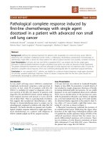
3. Technical aspects of the discrepant KRAS
sequencing results (see Fig. 2):
Potentially, the initial KRAS wildtype status detected
in 2010 could be the effect of a false negative
sequencing result. Both KRAS analyses in the tumor
tissue of the patient reported here were performed
Baechmann et al. BMC Cancer (2017) 17:374
Page 4 of 5
PDAC, future translational trials in pancreatic cancer
that evaluate a broad range of novel biomarkers should,
at least to our opinion, include a repeated biomarker
assessment during the course of disease within their
prospective study protocols. Novel promising techniques like liquid biopsy approaches may thereby help
to overcome the limitations of obtaining tumor tissue
safely in PDAC [25]. As it may be difficult to obtain
sufficient tumor tissue in PDAC by percutaneous- or
endosonography-guided biopsy techniques, a sampling
error may occur specifically in the light of tumor heterogeneity. In that context, liquid biopsy techniques
may also eventually help to overcome these limitations.
Abbreviations
5-FU: 5-fluorouracil; ALK: Anaplastic lymphoma kinase; CA19–9: Carbohydrate
antigen 19–9; CDX2: Caudal type homeo-box transcription factor 2;
CK: Cytokeratin; CRC: Colorectal cancer; CT: Computed tomography;
EGFR: Epidermal growth factor receptor; ERBB2: Human epidermal growth
factor receptor 2; FFPE: Formalin fixed paraffin embedded;
FOLFIRINOX: Folinic acid, 5-FU, irinotecan, oxaliplatin; Gy: Gray; KRAS: Kirsten
rat sarcoma viral oncogene homologue; nab-paclitaxel: Nanoparticle
albumin-bound paclitaxel; OS: Overall survival; PDAC: Pancreatic ductal
adenocarcinoma; UICC: International union against cancer
Fig. 2 Pyrograms comparing the tumors KRAS exon 2, codon 12
mutational status in a October 2010 (wildtype sequence GGTGGC)
and b October 2014 (point mutation p.G12 V, c. 35 G > T,
sequence GTTGGC)
in the same specialized and certified laboratory for
molecular pathology. For both analyses, formalin
fixed paraffin embedded (FFPE) tumor tissue was
microdissected under visual control using a
microscope to reduce contamination by adjacent
normal tissue. In both situations, sufficient tumor
tissue was available: In 2010 a subtotally infiltrated
lymph node metastasis, 22 mm in diameter,
containing insignificant residual lymphatic tissue
and in 2014 whole tumor resection tissue was used
for analysis. Moreover, the pyrosequencing assay
employed here is highly sensitive and requires only
10% of tumor DNA in the whole DNA extracted to
reliably detect the KRAS mutational status [24].
Thus, a false negative sequencing result is a very
unlikely event to explain the discrepancy in the
present case.
Conclusions
KRAS mutational status may change during the course
of disease in PDAC. Thus, in well-defined clinical scenarios (e. g. relapse after surgery in curative intent, disease progression during/after chemotherapy, unusual
clinical course) a re-assessment of the KRAS status
should be discussed, specifically within the setting of
controlled clinical and translational trials. As KRAS is
not yet established as a clinically relevant biomarker in
Acknowledgments
We thank all the lab technicians at the Institute of Pathology for their
excellent technical support.
Funding
SO is supported by grants from the Friedrich-Baur-Stiftung, Munich and the
association for the promotion of research and science at the medical faculty
LMU (wifomed), Munich.
Availability of data and materials
Not applicable.
Authors’ contributions
SB, SO, AR, TK and AJ did the pathological investigations and the molecularpathological analyses of the reported case. MH, SK, DPM, VH and SB were
the treating oncologists. JW was the surgeon who performed the pancreaticoduodenectomy. SB, SO, VH, AJ and SB designed the study, collected the
clinical data and drafted the manuscript. All authors have read and approved
the manuscript of this case report.
Authors´ information
The authors are experienced pathologists, oncologists or surgeons involved
in the multidisciplinary management of pancreatic cancer patients at the
comprehensive cancer center of a large tertiary care university hospital.
Competing interests
The authors declare that they have no competing interests.
Consent for publication
The patient reported here was included in a single-center translational study
protocol of the Ludwig-Maximilians-University of Munich, named “The Informative Patient” (Patient number 1303). Within that protocol, the patient
gave written informed consent for data analysis and publication. Additionally,
by signing the official consent form provided by BMC Cancer ( the patient gave written informed consent for publication of her
data in form of this case report. Both a copy of the original informed consent
for study participation, data analysis and publication (in German language only)
as well as a copy of the original BMC Cancer consent to publish form are
available for review through the editors of this journal.
Baechmann et al. BMC Cancer (2017) 17:374
Ethics approval and consent to participate
The present translational study protocol was approved by the local ethics
committee of the Ludwig-Maximilians-University of Munich (approval
number 284–10).
Author details
1
Institute of Pathology, Ludwig-Maximilians University of Munich, Munich,
Germany. 2Department of Internal Medicine III and Comprehensive Cancer
Center, Klinikum Grosshadern, Ludwig-Maximilians University of Munich,
Marchioninistr. 15, 81377 Munich, Germany. 3Department of General, Visceral,
Vascular and Transplantation Surgery, Klinikum Grosshadern,
Ludwig-Maximilians-University of Munich, Munich, Germany. 4DKTK, German
Cancer Consortium, German Cancer Research Center (DKFZ), Heidelberg,
Germany.
Received: 10 March 2016 Accepted: 19 May 2017
References
1. Siegel RL, Miller KD, Jemal A. Cancer statistics, 2016. CA: A cancer journal for
clinicians; 2015.
2. Eser S, et al. Oncogenic KRAS signalling in pancreatic cancer. Br J Cancer.
2014;111(5):817–22.
3. Burris HA 3rd, et al. Improvements in survival and clinical benefit with
gemcitabine as first-line therapy for patients with advanced pancreas
cancer: a randomized trial. J Clin Oncol. 1997;15(6):2403–13.
4. Heinemann V, et al. Randomized phase III trial of gemcitabine plus cisplatin
compared with gemcitabine alone in advanced pancreatic cancer. J Clin
Oncol. 2006;24(24):3946–52.
5. Heinemann V, Haas M, Boeck S. Systemic treatment of advanced pancreatic
cancer. Cancer Treat Rev. 2012;38(7):843–53.
6. Goldstein, D., et al., nab-Paclitaxel plus gemcitabine for metastatic
pancreatic cancer: long-term survival from a phase III trial. Journal of the
National Cancer Institute, 2015. 107(2): p. dju413.
7. Shin SH, et al. Genetic alterations of K-ras, p53, c-erbB-2, and DPC4 in
pancreatic ductal adenocarcinoma and their correlation with patient
survival. Pancreas. 2013;42(2):216–22.
8. Sinn BV, et al. KRAS mutations in codon 12 or 13 are associated with worse
prognosis in pancreatic ductal adenocarcinoma. Pancreas. 2014;43(4):578–83.
9. Miglio U, et al. KRAS mutational analysis in ductal adenocarcinoma of the
pancreas and its clinical significance. Pathology-Research and Practice. 2014;
210(5):307–11.
10. Lee J, et al. Impact of epidermal growth factor receptor (EGFR) kinase
mutations, EGFR gene amplifications, and KRAS mutations on survival of
pancreatic adenocarcinoma. Cancer. 2007;109(8):1561–9.
11. Boeck S, et al. EGFR pathway biomarkers in erlotinib-treated patients with
advanced pancreatic cancer: translational results from the randomised,
crossover phase 3 trial AIO-PK0104. Br J Cancer. 2013;108(2):469–76.
12. Kruger S, et al. Translational research in pancreatic ductal adenocarcinoma:
current evidence and future concepts. World J Gastroenterol: WJG. 2014;
20(31):10769.
13. Kim ST, et al. Impact of KRAS mutations on clinical outcomes in pancreatic
cancer patients treated with first-line gemcitabine-based chemotherapy.
Mol Cancer Ther. 2011;10(10):1993–9.
14. Boeck S, et al. KRAS mutation status is not predictive for objective response
to anti-EGFR treatment with erlotinib in patients with advanced pancreatic
cancer. J Gastroenterol. 2013;48(4):544–8.
15. Allegra, C.J., et al., Extended RAS gene mutation testing in metastatic
colorectal carcinoma to predict response to anti–epidermal growth factor
receptor monoclonal antibody therapy: American Society of Clinical
Oncology provisional clinical opinion update 2015. Journal of clinical
Oncology, 2015: p. JCO 2015.63. 9674.
16. Wolff AC, et al. Recommendations for human epidermal growth factor
receptor 2 testing in breast cancer: American Society of Clinical Oncology/
College of American Pathologists clinical practice guideline update. Arch
Pathol Lab Med. 2013;138(2):241–56.
17. Misale S, et al. Emergence of KRAS mutations and acquired resistance to
anti-EGFR therapy in colorectal cancer. Nature. 2012;486(7404):532–6.
18. Diaz LA Jr, et al. The molecular evolution of acquired resistance to targeted
EGFR blockade in colorectal cancers. Nature. 2012;486(7404):537–40.
Page 5 of 5
19. Gerlinger M, et al. Intratumor heterogeneity and branched evolution
revealed by multiregion sequencing. N Engl J Med. 2012;366(10):883–92.
20. Birkbak NJ, Hiley CT, Swanton C. Evolutionary precision medicine: a role for
repeat epidermal growth factor receptor analysis in ALK-rearranged lung
adenocarcinoma? J Clin Oncol. 2015;33(32):3681–3.
21. Cai, W., et al., Intratumoral heterogeneity of ALK-rearranged and ALK/EGFR
coaltered lung adenocarcinoma. Journal of Clinical Oncology, 2015: p. JCO.
2014.58. 8293.
22. Embuscado EE, et al. Immortalizing the complexity of cancer metastasis:
genetic features of lethal metastatic pancreatic cancer obtained from rapid
autopsy. Cancer biology & therapy. 2005;4(5):548–54.
23. Yachida S, et al. Distant metastasis occurs late during the genetic evolution
of pancreatic cancer. Nature. 2010;467(7319):1114–7.
24. Ogino S, et al. Sensitive sequencing method for KRAS mutation detecting
by pyrosequencing. The Journal of Molecular Diagnostics. 2005;7(3):413–21.
25. Kinugasa H, et al. Detection of K-ras gene mutation by liquid biopsy in
patients with pancreatic cancer. Cancer. 2015;121(13):2271–80.
Submit your next manuscript to BioMed Central
and we will help you at every step:
• We accept pre-submission inquiries
• Our selector tool helps you to find the most relevant journal
• We provide round the clock customer support
• Convenient online submission
• Thorough peer review
• Inclusion in PubMed and all major indexing services
• Maximum visibility for your research
Submit your manuscript at
www.biomedcentral.com/submit