A phase 2 randomized trial to evaluate the impact of a supervised exercise program on cardiotoxicity at 3 months in patients with HER2 overexpressing breast cancer undergoing adjuvant
Bạn đang xem bản rút gọn của tài liệu. Xem và tải ngay bản đầy đủ của tài liệu tại đây (749.38 KB, 11 trang )
Jacquinot et al. BMC Cancer (2017) 17:425
DOI 10.1186/s12885-017-3420-4
STUDY PROTOCOL
Open Access
A phase 2 randomized trial to evaluate the
impact of a supervised exercise program
on cardiotoxicity at 3 months in patients
with HER2 overexpressing breast cancer
undergoing adjuvant treatment by
trastuzumab: design of the CARDAPAC
study
Quentin Jacquinot1,2,3* , Nathalie Meneveau3, Marion Chatot4, Franck Bonnetain5, Bruno Degano2,6,
Malika Bouhaddi2,6, Gilles Dumoulin2,7, Dewi Vernerey5, Xavier Pivot3 and Fabienne Mougin1,2
Abstract
Background: The overexpression of human epidermal growth factor receptor-2 (HER2) in breast cancer is a poor
prognosis. Trastuzumab improves overall survival but is associated with cardiotoxicity, especially a decline in left
ventricular ejection fraction (LVEF). In addition, chemotherapy and radiotherapy increase fatigue and pain, decrease
physical capacity and health-related quality of life. To date, no study has evaluated the benefits of physical activity
on the side effects of treatment in patients with HER2 positive breast cancer. The aim of this study is to evaluate
the impact of 3 months’ exercise intervention on myocardial function and in particular on the rate of cardiotoxicity.
Methods: This multicenter, randomized clinical trial will include 112 patients treated by adjuvant trastuzumab for
HER2 positive breast cancer to investigate the effects of a 3 months’ supervised exercise program (intermittent
exercise, combining moderate and high intensities; 55 minutes duration, 3 times per week), on the rate of
cardiotoxicity [defined by either a decrease of the LVEF under 50% or an absolute drop of LVEF of 10%] between
baseline and at 3 months and on strength, aerobic capacity, metabolic, inflammatory and hormonal parameters.
Health-related quality of life, fatigue, pain and level of physical activity will also be assessed. Participants are
randomly allocated to one of the two groups (“training group” vs “standard oncological care”). Performance-based
and self-reported outcomes are assessed at baseline, at the end of supervised exercise program and at six months
follow-up.
(Continued on next page)
* Correspondence:
1
UPFR des Sports, Université de Franche-Comté, 31 chemin de l’Epitaphe,
25000 Besançon, France
2
EA 3920: Marqueurs pronostiques et facteurs de regulation des pathologies
cardiaques et vasculaires, CHU Jean-Minjoz, 25000 Besançon, France
Full list of author information is available at the end of the article
© The Author(s). 2017 Open Access This article is distributed under the terms of the Creative Commons Attribution 4.0
International License ( which permits unrestricted use, distribution, and
reproduction in any medium, provided you give appropriate credit to the original author(s) and the source, provide a link to
the Creative Commons license, and indicate if changes were made. The Creative Commons Public Domain Dedication waiver
( applies to the data made available in this article, unless otherwise stated.
Jacquinot et al. BMC Cancer (2017) 17:425
Page 2 of 11
(Continued from previous page)
Discussion: Although physical exercise is recommended to reduce the side effects of adjuvant treatments in breast
cancer patients, no randomized study has been conducted to assess the benefits of a physical training program in
patients with HER2 overexpressing breast cancer. Cardiac toxicity of trastuzumab may be minimized with an
exercise program combining high and moderate intensities. This type of program may be safe, feasible and
effective but also increase cardiorespiratory fitness and improve health-related quality of life. If these benefits are
confirmed, this exercise intervention could be systematically proposed to patients during the course of treatment
by trastuzumab in addition to standard oncological care.
Trial registration: National Clinical Trials Number (NCT02433067); Registration 28 april 2015.
Keywords: Breast cancer, HER2 overexpression, Cardiotoxicity, Exercise, Study protocol, Supportive care
Background
Breast cancer is the most frequently diagnosed cancer
and the leading cause of cancer death among females
[1]. In France, with 48,763 new cases reported in 2012,
breast cancer represents 31.5% of all incident cancers in
women, and almost 14% of all incident cancers in both
sexes [2]. Breast cancer also causes more deaths in
women, with 11,886 estimated deaths in France [2]. The
overexpression of human epidermal growth receptor 2
(HER2) proteins concerns approximately one third of
breast cancer patients [3, 4]. This overexpression has
historically been associated with poorer disease-free and
overall survival [3, 5]. However, targeted treatment using
monoclonal antibodies against HER2 expression, such as
trastuzumab, in addition to standard chemotherapy is
associated with substantial improvements in disease-free
survival and overall survival [6–8]. However, these
agents are associated with cardiotoxicity but mechanisms are still unknown [3, 9]. Cardiotoxicity is the main
side effect and is defined by either a decrease of the
LVEF under 50% (this decrease was independent from
the baseline value) or an absolute drop of LVEF of 10%
[6, 10, 11]. Indeed, the rates of heart failure and asymptomatic decline of left ventricular ejection fraction
(LVEF) have been reported to range from 0.4 to 4.1%,
and 3 to 18%, respectively in this indication [7, 12]. In
addition to cardiotoxicity, chemotherapy and radiotherapy also engender other side effects including weight
loss or gain [13], fatigue [14], muscle wasting, reduction
of physical fitness [15] as well as impaired exercise
capacity with a VO2 peak reportedly 27% below agematched healthy sedentary women [16]. This in turn can
have negative impacts on activities of daily living and
health-related quality of life [17].
Physical exercise programs are increasingly being
recognized as an effective strategy to counteract the
adverse effects of cancer therapy, such as a decline of
cardiorespiratory fitness [18], muscle strength [19],
immune function [20] and quality of life [21]. Nonetheless, to date, no consensus exists regarding the type and
intensity of exercise that is most effective during
treatment. Waart et al. [22] reported that low-intensity
program may be easier for patients to follow during
chemotherapy, whereas moderate-to high-intensity
programs may be most effective in minimizing decline in
cardiorespiratory fitness, muscle strength, and in limiting
fatigue and symptom burden. To the best of our
knowledge, only Haykowsky et al. [23] have investigated the effects of physical exercise on myocardial
function in patients with HER2 positive breast cancer,
and they showed that adjuvant trastuzumab therapy is
associated with left ventricular (LV) dilation and a
reduction in LVEF despite aerobic exercise training.
According to the authors, the intensity of their
program was inadequate as a stimulus to prevent LV
remodelling. Indeed, the intensity of the exercise
would be an important element to reshape the LV.
High intensity activity (95% of maximum heart rate)
would appear to be effective in remodelling of LV in
patients with heart failure. [24] However, it’s difficult
to know whether patients treated with chemotherapy
and trastuzumab in adjuvant can perform and tolerate
this high intensity exercise to remodel the LV.
Therefore, the purpose of this study is to evaluate, in
patients with HER2 positive breast cancer and treated exclusively by trastuzumab, the impact on cardiac function
(as assessed by LVEF) of an individualized, intermittent
aerobic exercise training regimen (55 min, 3 times a
week), comprising both moderate and high intensity exercise, for a period of 3 months. Secondary objectives are to
evaluate the effect of this supervised exercise program on
other parameters such as longitudinal strain, ventricular
volumes and mass, body composition (lean and fat mass),
cardiorespiratory fitness, quadriceps strength and metabolic, inflammatory and hormonal variables. Furthermore,
quality of life, fatigue, pain and level of physical activity
are also assessed.
We hypothesize that a supervised exercise program
will maintain at a constant level or increase LVEF and
improve myocardial parameters as longitudinal strain,
volumes and mass of ventricles. Exercise increases quadriceps strength and cardio-respiratory fitness, with an
Jacquinot et al. BMC Cancer (2017) 17:425
improvement in metabolic, hormonal and inflammatory
variables. We purport that these modifications will be
accompanied by a decline in fatigue and pain, and by
improved quality of life. Moreover, we hypothesize that
patients who participate in the supervised exercise program will maintain these benefits at the three-month
follow-up.
If this program confirms these beneficial effects, supervised exercise interventions could be systematically
proposed to patients with HER2 positive breast cancer,
in addition to standard oncological care, to reduce the
side effects of trastuzumab and facilitate the return to
social, family and professional life.
Methods
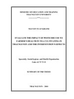
The design of the trial is displayed in Fig. 1. The study
has been approved by the ethics committee and the
National Health Products Safety Agency (P/2014/241).
Patient recruitment and data collection started in April,
2015. Financial support for this work is provided by the
Ligue Contre le Cancer (CCIR-GE).
Fig. 1 Flowchart of the study design
Page 3 of 11
Recruitment and inclusion
In total, 112 patients are being recruited for this study.
All potentially eligible patients, followed-up in the
department of medical oncology for HER2 positive
breast cancer, are identified during multidisciplinary
team meetings with oncologists, surgeons and radiotherapists in one of the eight public hospitals in the region
of Franche-Comté (eastern France). The inclusion and
exclusion criteria are detailed in Table 1. Eligible patients
are informed about the study, and given an information
leaflet. Before inclusion, patients with HER2–positive
breast cancer must have undergone surgery, and chemotherapy plus radiotherapy if indicated. The choice of
type of surgery, as well as the chemotherapy and radiotherapy regimens are left at the discretion of the physician. Patients are included when they receive adjuvant
treatment with trastuzumab (administrated every 3 weeks
for a total of 18 injections) (Fig. 2). At the inclusion visit,
the signed informed consent is retrieved and a clinical
examination is carried out. The medical history, use of
analgesics (type, level, dose) and questionnaires on lifestyle habits (smoking, alcohol) are also recorded.
Jacquinot et al. BMC Cancer (2017) 17:425
Page 4 of 11
Table 1 Inclusion and exclusion criteria
Inclusion criteria
- Patients aged 18 to 85 years
- First HER2 positive breast cancer, confirmed histologically
- WHO Performance status ≤1
- Completed chemo-radiotherapy
- Normal renal function (creatinine clearance ≥60 ml.min-1)
- Normal heart function with LVEF ≥50% (As assessed by
echocardiography dating from less than 3 months previously)
- Normal liver function (normal ASAT and ALAT)
- Certificate of non-contraindication to the practice of
physical activity
- Active contraception or menopaused
Randomization
After signing the consent, patients are randomized to
the Training Group (TG) or the Control Group (CG).
Randomization is conducted in open manner, in a 1:1
ratio and performed according to the minimization
technique with stratification (eRandomisation software
Tenalea®) by age (18–30 years vs 30–50 vs 50–65 years)
and global health score on the QLQ-C30 questionnaire
(0–30 vs 30–50 vs 50–70 vs > 70).
Study outcomes
All evaluations are carried out at inclusion (T0), and at
three (T3) and 6 months (T6) of the follow up period.
Between T0 and T3, both groups followed standard
oncological care either without (control group: CG) or
with (training group: TG) a supervised exercise program
(3 times/week). During the follow-up period (T3-T6),
Exclusion criteria
- HER2 negative Breast cancer
- Patients with metastases
- Heart failure (LVEF ≤50%)
- Resting oxygen saturation (SaO2) ≤ 92%
- Autoimmune disease (systemic lupus erythematosus, rheumatoid arthritis)
- Symptomatic osteoarthritis, cardiovascular disease (angina or uncontrolled
hypertension) or lung disease (chronic obstructive pulmonary disease)
- Patients suffering from malnutrition (body mass index <18 kg.m−2) or
weight loss of >10% during the previous 3 months
- Patients with psychiatric or cognitive disorders deemed unsuitable for
physical activity
- Pregnant or breastfeeding patients
both groups followed standard oncological care but no
supervised physical activity. An assessment of a level of
physical activity for 7 days by an actimeter is achieved
between T0-T3 and T3-T6 without guidelines is
imposed.
Primary endpoint
Cardiac toxicity is defined by either a decrease of the
LVEF under 50% (this decrease was independent from
the baseline value) or an absolute drop of LVEF of 10%
from baseline. LVEF is assessed by a transthoracic
doppler echocardiography and is performed at T0, T3
and T6 according to the American Society of Echocardiography recommendations [25] with a Philips echocardiography machine (Philips iE 33 or EPIQ7, Philips
Healthcare, Andover, MA, USA) and a 2.5 MHz probe.
Measurements are made on 3 representative beats and
Fig. 2 Representation of treatment schedule and study inclusion for patients with HER2 positive breast cancer
Jacquinot et al. BMC Cancer (2017) 17:425
the mean of the results is recorded. Standard echocardiographic analysis included two-dimensional, M-mode,
and Doppler flow measurements. All echocardiograms
are carried out by the same observer, who is blinded to
the clinical data. LVEF is measured in the apical 4- and
2-chamber views using Simpson’s biplane rule and with
TM measurement in 2D mode in the left parasternal
view according to the American Society of Echocardiography guidelines [26].
Secondary endpoints
Other myocardial and valve function
All other measurements are assessed by transthoracic
Doppler echocardiography as described above according
to the American Society of Echocardiography guidelines
[25]. 2D images are acquired in the apical and parasternal
axes and subcostal view, with the latter also used for Mmode imaging. Valve regurgitation is assessed and graded
in accordance with consensus criteria based on Doppler
and M-mode imaging [27, 28]. LV mass is calculated as
the product of myocardial density and myocardial volume.
Peak systolic longitudinal strain is calculated by averaging
the values of peak systolic strain in the basal, midventricular and apical segments in the 4 and 2-chamber views
[29]. Left and right ventricular fractional shortenings are
calculated as the percentage drop in right ventricular outflow tract diameter in systole with respect to diastole [30].
Page 5 of 11
out by the same technician following the guidelines of
the American Thoracic Society [31] using a spirometer
(CPFS/D, Medical Graphics, Strasbourg, France) which
is a device that measures the flow rate of instantaneous
gas mouth open circuit. It therefore measures the flow
rates and the patient’s lung volumes. For this study, a
flow-volume loop is performed during which the following parameters are evaluated: forced vital capacity
(FVC), forced expiratory volume in one second (FEV1),
FEV1/FVC ratio, and maximal expiratory flow (MEF) at
25%(MEF25), 50% (MEF50), 75%, (MEF75) and 25–75%
(MEF25–75) of FVC. These data are necessary before a
maximal graded test.
Cardiorespiratory fitness
The height is determined to the nearest 0.01 m using a
calibrated length board. Body mass is measured to the
nearest 0.1 kg using a calibrated. Body mass and height
are measured bare-foot while wearing underwear. Body
mass index (BMI) is calculated as body mass divided by
height squared (kg.m−2). Waist circumference (WC) is
measured to the nearest 0.5 cm in a standing position
with a standard non-elastic tape that was applied
horizontally midway between the last rib and the superior iliac crest. Body composition is determined by the
skinfold method (Harpenden® skinfold caliper, Baty
International, Burgess Hill, England). The measurement
is performed by the same operator 3 times on the same
fold (the fold biceps, supra iliac fold, fold subscapularis,
the triceps fold) and the average of 3 values is taken into
account. The body composition also is evaluated on
fasting subjects, lying flat for 15 min, not having drunk
and having performed no physical effort for at least 12 h
by multifrequency (5, 50, 100, 200 kHz) bioelectric
impedance (Z-Metrix®, BioparHom, Bourget du lac,
France), a simple, rapid, and non-invasive assessment.
Patients performed a maximal graded exercise test,
under a cardiologist’s supervision, using a cycle ergometer (Ergoselect 200; Ergoline; Bitz, Germany) at T0, T3
and T6. After an initial warm-up period of 3 min (30
watts), exercise begins at a power output of 10 W every
minute until exhaustion. The cadence is maintained
between 50 rpm and 70 rpm. Multichannel electrocardiograms (ECGs) (CASE P2, GE Healthcare, Buckinghamshire, UK) are monitored online before, during
exercise and recovery to follow heart rate. During
exercise, the subjects were connected to a gas analyzer
system (MGC-CPX System; MGC Diagnostics Corporation, Saint Paul, MN, USA), which was calibrated using
gases of known concentration. Expiratory gases were
sampled and analyzed for each 20 s period. The variables
determined are: rate of oxygen consumption (V˙O2) and
carbon dioxide production (V˙CO2), respiratory exchange ratio (V˙CO2/V˙O2), and ventilation per minute
(V˙E). The ventilatory threshold 1 (VT1) and 2 (VT2)
are assessed from V˙E, the relation between V˙CO2/
V˙O2, V˙E⁄V˙O2, V˙E⁄V˙CO2 and power output by three
experts in a blind fashion using the V-slope method. The
mean of the two closest values is taken as the ventilator
threshold and the corresponding power output (W˙VT)
was registered [32] and mechanical power (Watts) corresponding to the ventilatory threshold 1 and 2 (VT1 and
VT2) are used for the rehabilitation.
Blood gas (SaO2, PaO2, PaCO2, pH and Excess Bases)
and lactate levels are collected by microwave method on a
sample taken by incision of the earlobe at rest, at maximal
effort and after 5 min recovery [33]. To confirm that exhaustion is reached, two of the three following criteria
must be met: a drop in cadence below 50 rpm, a respiratory exchange ratio value exceeding 1.0, attainment of
100% of age-predicted maximal HR (220-age).
Resting respiratory function
Maximum voluntary force of the leg extensor
Resting respiratory function is explored by the analysis
of breathed air. All pulmonary function tests are carried
Maximum voluntary strength of quadriceps is performed
sitting, with the knee flexed to 90° with a strain gauge
Anthropometric and body composition
Jacquinot et al. BMC Cancer (2017) 17:425
(SENSY’s load cell 2712). The force was measured on
the right leg and then the left leg, only the value of the
dominant leg was reported. To obtain a valid measurement, it is necessary that the patient’s strength reaches a
plateau and is maintained at least 5 s. Three repeats are
performed and the best of the three is retained. The
signal from the strain gauge is sent to a Power lab 26 T®
series with dual bioamplifier (AD Instruments United
Kingdom, model No. ML4856) and the data are analyzed
with LabChart 8 Pro Software®.
Biological and hormonal parameters
Blood samples are collected in the morning (8:00 to 10:00)
after an overnight fast, by venous puncture. Plasma is
separated by centrifugation (15 min at 3500 rpm) and
aliquots are stored at −80 °C until biochemical analysis.
Plasma glucose, cholesterol, HDL-cholesterol, LDLcholesterol, triglycerides, C-reactive protein (CRP),
interleukin-6 (IL-6), tumor necrosis factor-alpha (TNF-α),
insulin, insulin-like growth factor (IGFs), leptin and adiponectin are measured. Homeostatic model assessment
(HOMA) is calculated as [fasting insulin (mU/l) x fasting
glucose(mmol/l)/22.5] and used to estimate insulin resistance [34].
Questionnaires
All questionnaires are completed prior to the completion of each maximal graded test (T0, T3 and T6).
Health-related quality of life and body image The
cancer-specific Health-related Quality of Life Questio
nnaire EORTC QLQ-C30 is a self-administered,
validated questionnaire to assess HRQoL in cancer patients [35]. It contains 30 items covering five functional
scales (physical, role, cognitive, emotional, and social),
three symptom scales (fatigue, pain, and nausea and
vomiting), and a global health and quality-of-life scale.
The remaining single items assess additional symptoms
commonly reported by cancer patients (dyspnea, appetite loss, sleep disturbance, constipation, and diarrhea),
as well as the perceived financial impact of the disease
and treatment.
In addition, the 23-item breast cancer specific module
EORTC QLQ-BR23 is completed, assessing 8 dimensions specific to breast cancer patients: four functional
scales (body image, sexual functioning and enjoyment,
future perspective) and four symptomatic scales (arm
symptoms, breast symptoms, systemic therapy side effect
and hair loss). For the present study, only the body
image items are compiled.
Fatigue The French version “Multidimensional Fatigue
Inventory 20” (MFI 20) validated by Gentile et al., (2003)
is a 20-item self-report instrument, designed to measure
Page 6 of 11
fatigue [36]. It covers the following dimensions: general
fatigue, physical fatigue, mental fatigue, reduced motivation and reduced activity. This instrument was tested
for its psychometric properties in cancer patients receiving radiotherapy. Patients assess themselves on a scale of
5 levels, from 1 to 5, according to the fatigue experienced the day before the questionnaire was completed.
Pain The Brief Pain Inventory short form (BPI-SF) is a
validated, widely used, self-administered questionnaire
developed to assess the pain severity and pain interference during the previous 24 h [37]. The BPI-SF includes
diagrams of the front and back of the body, four items
to capture the variability of pain over time: pain at its
“worst,” “least,” “average,” and “now” (current pain), the
seven items relating to the interference of pain with various daily activities, including general activity, walking,
work, mood, enjoyment of life, relations with others and
sleep rated on 0–10 scale, as well as a question about
percentage of pain relief by analgesics.
Level of physical activity
The International Physical Activity Questionnaire
(IPAQ) questionnaire and an actimeter (Actigraph®, Fort
Walton Beach Florida, USA) are used to evaluate the
level of physical activity.
The IPAQ validated in French assesses overall physical
activity the last 7 days (leisure time physical activity, sport,
physical activity at work, activities of daily living, transportation) in adult populations aged 15 to 69 years [38]. The
items are structured to provide separate scores on walking, moderate-intensity and vigorous-intensity activities as
well as a combined total score to describe overall level of
activity. Responses are converted to Metabolic Equivalent
Task minutes per week (MET.min.week−1) according to
the IPAQ scoring protocol [39].
The Actigraph® allows measurement of the overall
activity of an individual in the activities of daily living. It
is a small accelerometer which measures accelerations
from 0.05 to 2.00 G [40]. These accelerations are scored
in counts per minute that provide information about the
duration and intensity of activity. Patients wear the
accelerometer on the right hip for 7 consecutive days
during the period T0-T3 and between T3-T6. The
Actigraph® does not give any form of feedback to the
participants.
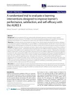
Supervised exercise program
The supervised exercise program consists of three 45-min
exercise sessions a week for 12 weeks. Each training
session includes 9 successive work bouts of 5 min each.
During each work bout, a 4-min period of moderate work
(base) is followed by a 1-min period of intense work
(peak). Initially, the base is set at the first ventilatory
Jacquinot et al. BMC Cancer (2017) 17:425
threshold (VT1) obtained on the initial maximal exercise
test, and the peak is set at the second ventilatory threshold
(VT2). The program starts with a 5-min warm-up at intensity equal to ½ VT1. Then, the intensity of each training
session is designed to lead up to almost 80% of maximum
heart rate at the end of the peak. The peak and base loads
are alternately readjusted by 10 W when the heart rate recorded at the end of the session is 10 to 12 beats/min
below the target heart rate. Hence, each training session
corresponds to the maximal endurance intensity that the
subject is able to maintain for 45 min. Active recovery is
carried out at ½ VT1 for 5 min (Fig. 3).
Sample size
The primary objective of this phase II, randomized,
prospective, multicenter; non-comparative trial is to
evaluate the rate of cardiotoxicity at 3 months (a
decrease of the LVEF under 50% or an absolute drop of
LVEF of 10% from baseline) of patients with HER2 overexpressing breast cancer undergoing adjuvant treatment
by trastuzumab. According to Fleming one-stage design
with a one-sided 5% type I error and power of 90%, 53
patients in the training arm will need to be randomized
in order to test the following hypotheses:
– H0 (null): a cardiotoxicity rate at 3 months of 75%
(uninteresting to pursue any further investigation)
– H1 (alternative): a cardiotoxicity rate at 3 months
of 90% (warrants further investigation in a
phase III trial).
The hypotheses regarding an anticipated cardiotoxicity
rate at 3 months of 90% and an uninteresting rate of
75% is based on the observed cardiotoxicity rate in randomized clinical trials who has been to be approximately
13 to 27% according the molecules used for chemotherapy [9]. The usual care arm will serve as calibration that
the populations in the two arms are similar and to
validate the H0 hypothesis: no statistical comparison is
planned between the two arms.
In the training group, after recruitment of the 53 patients with a 3-month follow-up from randomization:
Fig. 3 Supervised exercise program
Page 7 of 11
– if 44 or less than 44 patients are free of
cardiotoxicity at 3 months (83.0%), the supervised
exercise provided in the training arm could be
declared uninteresting.
– if 45 or more than 45 patients are free of
cardiotoxicity at 3 months (84.9%), the supervised
exercise could be declared interesting for further
phase III evaluation.
The probability to conclude for inefficacy at the end
whereas p = 90.0% is β = 7.8%. The probability to
conclude for efficacy at the end whereas p = 75.0% is
α = 6.1%.
Overall, 53 patients will be included in both arms: 106
patients need to be randomized. With an expected 5%
rate of patients not evaluable at 3 months or drop out
patients, it will be necessary to include a total of 112
(106*1.05) patients (Training arm: N = 56; Usual care
arm: N = 56).
Statistical analyses
A final statistical plan will be written before data frozen.
A specific statistical plan dedicated to HRQoL analyses
will be also written before data frozen. The statistical
analyzes will be carried out with software SAS® v9.2
(SAS Institute Inc., Cary, NC, USA). Clinical and
demographic data will be described using rules form.
The statistical parameters mean, median, SD, interquartile range and range will be presented for continuous
baseline variables. For categorical baseline variables,
corresponding frequencies (n, %) will be calculated. All
baseline variables will be summarized by treatment arm.
Means and medians will be compared using Student’s ttest and Wilcoxon test, respectively. Proportions will be
compared with Chi2 test (or Fisher’s exact test, if
appropriate).
The primary analysis will be performed in modified
intention-to-treat (mITT) population, i.e. including all
evaluable randomized patients regardless of their eligibility and supervised exercise received. The results will be
reported according to the randomized treatment.
Jacquinot et al. BMC Cancer (2017) 17:425
Confirmative analyses will be conducted firstly in the
ITT population (not assessable patients and patients
with drop out between randomization and 3 months will
be considered as progressive) and secondly, in the Per
Protocol population defined as patients who have
received at least a part of the supervised exercise and
presenting no major deviations from the protocol.
Patient compliance to the supervised exercise program
will also be evaluated by the ratio of the total number of
imposed sessions (n = 36) to the number of really performed sessions.
Correlation analyses will be performed to determine
whether the level of cardiorespiratory fitness is significantly associated with the changes in cardiac function
over time (longitudinal strain, volume), muscle strength,
fatigue, pain, quality of life and metabolic, hormonal and
inflammatory responses. Similarly, we will investigate
whether program effectiveness varies significantly as a
function of patients’ background characteristics (age,
weight, type of chemotherapy), and particularly those
variables assessing level of activity, and patient compliance to the supervised exercise program.
The time to HRQOL score deterioration will be
estimated as a modality of longitudinal HROQL analysis.
It will be defined as the time from inclusion in the study
to the occurrence of the first clinically significant deterioration of 5 point at least of the HRQOL score as compared to the baseline score [41]. Dimensions targeted
will be the physical functioning, emotional functioning
and fatigue scales of the QLQ-C30. All other dimensions
of HRQOL will be also analyzed. Patients with no
follow-up measure will be censored just after baseline
(Day 1). Patients with no deterioration before their
drop-out are censored at the time of the last HRQoL
assessment. TTD curves were estimated using the
Kaplan-Meier method and described using median and
its 95% CI. Some univariate and multivariate analysis
will be performed as exploratory purpose only in order
to investigate factors potentially influencing the TTD.
Other definitions of TTD will be explored as sensitivity
purpose varying the MCID and including death as an
event.
Discussion
Several studies have highlighted the benefits of physical
activity during adjuvant treatments in breast cancer in
reducing side effects, and have reported an improvement
in cardiorespiratory fitness [18, 42, 43], muscle strength
[18, 19], immune-function [20], body composition [44,
45] as well as quality of life [21] and fatigue [21, 46–48].
To the best of our knowledge, CARDAPAC is the first
randomized study to assess the effect of a supervised
exercise program on the incidence of cardiotoxicity induced by trastuzumab in patients with HER2 positive
Page 8 of 11
breast cancer. Only Haykowsky et al. [23] have demonstrated left ventricular dilation and a reduction in LVEF
in HER2 positive breast cancer despite aerobic exercise
training. Furthermore, theses authors did not observe an
improvement in exercise capacity (power output, VO2,
heart rate, perceived effort) after the aerobic training
intervention performed during the first 4 months of
trastuzumab therapy, 3 days per week, for between
30 min to 60 min. They suggest that the intensities of
the aerobic program (heart rate equal to 60% to 90% of
peak oxygen consumption) and the number of sessions
were not only insufficient to prevent left ventricular remodelling, but also to achieve beneficial training adaptations. However, this type of aerobic training program could
prevent the expected decline in peak oxygen consumption
that occurs during the first 4 months of trastuzumab treatment, as previously reported by Peels et al. [49].
Unlike the study of Haykowsky et al., in which the
exercise programme took place during the first 4 months
trastuzmab therapy, our study includes patients at the
end of their adjuvant treatments (chemotherapy and
radiotherapy) and consequently limits potential bias
from the side effects of these treatments. Furthermore, it
also includes two randomized arms with a control group,
and a training group who perform a supervised, individualized, intermittent exercise regime combining both
moderate and high intensities, over a period of 3 months.
The CARDAPAC study is the first randomized controlled trial in this indication, and has several strengths,
including the multicentre nature of the trial, the large
sample size, and the supervised, individualized exercise
program.
Given the lack of recommendations for exercise during
adjuvant breast cancer treatments (particularly regarding
type, duration, or frequency), we propose a specific
endurance training in our training group called the
Square-Wave Endurance Exercise Test (SWEET) validated by Gimenez et al. [50]. This bi-level training aims
to replicate interval training sessions (moderate and high
intensity) adjusted according to the physical characteristics of the subjects. It has previously been proposed with
healthy subjects [50], but also with patients suffering
from cardiovascular [51, 52] or other diseases [53, 54].
SWEET is quite efficient in increasing maximal oxygen
consumption, endurance capacity, and/or force and
endurance of the leg muscles.
This study was designed to reduce the side effects
of previous adjuvant therapies and the cardiotoxicity
of trastuzumab in keeping at a constant level or in
increasing of the LVEF. In addition, it meets the goals
of supportive care, which are to improve quality of
life during treatment and facilitate the post-treatment
phase with maintenance of the program benefits in
the long term.
Jacquinot et al. BMC Cancer (2017) 17:425
Finally, in case beneficial effects are observed at the end
of the exercise intervention, it is of interest to investigate
whether these benefits are sustained over a longer period
of time. Therefore, we intend to follow-up all patients for
3 months after the exercise intervention to assess physical
fitness, fatigue, quality of life and level of physical activity.
It should be noted that during the study the level of
physical activity and body composition are assessed
which may lead patients to modify their eating habits
and lifestyle (level of physical activity). Nevertheless, we
do not anticipate that this will take place in a structured
or systematic way, and thus the planned comparisons
(between the training groups and the control group) will
still be valid.
To summarize, the use of targeted therapies in the
treatment of cancer in constantly increasing. Substantial
improvements in disease-free and overall survival have
been reported with these therapies, particularly trastuzumab. Although there are fewer side effects than with the
surgery, chemotherapy and radiotherapy must not underestimate the side effects of these targeted therapies.
If this study shows that physical activity, combining
high and moderate intensities, can reduce or minimize
the cardiotoxicity of trastuzumab, increase cardiorespiratory fitness and health-related quality of life. This
exercise intervention could be systematically proposed
to patients with HER2 positive breast cancer in addition
to standard oncological care.
Abbreviations
2D mode: Two-dimensionel doppler echocardiography; ALAT: Alanine
aminotransferase; ASAT: Aspartate transaminase; BMI: Body mass index; BPISF: Brief pain inventory-short form; BR23: The 23-item breast cancer specific
module; CG: Control group; CRP: C-reactive protein; ECGs: Electrocardiogram;
EORTC: European organization for research and treatment of cancer;
FEV: Forced expiratory volume; FEV1: Force expiratory volume in 1 s;
FVC: Vital capacity; HDL-cholesterol: Hight density lipoprotein; HER2: Human
epidermal growth factor receptor 2; HOMA: Homeostatic model assessment;
HR: Heart rate; HRQOL: Health-related quality of life; IGFs: Insulin-like growth
factor; IL-6: Interleukin-6; IPAQ: International physical activity questionnaire;
ITT: Intention-to-treat; LDL-cholesterol: Low density lipoprotein; LV: Left
ventricular; LVEF: Left ventricular ejection fraction; MCID: Minimal clinically
importante difference; MET.min.week−1: Metabolic equivalent task minutes
per week; MFI 20: Multidimensional fatigue inventory 20; mITT: modified
intention-to-treat; PaCO2: Partial pressure of carbon dioxide in the arterial
blood; PaO2: Partial pressure of oxygen in arterial blood; pH: Hydrogen
potential; QLQ-C30: Quality of life questionnaire core 30; SaO2: Oxygen
arterial saturation; SD: Standard deviation; SWEET: Square-Wave Endurance
Exercise Test; TG: Training group; TM: Time-motion mode; TNF-α: Tumor
necrosis factor-alpha; TTD: Time to deteriation; VCO2: Carbon dioxide flow;
VE: Ventilation rate; VE/VCO2: Respiratory equivalents in carbon dioxide; VE/
VO2: Respiratory equivalent in oxygen; VO2 peak: Peak oxygen uptake;
VO2: Oxygen consumption; VT1 and VT2: Ventilatory thresholds 1 and 2;
W: Watts; W˙VT: Power output; WC: Waist circumference; WHO: World health
organization
Acknowledgements
The authors would like to thank Fiona Ecarnot for editorial assistance in the
preparation of the manuscript.
Page 9 of 11
Funding
This study is supported by the Ligue Contre le Cancer (CCIR-GE)(N°
8FI11826PYRO) through its 2014 Reasearch Grant. The funds allocated by the
Ligue Contre le Cancer relate primarily to the running and equipment costs,
excluding expenses, participation in symposia or congresses or any other
expenses not directly related to the completion of the research project or
not directly induced by the development of the project.
Availability of data and materials
Not applicable.
Authors’ contributions
QJ, NM, FB, XP and FM conception, design, trial protocol and initiation of the
project; MB, GD, QJ and FM conception and supervision of immunological
analyses; MB, GD, QJ and FM supervision of bio specimen collection and
analyses; QJ, BD and FM conception and supervision of training interventions
and physical performance diagnostics; MC realization of all echocardiography
for the study; QJ, NM, BD, XP and FM study coordinators, performs end point
assessments; NM, MC, BD and XP study physicians; QJ, DV and FB study and
data management; FB et DV drafted the sample size and statistical analyses
parts; QJ and FM drafted and finalized the manuscript. NM, BD and XP
medical advices. All authors have read and approved the final manuscript.
Competing interests
The authors declare that they have no competing interests.
Consent for publication
Not applicable.
Ethics approval and consent to participate
The study was approved by the ethics committee and the French National
Health Products safety agency (P/2014/241) the 27 march 2015. This study is
registered at the National Clinical Trials (NCT02433067) since the 28 April 2015.
Author details
1
UPFR des Sports, Université de Franche-Comté, 31 chemin de l’Epitaphe,
25000 Besançon, France. 2EA 3920: Marqueurs pronostiques et facteurs de
regulation des pathologies cardiaques et vasculaires, CHU Jean-Minjoz, 25000
Besançon, France. 3Service d’Oncologie Médicale, CHU Jean-Minjoz, 25000
Besançon, France. 4Service de Cardiologie, CHU Jean-Minjoz, 25000
Besançon, France. 5INSERM UMR 1098: Unité de méthodologie et de qualité
de vie en cancérologie, CHU Jean-Minjoz, 25000 Besançon, France.
6
Physiologie-Explorations Fonctionnelles, CHU Jean-Minjoz, 25000 Besançon,
France. 7Laboratoire de Biochimie Endocrinienne et Métabolique, CHU
Jean-Minjoz, 25000 Besançon, France.
Received: 14 March 2017 Accepted: 9 June 2017
References
1. Jemal A, Bray F, Center MM, Ferlay J, Ward E, Forman D. Global cancer
statistics. CA Cancer J Clin. 2011;61:69–90.
2. INCa. Les données - Institut National Du cancer. 2012. http://www.e-cancer.
fr/Expertises-et-publications/Catalogue-des-publications/Les-cancers-enFrance-en-2014-L-essentiel-des-faits-et-chiffres. Accessed 9 Mar 2016.
3. Slamon DJ, Clark GM, Wong SG, Levin WJ, Ullrich A, McGuire WL. Human
breast cancer: correlation of relapse and survival with amplification of the
HER-2/neu oncogene. Science. 1987;235:177–82.
4. Bilous M, Ades C, Armes J, Bishop J, Brown R, Cooke B, et al. Predicting the
HER2 status of breast cancer from basic histopathology data: an analysis of
1500 breast cancers as part of the HER2000 international study. Breast Edinb
Scotl. 2003;12:92–8.
5. Cobleigh MA, Vogel CL, Tripathy D, Robert NJ, Scholl S, Fehrenbacher L, et
al. Multinational study of the efficacy and safety of humanized anti-HER2
monoclonal antibody in women who have HER2-overexpressing Metastatic
breast cancer that has progressed after chemotherapy for Metastatic
disease. J Clin Oncol 1999;17:2639–2639.
6. Smith I, Procter M, Gelber RD, Guillaume S, Feyereislova A, Dowsett M, et al.
2-year follow-up of trastuzumab after adjuvant chemotherapy in HER2positive breast cancer: a randomised controlled trial. Lancet Lond Engl.
2007;369:29–36.
Jacquinot et al. BMC Cancer (2017) 17:425
7.
8.
9.
10.
11.
12.
13.
14.
15.
16.
17.
18.
19.
20.
21.
22.
23.
24.
25.
26.
Romond EH, Perez EA, Bryant J, Suman VJ, Geyer CEJ, Davidson NE, et al.
Trastuzumab plus adjuvant chemotherapy for operable HER2-positive breast
cancer. N Engl J Med. 2005;353:1673–84.
Slamon DJ, Leyland-Jones B, Shak S, Fuchs H, Paton V, Bajamonde A,
et al. Use of chemotherapy plus a monoclonal antibody against HER2
for Metastatic breast cancer that overexpresses HER2. N Engl J Med.
2001;344:783–92.
Seidman A, Hudis C, Pierri MK, Shak S, Paton V, Ashby M, et al. Cardiac
dysfunction in the trastuzumab clinical trials experience. J. Clin. Oncol. Off. J.
Am. Soc. Clin. Oncologia. 2002;20:1215–21.
Procter M, Suter TM, de Azambuja E, Dafni U, van Dooren V, Muehlbauer S,
et al. Longer-term assessment of trastuzumab-related cardiac adverse
events in the Herceptin adjuvant (HERA) trial. J Clin Oncol Off J Am Soc Clin
Oncol. 2010;28:3422–8.
Piccart-Gebhart MJ, Procter M, Leyland-Jones B, Goldhirsch A, Untch M,
Smith I, et al. Trastuzumab after adjuvant chemotherapy in HER2-positive
breast cancer. N Engl J Med. 2005;353:1659–72.
Suter TM, Procter M, Veldhuisen DJ van, Muscholl M, Bergh J, Carlomagno
C, et al. Trastuzumab-associated cardiac adverse effects in the Herceptin
adjuvant trial. J Clin Oncol 2007;25:3859–3865.
Goodwin PJ, Ennis M, Pritchard KI, McCready D, Koo J, Sidlofsky S, et al.
Adjuvant treatment and onset of menopause predict weight gain after
breast cancer diagnosis. J. Clin. Oncol. Off. J. Am. Soc. Clin. Oncologia.
1999;17:120–9.
Curt GA, Breitbart W, Cella D, Groopman JE, Horning SJ, Itri LM, et al. Impact
of cancer-related fatigue on the lives of patients: new findings from the
fatigue coalition. Oncologist. 2000;5:353–60.
Courneya KS. Exercise in cancer survivors: an overview of research. Med Sci
Sports Exerc. 2003;35:1846–52.
Peel JB, Sui X, Adams SA, Hébert JR, Hardin JW, Blair SN. A prospective study
of cardiorespiratory fitness and breast cancer mortality. Med Sci Sports
Exerc. 2009;41:742–8.
Kayl AE, Meyers CA. Side-effects of chemotherapy and quality of life
in ovarian and breast cancer patients. Curr Opin Obstet Gynecol.
2006;18:24–8.
Courneya KS, Segal RJ, Mackey JR, Gelmon K, Reid RD, Friedenreich CM, et
al. Effects of aerobic and resistance exercise in breast cancer patients
receiving adjuvant chemotherapy: a multicenter randomized controlled trial.
J Clin Oncol. 2007;25:4396–404.
Cheema B, Gaul CA, Lane K, Fiatarone Singh MA. Progressive resistance
training in breast cancer: a systematic review of clinical trials. Breast Cancer
Res Treat. 2008;109:9–26.
Galvão DA, Newton RU. Review of exercise intervention studies in cancer
patients. J. Clin. Oncol. Off. J. Am. Soc. Clin. Oncologia. 2005;23:899–909.
Mutrie N, Campbell AM, Whyte F, McConnachie A, Emslie C, Lee L, et al.
Benefits of supervised group exercise programme for women being treated
for early stage breast cancer: pragmatic randomised controlled trial. BMJ.
2007;334:517.
van Waart H, Stuiver MM, van Harten WH, Geleijn E, Kieffer JM, Buffart LM, et
al. Effect of Low-Intensity Physical Activity and Moderate- to High-Intensity
Physical Exercise During Adjuvant Chemotherapy on Physical Fitness,
Fatigue, and Chemotherapy Completion Rates: Results of the PACES
Randomized Clinical Trial. J. Clin. Oncol. 2015;JCO.2014.59.1081.
Haykowsky MJ, Mackey JR, Thompson RB, Jones LW, Paterson DI. Adjuvant
trastuzumab induces ventricular remodeling despite aerobic exercise
training. Clin. Cancer res. Off. J. Am. Assoc. Cancer Res. 2009;15:4963–7.
Wisløff U, Støylen A, Loennechen JP, Bruvold M, Rognmo Ø, Haram PM, et
al. Superior cardiovascular effect of aerobic interval training versus
moderate continuous training in heart failure patients. Circulation.
2007;115:3086–94.
Lang RM, Bierig M, Devereux RB, Flachskampf FA, Foster E, Pellikka PA, et al.
Recommendations for chamber quantification: a report from the American
Society of Echocardiography’s guidelines and standards committee and the
chamber quantification writing group, developed in conjunction with the
European Association of Echocardiography, a branch of the European
Society of Cardiology. J Am Soc Echocardiogr Off Publ Am Soc
Echocardiogr. 2005;18:1440–63.
Gottdiener JS, Bednarz J, Devereux R, Gardin J, Klein A, Manning WJ, et al.
American Society of Echocardiography recommendations for use of
echocardiography in clinical trials. J Am Soc Echocardiogr.
2004;17:1086–119.
Page 10 of 11
27. Jones EC, Devereux RB, Roman MJ, Liu JE, Fishman D, Lee ET, et al.
Prevalence and correlates of mitral regurgitation in a population-based
sample (the strong heart study). Am J Cardiol. 2001;87:298–304.
28. Zoghbi WA, Enriquez-Sarano M, Foster E, Grayburn PA, Kraft CD, Levine RA,
et al. Recommendations for evaluation of the severity of native valvular
regurgitation with two-dimensional and doppler echocardiography.
J Am Soc Echocardiogr. 2003;16:777–802.
29. Sawaya H, Sebag IA, Plana JC, Januzzi JL, Ky B, Cohen V, et al. Early
detection and prediction of cardiotoxicity in chemotherapy-treated patients.
Am J Cardiol. 2011;107:1375–80.
30. Anavekar NS, Gerson D, Skali H, Kwong RY, Kent Yucel E, Solomon SD. Twodimensional assessment of right ventricular function: an echocardiographic–
MRI correlative study. Echocardiography. 2007;24:452–6.
31. Standardization of spirometry–1987 update. Statement of the American
Thoracic Society. Am. Rev. Respir. Dis. 1987;136:1285–98.
32. Wasserman K. The anaerobic threshold: definition, physiological significance
and identification. Adv Cardiol. 1986;35:1–23.
33. Godfrey S, Wozniak ER, Courtenay Evans RJ, Samuels CS. Ear lobe blood
samples for blood gas analysis at rest and during exercise. Br J Dis Chest.
1971;65:58–64.
34. Matthews DR, Hosker JP, Rudenski AS, Naylor BA, Treacher DF, Turner RC.
Homeostasis model assessment: insulin resistance and beta-cell function
from fasting plasma glucose and insulin concentrations in man.
Diabetologia. 1985;28:412–9.
35. Aaronson NK, Ahmedzai S, Bergman B, Bullinger M, Cull A, Duez NJ, et al.
The European Organization for Research and Treatment of cancer QLQ-C30:
a quality-of-life instrument for use in international clinical trials in oncology.
J Natl Cancer Inst. 1993;85:365–76.
36. Gentile S, Delarozière JC, Favre F, Sambuc R, San Marco JL. Validation of the
French “multidimensional fatigue inventory” (MFI 20). Eur J Cancer Care
(Engl). 2003;12:58–64.
37. Larue F, Colleau SM, Brasseur L, Cleeland CS. Multicentre study of cancer
pain and its treatment in France. BMJ. 1995;310:1034–7.
38. Gauthier AP, Lariviere M, Young N. Psychometric properties of the IPAQ: a
validation study in a sample of northern Franco-Ontarians. J. Phys. Act.
Health. 2009;6 Suppl 1:S54–60.
39. Craig CL, Marshall AL, Sjöström M, Bauman AE, Booth ML, Ainsworth BE, et
al. International physical activity questionnaire: 12-country reliability and
validity. Med Sci Sports Exerc. 2003;35:1381–95.
40. Bassett DR, Ainsworth BE, Swartz AM, Strath SJ, O’Brien WL, King GA. Validity
of four motion sensors in measuring moderate intensity physical activity.
Med Sci Sports Exerc. 2000;32:S471–80.
41. Hamidou Z, Dabakuyo TS, Mercier M, Fraisse J, Causeret S, Tixier H, et
al. Time to deterioration in quality of life score as a modality of
longitudinal analysis in patients with breast cancer. Oncologist.
2011;16:1458–68.
42. Mock V, Burke MB, Sheehan P, Creaton EM, Winningham ML, McKenneyTedder S, et al. A nursing rehabilitation program for women with breast
cancer receiving adjuvant chemotherapy. Oncol Nurs Forum. 1994;21:899–907.
discussion 908
43. Segal R, Evans W, Johnson D, Smith J, Colletta S, Gayton J, et al. Structured
exercise improves physical functioning in women with stages I and II breast
cancer: results of a randomized controlled trial. J Clin Oncol Off J Am Soc
Clin Oncol. 2001;19:657–65.
44. Freedman RJ, Aziz N, Albanes D, Hartman T, Danforth D, Hill S, et al. Weight
and body composition changes during and after adjuvant chemotherapy in
women with breast cancer. J Clin Endocrinol Metab. 2004;89:2248–53.
45. Schwartz AL, Winters-Stone K, Gallucci B. Exercise effects on bone mineral
density in women with breast cancer receiving adjuvant chemotherapy.
Oncol Nurs Forum. 2007;34:627–33.
46. Velthuis MJ, Agasi-Idenburg SC, Aufdemkampe G, Wittink HM. The effect of
physical exercise on cancer-related fatigue during cancer treatment: a
meta-analysis of randomised controlled trials. Clin Oncol R Coll Radiol G B.
2010;22:208–21.
47. Kuchinski A-M, Reading M, Lash AA. Treatment-related fatigue and exercise
in patients with cancer: a systematic review. Medsurg Nurs. Off J Acad MedSurg Nurses. 2009;18:174–80.
48. Knols R, Aaronson NK, Uebelhart D, Fransen J, Aufdemkampe G. Physical
exercise in cancer patients during and after medical treatment: a systematic
review of randomized and controlled clinical trials. J Clin Oncol Off J Am
Soc Clin Oncol. 2005;23:3830–42.
Jacquinot et al. BMC Cancer (2017) 17:425
Page 11 of 11
49. Peel AB, Thomas SM, Dittus K, Jones LW, Lakoski SG. Cardiorespiratory
fitness in breast cancer patients: a call for normative values. J Am Heart
Assoc. 2014;3:e000432.
50. Gimenez M, Servera E, Saunier C, Lacoste J. Square-Wave endurance
exercise test (SWEET) for training and assessment in trained and untrained
subjects. Eur J Appl Physiol. 1982;49:369–77.
51. Geny B, Saini J, Mettauer B, Lampert E, Piquard F, Follenius M, et al. Effect of
short-term endurance training on exercise capacity, haemodynamics and
atrial natriuretic peptide secretion in heart transplant recipients. Eur J Appl
Physiol. 1996;73:259–66.
52. Lampert E, Mettauer B, Hoppeler H, Charloux A, Charpentier A, Lonsdorfer J.
Skeletal muscle response to short endurance training in heart transplant
recipients. J Am Coll Cardiol. 1998;32:420–6.
53. Tordi N, Dugue B, Klupzinski D, Rasseneur L, Rouillon JD, Lonsdorfer J.
Interval training program on a wheelchair ergometer for paraplegic
subjects. Spinal Cord. 2001;39:532–7.
54. Gimenez M, Predine E, Marchand M, Servera E, Ponz JL, Polu JM.
Implications of lower- and upper-limb training procedures in patients with
chronic airway obstruction. Chest. 1992;101:279S–88S.
Submit your next manuscript to BioMed Central
and we will help you at every step:
• We accept pre-submission inquiries
• Our selector tool helps you to find the most relevant journal
• We provide round the clock customer support
• Convenient online submission
• Thorough peer review
• Inclusion in PubMed and all major indexing services
• Maximum visibility for your research
Submit your manuscript at
www.biomedcentral.com/submit