Potential of extravasated platelet aggregation as a surrogate marker for overall survival in patients with advanced gastric cancer treated with preoperative docetaxel, cisplatin and S-1: A
Bạn đang xem bản rút gọn của tài liệu. Xem và tải ngay bản đầy đủ của tài liệu tại đây (925.24 KB, 11 trang )
Saito et al. BMC Cancer (2017) 17:294
DOI 10.1186/s12885-017-3279-4
RESEARCH ARTICLE
Open Access
Potential of extravasated platelet
aggregation as a surrogate marker for
overall survival in patients with advanced
gastric cancer treated with preoperative
docetaxel, cisplatin and S-1: a retrospective
observational study
Hiroto Saito, Sachio Fushida*, Tomoharu Miyashita, Katsunobu Oyama, Takahisa Yamaguchi, Tomoya Tsukada,
Jun Kinoshita, Hidehiro Tajima, Itasu Ninomiya and Tetsuo Ohta
Abstract
Background: The theory of extravasated platelet aggregation in cancer lesions was recently introduced. We
investigated the association of platelet aggregation in gastric cancer stroma with clinicopathological features,
chemotherapeutic response, pathological response, and survival.
Methods: The study comprised 78 patients with advanced gastric cancer who had undergone gastrectomy with or
without combination of docetaxel, cisplatin and S-1 (DCS) as preoperative chemotherapy between 2005 and 2014.
The patients were divided into two groups: patients who had received preoperative DCS therapy forming the p-DCS
group and patients who had not received preoperative DCS therapy forming the control group. The 39 patients in the
control group had received gastrectomy and postoperative chemotherapy of S-1 alone. Platelet aggregation in biopsy
specimens before preoperative DCS therapy in the p-DCS group and at the time of diagnosis in the control group
were evaluated using CD42b immunohistochemical staining.
Results: Twenty-four patients in the p-DCS group and 19 in the control group were found to have platelet
aggregation in their cancer stroma. Patients with histologically confirmed platelet aggregation had significantly higher
rates of chemoresistance (58.3%) than those without platelet aggregation (20.0%) (P = 0.019). According to multivariate
analysis, CD42b expression (odds ratio: 5.102, 95% confidence interval: 1.039–25.00, P = 0.045) was correlated with
chemoresistance. CD42b expression and histological non-responder status were both significantly correlated with poor
overall survival (OS) (P = 0.012, P = 0.016); however, RECIST was not correlated with OS. In the control group, CD42b
expression was also significantly correlated with poor overall survival (OS) (P = 0.033). In the p-DCS group, according to
multivariate analysis, male sex (hazard ratio: 0.281, 95% confidence interval: 0.093–0.846, P = 0.024) was correlated with
good prognosis and CD42b expression (hazard ratio: 4.406, 95% confidence interval: 1.325–14.65, P = 0.016) with poor
prognosis.
(Continued on next page)
* Correspondence:
Department of Gastroenterological Surgery, Division of Cancer Medicine,
Graduate School of Medical Science, Kanazawa University, 13-1 Takara-machi,
Kanazawa, Ishikawa 920-8641, Japan
© The Author(s). 2017 Open Access This article is distributed under the terms of the Creative Commons Attribution 4.0
International License ( which permits unrestricted use, distribution, and
reproduction in any medium, provided you give appropriate credit to the original author(s) and the source, provide a link to
the Creative Commons license, and indicate if changes were made. The Creative Commons Public Domain Dedication waiver
( applies to the data made available in this article, unless otherwise stated.
Saito et al. BMC Cancer (2017) 17:294
Page 2 of 11
(Continued from previous page)
Conclusions: This study suggests that platelets in gastric cancer stroma may create a favorable microenvironment for
chemoresistance. CD42b immunohistochemical staining of biopsy specimens is a promising candidate for being a
prognostic marker in patients with gastric cancer.
Keywords: Gastric cancer, Platelets, Preoperative chemotherapy, Chemoresistance, Surrogate marker
Background
An estimated 951,600 new cases of gastric cancer and
723,100 deaths occurred in 2012 [1]. Although the incidence of gastric cancer has decreased in recent decades,
it remains one of the leading causes of cancer-related
death in East Asia. S-1 is an effective postoperative
chemotherapy for East Asian patients who have undergone a D2 dissection for locally advanced gastric cancer
[2]. Multimodality treatment, including chemotherapy
and surgery, has reduced gastric cancer mortality and
improved quality of life. Some studies [3–7] have suggested that preoperative chemotherapy followed by surgery is improves long-term prognosis of advanced
gastric cancer. However, there are no established biomarkers for screening the efficacy of preoperative or
postoperative chemotherapy.
Two methods are currently available for evaluating
tumor responses to chemotherapy. The Response
Evaluation Criteria in Solid Tumors (RECIST) [8] have
been widely used to evaluate tumor responses. However,
RECIST cannot always be used in the preoperative setting because there may be no measurable lesions in patients with resectable gastric cancer. In contrast,
histological evaluation of the primary tumors is commonly used after surgery for the patients treated with
preoperative chemotherapy. Some studies have reported
that histological evaluation yields more valid response
criteria of preoperative treatment than RECIST [9, 10].
Platelets are primarily recognized as key regulators of
thrombosis and hemostasis. Bambace and Holmes [11]
have reported that platelets are linked to key steps in
cancer progression and metastasis. After tumor cells migrate into the bloodstream, they induce platelet aggregation and the platelet-coating protects tumor cells from
immune surveillance and shear stress. Platelets also facilitate cancer cell adherence to vascular endothelial
cells, which leads to extravasation into the stroma and
formation of secondary tumors [12]. However, there are
few reports regarding the role of platelets in primary tumors. Qi et al. [13] reported that platelet aggregation
within colorectal cancers is associated with tumor stage
and lymph node metastasis. Mikami et al. [14] showed
that interactions between platelets and gastric cancer
cells increase tumor proliferation.
A theory of extravasated platelet aggregation (EPA) in
primary cancer lesions was recently introduced [15].
Several studies have focused attention on the central role
of platelet interaction with cancer cells and the immune
system in promoting tumor progression and distant
spread through release of growth factors such as transforming growth factor (TGF)-β, vascular endothelial
growth factor A, and platelet-derived growth factor into
the microenvironment [15]. TGF-β enhances epithelial–
mesenchymal transition (EMT) in cancer cells [16] and
EMT promotes invasiveness, metastasis, and chemoresistance [17].
To clarify the presence of factors that affect chemoresistance in the cancer microenvironment, we focused on
EPA in biopsy specimens from primary tumor of gastric
cancer patients who treated with preoperative or postoperative chemotherapy.
Methods
Inclusion and exclusion criteria
Seventy-eight patients with advanced gastric cancer who
had undergone gastrectomy between 2005 and 2014
were retrospectively evaluated. Thirty-nine of them had
received preoperative DCS therapy (p-DCS group),
whereas the remaining 39 had not received any preoperative chemotherapy (control group). The 39 patients
in the control group had, however, received gastrectomy
and postoperative chemotherapy of S-1 alone. Eligibility
criteria were as follows: clinical Stage III and resectable
Stage IV gastric cancer with fewer than three peripheral
hepatic and para-aortic lymph node (PAN) metastases
[18] in accordance with the Japanese Classification of
Gastric Carcinoma (JCGC), 3rd English edition [19], PAN
metastasis being defined as clearly enlarged (≥ 10 mm) on
enhanced computed tomography (CT) scans with 2.5 mm
slice thickness; absence of peritoneal metastasis on staging
laparoscopy; age 20–80 years; Eastern Cooperative
Oncology Group (ECOG) performance status 0 or 1; no
prior chemotherapy or radiotherapy; no prior gastrectomy; no detected bleeding from primary lesion; good oral
intake; and adequate hematological, hepatic, and renal
function.
Patients were excluded for any of the following reasons:
apparent infection; serious comorbidity such as cardiovascular disease, pulmonary fibrosis, pneumonia, bleeding
tendency, uncontrolled hypertension, poorly controlled
diabetes mellitus, and other serious medical conditions;
synchronous or metachronous active malignancy; central
Saito et al. BMC Cancer (2017) 17:294
nervous system disorder; history of severe drug-induced
allergy; and pregnancy or breastfeeding.
Treatment
In the p-DCS group, patients had received two cycles of
preoperative chemotherapy consisting of 35 mg/m2 docetaxel as a 1-h intravenous infusion on days 1 and 15;
35 mg/m2 cisplatin as a 2-h intravenous infusion on
days 1 and 15 with hyperhydration; and 40 mg/m2 S-1
twice daily on days 1–14 every 4 weeks. At least
4 weeks after the completion of two cycles of DCS
therapy, curative gastrectomy and D2 lymphadenectomy plus PAN dissection (PAND) and hepatectomy
had been performed. Lymph node dissection was performed in patients with PAN metastasis diagnosed by
enhanced helical CT, which was defined as lymph
node station No. 16a2 and b1 (16a2b1PAN) between
the upper margin of the celiac artery and lower border
of the inferior mesenteric artery [19].
In the control group, administration of S-1 was started
within 6 weeks after gastrectomy and continued for
1 year. The treatment regimen consisted of 6-week cycles in which, in principle, 40 mg/m2 S-1 twice daily was
given for 4 weeks and no chemotherapy was given for
the following 2 weeks [2, 20].
Response evaluation
After the second course of preoperative DCS therapy,
the amount of tumor shrinkage was evaluated based on
thin-slice helical CT and the tumor response classified
into one of the following four categories in accordance
with RECIST [8]: complete response (CR), disappearance
of all target lesions; partial response (PR), ≥30% decrease
in the combined diameters of target lesions; progressive
disease (PD), ≥20% increase in the combined diameters
of target lesions; and stable disease (SD), neither sufficient shrinkage to qualify for PR nor sufficient increase
to qualify for PD. Patients with CR and PR were
regarded as RECIST responders.
In the p-DCS group, the resected specimens were histologically evaluated, and tumor response evaluated according to the histological criteria in JCGC, 3rd English
edition [19]. The histological evaluation criteria were classified into one of the following five categories according to
the proportion of the tumor affected by degeneration or
necrosis: grade 3, no viable tumor cells remaining; grade
2, viable tumor cells remaining in less than one-third of
the tumorous area; grade 1b, viable tumor cells remaining
in more than one-third but less than two-thirds of the tumorous area; grade 1a, viable tumor cells occupying more
than two-thirds of the tumorous area; and grade 0, no evidence of therapeutic effect.
Ten percent or 50% residual tumor per tumor bed has
been used as the cutoff percentage in Western countries,
Page 3 of 11
in accordance with the criteria proposed by Becker et al.
[21]. In contrast, a cutoff of 33% or 67% viable tumor
cells per tumor bed is commonly used in Asian countries, in accordance with the definition in JCGC, 3rd
English edition [19]. Although the definition of a histological response is controversial, Kurokawa et al. [9, 10]
have evaluated the results when histological responses
were classified as Grade 2 or 3 and found that the results
were similar to Grades 1b, 2 or 3. In this study, a histological response was defined as less than one-third of viable tumor cells (grade 2 or 3). All resected specimens
were examined by the same pathologist, who assessed
the extent of residual disease, disease stage, and effect of
chemotherapy according to the criteria of JCGC, 3rd
English edition [19].
Immunohistochemical examination
In the p-DCS group, primary cancer lesions were biopsied by esophagogastroduodenoscopy (EGD) before
commencement of preoperative chemotherapy. In the
control group, biopsies were performed by EGD on
diagnosis. Biopsies were taken from the edge of ulcerations associated with gastric cancer, not from the bases
of such ulceration. More than five biopsy specimens
were collected from each patient and evaluated immunohistochemically. Immunohistochemistry using 3-μmthick, 10% formalin-fixed, paraffin-embedded tissue
sections was performed using Dako Envision System
dextran polymers conjugated to horseradish peroxidase
(Dako, Carpinteria, CA, USA) to prevent any endogenous biotin contamination. The specimens were deparaffinized in xylene and rehydrated in a graded ethanol
series. Endogenous peroxidase was blocked by immersing sections in 3% H2O2 in 100% methanol for 20 min
at room temperature. Antigen retrieval was activated
by microwaving sections at 95 °C for 10 min in
0.001 M citrate buffer (pH 7.6). After blocking the endogenous peroxidase, sections were incubated with
Protein Block Serum-Free (Dako) at room temperature
for 10 min to block nonspecific staining. Subsequently,
sections were incubated for 2 h at room temperature
with a 1:100 diluted anti-platelet antibody (anti-CD42b
rabbit monoclonal, EPR6995; Abcam, Tokyo, Japan); a
1:50 diluted anti-podoplanin antibody (anti-D2–40
mouse monoclonal, Code IR072/IS072; Dako, Tokyo,
Japan); a 1:50 diluted anti-forkhead box (FOX)P3
antibody (anti-FOXP3 mouse monoclonal, 236A/E7;
Abcam), and a 1:50 diluted anti-SNAIL antibody (antiSNAIL rabbit polyclonal antibody, ab180714; Abcam).
Peroxidase activity was detected using 3-amino-9ethylcarbazole enzyme substrate. Sections were incubated
in Tris-buffered saline without primary antibodies as
negative controls. Samples were faintly counterstained
with Meyer hematoxylin.
Saito et al. BMC Cancer (2017) 17:294
Evaluation of immunostaining
To evaluate the expression of CD42b in the biopsy specimens, the immunostained cells in five non-overlapping
intratumoral fields were counted at 400× magnification.
The average expression of CD42b was evaluated: ≥10%
was defined as positive and <10% as negative [22]. In the
biopsy specimens stained by D2–40, the immunostained
cells were counted at 200× magnification. The percentage of podoplanin-positive (PP) cells and staining intensity (SI) were evaluated and an immunoreactivity score
(IRS) calculated for each tumor as IRS = PP × SI (0
negative, 1–3 weak, 4–7 moderate, and 8–15 high).
Scores were allocated as follows: 0 PP 0%, 1 PP 1%–20%,
2 PP 21%–40%, 3 PP 41%–60%, 4 PP 61%–80%, and 5
PP 81%–100%; and 0 SI negative, 1 weak, 2 moderate,
and 3 strong. For IRS, ≥4 was defined as positive and <3
as negative [23].
For analysis of SNAIL, IRS was calculated by multiplication of intensity (0–3) by the percentage of stained
cells (0–4). Tissue samples with scores of 0 were classified as SNAIL negative and those with scores of 1–12 as
SNAIL positive [24].
To evaluate infiltration of FOXP3, five nonoverlapping intratumoral fields were counted at 400×
magnification and the mean number per field defined
as the number of FOXP3 infiltrates for the tumor.
The average number of FOXP3-positive T cells was
evaluated; ≥5.5 being defined as positive and <5.5 as
negative [25].
Statistical analysis
Fisher’s exact test was used to measure the statistical
significance of correlations between CD42b expression and chemotherapeutic response. Patient survival
was calculated by the Kaplan–Meier method and the
log-rank test was used to compare the survival rates
between subgroups. Variables found to have possible
associations with chemoresistance and prognosis by
univariate analysis (P < 0.10) were subjected to
multivariate analysis using multi logistic regression
analysis and the Cox proportional hazards regression
model, respectively. Statistical significance was set at
P < 0.05. Data management and statistical analysis
were performed using SPSS version 23 (SPSS, Chicago,
IL, USA).
Results
Patient characteristics
From 2005 to 2014, 78 patients with advanced gastric
cancer were found to be eligible, 39 of whom had received preoperative DCS therapy followed by curative
gastrectomy with D2 lymphadenectomy plus PAND
and/or hepatectomy (p-DCS group). The remaining 39
patients had not received preoperative DCS therapy
Page 4 of 11
prior to undergoing curative gastrectomy with D2
lymphadenectomy plus hepatectomy and had received
postoperative chemotherapy of S-1 alone (control
group). Patient characteristics are summarized in Table 1.
In the p-DCS group, baseline CT showed that 16 (41%)
had PAN metastases and nine (23%) hepatic metastases.
The tumor stages were as follows: 13 (33%) clinical Stage
III and 26 (67%) clinical Stage IV. In the control group,
baseline CT showed that none had PAN metastases and
one (3%) had hepatic metastases. The tumor stages were
as follows: 38 (97%) clinical Stage III and one (3%) clinical
Stage IV.
Response rates
The responses to preoperative DCS therapy were
assessed by RECIST and histological evaluation criteria
(Table 1). The response rates were 74% with RECIST
and 56% with histological criteria.
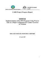
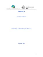
CD42b and podoplanin expression
In the p-DCS group, biopsy specimens were obtained
from the primary gastric cancers before commencing
preoperative chemotherapy. Expression of CD42b, a
platelet marker, was observed around cancer-associated
fibroblasts (CAFs) in the biopsy specimens (Fig. 1a) and
podoplanin expression was found on the membranes of
CAFs (Fig. 1b).
Relationship between CD42b expression and
histopathological variables
There were no significant associations between CD42b
expression and Borrmann macroscopic type, tumor differentiation, clinical T stage, clinical N stage, PAN metastases, or hepatic metastases in either group (Tables 2
and 3).
In the p-DCS group, CD42b positivity was seen in 24
(62%) patients, including 10 (26%) histological responders and 14 (36%) non-responders. There were 15
(38%) CD42b-negative patients, including 12 (31%)
histological responders and three (7%) non-responders.
CD42b-positive patients had significantly higher rates of
chemoresistance (58%) than CD42b-negative patients
(20%) (P = 0.019).
Univariate analysis of expression of three factors
(CD42b, SNAIL, and FOXP3) that are reportedly associated with chemoresistance showed significant associations between CD42b expression (P = 0.025) and SNAIL
expression (P = 0.029) and chemoresistance (Table 4).
These two variables were therefore considered to be potential predictors of chemoresistance and were subjected
to multivariate analysis, which identified a correlation
between CD42b expression and chemoresistance (odds
ratio: 5.102, 95% confidence interval: 1.039–25.00,
P = 0.045) (Table 4).
Saito et al. BMC Cancer (2017) 17:294
Page 5 of 11
Table 1 Patient characteristics according to treatment group
and response to preoperative DCS therapy evaluated by RECIST
and histological evaluation criteria
Characteristic
p-DCS group Control group
Number of patients
39
39
63.6 (30–78)
67.0 (41–80)
32
25
Age, yr.; median (range)
Gender
ECOG performance status
Borrmann macroscopic type
Differentiation
Clinical T stage
Clinical N stage
Clinical stage
PAN metastasis
Hepatic metastasis
RECIST
Male
Female
7
14
≥1
2
0
0
37
39
1
0
1
2
14
10
3
21
16
4
1
10
5
3
2
Diffuse
SNAIL expression
In the p-DCS group, the EMT marker SNAIL was
mainly expressed in the nuclei of cancer cells. Positive
SNAIL expression was found in 30/39 cases (77%)
(Fig. 1c); however, SNAIL expression was not correlated
with CD42b expression (P = 0.230). There was a significant relationship between SNAIL expression and chemoresistance (P = 0.026) but no significant relationship
between SNAIL expression and OS (P = 0.248).
FOXP3 expression
In the p-DCS group, the regulatory T (Treg) cell marker
FOXP3 was found in 7/39 cases (18%) (Fig. 1d). FOXP3
expression was not significantly correlated with CD42b
expression (P = 0.686), chemoresistance (P = 0.205), or
OS (P = 0.698).
18
28
Intestinal 21
11
Survival curves according to chemotherapy response
cT0
0
0
cT1
0
0
cT2
5
5
cT3
13
16
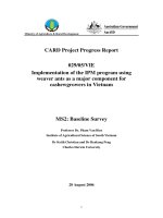
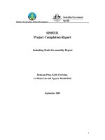
Overall survival (OS) curves for the patients in the both
groups are shown in Fig. 2. In the p-DCS group, comparison of survival rates in RECIST responders and nonresponders by log-rank test revealed no significant difference in prognosis (P = 0.212) (Fig. 2a). In contrast,
OS was significantly longer in histological responders
than non-responders (P = 0.016) (Fig. 2b) and in
CD42b-negative than CD42b-positive patients (P = 0.012)
(Fig. 2c). In the control group, the OS was significantly
longer for CD42b-negative than CD42b-positive patients
(P = 0.033) (Fig. 2d).
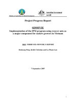
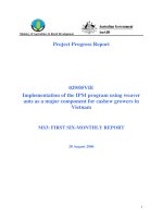
Relapse-free survival curves for the patients in the
both groups are shown in Fig. 3. In the p-DCS group,
there was no significant difference in prognosis between the RECIST responders and non-responders
(P = 0.112) (Fig. 3a). Histological evaluation and
CD42b expression showed that relapse-free survival
was significantly longer in responders than nonresponders (P = 0.004, P = 0.013, respectively) (Fig. 3b, c).
In the control group, the relapse-free survival was significantly longer in CD42b-negative than in CD42b-positive
patients (P = 0.015) (Fig. 3d).
In the p-DCS group, univariate analysis showed that
histological findings (P = 0.023) and CD42b expression
(P = 0.021) were significantly associated with OS. The
four variables (sex, hepatic metastasis, histological evaluation, and CD42b expression) that were found to be significant by univariate analysis and therefore had
prognostic potential (P < 0.10) were subjected to multivariate analysis. Multivariate analysis identified that male
sex (hazard ratio: 0.281, 95% confidence interval: 0.093–
0.846, P = 0.024) was correlated with good prognosis
and CD42b expression (hazard ratio: 4.406, 95% confidence interval: 1.325–14.65, P = 0.016) with poor prognosis (Table 5).
cT4
21
18
cN0
2
0
cN1
2
6
cN2
21
18
cN3
14
15
0
0
0
I
0
0
II
0
0
III
13
38
IV
26
1
(+)
16
0
(−)
23
0
(+)
9
1
(−)
30
38
CR
0
-
PR
29
-
SD
8
-
PD
2
-
3
-
19
-
1b
4
-
1a
11
-
0
2
-
Histological evaluation criteria 3
(Grade)
2
CR complete response, DCS docetaxel, cisplatin, and S-1, ECOG Eastern
Cooperative Oncology Group, PAN para-aortic lymph node, PD progressive
disease, PR partial response, RECIST Response Evaluation Criteria in Solid
Tumors, SD stable disease
Saito et al. BMC Cancer (2017) 17:294
Page 6 of 11
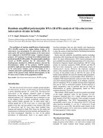
Fig. 1 Representative photomicrograph of pretreatment biopsy specimens from advanced gastric cancer lesion. a: Immunohistological images of
CD42b-positive platelets. Extravasated platelet aggregation (EPA) is mainly seen in the cancer stroma. Cancer-associated fibroblasts (CAFs) with
platelet aggregation were observed. b: CAFs in gastric cancer stroma showing D2–40 expression on the membrane, whereas the cancer cells are
negative for D2–40 expression. c: SNAIL-positivity expressed in the nuclei of cancer cells. d: Weak expression of forkhead box P3
Table 2 Relationship between CD42b expression and
histopathological variables in the p-DCS group
Variables
CD42b (+)
CD42b (−)
24
14
Borrmann macroscopic
type
Non-type 4
Type 4
0
1
Differentiation
Diffuse
11
7
Clinical T stage
Clinical N stage
PAN metastasis
Hepatic metastasis
Intestinal
13
8
0
0
0
1
0
2
5
3
4
Table 3 Relationship between CD42b expression and
histopathological variables in the control group
P value
Variables
CD42b (+)
CD42b (−)
P value
4
6
0.394
0.385
Borrmann macroscopic
type
Non-type 4
Type 4
15
14
0.959
Differentiation
Diffuse
13
15
0.140
Clinical T stage
Intestinal
6
5
0
0
0
0
1
0
0
0
2
5
0
7
6
3
5
11
12
9
4
9
9
0
2
0
0
0
0
1
1
1
1
3
3
2
12
9
2
7
11
3
9
5
3
9
6
(+)
9
7
(−)
15
8
(+)
6
3
(−)
18
12
DCS docetaxel, cisplatin, and S-1, PAN para-aortic lymph node
0.436
0.571
0.519
Clinical N stage
PAN metastasis
Hepatic metastasis
(+)
-
-
(−)
-
-
(+)
1
0
(−)
18
20
DCS docetaxel, cisplatin, and S-1, PAN para-aortic lymph node
0.460
0.202
0.307
-
0.487
Saito et al. BMC Cancer (2017) 17:294
Page 7 of 11
Table 4 Univariate/multivariate analyses of factors that are reportedly associated with chemoresistance in the p-DCS group
Univariate analysis
CD42b expression
SNAIL expression
FOXP3 expression
Multivariate analysis
No. of patients
OR
95% CI
P value
OR
95% CI
P value
≥10%
24
5.587
(1.245–25.00)
0.025
5.102
(1.039–25.00)
0.045
<10%
15
(+)
30
6.993
(1.222–40.00)
0.029
6.289
(0.988–40.00)
0.052
(−)
9
(+)
7
4.167
(0.696–29.94)
0.118
(−)
32
Variable
CI confidence interval, FOXP3 forkhead box P3, OR odds ratio
Discussion
S-1 is a standard postoperative chemotherapy for patients who have undergone curative gastrectomy and D2
lymphadenectomy for locally advanced gastric cancer in
Japan [20]. DCS therapy has been found to be effective
in several trials [26–28] and is expected to become the
next standard regimen for advanced gastric cancer in
Japan because it results in a sufficient R0 resection rate
and good histological response rate. According to multivariate analysis, expression of CD42b, a platelet marker,
in our biopsy specimens from advanced gastric cancer
with preoperative DCS therapy was significantly associated with chemoresistance. In the p-DCS group, the
prognosis was significantly longer in the CD42b-negative
than the CD42b-positive patients and histological responders had significantly longer survival than the nonresponders. According to multivariate analysis, male sex
and CD42b expression were significantly associated with
OS. Similarly, in the control group, the OS was significantly longer in CD42b-negative than in CD42b-positive
patients.
In the p-DCS group, the reasons for a significantly association between male sex and better prognosis remain
uncertain. However, one possible reason is that our findings were affected by the numbers of male (32) and female (seven) patients. Also, 13/32 (41%) men had died,
compared with 6/7 (86%) women. The female mortality
rate (86%) may have influenced the association between
male sex and better prognosis. Although there was a significant association between male sex and OS in this
study, it was considered of no particular importance.
Although Takahari et al. [29] have proposed a novel
prognostic index consisting of four factors (performance
status ≥1, ≥two metastatic sites, no prior gastrectomy,
and high serum alkaline phosphatase concentration), this
index was considered unsuitable for our cases (data not
shown).
It has been suggested that platelets are one of the factors promoting cancer migration, infiltration, and metastasis [30]. Although intravasated platelet aggregation has
focused attention on EMT, EPA has been less noticeable.
Hematoxylin and eosin staining cannot be used to confirm the presence of EPA in cancer stroma because
platelets lack nuclei. EPA signifies platelet aggregation in
the extravascular space, in which there are usually no
platelets, and these platelets release microparticles into
Fig. 2 Overall survival curves for responders and non-responders in the p-DCS group and CD42b expression in the both groups. a: RECIST
responders (P = 0.212; log-rank test). b: Histological responders (P = 0.016; log-rank test). c: CD42b expression in the p-DCS group (P = 0.012;
log-rank test). d: CD42b expression in the control group (P = 0.033; log-rank test). DCS, docetaxel, cisplatin and S-1; RECIST, Response Evaluation
Criteria in Solid Tumors
Saito et al. BMC Cancer (2017) 17:294
Page 8 of 11
Fig. 3 Relapse-free survival curves for responders and non-responders in the p-DCS group and CD42b expression in the both groups. a: RECIST
responders (P = 0.112; log-rank test). b: Histological responders (P = 0.004; log-rank test). c: CD42b expression in the p-DCS group (P = 0.013;
log-rank test). d: CD42b expression in the control group (P = 0.015; log-rank test). DCS, docetaxel, cisplatin, and S-1; RECIST, Response Evaluation
Criteria in Solid Tumors
Table 5 Univariate/multivariate analyses of factors associated with prognosis in the p-DCS group
Univariate analysis
Age (years)
Gender
ECOG performance status
Borrmann macroscopic type
Differentiation
PAN metastasis
Hepatic metastasis
RECIST
Histological evaluation
CD42b expression
Podoplanin expression
SNAIL expression
FOXP3 expression
Multivariate analysis
No. of patients
HR
95% CI
P value
≥70
15
1.470
(0.607–3.560)
0.393
<70
24
Male
32
0.409
(0.156–1.075)
0.070
Female
7
≥1
2
0.894
(0.119–6.698)
0.913
0
37
Non-type 4
38
0.452
(0.059–3.439)
0.443
0.758
(0.310–1.854)
0.543
1.869
(0.539–4.854)
0.201
2.508
(0.993–6.333)
0.052
1.769
(0.705–4.439)
0.225
2.84
(1.152–7.000)
3.644
Variable
Type 4
1
Diffuse
18
Intestinal
21
(+)
16
(−)
23
(+)
9
(−)
30
SD, PD
10
CR, PR
29
0, 1a, 1b
17
2, 3
22
≥10%
24
<10%
15
(+)
28
(−)
11
(+)
30
(−)
9
(+)
7
(−)
32
HR
95% CI
P value
0.281
(0.093–0.846)
0.024
1.718
(0.530–5.570)
0.367
0.023
1.938
(0.612–6.129)
0.260
(1.213–10.95)
0.021
4.406
(1.325–14.65)
0.016
1.411
(0.512–3.889)
0.505
1.736
(0.664–4.539)
0.261
1.272
(0.369–4.386)
0.703
CI confidence interval, CR complete response, ECOG Eastern Cooperative Oncology Group, FOXP3 forkhead box P3, HR hazard ratio, PD progressive disease,
PR partial response, RECIST Response Evaluation Criteria in Solid Tumors, SD stable diseases
Saito et al. BMC Cancer (2017) 17:294
the surrounding environment. Platelets contain high
concentrations of TGF-β, which is secreted by activated
platelets [31, 32]. TGF-β enhances invasion, metastasis,
and chemoresistance in cancer stroma through induction of EMT [32]. One study has suggested that the
EMT marker SNAIL is associated with chemoresistance
[17] and we found a significant relationship between
SNAIL expression and chemoresistance in our study.
However, we did not find a significant relationship between CD 42b expression and SNAIL expression. A possible explanation for the lack of correlation between
SNAIL expression and CD42b expression is that many
factors can induce SNAIL expression in cancer microenvironments. Not only TGF-β signal but also other signaling pathways such as Notch, Wnt, Hedgehog, AKTmTOR, MAPK/ERK, and NF-kB pathways can induce
SNAIL expression [33]. This may explain why we found
no correlation between SNAIL expression and CD42b
expression.
Oshimori et al. [34] have reported that the distribution
of TGF-β coincides with vasculature and monocytic
myeloid cells in tumor microenvironments and that
TGF-β signaling is at the root of cancer heterogeneity.
The heterogeneity of cancer cells is also related to chemoresistance, distant metastasis, malignant transformation, and cancer recurrence. Our findings suggest that
the presence of EPA in the cancer microenvironment induces a concentration gradient of TGF-β, resulting in
heterogeneity of cancer and stromal cells.
TGF-β also enhances induction of immune tolerance
by Treg cell infiltration into cancer stroma, which
contributes to chemoresistance [35]. TGF-β-induced
FOXP3+ Treg cells participate in maintenance of immunosuppression [36, 37] and play critical roles in chemoresistance [35]. Myeloid-derived suppressor cells
(MDSCs) may mediate the development of Treg cells
through a combination of pathways dependent on TGFβ [38–40]. Expression of the Treg cell marker FOXP3
contributes to immune tolerance [33, 34] and chemoresistance [35]; however, we found no relationship between
FOXP3 expression and chemoresistance in our study.
Because there is a close relationship between MDSCs
and Treg cell induction, when MDSCs are blocked by
docetaxel [41] and 5-fluorouracil [42], the number and
function of Treg cells decrease and anti-tumor immune
responses recover. This explains why expression of the
Treg cell marker FOXP3 was not associated with chemoresistance in our study.
This study had some limitations. First, histological
evaluation is more subjective than RECIST; therefore,
there may have been some issues with inter-rater reliability. Evaluation of residual tumor volume may vary between pathologists because there is no consensus on a
morphological definition of viable cancer cells. Moreover,
Page 9 of 11
in poorly differentiated adenocarcinomas the interface between tumor and stroma is unclear because of poor formation of the ducts and alveolar structures and fibrosis of
stroma. Second, there is a concern about heterogeneity of
tumor characteristics. In an attempt to minimize the effects of histological heterogeneity of our patients’ gastric
cancers, we performed as evaluated expression of CD42b
in available resected specimens and biopsies. Third, this
study enrolled the patients who had received preoperative
DCS therapy and postoperative chemotherapy of S-1
alone. Future studies should evaluate CD42b expression in
patients undergoing standard regimen such as S-1 plus
cisplatin or the few available second-line therapies. Fourth,
our study was small, retrospective, and conducted in a single institution; therefore, further larger, multi-center studies are required to validate our results.
Conclusions
Our findings indicate that EPA in gastric cancer biopsy
specimens is associated with OS, suggesting that EPA
could become a new prognostic factor for OS. Moreover,
EPA could be a predictor of response to both preoperative and postoperative setting and could therefore be
used to guide changes in dosage or other regimens.
CD42b immunohistochemistry may be useful not only
for preoperative or postoperative chemotherapy but also
for chemotherapy for unresectable recurrent gastric cancer. Further studies are needed to investigate the relationship between CD42b expression and unresectable
recurrent gastric cancer. We believe our study is the first
report of an association between EPA and prognosis of
advanced gastric cancer.
Abbreviations
CAF: Cancer-associated fibroblast; CR: Complete response; CT: Computed
tomography; DCS: Docetaxel, cisplatin, and S-1; ECOG: Eastern Cooperative
Oncology Group; EGD: Esophagogastroduodenoscopy; EMT: Epithelial–
mesenchymal transition; EPA: Extravasated platelet aggregation;
FOXP3: Forkhead box P3; HR: Hazard ratio; IRS: Immunoreactivity score;
JCGC: Japanese Classification of Gastric Carcinoma; MDSCs: Myeloidderived suppressor cells; OR: Odds ratio; OS: Overall survival; PAN: Paraaortic lymph node; PAND: Para-aortic lymph node dissection; PD: Progressive
disease; p-DCS: Preoperative DCS therapy; PP: Podoplanin-positive; PR: Partial
response; RECIST: Response Evaluation Criteria in Solid Tumors; SD: Stable
disease; SI: Staining intensity; TGF-β: Transforming growth factor-β; Treg
cell: Regulatory T cell
Acknowledgements
Not applicable.
Funding
The authors declare that this study was not funded.
Availability of data and materials
All data generated or analyzed during this study are included in this
published article.
Authors’ contributions
HS performed the majority of procedures, participated in the design of the
study, performed the statistical analyses, and drafted the manuscript. SF
participated in the design of the study and helped draft the manuscript. TM,
Saito et al. BMC Cancer (2017) 17:294
KO, TY, TT and JK assisted with procedures. HT, IN, and TO participated in
study design and coordination. All authors read and approved the final
manuscript.
Competing interests
The authors declare that they have no competing interests.
Consent for publication
Not applicable.
Ethics approval and consent to participate
All procedures followed were in accordance with the ethical standards of
the responsible committees on human experimentation (institutional and
national) and with the Helsinki Declaration of 1964 and later versions. This
study was approved by the Institutional Review Board of Kanazawa
University Graduate School of Medical Sciences (Permission number 1840–1).
Written informed consent was obtained from all patients.
Publisher’s Note
Springer Nature remains neutral with regard to jurisdictional claims in
published maps and institutional affiliations.
Received: 17 October 2016 Accepted: 12 April 2017
References
1. Torre LA, Bray F, Siegel RL, Ferlay J, Lortet-Tieulent J, Jemal A. Global cancer
statistics, 2012. CA Cancer J Clin. 2015;65(2):87–108.
2. Sakuramoto S, Sasako M, Yamaguchi T, Kinoshita T, Fujii M, Nashimoto A,
Furukawa H, Nakajima T, Ohashi Y, Imamura H, et al. Adjuvant chemotherapy
for gastric cancer with S-1, an oral fluoropyrimidine. N Engl J Med.
2007;357(18):1810–20.
3. Fushida S, Fujimura T, Oyama K, Yagi Y, Kinoshita J, Ohta T. Feasibility and
efficacy of preoperative chemotherapy with docetaxel, cisplatin and S-1 in
gastric cancer patients with para-aortic lymph node metastases. Anti-Cancer
Drugs. 2009;20(8):752–6.
4. Oyama K, Fushida S, Kinoshita J, Makino I, Nakamura K, Hayashi H,
Nakagawara H, Tajima H, Fujita H, Takamura H, et al. Efficacy of preoperative chemotherapy with docetaxel, cisplatin, and S-1 (DCS therapy)
and curative resection for gastric cancer with pathologically positive paraaortic lymph nodes. J Surg Oncol. 2012;105(6):535–41.
5. Kinoshita J, Fushida S, Tsukada T, Oyama K, Okamoto K, Makino I, Nakamura K,
Miyashita T, Tajima H, Takamura H, et al. Efficacy of conversion gastrectomy
following docetaxel, cisplatin, and S-1 therapy in potentially resectable stage IV
gastric cancer. Eur J Surg Oncol. 2015;41(10):1354–60.
6. Tsuburaya A, Mizusawa J, Tanaka Y, Fukushima N, Nashimoto A, Sasako M.
Neoadjuvant chemotherapy with S-1 and cisplatin followed by D2
gastrectomy with para-aortic lymph node dissection for gastric cancer
with extensive lymph node metastasis. The British journal of surgery.
2014;101(6):653–60.
7. Wang Y, Yu YY, Li W, Feng Y, Hou J, Ji Y, Sun YH, Shen KT, Shen ZB, Qin XY,
et al. A phase II trial of Xeloda and oxaliplatin (XELOX) neo-adjuvant
chemotherapy followed by surgery for advanced gastric cancer patients
with para-aortic lymph node metastasis. Cancer Chemother Pharmacol.
2014;73(6):1155–61.
8. Eisenhauer EA, Therasse P, Bogaerts J, Schwartz LH, Sargent D, Ford R,
Dancey J, Arbuck S, Gwyther S, Mooney M, et al. New response evaluation
criteria in solid tumours: revised RECIST guideline (version 1.1). Eur J Cancer.
2009;45(2):228–47.
9. Kurokawa Y, Shibata T, Ando N, Seki S, Mukaida H, Fukuda H. Which is the
optimal response criteria for evaluating preoperative treatment in esophageal
cancer: RECIST or histology? Ann Surg Oncol. 2013;20(9):3009–14.
10. Kurokawa Y, Shibata T, Sasako M, Sano T, Tsuburaya A, Iwasaki Y, Fukuda H.
Validity of response assessment criteria in neoadjuvant chemotherapy for
gastric cancer (JCOG0507-a). Gastric Cancer. 2014;17(3):514–21.
11. Bambace NM, Holmes CE. The platelet contribution to cancer progression.
Journal of thrombosis and haemostasis: JTH. 2011;9(2):237–49.
12. Tsuruo T, Fujita N. Platelet aggregation in the formation of tumor
metastasis. Proceedings of the Japan Academy Series B, Physical and
biological sciences. 2008;84(6):189–98.
Page 10 of 11
13. Qi C, Li B, Guo S, Wei B, Shao C, Li J, Yang Y, Zhang Q, Li J, He X, et al. Pselectin-mediated adhesion between platelets and tumor cells promotes
Intestinal tumorigenesis in Apc(min/+) mice. Int J Biol Sci. 2015;11(6):679–87.
14. Mikami J, Kurokawa Y, Takahashi T, Miyazaki Y, Yamasaki M, Miyata H,
Nakajima K, Takiguchi S, Mori M, Doki Y. Antitumor effect of antiplatelet
agents in gastric cancer cells: an in vivo and in vitro study. Gastric Cancer.
2016;19(3):817–26.
15. Miyashita T, Tajima H, Makino I, Nakagawara H, Kitagawa H, Fushida S,
Harmon JW, Ohta T. Metastasis-promoting role of extravasated platelet
activation in tumor. J Surg Res. 2015;193(1):289–94.
16. Iwatsuki M, Mimori K, Yokobori T, Ishi H, Beppu T, Nakamori S, Baba H, Mori M.
Epithelial-mesenchymal transition in cancer development and its clinical
significance. Cancer Sci. 2010;101(2):293–9.
17. Foroni C, Broggini M, Generali D, Damia G. Epithelial-mesenchymal
transition and breast cancer: role, molecular mechanisms and clinical
impact. Cancer Treat Rev. 2012;38(6):689–97.
18. Yoshida K, Yamaguchi K, Okumura N, Tanahashi T, Kodera Y. Is conversion
therapy possible in stage IV gastric cancer: the proposal of new biological
categories of classification. Gastric Cancer. 2016;19(2):329–38.
19. Japanese classification of gastric carcinoma. 3rd English edition. Gastric
Cancer. 2011;14(2):101–12.
20. Sasako M, Sakuramoto S, Katai H, Kinoshita T, Furukawa H, Yamaguchi T,
Nashimoto A, Fujii M, Nakajima T, Ohashi Y. Five-year outcomes of a
randomized phase III trial comparing adjuvant chemotherapy with S-1
versus surgery alone in stage II or III gastric cancer. J Clin Oncol.
2011;29(33):4387–93.
21. Becker K, Mueller JD, Schulmacher C, Ott K, Fink U, Busch R, Bottcher K,
Siewert JR, Hofler H. Histomorphology and grading of regression in
gastric carcinoma treated with neoadjuvant chemotherapy. Cancer.
2003;98(7):1521–30.
22. Ishikawa S, Miyashita T, Inokuchi M, Hayashi H, Oyama K, Tajima H,
Takamura H, Ninomiya I, Ahmed AK, Harman JW, et al. Platelets
surrounding primary tumor cells are related to chemoresistance. Oncol
Rep. 2016;36(2):787–94.
23. Tong L, Yuan S, Feng F, Zhang H. Role of podoplanin expression in
esophageal squamous cell carcinoma: a retrospective study. Dis Esophagus.
2012;25(1):72–80.
24. Keck B, Wach S, Goebell PJ, Kunath F, Bertz S, Lehmann J, Stockle M,
Taubert H, Wullich B, Hartmann A. SNAI1 protein expression is an
independent negative prognosticator in muscle-invasive bladder cancer.
Ann Surg Oncol. 2013;20(11):3669–74.
25. Oda N, Shimazu K, Naoi Y, Morimoto K, Shimomura A, Shimoda M,
Kagara N, Maruyama N, Kim SJ, Noguchi S. Intratumoral regulatory T
cells as an independent predictive factor for pathological complete
response to neoadjuvant paclitaxel followed by 5-FU/epirubicin/
cyclophosphamide in breast cancer patients. Breast Cancer Res Treat.
2012;136(1):107–16.
26. Nakayama N, Koizumi W, Sasaki T, Higuchi K, Tanabe S, Nishimura K, Katada C,
Nakatani K, Takagi S, Saigenji K. A multicenter, phase I dose-escalating study of
docetaxel, cisplatin and S-1 for advanced gastric cancer (KDOG0601).
Oncology. 2008;75(1–2):1–7.
27. Sato Y, Takayama T, Sagawa T, Takahashi Y, Ohnuma H, Okubo S, Shintani N,
Tanaka S, Kida M, Sato Y, et al. Phase II study of S-1, docetaxel and cisplatin
combination chemotherapy in patients with unresectable metastatic gastric
cancer. Cancer Chemother Pharmacol. 2010;66(4):721–8.
28. Hirakawa M, Sato Y, Ohnuma H, Takayama T, Sagawa T, Nobuoka T,
Harada K, Miyamoto H, Sato Y, Takahashi Y, et al. A phase II study of
neoadjuvant combination chemotherapy with docetaxel, cisplatin, and
S-1 for locally advanced resectable gastric cancer: nucleotide excision
repair (NER) as potential chemoresistance marker. Cancer Chemother
Pharmacol. 2013;71(3):789–97.
29. Takahari D, Boku N, Mizusawa J, Takashima A, Yamada Y, Yoshino T,
Yamazaki K, Koizumi W, Fukase K, Yamaguchi K, et al. Determination of
prognostic factors in Japanese patients with advanced gastric cancer using
the data from a randomized controlled trial, Japan clinical oncology group
9912. Oncologist. 2014;19(4):358–66.
30. Lowe KL, Navarro-Nunez L, Watson SP. Platelet CLEC-2 and podoplanin in
cancer metastasis. Thromb Res. 2012;129:S30–7.
31. Assoian RK, Komoriya A, Meyers CA, Miller DM, Sporn MB. Transforming
growth factor-beta in human platelets. Identification of a major storage site,
purification, and characterization. J Biol Chem. 1983;258(11):7155–60.
Saito et al. BMC Cancer (2017) 17:294
Page 11 of 11
32. Labelle M, Begum S, Hynes RO. Direct signaling between platelets and
cancer cells induces an epithelial-mesenchymal-like transition and promotes
metastasis. Cancer Cell. 2011;20(5):576–90.
33. Du B, Shim JS. Targeting Epithelial-Mesenchymal Transition (EMT) to
Overcome Drug Resistance in Cancer. Molecules (Basel, Switzerland). 2016;
21(7):965–79.
34. Oshimori N, Oristian D, Fuchs E. TGF-beta promotes heterogeneity and drug
resistance in squamous cell carcinoma. Cell. 2015;160(5):963–76.
35. Liu H, Zhang T, Ye J, Li H, Huang J, Li X, Wu B, Huang X, Hou J. Tumorinfiltrating lymphocytes predict response to chemotherapy in patients with
advance non-small cell lung cancer. Cancer immunology, immunotherapy:
CII. 2012;61(10):1849–56.
36. Nishikawa H, Sakaguchi S. Regulatory T cells in tumor immunity. Int J
Cancer. 2010;127(4):759–67.
37. Winkler I, Wilczynska B, Bojarska-Junak A, Gogacz M, Adamiak A, Postawski K,
Darmochwal-Kolarz D, Rechberger T, Tabarkiewicz J. Regulatory T
lymphocytes and transforming growth factor beta in epithelial ovarian
tumors-prognostic significance. J Ovarian Res. 2015;8:39.
38. Huang B, Pan PY, Li Q, Sato AI, Levy DE, Bromberg J, Divino CM, Chen SH.
Gr-1+CD115+ immature myeloid suppressor cells mediate the development
of tumor-induced T regulatory cells and T-cell anergy in tumor-bearing
host. Cancer Res. 2006;66(2):1123–31.
39. Diaz-Montero CM, Finke J, Montero AJ. Myeloid-derived suppressor cells in
cancer: therapeutic, predictive, and prognostic implications. Semin Oncol.
2014;41(2):174–84.
40. Serafini P, Mgebroff S, Noonan K, Borrello I. Myeloid-derived suppressor cells
promote cross-tolerance in B-cell lymphoma by expanding regulatory T
cells. Cancer Res. 2008;68(13):5439–49.
41. Kodumudi KN, Woan K, Gilvary DL, Sahakian E, Wei S, Djeu JY. A novel
chemoimmunomodulating property of docetaxel: suppression of
myeloid-derived suppressor cells in tumor bearers. Clin Cancer Res.
2010;16(18):4583–94.
42. Vincent J, Mignot G, Chalmin F, Ladoire S, Bruchard M, Chevriaux A, Martin F,
Apetoh L, Rebe C, Ghiringhelli F. 5-fluorouracil selectively kills tumor-associated
myeloid-derived suppressor cells resulting in enhanced T cell-dependent
antitumor immunity. Cancer Res. 2010;70(8):3052–61.
Submit your next manuscript to BioMed Central
and we will help you at every step:
• We accept pre-submission inquiries
• Our selector tool helps you to find the most relevant journal
• We provide round the clock customer support
• Convenient online submission
• Thorough peer review
• Inclusion in PubMed and all major indexing services
• Maximum visibility for your research
Submit your manuscript at
www.biomedcentral.com/submit