Diaphragmatic surgery and related complications in primary cytoreduction for advanced ovarian, tubal, and peritoneal carcinoma
Bạn đang xem bản rút gọn của tài liệu. Xem và tải ngay bản đầy đủ của tài liệu tại đây (773.52 KB, 9 trang )
Ye et al. BMC Cancer (2017) 17:317
DOI 10.1186/s12885-017-3311-8
RESEARCH ARTICLE
Open Access
Diaphragmatic Surgery and Related
Complications In Primary Cytoreduction for
Advanced Ovarian, Tubal, and Peritoneal
Carcinoma
Shuang Ye1,2, Tiancong He1,2, Shanhui Liang1,2, Xiaojun Chen1,2, Xiaohua Wu1,2, Huijuan Yang1,2*
and Libing Xiang1,2*
Abstract
Background: To evaluate the procedures and complications of diaphragm peritonectomy (DP) and diaphragm
full-thickness resection (DFTR) during primary cytoreduction for advanced stage epithelial ovarian cancer.
Methods: All the patients with epithelial ovarian carcinoma who underwent diaphragm procedures at our
institution between January 2009 and August 2015 were identified. Clinicopathological data were retrospectively
collected from the patients’ medical records. Postoperative morbidities were assessed according to the Memorial
Sloan-Kettering Cancer Center (MSKCC) grading system.
Results: A total of 150 patients were included in the study. The majority of the patients had ovarian cancer (96%),
stage IIIC disease (76%) and serous histology (89.3%). DP and DFTR were performed in 124 (82.7%) and 26 (17.3%)
patients, respectively. A total of 142 upper abdominal procedures in addition to the diaphragmatic surgery were
performed in 77 (51.3%) patients. No macroscopic residual disease was observed in 35.3% of the patients, while
84% of the total patient cohort had residual disease ≤1 cm. The overall incidence of at least one major morbidity
(MSKCC grades 3–5) was 18.0%, whereas pleural effusions (33.3%), pneumonia (15.3%) and pneumothorax (7.3%)
were the most commonly reported morbidities. The rate of postoperative pleural drainage was 14.6% in total, while
half the patients in the DFTR group received drainage intraoperatively (11.5%) and postoperatively (38.5%). The
incidence of postoperative pleural effusion was associated with stage IV disease (hazard ratio [HR], 17.2; 95%
confidence interval [CI]: 4.5–66.7; P < 0.001), DFTR (HR, 4.9; 95% CI: 1.2–19.9; P = 0.028) and a long surgery time
(HR, 15.4; 95% CI: 4.3–55.5; P < 0.001).
Conclusions: Execution of DP and DFTR as part of an extensive upper abdominal procedure resulted in an acceptable
morbidity rate. Pleural effusion, pneumonia and pneumothorax were the most common pulmonary morbidities. The
pleural drainage rate was not high enough to justify prophylactic chest tube placement for all the patients. However,
patients who underwent DFTR merited special consideration for intraoperative prophylactic drainage.
Keywords: Ovarian carcinoma, Diaphragm, Surgery, Complications
* Correspondence: ;
1
Department of Gynecologic Oncology, Fudan University Shanghai Cancer
Center, Shanghai 200032, China
Full list of author information is available at the end of the article
© The Author(s). 2017 Open Access This article is distributed under the terms of the Creative Commons Attribution 4.0
International License ( which permits unrestricted use, distribution, and
reproduction in any medium, provided you give appropriate credit to the original author(s) and the source, provide a link to
the Creative Commons license, and indicate if changes were made. The Creative Commons Public Domain Dedication waiver
( applies to the data made available in this article, unless otherwise stated.
Ye et al. BMC Cancer (2017) 17:317
Background
Epithelial ovarian carcinoma is the most lethal gynecologic malignancy [1]. A recent publication from China
reported that approximately 52,100 new cases of ovarian
cancer were diagnosed in 2015 and that 22,500 women
will die from this disease [2]. Most patients present with
advanced stage disease, and optimal cytoreduction has
been shown to be the cornerstone of effective treatment
[3, 4]. Patients with advanced ovarian cancer often develop metastatic disease in the upper abdominal region,
and extensive upper abdominal procedures are advocated as part of the surgical armamentarium [5]. Of
note, it is estimated that nearly 40% of patients with
ovarian cancer have widespread disease in the diaphragm [6, 7].
In the past decade, several important studies (primarily
from the United States and European countries) focusing
on surgical diaphragm procedures have been published
[8–18]. In some of these studies, ablative procedures
(e.g., Cavitron ultrasound aspiration or argon beam coagulation) were also included [13–16].
In China, only a few gynecologic oncologists are willing to perform extensive upper abdominal surgery due
to either a lack of the relevant surgical skills or the intense patient-physician relationship [19]. Since January
2009, upper abdominal procedures have been incorporated into the primary cytoreduction at the Fudan
University Shanghai Cancer Center. Our previous publication reported that the overall number of major
complications accompanying radical upper abdominal
surgery were acceptable [19]. The current study was
conducted to specifically assess diaphragmatic surgery
in primary cytoreduction for patients with advanced
ovarian, tubal and peritoneal cancer. The perioperative
complications were also evaluated in relation to diaphragm surgery.
Page 2 of 9
Methods
Study patients
This study was approved by the institutional review
board (SCCIRB-090371-2). After we searched the electronic medical record database, we identified all the patients with epithelial ovarian cancer who underwent
either diaphragm peritonectomy (DP; stripping) or diaphragm full-thickness resection (DFTR) in primary
cytoreduction between January 2009 and August 2015.
A comprehensive retrospective review of available medical documentation was performed by two gynecologic
oncologists. All the included patients provided their
written informed consent.
Diaphragmatic surgery
The incision was extended to the xiphoid process for adequate exposure and space. A fixed retraction device
was employed to elevate the costal margin. Falciform
ligament dissection was an essential procedure for providing extensive exposure for diaphragm exploration.
Dissection was extended to the coronary and triangular
ligaments for complete liver mobilization. After the lesions were evaluated, either DP or DFTR was performed
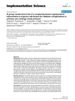
on the basis of muscle infiltration. DP (Fig. 1 a-c) is defined as dissection of the overlying peritoneum, while
DFTR (Fig. 1 d-f ) refers to resection of the diaphragm
muscle inclusive of the overlying peritoneum and pleura.
The extent of the procedure was determined by the distribution of the tumor lesions. Monopolar cautery was
applied to perform the diaphragmatic procedure. Several
key points of the specific surgical technique are mentioned here. Before diaphragm resection, the diaphragm
is gripped with several clamps to separate the diaphragm
muscle from the overlying lung tissue. After resection,
exploration of the pleural cavity with fingers is routinely
performed in order to confirm the extent of resection.
Fig. 1 Diaphragm peritonectomy (a-c) and full-thickness resection (d-f). a-b represents the status before and after peritonectomy. c shows the
sample. d presents that the tumor infiltrated into the diaphragm muscle and the nodule in the pleural cavity was pointed out by arrow. e illustrates
the diaphragm after resection and repair. f shows the diaphragm sample
Ye et al. BMC Cancer (2017) 17:317
With regard to DP, the central tendon merits special attention to avoid incidental rupture of this weak structure
as much as possible. Identification and protection of the
right hepatic vein is also significant because this vein
drains into the anterior surface of inferior vena cava at
the level where coronary ligaments reflect off the liver
capsule. Therefore, special attention should be taken to
avoid injury to these major vessels during the dissection.
It is also essential to be cautious and avoid tearing the
right hepatic vein when pushing downward on the liver.
The diaphragm defect was closed with a large-caliber
un-absorbable suture. The anesthesiologist was asked to
give the patient maximal inspiration, and the final diaphragmatic suture was tied down. After the diaphragmatic
surgery, a bubble test was performed to identity any possible defect in the diaphragm [20]. The application of
either mesh reconstruction or a prophylactic chest tube
was at the discretion of the operating surgeons. Transdiaphragmatic thoracic exploration (TDDE) was performed in some patients [21]. Specific indications for
TDDE were presented in the previous publication. The
definition of the extended procedures in addition to the
diaphragmatic surgery was in line with our previous publication [19].
Perioperative morbidities
Perioperative morbidities and mortality were defined as
any adverse events within 30 days of surgery that were
related to treatment. All perioperative complications in
the current series were graded according to the Memorial Sloan-Kettering Cancer Center (MSKCC) surgical
secondary events grading system [22, 23]. Grade 3–5
complications are those that lead to invasive reoperation,
unplanned intensive care unit (ICU) admission, chronic
disability or death [23].
Patients presenting with no physical signs or symptoms of pulmonary complications were exempted from
subsequent routine chest radiographs. The definition of
ipsilateral effusions was effusions on the same side as
the diaphragm operation. In patients with pleural effusions preoperatively, an increase in the size of the effusion (comparison of chest X-ray before and after
operation if indicated) was included as a positive finding.
The laterality and size (small, moderate or large as determined by imaging modality) of the effusions were
recorded.
Data collection and statistical analyses
In our center, preoperative work up for patients highly
suspicious for ovarian cancer involved serum tumor
marker, a comprehensive radiologic imaging (thorax/abdomen/pelvis), and gastroscopy and colonoscopy if necessary. Patient-, disease- and surgery-related information
Page 3 of 9
was extracted from the patients’ medical records. The data
collection included age at diagnosis, primary site of disease, body mass index (BMI, calculated as weight (kg)/
[height (m)]2), histological subtype, International Federation of Gynecology and Obstetrics (FIGO) stage [24], the
presence of ascites and pleural effusion at the time of disease diagnosis, and administration of neoadjuvant chemotherapy. Preoperative laboratory values, including serum
protein (i.e., total protein and albumin), and serum cancer
antigen 125 (CA-125), were also recorded. The surgeryrelated parameters were listed as follows: operation radicality, distribution of diaphragm implants, diaphragm surgery type (DP or DFTR), perforation into the pleural
cavity, mesh application during diaphragm repair, prophylactic chest tube placement, residual disease, operation
time, estimated blood loss (EBL), intra-operative transfusion, ICU stay, postoperative complications, and time
interval from surgery to chemotherapy. Preoperative
plural or peritoneal effusions were drained only if the patients had any related symptoms. In concordance with the
Gynecologic Cancer InterGroup (GCIG) consensus, optimal cytoreduction refers to no macroscopic residual disease [25].
Parametric Student’s t-tests were employed in evaluating continuous variables, while chi-square tests were
used for the categorical variables. The associations between different variables were evaluated using univariate
and multivariate logistic regression analyses, and the
hazard ratio (HR) with 95% confidence interval (CI) was
calculated. All of the P values reported were two-sided,
and a value of P < 0.05 was considered statistically significant. Statistical Package for Social Science (SPSS)
(Version 17.0, SPSS, Inc., Chicago, IL, USA) and GraphPad Prism (Version 5.0, GraphPad Software, Inc., La
Jolla, CA, USA) were used for all the analyses.
Results
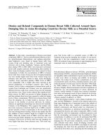
A total of 150 patients underwent diaphragmatic surgery. Figure 2 highlights the increasing application of
diaphragmatic surgery at our institution over the past 6
years. The patient characteristics of the entire cohort are
shown in Table 1. The median age was 55 years (range,
25–77 years). The majority of the patients had ovarian
cancer (96%), FIGO stage IIIC tumor (76%) and serous
histology (89.3%). Neoadjuvant chemotherapy was administered in 14 (9.3%) patients. Ascites was present in
94% of the patients, and the median volume was
2000 mL (range, 20–7300 mL). Before surgery, it was
noted that 47 (31.3%) patients had pleural effusions,
which were distributed as right-sided (9, 6.0%), left-sided
(9, 6.0%), and bilateral (29, 19.3%). Among these patients, seven symptomatic patients (4.7%) underwent
preoperative pleural drainage.
Ye et al. BMC Cancer (2017) 17:317
Page 4 of 9
Fig. 2 Trends in utilizing diaphragmatic procedures for advanced
ovarian, tube, and peritoneal carcinoma at the Fudan University
Shanghai Cancer Center. Abbreviations: DP = diaphragm peritonectomy;
DFTR = diaphragm full-thickness resection; DFTR% = percentage of
DFTR in the total population
Table 1 Patient baseline characteristics of the entire cohort
Variables
Median age (range), years
55 (25–77)
Median body mass index (range), kg/m2
22.7 (15.0–32.9)
Site of disease
Ovary (%)
144 (96%)
Fallopian tube/peritoneum (%)
6 (4%)
Neo-adjuvant chemotherapy (%)
14 (9.3%)
Preoperative laboratory values
Median CA-125 (range), U/mL
1166 (57–5502)
Median total protein (range), g/dL
7.1 (4.1–9.6)
Median albumin (range), g/dL
3.9 (2.4–8.2)
Tumor stage
IIIC (%)
114 (76%)
IV (%)
36 (24%)
Histology
Serous (%)
134 (89.3%)
Non-serous (%)
16 (10.7%)
Presence of pleural effusion before surgery (%)
47 (31.3%)
Right
9 (6.0%)
Left
9 (6.0%)
Bilateral
29 (19.3%)
Preoperative pleural drainage (%)
7 (4.7%)
Presence of ascites at surgery (%)
141 (94%)
Median ascites volume (range), mL
2000 (20–7300)
Type of diaphragm surgery
Diaphragm peritonectomy (%)
124 (82.7%)
Diaphragm full-thickness resection (%)
26 (17.3%)
Abbreviations: CA-125 cancer antigen 125
DP and DFTR were performed in 124 (82.7%) and 26
(17.3%) patients, respectively. Table 2 lists the specific
surgical procedures and outcomes based on the diaphragmatic surgery stratification. The diaphragm lesions
were predominantly right-sided (63.3%) followed by
bilateral (36.0%). We did notice one case with only left
hemidiaphragm involvement. The diaphragm was
opened during 65 (43.4%) procedures, while TDDE was
performed in 34 (22.7%) patients. Of these 34 patients,
suspicious pleural lesions were noted in 21 (61.8%) patients; therefore, a biopsy was collected. Mesh was utilized in four patients (2.7%) when closing the diaphragm
opening after DFTR. Intraoperative chest tube placement was conducted in eight (5.3%) patients: five (4.0%)
in the DP group and three (11.5%) in the DFTR group
(P = 0.285). Extended procedures in addition to the diaphragmatic surgery were performed in 77 (51.3%) patients, resulting in a total of 142 procedures. The
specific details of these procedures are shown in Table 2.
The debulking results were 53 (35.3%) patients with no
gross residual disease, 73 (48.7%) with gross residual disease ≤1 cm, and 24 (16.0%) with gross residual disease
>1 cm. The two patient groups (DP vs. DFTR) had no
difference with regard to the extended procedures and
cytoreduction outcomes.
The median operation time was 180 min (range, 60–
330 min), while the median blood loss was 900 mL
(range, 100–5300 mL). Intraoperatively, 88% of the patients received a transfusion, and the median volume
transfused was 4.0 units (range, 1–15 units). In all, 46
(30.7%) patients had a planned transient postoperative
ICU stay. For the entire cohort, the median time from
surgery to chemotherapy was 14 days (range, 6–40 days).
No significant difference was observed between the DP
and DFTR groups with regard to these characteristics
(P = 0.272).
Table 3 is a comprehensive review of the postoperative
complications. Pleural effusions and pneumothorax occurred in 50 (33.3%) and 11 (7.3%) patients, respectively.
Ten of the 11 patients had concurrent effusions, while
only one patient developed an exclusive pneumothorax. In
other words, a total of 51 patients developed postoperative
pleural effusions and/or pneumothorax. Pneumonia was
the main concurrent finding based on the postoperative
imaging. Neither diaphragmatic hernia nor hydrothorax
was observed in either group. Regarding the MSKCC
grading system, there were 82 mild (Grade 1–2) and 27
severe (Grade 3–5) adverse events in the entire cohort.
The specific details of the severe complications are listed
as follows: symptomatic pleural effusion requiring drainage (21, 14.0%), symptomatic pneumothorax requiring a
thoracostomy tube (1, 0.6%), right hepatic vein rupture requiring intra-operative repair and transfusion (1, 0.6%),
bleeding requiring return to the operating room (1, 0.6%),
Ye et al. BMC Cancer (2017) 17:317
Page 5 of 9
Table 2 Surgical procedures and outcomes based on type of diaphragm surgery
Cohort
(n = 150)
DP
(n = 124)
DFTR
(n = 26)
P
0.811
Laterality of diaphragm lesions (%)
Right
95 (63.3%)
78 (62.9%)
17 (65.4%)
Left
1 (0.7%)
1 (0.8%)
0
Bilateral
54 (36.0%)
45 (36.3%)
9 (34.6%)
Perforation into pleural cavity (%)
65 (43.3%)
39 (31.5%)
26 (100%)
<0.001
TDDE (%)
34 (22.7%)
21 (16.9%)
13 (50.0%)
<0.001
Pleural nodule biopsy performed (%)
21 (14.0%)
15 (12.1%)
6 (23.1%)
0.248
Prophylactic chest tube placement (%)
8 (5.3%)
5 (4.0%)
3 (11.5%)
0.285
Mesh utilization in diaphragm repair (%)
4 (2.7%)
0
4 (15.4%)
<0.001
5 (3.3%)
5 (4.0%)
0
0.660
Extended procedures performed (%)
Small bowel resection
Large bowel resection
48 (32.0%)
37 (29.8%)
11 (42.3%)
0.215
Splenectomy
18 (12.0%)
14 (11.3%)
4 (15.4%)
0.801
Partial pancreatectomy
5 (3.3%)
4 (3.2%)
1 (3.8%)
1.000
Partial hepatic resection
4 (2.7%)
4 (3.2%)
0
0.796
Resection of porta hepatis tumor
4 (2.7%)
3 (2.4%)
1 (3.8%)
1.000
Cholecystectomy
1 (0.7%)
1 (0.8%)
0
1.000
Stomia
15 (10.0%)
12 (9.7%)
3 (11.5%)
1.000
Resection of the tumor on the liver surface
23 (15.3%)
20 (16.1%)
3 (11.5%)
0.771
Resection of the tumor on the stomach surface
13 (8.7%)
10 8.1(%)
3 (11.5%)
0.850
Resection of the tumor in the gallbladder fossa
6 (4.0%)
6 (4.8%)
0
0.552
Complete cytoreduction
53 (35.3%)
42 (33.9%)
11 (42.3%)
0.649
Residual disease ≤1 cm
73 (48.7%)
61 (49.2%)
12 (46.2%)
180 (60–330)
180 (60–330)
195 (90–270)
Residual disease (%)
Median operation time (range), minutes
0.576
Median EBL (range), mL
900 (100–5300)
900 (100–5300)
1000 (200–2200)
0.802
Intraoperative blood transfusion (%)
132 (88%)
109 (87.9%)
23 (88.5%)
0.937
Median transfusion amount (range), units
4.0 (1.0–15.0)
4.5 (1–15)
4 (2–13)
0.286
Planned intensive care unit stay (%)
46 (30.7%)
39 (30.6%)
8 (30.8%)
0.649
Median time interval from surgery to chemotherapy (range), days
14 (6–40)
14 (6–40)
14 (6–34)
0.272
Note: Values in bold are statistically significant.
Abbreviations: DP diaphragm peritonectomy, DFTR diaphragm full-thickness resection, TDDE trans-diaphragmatic thoracic exploration, EBL estimated blood loss
pancreatic leak requiring drainage (1, 0.6%), intestinal perforation requiring return to the operating room (1, 0.6%),
and wound dehiscence resulting in delayed repair (1,
0.6%). There was no mortality (MSKCC grade 5) within
30 days of surgery. Patients in the DFTR group were more
likely to have postoperative pleural effusion (69.2% vs.
25.8%, P < 0.001) and pleural drainage (38.5% vs. 8.9%,
P < 0.001). No significant difference was observed with
the other morbidities.
We further evaluated the application of postoperative
thoracentesis and thoracostomy tube placement. Pleural
effusions and pneumothorax were most commonly diagnosed on postoperative day (POD) 3 (range, 1–16 days).
It was worth mentioning that of the 51 patients with
postoperative pleural effusions and/or pneumothorax,
only 21 (41.2%) patients had pulmonary-related symptoms. A total of 22 pleural drainages were performed in
the 21 patients primarily by thoracentesis (20/22, 90.9%).
Pleural puncture was performed at a median of 3 days
(range, 2–13 days) postoperatively. Two patients required thoracostomy tube placement for pulmonary
complications, which warrants discussion. The first patient presented with diffuse lesions in the right hemidiaphragm (approximately 8 × 6 cm) that infiltrated into
the diaphragm muscle. She underwent DFTR and
pleural nodule biopsy without either a prophylactic chest
Ye et al. BMC Cancer (2017) 17:317
Page 6 of 9
Table 3 Perioperative surgical complications based on type of diaphragm surgery
Cohort
(n = 150)
DP
(n = 124)
DFTR
(n = 26)
P
All complications
Ipsilateral pleural effusion (%)
50 (33.3%)
32 (25.8%)
18 (69.2%)
<0.001
Ipsilateral pneumothorax (%)
11 (7.3%)
8 (6.5%)
3 (11.5%)
0.623
Pulmonary embolism (%)
2 (1.3%)
2 (1.6%)
0
1.000
Pneumonia (%)
23 (15.3%)
20 (16.1%)
3 (11.5%)
0.771
Right hepatic vein rupture
1 (0.6%)
0
1 (3.8%)
1.000
Sub-diaphragmatic abscess
1 (0.6%)
1 (0.8%)
0
1.000
Postoperative bleeding (%)
1 (0.6%)
1 (0.8%)
0
1.000
Bowel obstruction (%)
10 (6.7%)
9 (7.3%)
1 (3.8%)
0.825
Pancreatic leak
1 (0.6%)
1 (0.8%)
0
1.000
Intestinal perforation
1 (0.6%)
1 (0.8%)
0
1.000
Heart arrhythmia
1 (0.6%)
1 (0.8%)
0
1.000
Wound infection/dehiscence
5 (3.3%)
3 (2.4%)
2 (7.7%)
0.447
Vaginal cuff infection
1 (0.6%)
1 (0.8%)
0
1.000
Urinary tract infection
1 (0.8%)
1 (0.8%)
0
1.000
Grade 1–2 (%)
82 (54.7%)
66 (53.2%)
16 (61.5%)
0.010
Grade 3–5 (%)
27 (18.0%)
15 (12.1%)
12 (46.2%)
Symptomatic pleural effusion requiring drainage
21 (14.0%)
11 (8.9%)
10 (38.5%)
<0.001
Symptomatic pneumothorax requiring thoracostomy tube
1 (0.6%)
0
1 (3.8%)
1.000
Right hepatic vein rupture requiring intra-operative repair and transfusion
1 (0.6%)
0
1 (3.8%)
1.000
Bleeding requiring return to operating room
1 (0.6%)
1 (0.8%)
0
1.000
Pancreatic leak requiring drainage
1 (0.6%)
1 (0.8%)
0
1.000
Intestinal perforation requiring return to the operating room
1 (0.6%)
1 (0.8%)
0
1.000
Wound dehiscence requiring delayed repair
1 (0.6%)
1 (0.8%)
0
1.000
MSKCC grading
Grade 3–5 complicationsa
Abbreviations: DP diaphragm peritonectomy, DFTR diaphragm full-thickness resection, MSKCC Memorial Sloan Kettering Cancer Center
Note:
1. Percentages are not additive as multiple procedures might be performed on the same patient.
2. Values in bold are statistically significant.
a
Severe complications leading to invasive radiologic intervention/re-operation/unplanned ICU admission (grade 3), chronic disability (grade 4), or death (grade 5).
tube or mesh reconstruction. On POD 1, the bedside
chest film revealed a right-side pneumothorax with the
lung compression of nearly 30%. The patient received a
thoracic surgical consultation, and a thoracostomy tube
was placed on the same day. On POD 2, bilateral pleural
effusions were noted in the bedside ultrasonography,
and thoracentesis was performed. The second patient received right-sided DP for multiple small nodules without
either a pleural opening or chest tube placement. On
POD 4, the patient experienced sudden onset chest distress and dyspnea. Pulse oxygen saturation was approximately 97% on 3 L/min of nasal cannula oxygenation.
Computer tomography pulmonary angiography revealed
a large amount of ipsilateral hydrothorax instead of pulmonary embolism. A thoracostomy tube was inserted
into the sixth intercostal space.
Table 4 illustrates the possible risk factors for pleural
effusion and drainage after diaphragmatic surgery. After
multivariate analysis, stage IV disease (HR, 17.2; 95% CI:
4.5–66.7; P < 0.001), DFTR (HR, 4.9; 95% CI: 1.2–19.9;
P = 0.028) and a long operating time (HR, 15.4; 95% CI:
4.3–55.5; P < 0.001) retained their statistical significance.
In contrast, DFTR (HR, 5.9; 95% CI: 1.5–23.6; P = 0.011)
and TDDE (HR, 28.3: 95% CI: 4.9–160.8; P < 0.001) were
found to be predictive factors for pleural drainage.
Discussion
In the current series, we analyzed the results of patients
with stage IIIC–IV ovarian carcinoma who underwent
diaphragmatic procedures for primary cytoreduction. To
the best of our knowledge, the present study is one of
Ye et al. BMC Cancer (2017) 17:317
Page 7 of 9
Table 4 Univariate and multivariate analysis of factors predictive of postoperative ipsilateral plural effusion and drainage after
diaphragm surgery
Univariate
Parameters
Pleural effusions
Pleural drainage
P value
P value
Age (>55 years old)
0.908
0.080
Tumor stage (IV vs. IIIC)
<0.001
0.009
Preoperative pleural effusion
0.215
0.831
Ascites >2000 mL
0.558
0.878
CA-125 > 1166 U/mL
0.729
0.482
Albumin <3.9 g/dL
0.568
0.815
Diaphragmatic surgery (DFTR vs. DP)
<0.001
<0.001
Perforation into pleural cavity
0.996
0.996
TDDE
0997
<0.001
Pleural nodule biopsy
0.998
0.995
Operating time > 180 min
<0.001
0.015
Estimated blood loss >900 mL
0.249
0.442
Intraoperative blood transfusion >4.0 units
0.016
0.719
Pleural effusions
Pleural drainage
Multivariate
Parameters
Tumor stage (IV vs. IIIC)
HR (95% CI)
P value
HR (95% CI)
P value
17.2 (4.5–66.7)
<0.001
4.5 (0.8–25.7)
0.086
Diaphragmatic surgery (DFTR vs. DP)
4.9 (1.2–19.9)
0.028
5.9 (1.5–23.6)
0.011
TDDE
/
/
28.3 (4.9–160.8)
<0.001
Operating time > 180 min
15.4 (4.3–55.5)
<0.001
2.2 (0.6–8.6)
0.242
Intraoperative blood transfusion >4.0 units
0.7 (0.2–2.3)
0.581
/
/
Note: Values in bold are statistically significant.
Abbreviations: CA-125 cancer antigen 125, DP diaphragm peritonectomy, DFTR diaphragm full-thickness resection, TDDE trans-diaphragmatic thoracic exploration,
HR hazard ratio, CI confidence interval.
the largest series and the first study from a Chinese academic center [7, 26].
Adequate exposure of the diaphragm is the very first
and critical step in not only assessing tumor resectability
but also performing the procedure. Involvement of the
right hemidiaphragm was extraordinarily common in
previous publications [12, 13, 27] as well as in our study.
Based on our clinical observation (although without supporting data available), bulky tumors are frequently identified in the area where the diaphragmatic peritoneum is
reflected to the capsule of the posterior region of the
right liver lobe. Given that large-volume disease on the
right hemidiaphragm is obscured by the right liver, liver
mobilization and medial retraction of the liver allow exposure of the diaphragmatic lesion. Dr. Chi from
MSKCC mentioned that tumor implantation on the left
diaphragm could be more easily resected without fully
mobilizing the liver, although in some cases, splenectomy might be necessary [20]. In our experience, adequate mobilization and extensive knowledge of the
upper abdominal anatomy are fundamental to a successful diaphragm operation.
Concerning the type of diaphragmatic surgery, we did
not include ablation and coagulation procedures in this
study. The two techniques (DP and DFTR) described are
not comparable given that surgeons do not have a choice
in which technique to perform. In our cohort, more patients underwent diaphragm stripping (82.7% vs. 17.3%
for DP and DFTR, respectively), which might be explained that ovarian cancer tended to be superficially
spread within the peritoneal surface [7]. Patients in the
two groups were similar in terms of surgical procedures
and outcomes.
The most commonly encountered adverse events were
new or increased pleural effusions. The rate of intraoperative chest tube placement was 5.3%, and postoperative
pleural drainage accounted for 14.6% in the entire population. Thus, we do not feel that this rate is high enough
to routinely place a prophylactic chest tube in all of the
patients during the operation. However, when it comes
Ye et al. BMC Cancer (2017) 17:317
to DFTR alone, half the patients underwent drainage
during the operation for prophylactic purpose (11.5%)
and after the operation for relieving the symptom
(38.5%). Therefore, intraoperative prophylactic tube
placement should be considered for the subgroup patients. Among the 292 included patients (197 DP and 75
DFTR) from a recent systematic review, the estimated
pleural effusion rates after DP and DFTR were 43% and
53%, respectively, while the need for pleural punctures
or chest tube placement after DP and DFTR was 4% and
9%, respectively [7]. In the MSKCC study, the rate of ipsilateral effusions was 58%, and the overall rate of either
postoperative thoracentesis or chest tube placement was
15% [10]. Researchers have investigated possible predictive factors for postoperative effusions in different populations [10, 13, 16, 17, 28]. The following parameters
retained significance upon multivariate analysis: liver
mobilization [10, 13], entry into the pleural space during
DP [28], and the size of the diaphragmatic resection [13,
16]. Given that we routinely divide the hepatic ligaments
to mobilize the liver, we were unable to test this association in our own series. Based on our data, stage IV disease, DFTR and a long operating time (>180 min)
correlated with postoperative pleural effusions with statistical significance. Researchers from Italy also noticed
that patients who underwent DFTR were more likely to
have postoperative pleural effusions compared to patients who underwent DP [29]. The duration of surgery
was recognized as a common risk factor for postoperative complications [10]. It has been proposed that complications due to diaphragmatic surgery result from the
transfer of ascites to the pleural cavity rather than as primary thoracic processes [10, 15]. Diaphragm defects, the
presence of ascites, extended exposure of the diaphragmatic bare area after liver mobilization and postoperative release of either VEGF or inflammatory mediators
were suggested was possible mechanisms for pleural effusion [10, 15].
Despite the high reported rate of pneumothorax,
symptomatic pneumothorax requiring intervention was
quite low in our series. One of the reasons for this finding might be that residual pneumothorax was not evacuated by a catheter prior to diaphragmatic closure. Since
2015, we have included an intentional evacuation of the
pneumothorax by suctioning as part of the surgical
procedure.
When interpreting the results of this study, several potential limitations must be addressed. Firstly, this study
has inherent bias pertaining to its retrospective design.
Secondly, survival information was not evaluated in the
present series. The short follow-up period and the number of patients who underwent multiple radical procedures were two reasons that we did not attempt to
assess the survival outcome. Thirdly, the multivariate
Page 8 of 9
model assessing the predictive factors for postoperative
drainage might overfit with factors given the few number
of drainage events. Last but not least, the subject data
were collected from a tertiary referral center, and although the cohort contained a relatively large sample
size, the results might not be generalizable to all of the
patients in China. For now, the application of upper abdominal procedures (including diaphragm surgery) for
patients with ovarian cancer in China remains unclear
because there are few publications focused on this issue.
The importance of not only surgical skills and experience but also the high-quality surgical care delivered by
a multidisciplinary team has been increasingly emphasized [16]. Referrals should be considered at institutions
where the necessary treatments are unavailable.
Conclusions
The performance of DP and DFTR as part of the extensive
upper abdominal operation resulted in an acceptable morbidity rate. Pleural effusion, pneumonia and pneumothorax were the most common morbidities, and the rate
of pleural drainage was not high enough to justify prophylactic chest tube placement for all the patients. However,
for patients who received DFTR, intraoperative prophylactic drainage should be considered.
Abbreviations
BMI: Body mass index; CA-125: Cancer antigen 125; CI: Confidence interval;
DFTR: Diaphragm full-thickness resection; DP: Diaphragm peritonectomy;
EBL: Estimated blood loss; FIGO: International Federation of Gynecology and
Obstetrics; HR: Hazard ratio; ICU: Intensive care unit; MSKCC: Memorial
Sloan-Kettering Cancer Center; TDDE: Trans-diaphragmatic thoracic exploration
Acknowledgements
Not Applicable.
This work was presented as oral-E poster at the 16th Biennial Meeting of the
International Gynecologic Cancer Society, Lisbon, Portugal, 30 October 2016.
Funding
No specific funding was received for this study.
Availability of data and material
The dataset supporting the conclusions of this article is available upon
request. Please contact Prof. Huijuan Yang ().
Authors’ contributions
Conception and design: SY, TH, SL, XC, XW, HY, LX; Collection and assembly
of data: SY, TH, SL, LX; Data analysis and interpretation: SY, XC, XW, HY, LX;
Manuscript writing: SY, XC, XW, HY, LX; Final approval of manuscript: SY, TH,
SL, XC, XW, HY, LX. All authors have read and approved the final version of
this manuscript.
Competing interests
The authors declare that they have no competing interests.
Consent for publication
Not applicable.
Ethics approval and consent to participate
This study was approved by the institutional review board at Fudan University
Shanghai Cancer Center. All the included patients provided their written
informed consent.
Ye et al. BMC Cancer (2017) 17:317
Page 9 of 9
Publisher’s Note
Springer Nature remains neutral with regard to jurisdictional claims in
published maps and institutional affiliations.
Author details
1
Department of Gynecologic Oncology, Fudan University Shanghai Cancer
Center, Shanghai 200032, China. 2Department of Oncology, Shanghai
Medical College, Fudan University, Shanghai 200032, China.
19.
20.
21.
Received: 30 January 2017 Accepted: 1 May 2017
22.
References
1. Jemal A, Bray F, Center MM, Ferlay J, Ward E, Forman D. Global cancer
statistics. CA Cancer J Clin. 2011;61:69–90.
2. Chen W, Zheng R, Baade PD, Zhang S, Zeng H, Bray F, et al. Cancer statistics
in China, 2015. CA Cancer J Clin. 2016;66:115–32.
3. Bristow RE, Tomacruz RS, Armstrong DK, Trimble EL, Montz FJ. Survival
effect of maximal cytoreductive surgery for advanced ovarian carcinoma
during the platinum era: a meta-analysis. J Clin Oncol. 2002;20:1248–59.
4. Chi DS, Eisenhauer EL, Lang J, Huh J, Haddad L, Abu-Rustum NR, et al. What
is the optimal goal of primary cytoreductive surgery for bulky stage IIIC
epithelial ovarian carcinoma (EOC)? Gynecol Oncol. 2006;103:559–64.
5. Barlin JN, Long KC, Tanner EJ, Gardner GJ, Leitao MM Jr, Levine DA, et al.
Optimal (warranted? Gynecol Oncol. 2013;130:284–8.
6. Benedetti Panici P, Di Donato V, Fischetti M, Casorelli A, Perniola G, Musella
A, et al. Predictors of postoperative morbidity after cytoreduction for
advanced ovarian cancer: Analysis and management of complications in
upper abdominal surgery. Gynecol Oncol. 2015;137:406–11.
7. Bogani G, Ditto A, Martinelli F, Lorusso D, Chiappa V, Donfrancesco C, et al.
Surgical Techniques for Diaphragmatic Resection During Cytoreduction in
Advanced or Recurrent Ovarian Carcinoma: A Systematic Review and Metaanalysis. Int J Gynecol Cancer. 2015;00:00–00.
8. Cliby W, Dowdy S, Feitoza SS, Gostout BS, Podratz KC. Diaphragm resection
for ovarian cancer: technique and short-term complications. Gynecol Oncol.
2004;94:655–60.
9. Aletti GD, Dowdy SC, Podratz KC, Cliby WA. Surgical treatment of
diaphragm disease correlates with improved survival in optimally debulked
advanced stage ovarian cancer. Gynecol Oncol. 2006;100:283–7.
10. Eisenhauer EL, D'Angelica MI, Abu-Rustum NR, Sonoda Y, Jarnagin WR,
Barakat RR, et al. Incidence and management of pleural effusions after
diaphragm peritonectomy or resection for advanced mullerian cancer.
Gynecol Oncol. 2006;103:871–7.
11. Einenkel J, Ott R, Handzel R, Braumann UD, Horn LC. Characteristics and
management of diaphragm involvement in patients with primary
advanced-stage ovarian, fallopian tube, or peritoneal cancer. Int J Gynecol
Cancer. 2009;19:1288–97.
12. Bashir S, Gerardi MA, Giuntoli RL 2nd, Montes TP, Bristow RE. Surgical
technique of diaphragm full-thickness resection and trans-diaphragmatic
decompression of pneumothorax during cytoreductive surgery for ovarian
cancer. Gynecol Oncol. 2010;119:255–8.
13. Fanfani F, Fagotti A, Gallotta V, Ercoli A, Pacelli F, Costantini B, et al. Upper
abdominal surgery in advanced and recurrent ovarian cancer: role of
diaphragmatic surgery. Gynecol Oncol. 2010;116:497–501.
14. Gouy S, Chereau E, Custodio AS, Uzan C, Pautier P, Haie-Meder C, et al.
Surgical procedures and morbidities of diaphragmatic surgery in patients
undergoing initial or interval debulking surgery for advanced-stage ovarian
cancer. J Am Coll Surg. 2010;210:509–14.
15. Tsolakidis D, Amant F, Van Gorp T, Leunen K, Neven P, Vergote I.
Diaphragmatic surgery during primary debulking in 89 patients with stage
IIIB-IV epithelial ovarian cancer. Gynecol Oncol. 2010;116:489–96.
16. Chereau E, Rouzier R, Gouy S, Ferron G, Narducci F, Bergzoll C, et al.
Morbidity of diaphragmatic surgery for advanced ovarian cancer:
retrospective study of 148 cases. Eur J Surg Oncol. 2011;37:175–80.
17. Kato K, Tate S, Nishikimi K, Shozu M. Assessment of intraoperative tube
thoracostomy after diaphragmatic resection as part of debulking surgery for
primary advanced-stage Mullerian cancer. Gynecol Oncol. 2013;131:32–5.
18. Soleymani Majd H, Ferrari F, Manek S, Gubbala K, Campanile RG, Hardern K,
et al. Diaphragmatic peritonectomy vs. full thickness resection with
pleurectomy during Visceral-Peritoneal Debulking (VPD) in 100 consecutive
23.
24.
25.
26.
27.
28.
29.
patients with stage IIIC-IV ovarian cancer: A surgical-histological analysis.
Gynecol Oncol. 2016;140:430–5.
Ren Y, Jiang R, Yin S, You C, Liu D, Cheng X, et al. Radical surgery versus
standard surgery for primary cytoreduction of bulky stage IIIC and IV
ovarian cancer: an observational study. BMC Cancer. 2015;15:583.
Kehoe SM, Eisenhauer EL, Chi DS. Upper abdominal surgical procedures:
liver mobilization and diaphragm peritonectomy/resection, splenectomy,
and distal pancreatectomy. Gynecol Oncol. 2008;111:S51–5.
Yin S, Jiang R, Wang P, Zang R. Role of Transdiaphragmatic Thoracic
Exploration in Bulky Stage IIIC Ovarian Cancer Patients Who Underwent
Diaphragmatic Surgery. Int J Gynecol Cancer. 2015;25:1392–7.
Martin RC 2nd, Brennan MF, Jaques DP. Quality of complication reporting in
the surgical literature. Ann Surg. 2002;235:803–13.
Chi DS, Franklin CC, Levine DA, Akselrod F, Sabbatini P, Jarnagin WR, et al.
Improved optimal cytoreduction rates for stages IIIC and IV epithelial
ovarian, fallopian tube, and primary peritoneal cancer: a change in surgical
approach. Gynecol Oncol. 2004;94:650–4.
FIGO Committee on Gynecologic Oncology. FIGO staging for carcinoma of
the vulva, cervix, and corpus uteri. Int J Gynaecol Obstet. 2014;125:97–8.
Stuart GC, Kitchener H, Bacon M, duBois A, Friedlander M, Ledermann J, et
al. 2010 Gynecologic Cancer InterGroup (GCIG) consensus statement on
clinical trials in ovarian cancer: report from the Fourth Ovarian Cancer
Consensus Conference. Int J Gynecol Cancer. 2011;21:750–5.
Papadia A, Morotti M. Diaphragmatic surgery during cytoreduction for
primary or recurrent epithelial ovarian cancer: a review of the literature.
Arch Gynecol Obstet. 2013;287:733–41.
Eisenhauer EL, Abu-Rustum NR, Sonoda Y, Levine DA, Poynor EA,
Aghajanian C, et al. The addition of extensive upper abdominal surgery to
achieve optimal cytoreduction improves survival in patients with stages IIICIV epithelial ovarian cancer. Gynecol Oncol. 2006;103:1083–90.
Dowdy SC, Loewen RT, Aletti G, Feitoza SS, Cliby W. Assessment of
outcomes and morbidity following diaphragmatic peritonectomy for
women with ovarian carcinoma. Gynecol Oncol. 2008;109:303–7.
Zapardiel I, Peiretti M, Zanagnolo V, Biffi R, Bocciolone L, Landoni F, et al.
Diaphragmatic surgery during primary cytoreduction for advanced ovarian
cancer: peritoneal stripping versus diaphragmatic resection. Int J Gynecol
Cancer. 2011;21:1698–703.
Submit your next manuscript to BioMed Central
and we will help you at every step:
• We accept pre-submission inquiries
• Our selector tool helps you to find the most relevant journal
• We provide round the clock customer support
• Convenient online submission
• Thorough peer review
• Inclusion in PubMed and all major indexing services
• Maximum visibility for your research
Submit your manuscript at
www.biomedcentral.com/submit