Functional analysis of fatty acid binding protein 7 and its effect on fatty acid of renal cell carcinoma cell lines
Bạn đang xem bản rút gọn của tài liệu. Xem và tải ngay bản đầy đủ của tài liệu tại đây (1.24 MB, 8 trang )
Takaoka et al. BMC Cancer (2017) 17:192
DOI 10.1186/s12885-017-3184-x
RESEARCH ARTICLE
Open Access
Functional analysis of fatty acid binding
protein 7 and its effect on fatty acid of
renal cell carcinoma cell lines
Naohisa Takaoka1*, Tatsuya Takayama2 and Seiichiro Ozono1
Abstract
Background: Renal cell carcinomas (RCCs) overexpress fatty acid binding protein 7 (FABP7). We chose to study the
TUHR14TKB cell line, because it expresses higher levels of FABP7 than other cell lines derived from renal carcinomas
(OS-RC-2, 786-O, 769-P, Caki-1, and ACHN).
Methods: FABP7 expression was detected using western blotting and real-time PCR. Cell proliferation was determined
using an MTS assay and by directly by counting cells. The cell cycle was assayed using flow cytometry. Cell migration
was assayed using wound-healing assays. An FABP7 expression vector was used to transfect RCC cell lines.
Results: The levels of FABP7 expressed by TUHR14TKB cells and their doubling times decreased during passage.
High-passage TUHR14TKB cells comprised fewer G0/G1-phase and more S-phase cells than low-passage cells. Cell
proliferation differed among subclones isolated from cultures of low-passage TUHR14TKB cells. The proliferation of
TUHR14TKB cells decreased when FABP7 was overexpressed, and the cell migration property of TUHR14TKB cells were
decreased when FABP7 was overexpressed. High concentrations of docosatetraenoic acid and eicosapentaenoic acid
accumulated in TUHR14TKB cells that overexpressed FABP7, and docosatetraenoic acid enhanced cell proliferation.
Conclusions: The TUHR14TKB cell line represents a heterogeneous population that does not express FABP7 when it
rapidly proliferates. The differences in FABP7 function between RCC cell lines suggests that FABP7 affects cell
proliferation depending on cell phenotype.
Keywords: FABP7, Renal cell carcinoma, Subculture, Cell proliferation, Cell migration, Docosatetraenoic acid
Background
Kidney cancer is the 15th most common malignancy
worldwide. In 2008, approximately 271,000 new cases
were diagnosed, and 116,000 patients died from this
disease [1]. These rates are approximately twice as high in
men as in women [1]. Renal cell carcinomas (RCCs) represent 91.6% of kidney cancers [2]. The identification of
molecular markers in body fluids, which can be used for
screening, diagnosis, follow-up, and monitoring drugbased therapy of patients with RCC, is one of the most
important challenges of cancer research [3]. In a search
for candidate markers of RCC, we identified the gene
(FABP7) encoding fatty acid binding protein 7 [4].
* Correspondence:
1
Department of Urology, Hamamatsu University School of Medicine, 1-20-1
Handayama, Higashi-ku, Hamamatsu, Shizuoka 431-3192, Japan
Full list of author information is available at the end of the article
Human FABP7 was first isolated from a library of fetal
brain complementary DNA (cDNA), and the FABP7 transcript is expressed specifically in adult human brain and
skeletal muscle [5]. Further, FABP7 is expressed more
abundantly during the early stages of maturation of the
brain [5]. RCCs overexpress FABP7 [4, 6–14], and FABP7
transcripts are present in the tumors or urine of patients
with RCC [9]. The role of FABP7 in inhibiting the proliferation of a breast cancer cell line suggests that it may act as a
tumor suppressor [15, 16]. In apparent contradiction to
this, inhibition of FABP7 expression by small interfering
RNAs (siRNAs) significantly reduces the proliferation of
certain human cancer cell lines [17–21], and overexpression of FABP7 stimulates the proliferation of RCC
cell lines [14]. Further, inhibition of FABP7 expression by
siRNAs significantly decreases the ability of certain human
cancer cell lines to migrate [17–19, 21–23]. Moreover,
© The Author(s). 2017 Open Access This article is distributed under the terms of the Creative Commons Attribution 4.0
International License ( which permits unrestricted use, distribution, and
reproduction in any medium, provided you give appropriate credit to the original author(s) and the source, provide a link to
the Creative Commons license, and indicate if changes were made. The Creative Commons Public Domain Dedication waiver
( applies to the data made available in this article, unless otherwise stated.
Takaoka et al. BMC Cancer (2017) 17:192
FABP7 enhances the migration of glioma cells [24], and
an antibody against FABP7 inhibits cell migration [25].
To better understand the role of FABP7 in RCC and
to attempt to resolve the conflicting findings summarized above, the present study aimed to analyze the
effects of FABP7 on the phenotypes of RCC cell lines,
with particular focus on the composition of the fatty
acids accumulating in cell lines that overexpress FABP7.
Methods
Reagents
Reagents and their sources were as follows: RPMI 1640
medium, Oligo(dT)12–18 Primer, SuperScript® III Reverse
Transcriptase, SYBR® Green PCR Master Mix, pENTR™/
D-TOPO® vector, Gateway® pT-Rex™-DEST30 vector, pTRex/GW-30/lacZ vector, pcDNA™6/TR vector, Lipofectamine® 2000 Transfection Reagent and blasticidin S HCl
(Thermo Fisher Scientific, Waltham, MA, USA); docosatetraenoic acid, eicosapentaenoic acid (EPA) (NU-CHEK
PREP, Inc.; Elysian, MN, USA); oligopeptides (Hokkaido
System Science, Sapporo, Hokkaido, Japan); Tris, dithiothreitol, sodium orthovanadate, phenylmethanesulfonyl
fluoride, and doxycycline hyclate (Sigma-Aldrich, St.
Louis, MO, USA); sodium chloride (Nacalai Tesque,
Kyoto, Japan); EDTA, sodium deoxycholate, sodium fluoride, sodium dodecyl sulfate (SDS), 4% paraformaldehyde
and crystal violet (Wako, Osaka, Japan); IGEPAL CA-630
(MP Biomedicals, Santa Ana, CA, USA); protease inhibitor cocktail tablet (Complete, Mini, EDTA-free), geneticin
(G418) (Roche Diagnostics GmbH, Mannheim, Germany);
and SacI, XhoI (Takara Bio Inc., Otsu, Shiga, Japan).
Cell culture
The 786-O cell line (CRL-1932) was purchased from the
American Type Culture Collection (Manassas, VA, USA).
The TUHR14TKB cell line (RCB1383) was provided by
RIKEN (Tsukuba, Ibaraki, Japan). Short tandem-repeat
typing was performed to confirm the identity of highpassage TUHR14TKB cells, and the data were verified
using the RIKEN short tandem-repeat database [26]. All
cell lines were grown in RPMI 1640 medium supplemented with 10% (v/v) or 1% fetal bovine serum (FBS)
(Nichirei Biosciences Inc., Tokyo, Japan). Cells were
cultured at 37 °C in a humidified atmosphere containing
5% CO2. Docosatetraenoic acid or EPA (100 mM each)
was dissolved in ethanol, and a 1:2000 dilution of each
fatty acid was added to the culture medium.
Cell cloning
Clones were isolated from low-passage cultures of
TUHR14TKB cells by plating the cells at limiting dilution in 96-well plates. The cells were serially diluted to
128 to 4 viable cells/mL, and 50 μL was added per each
Page 2 of 8
well of a 96-well plate. After incubation at 37 °C in a humidified atmosphere containing 5% CO2, single colonies
in the wells were expanded.
Real-time PCR analysis
Real-time PCR assays were performed using a modified version of the method described by Takaoka et
al. [27]. Cells were cultured in 10-cm dishes. Total
RNA was isolated from cultured cell lines using the
RNeasy Mini Kit (QIAGEN, Hilden, Germany) according
to the manufacturer’s instructions. Two micrograms of
RNA was reverse transcribed using SuperScript® III Reverse Transcriptase primed by 500 ng of Oligo(dT)12–18
Primer according to the manufacturer’s protocol. Realtime PCR analysis of FABP7 expression was performed
using an Applied Biosystems StepOnePlus (Thermo Fisher
Scientific). The final PCR reaction mix (20 μL) included
2 μL of each specific primer (5 μM), 1 μL of first-strand
cDNA, and 10 μL of SYBR® Green PCR Master Mix. Plasmids that encode FABP7 and TATA box binding protein
(TBP) were synthesized as described previously [27], and
standard curves for each gene were generated using seven
serial dilutions of plasmid templates (0.1 nM to 0.1 fM).
TBP was used as an internal control. Takaoka et al. [27]
and Jung et al. [28] reported the sequences of the primers
used to amplify FABP7 and TBP, respectively.
Western blotting
Western blotting was performed using a modified version of a published method [27]. Cells were cultured in
6-well culture plates or in 10-cm culture dishes. The
cells were detached using trypsin-EDTA, collected by
centrifugation, and washed once with phosphatebuffered saline (PBS). The pellets were lysed on ice for
30 min in RIPA buffer (50 mM Tris, pH 8.0, 150 mM
sodium chloride, 5 mM EDTA, 0.5% sodium deoxycholate, 1% IGEPAL CA-630, and 0.1% SDS) containing
2 mg/L sodium orthovanadate, 10 mM sodium fluoride,
1 mM phenylmethanesulfonyl fluoride, 2 mM dithiothreitol, and a protease inhibitor cocktail tablet. Lysates
were centrifuged for 10 min at 4 °C at 18,000×g. The supernatants were transferred to sterile microcentrifuge
tubes. Protein concentrations were determined using the
Bio-Rad Protein Assay Kit II (Bio-Rad, Hercules, CA,
USA). Cell extracts (20 μg) were electrophoresed through
an 18% (w/v) polyacrylamide-SDS gel. The proteins were
transferred electrophoretically onto a PVDF membrane
(GE Healthcare UK Ltd., Little Chalfont, Buckinghamshire
HP7 9NA, England), and the membrane was incubated
with 1 g/L of an FABP7 antibody (AF3166; R&D Systems,
Minneapolis, MN, USA) diluted 1:5000. Antibody-antigen
complexes were visualized using peroxidase-conjugated
anti-goat IgG (86,285; Jackson ImmunoResearch Laboratories, West Grove, PA) and Immobilon Western HRP
Takaoka et al. BMC Cancer (2017) 17:192
Substrate (Millipore, Billerica, MA, USA). A mouse monoclonal anti-α-tubulin antibody (T6074; Sigma-Aldrich, St.
Louis, MO, USA) served as an internal control.
Flow cytometry
Cells were plated in 10-cm culture dishes at a density of
2 × 10⁶ cells per plate and incubated for two days at 37 °C
in an atmosphere containing 5% CO2. After incubation,
the cells were harvested with trypsin/EDTA, washed once
with PBS, and then resuspended to 1 × 10⁶ cells/0.2 mL in
PBS containing 0.25% Triton X-100 for 5 min at room
temperature. Cellular DNA in each cell suspension was
stained using 0.6 mL of 50 mg/L propidium iodide for
10 min at room temperature. Cell-cycle analysis was performed using an EPICS-XL flow cytometer (BeckmanCoulter, Brea, CA, USA).
Vector construction
To generate FABP7 expression constructs, the FABP7
cDNA sequence was amplified using PCR with the
primers Full B-FABP F2 (5′-CACCATGGTGGAGGCT
TTCTGT) and Full B-FABP R3 (5′-TTATGCCTTCT
CATAGTGGCG). The PCR product was inserted into
pENTR™/D-TOPO® via TOPO cloning (Invitrogen, CA,
USA). The cloning vector (pENTR-FABP7) was transferred to the Gateway® pT-Rex™-DEST30 vector via gateway recombination (Invitrogen, CA, USA). The plasmid
generated (DEST30-FABP7) was verified by direct DNA
sequencing. The pT-Rex/GW-30/lacZ vector that expresses β-galactosidase (lacZ) served as a control.
Transfection
TUHR14TKB and 786-O cells were transfected with
2 μg of XhoI-digested pcDNA6/TR using FuGene® HD
transfection reagent (Promega, Madison, WI, USA). The
transfectants were cultured in RPMI 1640 medium containing 10% FBS and 5 mg/L blasticidin S HCl. The
pcDNA6™/TR transfectants (TUHR-TR, TUHR14TKB
pcDNA6™/TR transfectant; 786-O TR, 786-O pcDNA6™/
TR transfectant) were expanded and transfected in the
presence of Lipofectamine® 2000 transfection reagent
with 4 μg of DEST30-FABP7 or the empty vector pTRex/GW-30/lacZ digested with SacI. The transfectants
were cultured in RPMI 1640 medium containing 10%
FBS, 5 mg/L blasticidin S HCl, and 0.3 g/L G418, and
blasticidin- and G418-resistant cells were expanded. The
FABP7 or control-vector transfectants of TUHR-TR or
786-O TR were cultured in RPMI 1640 medium containing 10% FBS or 1% FBS with 5 mg/L blasticidin S HCl,
0.3 g/L G418, and 1 mg/L doxycycline hyclate for one to
three days and then subjected to western blotting or the
following assays: MTS, cell counting, or wound-healing.
SRL Inc. (Tokyo, Japan) performed the analyses of the
fatty acid composition of the TUHR-TR transfectants.
Page 3 of 8
Cell proliferation assay
Cells were plated in 96-well cell culture plates at 400
(786-O transfectant) or 2000 cells per well (low-passage
or high-passage TUHR14TKB or TUHR14TKB transfectants, respectively) in 100 μL of culture medium. The
plates were incubated at 37 °C in an atmosphere containing 5% CO2. The cells were analyzed using a CellTiter 96® AQueous One Solution Cell Proliferation Assay
Kit (Promega, Madison, WI, USA) according to the
manufacturer’s instructions. Absorbance (490 nm) was
measured one, two, and three days after cell plating.
Doubling times were determined from four replicate
samples per point.
Cell counts
TUHR14TKB transfectants were plated in 24-well cell
culture plates (10,000 cells per well) in 500 μL of RPMI
1640 medium containing 10% FBS with 5 mg/L blasticidin S HCl, 0.3 g/L G418, and 1 mg/L doxycycline
hyclate. The plates were incubated at 37 °C in an atmosphere containing 5% CO2. Cells on the plate were fixed
with 4% paraformaldehyde and stained with 0.1% crystal
violet one, two, and three days after plating. The numbers of cells were counted in five random fields using a
light microscope (×100).
Wound-healing assay
Cells (1 × 10⁶) were seeded in 24-well plates. After incubation overnight (786-O TR transfectant) or for one day
(TUHR-TR transfectant), an artificial wound was created
(0 h) using a 200-μL tip to introduce a gap in the confluent cell monolayer, and the culture medium was changed. Images were acquired at 0 h and 6 h (786-O TR
transfectant) or 16 h (TUHR-TR transfectant). The
wounded areas were measured before and after healing.
Data analysis
Cell proliferation and migration data were analyzed
using the Student t test. Statistical significance was defined as p < 0.05.
Results
Analyses of FABP7 expression and proliferation of
TUHR14TKB cells during passage in culture
High levels of FABP7 were detected during passages
6–8 of TUHR14TKB cells, but not during passages
16–18 (Fig. 1a). The levels of FABP7 expressed by
TUHR14TKB cells decreased by approximately fourfold between two cell passages (Fig. 1b). In contrast,
the doubling time of low-passage cells was approximately twice that of high-passage cells (Fig. 2a). The
doubling times differed among cells that were isolated
from individual colonies of low-passage TUHR14TKB
cells (Fig. 2a). Further, the percentage of S-phase cells
Takaoka et al. BMC Cancer (2017) 17:192
Page 4 of 8
Fig. 1 Expression of FABP7 during subculture of TUHR14TKB cells. The zero passage (0) was started when the cells were received from RIKEN.
TUHR14TKB cells were cultured in RPMI 1640 medium containing 10% FBS for one to two weeks, harvested when they reached confluence, and
assayed for FABP7 expression. a Western blot analysis of FABP7 expression. b Real-time PCR analysis of FABP7 expression
Fig. 2 Proliferation of TUHR14TKB cells during passage. a The doubling times of TUHR14TKB cells and its subclones were subjected to MTS assay.
Low and high passages are defined as TUHR14TKB cells 7–9 and 19–23 passages, respectively. “Subclones 1, 2, and 3” represents subclones from
low-passage TUHR14TKB cells. The assay was repeated three to five times, and the data represent the average value and standard deviation
(error bars). b The stages of the cell cycle of low- and high-passage TUHR14TKB cells and the subclones were determined using flow cytometry
Takaoka et al. BMC Cancer (2017) 17:192
Page 5 of 8
in high-passage TUHR14TKB cells increased and was accompanied by a decrease in the percentage of G0/G1phase cells compared with low-passage TUHR14TKB
cells (Fig. 2b).
Functional analysis of FABP7 in RCC cells
We transfected FABP7 low-expressing TUHR14TKB and
786-O cells with an FABP7 expression vector (Fig. 3a and b
and Additional file 1: Figure S1a and S1b). In the presence
of 10% FBS, the doubling time of TUHR14TKB cells that
overexpressed FABP7 was significantly longer than that of
cells transfected with the control vector (Fig. 4a and b).
Although TUHR14TKB cells transfected with the control
vector were able to proliferate, the cells that overexpressed
FABP7 were unable to proliferate in the presence of 1%
FBS (Additional file 2: Figure S2). Further, the percentage of
TUHR14TKB FABP7 in G2/M increased compared with
that of TUHR14TKB lacZ cells (Fig. 4c), indicating that
FABP7 induced the arrest of TUHR14TKB in G2. In contrast, overexpression of FABP7 stimulated the proliferation
Fig. 4 Effect of FABP7 on cell proliferation and cell cycle of
TUHR14TKB cells. TUHR14TKB cells were cultured in RPMI 1640
medium containing 10% FBS, 5 mg/L blasticidin S HCl, 0.3 g/L G418,
and 1 mg/L doxycycline hyclate. Assays to determine the rate of cell
proliferation: a MTS assay. The data represent the average and
standard deviation (error bars) of five experiments. b Cell counts.
The data represent the average and standard deviation (error bars)
of four experiments. c The stages of the cell cycles of TUHR14TKB
FABP7 and TUBR14TKB lacZ were determined using flow cytometry
of the 786-O cell line cultured in medium containing 1%
FBS (Additional file 1: Figure S1c).
Wound-healing assays revealed that TUHR14TKB
cells that overexpressed FABP7 migrated significantly
slower than TUHR14TKB cells transfected with the
control vector (Fig. 3c), although overexpression of
FABP7 did not affect the migration of 786-O cells
(Additional file 1: Figure S1d).
Effects of fatty acids on TUHR14TKB cells expressing FABP7
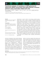
Fig. 3 Effect of FABP7 on the migration of TUHR14TKB cells.
TUHR14TKB cells were transfected with the FABP7 or lacZ expression
vector. a Western blot analysis of FABP7 expression by cells
transfected with the FABP7 vector or control (lacZ) vector. b Realtime PCR analysis of FABP7 expression in cells transfected with the
FABP7 vector or lacZ vector. c Wound-healing assays. TUHR14TKB
transfectants were cultured in RPMI 1640 medium containing with
10% FBS or 1% FBS with 5 mg/L blasticidin S HCl, 0.3 g/L G418, and
1 mg/L doxycycline hyclate. The data represent the average and
standard deviation (error bars) of four experiments
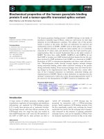
Although FABP7 binds to fatty acids, it does not catalyze
de novo fatty acid synthesis, suggesting that FABP7 expression leads to the accumulation of fatty acid in cells. Docosatetraenoic acid and EPA accumulated in TUHR14TKB cells
that expressed FABP7 (Fig. 5a). In contrast, other fatty acids
did not accumulate in TUHR14TKB cells that expressed
FABP7 (Additional file 3: Table S1). Therefore, we tested
the effects of docosatetraenoic acid or EPA on the proliferation of TUHR14TKB cells. The addition of docosatetraenoic acid significantly stimulated the proliferation of
TUHR14TKB cells that expressed β-galactosidase (Fig. 5b).
Takaoka et al. BMC Cancer (2017) 17:192
Page 6 of 8
Fig. 5 Effect of fatty acids on cell proliferation. a Docosatetraenoic acid or EPA concentration of TUHR14TKB cells transfected with the FABP7 or lacZ
expression vector. Four independent cell cultures were harvested, and the concentration of docosatetraenoic acid or EPA was determined. The data
represent the average and standard deviation. b Cell proliferation was assayed in the presence or absence of 50 μM docosatetraenoic acid or EPA in
RPMI 1640 medium containing 10% FBS, 5 mg/L blasticidin S HCl, 0.3 g/L G418, and 1 mg/L doxycycline hyclate. The doubling times of TUHR14TKB
lacZ cells were determined using an MTS assay. The data represent the average and standard deviation (error bars) of 12 experiments
Discussion
Human RCCs overexpress FABP7 [4, 6–14], indicating
that FABP7 might affect the progression of RCC.
Therefore, we studied FABP7 function using RCC cell
lines. In the present study, we show that the levels of
FABP7 dramatically decreased during passage of the
RCC cell line TUHR14TKB. Further, FABP overexpression differentially affected the proliferation of the
RCC cell lines analyzed here. Thus, overexpression of
FABP7 decreased the proliferation of TUHR14TKB
cells. In contrast, overexpression of FABP7 increased
the proliferation of 786-O cells.
FABP7 transcripts are expressed in 18 of 30 clear celltype RCC lesions but in only 4 of 19 RCC cell lines [6].
These results are consistent with our previous findings
that FABP7 is expressed in one (TUHR14TKB) of six
RCC cell lines [27]. We show here that the levels of
FABP7 decreased during the passage of TUHR14TKB
cells (Fig. 1). Further, TUHR14TKB cells proliferated
faster during continued passage (Fig. 2a), suggesting that
continued passage selected for cells that did not express FABP7 and therefore proliferated at an increased
rate. Moreover, the doubling times of subclones of
TUHR15TKB cells differed significantly (Fig 2a), which
is consistent with the loss of FABP7 expression during
attempts to establish cell lines from primary RCC
tumor tissue. In addition, glioblastoma neurospheres
express FABP7 at higher levels than those of adherent
cells derived from the same tumor [21]. Therefore,
conditions that favor the formation of spheres may
provide a selective advantage for primary RCC cells
that express FABP7.
Overexpression of FABP7 inhibited the proliferation
of TUHR14TKB cells (Fig. 4a and b), which is consistent with findings that FABP7 (referred to formerly
in the studies cited here as the protein encoded by
mammary-derived growth inhibitor-related gene) inhibits the proliferation of breast cancer cell lines [15, 16].
Further, high tumor-grade (G3 + G4) RCCs express
significantly lower levels of FABP7 mRNA than low-grade
(G1 + G2) RCCs [10], and FABP7 is highly expressed in
primary melanomas compared with metastatic melanomas [29, 30]. In contrast, knockdown of FABP7 expression inhibits the proliferation of melanoma cells [17,
18], an RCC cell line [19], a breast cancer cell line [20],
and glioblastoma cells [21]. Further, we show here that
FABP7 overexpression did not affect proliferation of
the 786-O cell line (Additional file 1: Figure S1c and
[14]), and down-regulation of FABP7 expression by
FABP7-specific siRNAs does not affect the proliferation of certain melanoma cells [17]. Interestingly,
FABP7 overexpression stimulated the proliferation of
786-O cells in medium containing 1% FBS (Additional
file 1: Figure S1c and [14]).
Takaoka et al. BMC Cancer (2017) 17:192
The present and previous studies demonstrate that the
effect of FABP7 on cell proliferation varies among cell
lines and with cell culture conditions. These findings may
be explained by the interaction of FABP7 with molecule(s)
that inhibit or enhance cell proliferation. Cancer is a
multistage disease, which develops through a succession
of mutations [31, 32]. Thus, FABP7 and other molecule(s)
may control cell proliferation through a similar mechanism. Another explanation for the inconsistencies among
studies of FABP7 function may be that FABP7 modulates
signaling networks that influence cell proliferation.
Down-regulation of FABP7 expression by siRNAs significantly reduces the migration of melanoma cell lines
[17, 18], an RCC cell line [19], breast cancer cells [20], and
malignant glioma cells [21–23]. Further, overexpression of
FABP7 enhances the migration of glioma cells [24]. In
contrast, FABP7 overexpression inhibited the migration of
TUHR14TKB cells that was revealed using a woundhealing assay (Fig. 3c). Thus, the effect of wound healing
may be related to the effect of proliferation.
Docosatetraenoic acid and EPA accumulated in
TUHR14TKB cells that expressed FABP7 (Fig. 5a and
Additional file 3: Table S1). Ligand-binding studies
conducted in vitro show that ω-3 EPA is the preferred
ligand of FABP7 [33]. Further, the addition of docosatetraenoic acid significantly increased cell growth (Fig. 5b),
suggesting that inhibition of the proliferation by FABP7 of
TUHR14TKB cells does not act through the accumulation
of docosatetraenoic acid by FABP7.
Conclusions
Our data lead us to conclude that the TUHR14TKB cell
line comprises a heterogeneous population and that cells
that do not express FABP7 grow faster and are therefore
selected during passage in culture. Further, our finding
that FABP7 inhibited the proliferation of TUHR14TKB
cells but stimulated the proliferation of 786-O cells cultured in medium with 1% FBS indicates that FABP7
function depends on cell type and culture conditions.
Additional files
Additional file 1: Figure S1. Effect of FABP7 overexpression on the
786-O cell line. 786-O cells were cultured for two days in RPMI 1640
medium containing 10% FBS, 5 mg/L blasticidin S HCl, 0.3 g/L G418, and
1 mg/L doxycycline hyclate. a, Western blot analysis of FABP7 expression
by cells transfected with the FABP7 vector or control (lacZ) vector. b,
Real-time PCR analysis of FABP7 expression of cells transfected with the
FABP7 vector or lacZ vector. c-d, The 786-O transfectants were cultured
in RPMI-1640 medium containing 10% FBS or 1% FBS with 5 mg/L
blasticidin S HCl, 0.3 g/L G418, and 1 mg/L doxycycline hyclate and
subjected to cell proliferation and migration assays. c, The doubling times
of cell transfected with the FABP7- or the lacZ-expression vector were
determined using an MTS assay. The data represent the average and
standard deviation (error bars) of five experiments. d, The migration of
786-O cells transfected with the FABP7- or lacZ-expression vector was
Page 7 of 8
determined using a wound-healing assay. The data represent the average
and standard deviation (error bars) of four experiments. (TIFF 2702 kb)
Additional file 2: Figure S2. Proliferation of TUHR14TKB cells
transfected with an FABP7 expression vector. The proliferation of cells
transfected with the FABP7 expression vector or lacZ expression vector
was determined using an MTS assay. The data represent of five
experiments. Transfectants were cultured in RPMI 1640 medium
containing 5 mg/L blasticidin S HCl, 0.3 g/L G418, and 1 mg/L
doxycycline hyclate with 1% FBS (a-b) or 10% FBS (c-d). a, c, TUHR14TKB
lacZ. b, d, TUHR14TKB FABP7. (TIFF 1521 kb)
Additional file 3: Table S1. Fatty acid concentrations of TUHR14TKB
transfectants. The concentrations of fatty acids accumulated by
TUHR14TKB cells transfected with vectors expressing FABP7 or lacZ were
assayed in four independent cultures. (XLSX 46 kb)
Abbreviations
cDNA: Complementary DNA; EPA: Eicosapentaenoic acid; FABP7: Fatty
acid binding protein 7; FBS: Fetal bovine serum; lacZ: β-galactosidase;
MTS: 3-(4,5-dimethylthiazol-2-yl)-5-(3-carboxymethoxyphenyl)-2-(4-sulfophenyl)2H–tetrazolium; PBS: Phosphate-buffered saline; RCC: Renal cell carcinoma;
SDS: Sodium dodecyl sulfate; siRNA: Small interfering RNA; TBP: TATA box
binding protein
Acknowledgements
We thank Kiyoshi Shibata (Equipment Center, Hamamatsu University School of
Medicine) for supporting the flow cytometry analysis, Hiromi Fujita (Urology,
Hamamatsu University School of Medicine) and Miki Miyazaki (Urology, Hamamatsu
University School of Medicine) for supporting the flow cytometry analysis and for
technical assistance, and DMC Corp [34] for editing the manuscript.
Funding
This research and editing the manuscript were supported by a Grant-in-Aid
for Scientific Research (C) 26,462,407 from the Ministry of Education, Culture,
Sports, Science, and Technology of Japan.
Availability of data and materials
All data generated or analysed during this study are included in this
published article and its Additional files.
Authors’ contributions
NT designed the study, performed the experiments, analyzed the data, and
wrote the manuscript. TT participated in designing the study, analyzing the
data, and editing the manuscript. SO participated in editing the manuscript.
All authors read and approved the final version of the manuscript.
Competing interests
The authors declare that they have no competing interests.
Consent for publication
Not applicable.
Ethics approval and consent to participate
Not applicable.
Publisher's Note
Springer Nature remains neutral with regard to jurisdictional claims in
published maps and institutional affiliations.
Author details
1
Department of Urology, Hamamatsu University School of Medicine, 1-20-1
Handayama, Higashi-ku, Hamamatsu, Shizuoka 431-3192, Japan. 2Department
of Urology, Jichi Medical University, 3311-1 Yakushiji, Shimotsuke, Tochigi
329-0498, Japan.
Takaoka et al. BMC Cancer (2017) 17:192
Received: 22 July 2016 Accepted: 9 March 2017
References
1. Ferlay J, Shin HR, Bray F, Forman D, Mathers C, Parkin DM. Estimates of
worldwide burden of cancer in 2008: GLOBOCAN 2008. Int J Cancer. 2010;
127(12):2893–917.
2. Chow WH, Dong LM, Devesa SS. Epidemiology and risk factors for kidney
cancer. Nat Rev Urol. 2010;7(5):245–57.
3. Pastore AL, Palleschi G, Silvestri L, Moschese D, Ricci S, Petrozza V, et al.
Serum and urine biomarkers for human renal cell carcinoma. Dis Markers.
2015;2015:251403.
4. Domoto T, Miyama Y, Suzuki H, Teratani T, Arai K, Sugiyama T, et al.
Evaluation of S100A10, annexin II and B-FABP expression as markers for
renal cell carcinoma. Cancer Sci. 2007;98(1):77–82.
5. Shimizu F, Watanabe TK, Shinomiya H, Nakamura Y, Fujiwara T. Isolation and
expression of a cDNA for human brain fatty acid-binding protein (B-FABP).
Biochim Biophys Acta. 1997;1354(1):24–8.
6. Seliger B, Lichtenfels R, Atkins D, Bukur J, Halder T, Kersten M, et al.
Identification of fatty acid binding proteins as markers associated with the
initiation and/or progression of renal cell carcinoma. Proteomics. 2005;5(10):
2631–40.
7. Skubitz KM, Zimmermann W, Kammerer R, Pambuccian S, Skubitz AP.
Differential gene expression identifies subgroups of renal cell carcinoma. J
Lab Clin Med. 2006;147(5):250–67.
8. Perroud B, Lee J, Valkova N, Dhirapong A, Lin PY, Fiehn O, et al. Pathway
analysis of kidney cancer using proteomics and metabolic profiling. Mol
Cancer. 2006;5:64.
9. Teratani T, Domoto T, Kuriki K, Kageyama T, Takayama T, Ishikawa A, et al.
Detection of transcript for brain-type fatty Acid-binding protein in tumor
and urine of patients with renal cell carcinoma. Urology. 2007;69(2):236–40.
10. Tölle A, Jung M, Lein M, Johannsen M, Miller K, Moch H, et al. Brain-type
and liver-type fatty acid-binding proteins: new tumor markers for renal
cancer? BMC Cancer. 2009;9:248.
11. Raimondo F, Salemi C, Chinello C, Fumagalli D, Morosi L, Rocco F, et
al. Proteomic analysis in clear cell renal cell carcinoma: identification
of differentially expressed protein by 2-D DIGE. Mol BioSyst. 2012;8(4):
1040–51.
12. Feng JY, Diao XW, Fan MQ, Wang PX, Xiao Y, Zhong X, et al. Screening of
feature genes of the renal cell carcinoma with DNA microarray. Eur Rev
Med Pharmacol Sci. 2013;17(22):2994–3001.
13. Tan C, Takayama T, Takaoka N, Fujita H, Miyazaki M, Sugiyama T, Ozono S.
Impact of Gender in Renal Cell Carcinoma: The Relationship of FABP7 and
BRN2 Expression with Overall Survival. Clin Med Insights Oncol. 2014;8:21–7.
14. Zhou J, Deng Z, Chen Y, Gao Y, Wu D, Zhu G, et al. Overexpression of
FABP7 promotes cell growth and predicts poor prognosis of clear cell renal
cell carcinoma. Urol Oncol. 2015;33(3):113. e9-17
15. Shi YE, Ni J, Xiao G, Liu YE, Fuchs A, Yu G, et al. Antitumor activity of the
novel human breast cancer growth inhibitor, mammary-derived growth
inhibitor-related gene. MRG Cancer Res. 1997;57(15):3084–91.
16. Wang M, Liu YE, Ni J, Aygun B, Goldberg ID, Shi YE. Induction of Mammary
Differentiation by Mammary-derived Growth Inhibitor- related Gene That
Interacts with an omega-3 Fatty Acid on Growth Inhibition of Breast Cancer
Cells. Cancer Res. 2000;60(22):6482–7.
17. Goto Y, Matsuzaki Y, Kurihara S, Shimizu A, Okada T, Yamamoto K, et al. A
new melanoma antigen fatty acid-binding protein 7, involved in
proliferation and invasion, is a potential target for immunotherapy and
molecular target therapy. Cancer Res. 2006;66(8):4443–9.
18. Slipicevic A, Jørgensen K, Skrede M, Rosnes AK, Trøen G, Davidson B,
Flørenes VA. The fatty acid binding protein 7 (FABP7) is involved in
proliferation and invasion of melanoma cells. BMC Cancer. 2008;8:276.
19. Tölle A, Krause H, Miller K, Jung K, Stephan C. Importance of brain-type fatty
acid binding protein for cell-biological processes in human renal carcinoma
cells. Oncol Rep. 2011;25(5):1307–12.
20. Liu RZ, Graham K, Glubrecht DD, Lai R, Mackey JR, Godbout R. A fatty acidbinding protein 7/RXRβ pathway enhances survival and proliferation in
triple-negative breast cancer. J Pathol. 2012;228(3):310–21.
21. De Rosa A, Pellegatta S, Rossi M, Tunici P, Magnoni L, Speranza MC, et al. A
radial glia gene marker, fatty acid binding protein 7 (FABP7), is involved in
proliferation and invasion of glioblastoma cells. PLoS One. 2012;7(12):e52113.
Page 8 of 8
22. Mita R, Coles JE, Glubrecht DD, Sung R, Sun X, Godbout R. B-FABPexpressing radial glial cells: the malignant glioma cell of origin? Neoplasia.
2007;9(9):734–44.
23. Mita R, Beaulieu MJ, Field C, Godbout R. Brain fatty acid-binding protein and
omega-3/omega-6 fatty acids: mechanistic insight into malignant glioma
cell migration. J Biol Chem. 2010;285(47):37005–15.
24. Liang Y, Diehn M, Watson N, Bollen AW, Aldape KD, Nicholas MK, et al.
Gene expression profiling reveals molecularly and clinically distinct subtypes
of glioblastoma multiforme. Proc Natl Acad Sci U S A. 2005;102(16):5814–9.
25. Liang Y, Bollen AW, Aldape KD, Gupta N. Nuclear FABP7 immunoreactivity is
preferentially expressed in infiltrative glioma and is associated with poor
prognosis in EGFR-overexpressing glioblastoma. BMC Cancer. 2006;6:97.
26. RIKEN BioResource Center: Pattern of STR-PCR of TUHR14TKB. http://www2.
brc.riken.jp/lab/cell/str_start.shtml?cell_no=RCB1383. Accessed 15 July 2016.
27. Takaoka N, Takayama T, Teratani T, Sugiyama T, Mugiya S, Ozono S. Analysis
of the regulation of fatty acid binding protein 7 expression in human renal
carcinoma cell lines. BMC Mol Biol. 2011;12:31.
28. Jung M, Ramankulov A, Roigas J, Johannsen M, Ringsdorf M, Kristiansen G,
Jung K. In search of suitable reference genes for gene expression studies of
human renal cell carcinoma by real-time PCR. BMC Mol Biol. 2007;8:47.
29. de Wit NJ, Rijntjes J, Diepstra JH, van Kuppevelt TH, Weidle UH, Ruiter DJ,
van Muijen GN. Analysis of differential gene expression in human
melanocytic tumour lesions by custom made oligonucleotide arrays. Br J
Cancer. 2005;92(12):2249–61.
30. Goto Y, Koyanagi K, Narita N, Kawakami Y, Takata M, Uchiyama A, et al.
Aberrant fatty acid-binding protein-7 gene expression in cutaneous
malignant melanoma. J Invest Dermatol. 2010;130(1):221–9.
31. Kinzler KW, Vogelstein B. Lessons from hereditary colorectal cancer. Cell.
1996;87(2):159–70.
32. Chaffer CL, Weinberg RA. How Does Multistep Tumorigenesis Really
Proceed? Cancer Discov. 2015;5(1):22–4.
33. Balendiran GK, Schnutgen F, Scapin G, Borchers T, Xhong N, Lim K, et al.
Crystal structure and thermodynamic analysis of human brain fatty acidbinding protein. J Biol Chem. 2000;275(35):27045–54.
34. DMC CorpDMC Corp. . Accessed 15 July 2016
Submit your next manuscript to BioMed Central
and we will help you at every step:
• We accept pre-submission inquiries
• Our selector tool helps you to find the most relevant journal
• We provide round the clock customer support
• Convenient online submission
• Thorough peer review
• Inclusion in PubMed and all major indexing services
• Maximum visibility for your research
Submit your manuscript at
www.biomedcentral.com/submit