Potential importance of protease activated receptor (PAR)-1 expression in the tumor stroma of non-small-cell lung cancer
Bạn đang xem bản rút gọn của tài liệu. Xem và tải ngay bản đầy đủ của tài liệu tại đây (3.78 MB, 8 trang )
Lin et al. BMC Cancer (2017) 17:113
DOI 10.1186/s12885-017-3081-3
RESEARCH ARTICLE
Open Access
Potential importance of protease activated
receptor (PAR)-1 expression in the tumor
stroma of non-small-cell lung cancer
Cong Lin1*, Christof J. Majoor2†, Joris J. T. H. Roelofs3†, Martijn D. de Kruif2,4, Hugo M. Horlings5,
Keren Borensztajn1,6,7 and C. Arnold Spek1
Abstract
Background: Protease activated receptor (PAR)-1 expression is increased in a variety of tumor cells. In preclinical
models, tumor cell PAR-1 appeared to be involved in the regulation of lung tumor growth and metastasis; however
the role of PAR-1 in the lung tumor microenvironment, which is emerging as a key compartment in driving cancer
progression, remained to be explored.
Methods: In the present study, PAR-1 gene expression was determined in lung tissue from patients with non-smallcell lung cancer (NSCLC) using a combination of publicly available RNA microarray datasets and in house-made
tissue microarrays including tumor biopsies of 94 patients with NSCLC (40 cases of adenocarcinoma, 42 cases
of squamous cell carcinoma and 12 cases of other type of NSCLC at different stages).
Results: PAR-1 gene expression strongly correlated with tumor stromal markers (i.e. macrophage, endothelial
cells and (myo) fibroblast markers) but not with epithelial cell markers. Immunohistochemical analysis confirmed the
presence of PAR-1 in the tumor stroma and showed that PAR-1 expression was significantly upregulated in malignant
tissue compared with normal lung tissue. The overexpression of PAR-1 in tumor stroma of NSCLC appeared
to be independent from tumor type, tumor stage, histopathological differentiation status, disease progression
and patient survival.
Conclusion: Overall, our data provide evidence that PAR-1 in NSCLC is mainly expressed on cells that constitute the
pulmonary tumor microenvironment, including vascular endothelial cells, macrophages and stromal fibroblasts.
Keywords: Protease activated receptor, NSCLC and tumor stroma
Background
Lung cancer is the leading cause of cancer related
death, with around 1.6 million deaths worldwide and
the mortality rates for lung cancer are still increasing
annually [1, 2]. Non-small-cell lung cancer (NSCLC),
the most common type of lung cancer, has a devastating
survival outcome. Traditional chemotherapy, including
predominantly platinum-based regimens, as first-line
standard treatment for NSCLC only shows a modest
prolongation of median and overall survival. Despite
* Correspondence:
†
Equal contributors
1
Center for Experimental and Molecular Medicine, Academic Medical Center,
Amsterdam 1105 AZ, The Netherlands
Full list of author information is available at the end of the article
aggressive multimodality therapy, 5-year survival rate
for patients with stage IV NSCLC at diagnosis is only
approximately 2% [3]. More recently, targeted therapies
showed efficacy in patients with advanced NSCLC who
have specific genetic alterations, like mutations of the
anaplastic lymphoma kinase gene or of the epidermal
growth factor receptor [1]. However, these available
molecular therapies can only be applied to selective
patients and the observed benefits are small, suggesting
that more in-depth studies of molecules that relate to
the pathogenesis of NSCLC is required.
Protease-activated receptor (PAR)-1 is a cell surface
seven-transmembrane G protein coupled receptor that
is activated by proteolytic cleavage. Removal of the Nterminal extracellular domain of PAR-1 reveals a new
© The Author(s). 2017 Open Access This article is distributed under the terms of the Creative Commons Attribution 4.0
International License ( which permits unrestricted use, distribution, and
reproduction in any medium, provided you give appropriate credit to the original author(s) and the source, provide a link to
the Creative Commons license, and indicate if changes were made. The Creative Commons Public Domain Dedication waiver
( applies to the data made available in this article, unless otherwise stated.
Lin et al. BMC Cancer (2017) 17:113
tethered ligand that binds to the body of PAR-1 and
activates transmembrane signaling to intracellular G
proteins, thereby leading to multiple pathophysiological
responses [4, 5]. Overexpression of PAR-1 has been
detected in various types of cancers, including ovarian,
breast, lung, prostate cancer and melanoma [6–10]. Importantly, elevated PAR-1 expression is closely associated
with diseases progression and overall survival in breast,
prostate, gastric cancer and melanoma [6, 8, 9, 11]. Moreover, tumor cell PAR-1 is recently identified as a
promising target to decrease lung cancer progression.
Indeed, PAR-1 pepducin inhibitors not only block the
migration of both primary and established lung cancer
cell lines, but also significantly limit lung tumor growth in
nude mice [10]. Moreover, melanoma growth and metastasis were significantly decreased in mice treated
with PAR-1 small interfering RNA (siRNA) [12].
During the last decade, the paradigm that tumor
growth solely relies on the malignant cells has shifted to
a more comprehensive view that tumor growth is
dependent on interactions between cancer cells and their
adjacent microenvironment, also known as the stroma
[13]. The tumor stroma, predominately composed of
basement membrane, fibroblasts, vasculature with endothelial cells, inflammatory cells and extra cellular matrix
proteins such as collagen and fibronectin [14], is indeed
emerging as a key player in promoting carcinogenesis by
modulating tumor growth, angiogenesis, invasion and
metastasis [15, 16]. Targeting the tumor stroma is consequently under intense investigation as novel treatment
strategy in cancer.
Interestingly, PAR-1 expression is not tumor cell
specific and PAR-1 is also expressed on key cell types
that constitute the tumor stroma such as endothelial
cells, fibroblasts and macrophages. Activation of PAR1 on these stromal cells leads to increased vascular
permeability, fibroblast activation, extracellular matrix
production and cytokine secretion, thereby potentially
driving tumor growth and metastasis [13]. In line with
these observations, colonic adenocarcinoma growth
was limited in PAR-1-deficient mice, suggesting the
importance of PAR-1 in the tumor microenvironment
[17]. In addition, pancreatic tumors in PAR-1 deficient
animals were significantly smaller compared with
tumors in wild type mice. Moreover, the same study also
showed that stromal cells drive tumor growth and
induce chemoresistance of pancreatic cancer in a
PAR-1 dependent manner [18]. Overall these data
point to an important role of stromal cell-associated
PAR-1 in tumor progression. However, the role of
stromal PAR-1 in lung cancer has not been explored
yet. In the present study, we examined PAR-1 expression in NSCLC stroma and assessed its correlation
with disease progression.
Page 2 of 8
Methods
Patients
Tissue microarrays (TMAs, triplicate cores per case)
were prepared with tumor sections obtained from
NSCLC patients during surgery according to the guidelines of the Medical Ethical Committee of the Academic
Medical Center of Amsterdam. The TMAs consist of
samples from 94 patients with NSCLC, including 40
cases of adenocarcinoma (ADC), 42 cases of squamous
cell carcinoma (SCC) and 12 cases of other type of
NSCLC at different stages (Table 1). On each TMA, 3
cases of healthy lung tissue (i.e. adjacent normal tissue)
were also included.
Mining of publically available RNA microarray dataset
The datasets were derived from Gene Expression Omnibus
( using the R2 microarray
analysis and visualization platform (). Correlation of gene expression between PAR-1 and markers of
different stromal cell types in NSCLC cancer patients were
derived by the R2 program from five different datasets,
including Bild (n = 114, GSE3141), Peitsch (n = 150,
GSE43580), EXPO (n = 121, GSE2109), Mao (n = 124,
GSE 31852) and Hou (n = 156, GSE 19188).
Table 1 Patient characteristics
Characteristic
Patient
N
%
Male
63
67
Median Age (Range)
66
Progression
26
36
Adenocarcinoma
40
42.5
Squamous cell carcinoma
42
44.7
Other type*
12
12.8
Well differentiated
6
10.4
(30–86)
Tumor type:
Tumor differentiation:
Less differentiated
30
51.7
Little differentiated
14
24.1
Poorly differentiated
8
13.8
I
53
57.6
II
28
30.4
III
10
10.9
IV
1
1.1
14
15.2
NSCLC stage:
Lymph node metastasis
*This group includes 2 large cell carcinoma patients, 10 patients with mixed
tumor types (for instance adenocarcinoma/bronchioloalveolar carcinoma)
Lin et al. BMC Cancer (2017) 17:113
Immunohistological analysis
Four-μm sections were first deparaffinized and rehydrated.
Endogenous peroxidase activity was quenched with 0.3%
H2O2 in methanol. PAR-1 staining was performed with a
primary antibody specific for PAR-1 (ATAP-2 ;1:200; SC13503, 24 h at 4 °C, Santa Cruz, San Diego, CA) [19, 20].
A horseradish peroxidase-conjugated polymer detection
system (ImmunoLogic, Duiven, the Netherlands) was
applied for visualization, using an appropriate secondary
antibody and diaminobenzidine staining. Specimens with
PAR-1 immunostaining were reviewed jointly at a multihead microscope by 2 investigators blinded to the patients’
clinical status. To evaluate immunohistochemical expression of PAR-1, the intensity of PAR-1 staining was graded
by consensus on a scale from 0 to 3 (0 = negative staining;
1 = weakly positive; 2 = moderately positive; 3 = strongly
positive). Slides were photographed with a microscope
equipped with a digital camera (Leica CTR500).
Statistics
Statistical analyses were conducted using GraphPad Prism
(GraphPad software, San Diego). Comparisons between
conditions were analyzed using two tailed unpaired t-tests
when the data were normally distributed; otherwise
Mann–Whitney analysis was performed. Results are
expressed as mean ± SEM, P values < 0.05 are considered
significant.
Results
PAR-1 gene expression is correlated with lung tumor
stroma activation
To explore the association of PAR-1 expression with the
NSCLC stroma, we correlated PAR-1 gene expression
levels with specific markers of different stromal cell types,
including macrophages, endothelial cells, epithelial cells
and (myo) fibroblasts in resected tumor specimens using
publicly available microarray datasets. To this end, 3
markers were selected for each stromal cell type, except
for (myo) fibroblasts for which we included markers of
differentiated fibroblasts and markers for extracellular
matrix (ECM) produced by myofibroblasts. Interestingly,
tumors with higher PAR-1 levels also displayed elevated
expression levels of markers for macrophages, endothelial
cells and (myo) fibroblasts on the microarrays. Using the
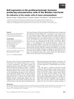
GSE3141 dataset (Fig. 1), PAR-1 gene expression was
correlated with all three markers for human monocytes
and macrophages, i.e. CD68 (p < 0.01), CD163 (p < 0.001)
and CD14 (p < 0.0001) [21]. Correlations with specific vascular endothelial cell markers (e.g. Platelet endothelial cell
adhesion molecule (PECAM)-1) and fibroblasts markers
(e.g. Vimentin (VIM) and fibroblast activation protein
alpha (FAP)) were also significant (p < 0.0001), with rvalues ranging from 0.2 to 0.7. The commonly used differentiation marker for fibroblasts ACTA2 (gene encoding
Page 3 of 8
for alpha-smooth muscle actin, α-SMA [22]) and markers
for prominent constituents of ECM deposition Collagen,
type I, alpha (COL1A1) and Fibronectin (FN1) were also
all correlated with PAR-1 gene expression in the NSCLC
specimens (all p < 0.01). Intriguingly, PAR-1 expression
did not correlate to epithelial (tumor) cell markers Epithelial cell adhesion molecule (EpCAM), Cadherin 1 (CDH1)
and Mucin 1 (MUC1). These observed correlations (and
lack of correlation in epithelial cells) were confirmed in
four additional independent microarray datasets from
NSCLC (Table 2). However, no correlation between PAR1 and stromal markers was observed in the healthy
control group included in the Hou et al. set (GSE19188),
suggesting the correlation between PAR-1 gene expression
and stroma activity specifically exists in tumor microenvironment. To confirm the identity of the stromal cell
types expressing PAR-1, we performed immunohistochemistry with different cell type markers on consecutive lung cancer slides. As shown in Additional file 1:
Figure S1, PAR-1 positive areas are also positive for
CD31 (endothelial marker), CD68 (macrophage marker)
and aSMA (myofibroblast marker).
PAR-1 is overexpressed in stroma of primary pulmonary
tumors on TMAs
To confirm the presence of PAR-1 in NSCLC stroma, we
next analyzed PAR-1 protein expression in tumor sections
using immunohistochemistry. Ninety-four patients with
pathologically confirmed diagnosis of NSCLC were included into this study. The median age at diagnosis was
66 years (range 30 to 86 years), and the majority of patients
had NSCLC stage I disease (n = 53, 57.6%). Six cases were
well differentiated (2 ADC, 1 SCC, 3 other types), 30 cases
were moderately differentiated (12 ADC, 18 SCC) and 22
cases were poorly differentiated (10 ADC, 11 SCC, 1 other
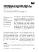
types) (Table 1). Overall, strong PAR-1 expression was seen
in stroma of all different types of NSCLC (ADC, SCC and
large-cell carcinoma) as opposed to weak PAR-1 staining
on control sections (Fig. 2). In line with our observations in
the tumor microarray datasets, the stromal cells (fibroblastlike cells, inflammatory cells and endothelial cells) were all
intensively stained for PAR-1, while cancer cells were negative for PAR-1 or showed only weak PAR-1 staining. Subsequent quantifications showed that 93 out of the 94 cases
had PAR-1 expression in the stroma, with an average score
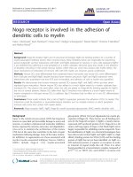
of 2, while 1 SSC patient was PAR-1 negative. Importantly,
the average PAR-1 score in control lungs was significantly
lower as in NSCLC stroma (average score of 1; Fig. 3a). As
shown in Fig. 3b, PAR-1 levels were similar in different
subtype of NSCLC (average scores of 2.11, 2.01 and 2.08
for ADC, SCC and other type of NSCLCs respectively).
Stromal PAR-1 expression levels did not correlate
with clinical variables like stage of NSCLC (Fig. 3c),
Lin et al. BMC Cancer (2017) 17:113
Page 4 of 8
Fig. 1 PAR-1 expression correlates with stromal markers in NSCLC patients. Scatter plot of PAR-1 gene (F2R) expression versus the expression of
specific macrophage (a), endothelial (b), epithelial (c) and (myo) fibroblast (d) markers in tumors derived from NSCLC patients (Bild microarray dataset;
GSE3141, n = 114). Linear regression analysis was used to determine the correlation coefficient, and p-values of significant correlations are
indicated in red
differentiation status (Fig. 3d), disease progression
(Fig. 3e) and overall survival (Fig. 3f ).
Discussion
One of the anticipated future treatment options for NSCLC
is to target the interactions between tumor and stromal
cells, since stromal cells provide additional signals that support tumor growth and invasion [1, 16]. In the present
study, we determined PAR-1 expression in NSCLC patients
and found high PAR-1 expression predominantly in the
tumor stroma compartment during early stage cancer. This
was reflected by the correlation of PAR-1 gene expression
Lin et al. BMC Cancer (2017) 17:113
Page 5 of 8
Table 2 Correlation of gene expression between PAR-1 and markers of different stromal cell types
Database
Bild (n=114)
Peitsch (n=150)
EXPO (n=121)
Mao (n=124)
Hou (n=91)
(nc=65)
Stroma
Macrophage
Endothelium
Epithelium
(Myo)Fibroblasts
Gene
P-value
R-value
P-value
R-value
P-value
R-value
P-value
R-value
Pn-value
Rn-value
Pnc-value
Rnc-value
CD68
6.4e-03
0.254
8.3e-04
0.270
0.03
0.198
0.03
0.191
2.5e-04
0.375
0.06
-0.238
CD14
2.9e-05
0.381
3.3e-07
0.402
0.02
0.213
0.82
0.020
6.7e-03
0.282
0.21
-0.157
CD163
2.0e-04
0.342
2.6e-05
0.336
0.05
0.175
1.5e-07
0.450
2.3e-03
0.316
0.24
-0.149
PECAM-1
1.8e-06
0.430
8.3e-05
0.316
2.0e-06
0.417
3.2e-06
0.404
0.36
0.098
0.45
-0.095
CDH5
3.3e-04
0.331
7.9e-03
0.216
5.1e-04
0.311
6.4e-07
0.429
0.02
0.239
0.15
0.183
vWF
1.8e-03
0.289
9.8e-05
0.313
5.7e-03
0.250
1.1e-05
0.383
1.5e-03
0.328
0.53
0.080
EpCAM
0.10
-0.154
2.0e-04
-0.299
0.04
-0.188
0.09
-0.151
1.3e-03
-0.333
0.19
0.166
CDH1
0.11
-0.149
1.8e-07
-0.410
0.62
-0.046
0.04
-0.182
0.05
-0.207
0.61
0.064
MUC1
0.99
0.001
0.33
0.079
0.96
0.005
0.07
-0.165
0.39
-0.091
0.15
-0.182
VIM
4.6e-13
0.612
2.6e-11
0.510
1.8e-03
0.281
4.9e-07
0.433
4.4e-08
0.536
0.70
0.048
PDGFRB
9.4e-09
0.506
8.1e-12
0.521
3.3e-05
0.368
1.6e-17
0.670
2.2e-05
0.429
0.02
0.286
FAP
2.4e-05
0.384
7.0e-12
0.522
9.2e-04
0.297
7.5e-08
0.460
2.1e-05
0.430
0.02
0.299
ACTA2
6.0e-08
0.481
4.2e-08
0.429
8.7e-06
0.392
8.9e-16
0.642
2.6e-03
0.312
0.20
0.159
COL1A1
5.7e-03
0.257
9.6e-07
0.388
1.0e-05
0.389
1.0e-12
0.585
0.24
0.124
0.05
0.249
FN1
6.4e-09
0.511
3.3e-07
0.402
7.8e-07
0.431
1.6e-10
0.535
1.9e-05
0.432
0.23
0.150
Selection of the datasets is based on the patient group size ≥50. Bild (n = 114, GSE3141), Peitsch (n = 150, GSE43580), EXPO (n = 121, GSE2109), Mao
(n = 124, GSE 31852), Hou (n = 91, control group nc = 65, GSE 19188). Linear regression analysis was used to determine p-value of correlation. Significant correlations are
indicated in red
with stroma markers like CD163, CD31 and vimentin, and
ECM proteins like collagen and fibronectin, as well as by a
significant increase in the intensity of PAR-1 staining in
stromal cells of tumor tissue compared with normal lung
tissue. Although it has been documented that upregulation
of PAR-1 expression appears in a variety of invasive cancers
of epithelial origin, our data do show that increased PAR-1
expression in NSCLC patients arises mainly in the tumor
stroma rather than in the epithelial cancer cells.
The observed PAR-1 expression pattern in NSCLC resembles that seen in other malignancies. In breast cancer,
PAR-1 expression, as shown by immunohistochemistry and
in situ hybridization, is observed in mast cells, macrophages, endothelial cells, and vascular smooth muscle cells
of the metastatic tumor microenvironment. Interestingly
however, PAR-1 expression is particularly increased in
stromal fibroblasts surrounding breast carcinoma cells as
opposed to low/negative expression in fibroblasts of healthy
or benign conditions [23]. Moreover, in prostate cancer
PAR-1 is predominantly expressed in peritumoral stroma.
In particular, PAR-1 is mainly expressed in myofibroblasts
and to a lower level in endothelial cells in isolated
capillaries around the malignant glands [24, 25].
The enrichment of PAR-1 expression in the stroma
surrounding the tumor may actually be clinically relevant.
Indeed, in the setting of pancreatic cancer, PAR-1 also
coincides with the expression pattern of the stromal
markers, such as vimentin, collagen I and α-SMA [18].
More importantly, PAR-1 promoted monocyte recruitment due to fibroblast dependent chemokine production,
thereby driving pancreatic tumor growth and chemoresistance [18]. In the context of lung cancers, the expression
of PAR-1 mRNA in alveolar walls with surface spreading
of neoplastic cells was shown to increase by 10-fold
compared with alveolar walls without surface spreading of
neoplastic cells, and stimulation of PAR-1 led to the
proliferation of alveolar capillary endothelial cells, pointing
to PAR-1 as a potential regulator in alveolar angiogenesis
[26]. Interestingly, accumulating evidence indicates that
PAR-1 also exerts pro-inflammatory and pro-fibrotic
functions through macrophages and fibroblasts during
pulmonary fibroproliferative disease progression [27–29],
which may also benefit tumor progression and metastasis.
Previous studies about PAR-1 in NSCLC focused on its
function in cancer cells. Indeed, multiple reports showed
that PAR-1 modulates lung cancer cell proliferation and
migration, thereby supporting tumor growth and invasion
[10, 30]. Hence, targeting PAR-1 to inhibit progression of
lung cancer cells seems to be an option for cancer therapy.
Recently, emphasis has shifted toward the tumor stroma
for novel therapeutic strategies and several approaches
targeting the stromal tissue in different types of cancers
Lin et al. BMC Cancer (2017) 17:113
Page 6 of 8
Fig. 2 Stromal PAR-1 expression is upregulated in NSCLC patients. Representative PAR-1 staining of normal lung tissue and tumor sections of NSCLC
patients (Pictures were taken with 100x magnification; Enlarged pictures were taken with 200x magnification). ADC indicates adenocarcinoma,
SCC indicates squamous cell carcinoma and LCC indicates large cell carcinoma. Tumor cells are indicated by (i) whereas inflammatory cells are
indicated by solid arrowheads, vascular endothelial cells are indicated by stars and fibroblasts-like cells and ECM are indicated by crosses
have been proved to be effective [31–33]. Our data
showing high stromal PAR-1 expression in NSCLC may
thus indicate stromal PAR-1 may be the main target of
the treatment for NSCLC. However, before drawing
conclusions on potential clinical implications of stromal
PAR-1 in NSCLC, it is important to elucidate the functional consequence of PAR-1 activation on stromal cells
with respect to lung cancer development.
In the present study, we observed that PAR-1 expression
is highly upregulated in the tumor stroma but not in
normal lung tissue, suggesting that PAR-1 may have a
diagnostic value in NSCLC. However, the increased PAR1 expression does not seem to correlate with diseases
progression, which indicates that stromal PAR-1 in lung
cancer is crucial for carcinogenesis but may not be a determinant factor for cancer progression. These results are
in line with a recent study by Erturk and colleagues, who
determined serum PAR-1 levels in 80 patients with lung
cancer [34]. Serum PAR-1 concentrations of lung cancer
patients were significantly increased as compared to controls (i.e. median values of 26.45 ng/mL and 0.07 ng/mL,
respectively), but serum PAR-1 levels did not correlate
with clinical variables and failed to predict prognosis of
the lung cancer patients. In apparent disagreement, other
studies using immunohistochemistry analysis showed that
PAR-1 may be a prognostic factor for poor prognosis in
Lin et al. BMC Cancer (2017) 17:113
Page 7 of 8
Fig. 3 Association of stromal PAR-1 expression with clinical parameters in NSCLC patients. a Stromal PAR-1 expression in healthy lung tissue and
in NSCLC. b Stromal PAR-1 expression in healthy lung tissue and in different types of NSCLC. c Stromal PAR-1 expression in healthy
lung tissue and in different stages of NSCLC. d Stromal PAR-1 expression according to the differentiation status of NSCLC, including
well differentiated, moderately differentiated and poorly differentiated. e Stromal PAR-1 expression in NSCLC patients with disease progression and in
patients with stable disease (no-progression). f Stromal PAR-1 expression of survivors and non-survivors of NSCLC. All data are expressed as
mean ± SEM, *P < 0.05, **P < 0.01 and *** P < 0.001
both early-stage and advanced stages (III and IV) of
NSCLC [35, 36]. Importantly however, these studies
analyzed tumor cell PAR-1 expression and did not address
PAR-1 expression in the stromal compartment.
Funding
This study was supported by grant from the Netherlands Organization for
Scientific Research (016.136.167). The funders had no role in study design,
data collection and analysis, decision to publish, or preparation of the
manuscript.
Conclusion
In summary, our data show PAR-1 is overexpressed in
the tumor stroma of NSCLC, but stromal PAR-1 expression levels do not correlate with disease progression
and/or overall survival.
Availability of data and materials
The microarray datasets analyzed during the current study were derived
from the Gene Expression Omnibus ( using
the R2 microarray analysis and visualization platform (). The
five microarray datasets include Bild (n = 114, GSE3141), Peitsch (n = 150,
GSE43580), EXPO (n = 121, GSE2109), Mao (n = 124, GSE 31852) and Hou
(n = 156, GSE 19188). The data obtained from TMAs and biopsies are
available upon reasonable request from the corresponding author.
Additional file
Additional file 1: Figure S1. Correlation of PAR-1 expression and specific
markers for endothelial cells, macrophages and myofibroblasts. Consecutive
lung cancer slides stained for PAR-1 (left panels), CD31 (endothelial marker),
CD68 (macrophage marker) and aSMA (myofibroblast marker). Please note
that due to the use of consecutive slides, the structure of the tissue in the
PAR-1 stained slide is somewhat different from the CD31, CD68 and aSMA
stained slides. Pictures were taken with 100x magnification. (TIF 7026 kb)
Abbreviations
ADC: Adenocarcinoma; CDH1: Cadherin-1; COL1A1: Collagen, type I, alpha;
ECM: Extracellular matrix; EpCAM: Epithelial cell adhesion molecule; FAP: Fibroblast
activation protein alpha; FN1: Fibronectin 1; MUC1: Mucin 1; NSCLC: Non-small-cell
lung cancer; PAR-1: Protease activated receptor-1; PECAM-1: Platelet endothelial
cell adhesion molecule-1; SCC: Squamous cell carcinoma; siRNA: Small interfering
RNA; TMA: Tissue microarray; VIM: Vimentin; α-SMA: Alpha-smooth muscle actin
Acknowledgements
Not applicable.
Authors’ contributions
CL conceived and designed the experiments, performed the experiments,
analyzed the data and wrote the manuscript; CJM performed part of the
experiments and analyzed the data; JJTHR performed part of the experiments
and analyzed the data; MDK analyzed the data; HMH performed part of the
experiments; KB analyzed the data and wrote the manuscript; CAS conceived
and designed the experiments, and was a major contributor in writing the
manuscript. All authors have read and approved the final manuscript.
Competing interests
The authors declare that they have no competing interests.
Consent for publication
Not applicable.
Ethics approval and consent to participate
This research project used anonymized human tissue (both NSCLC tumorous
and adjacent healthy tissue) that was removed from a patient during the
normal course of treatment and which was later made available for scientific
research (so-called ‘further use’ of human tissue). According to the Code of
Conduct for dealing responsibly with human tissue in the context of health
research (Human Tissue and Medical Research: Code of conduct for responsible
use drawn up by the Federation of Dutch Medical Scientific Societies in
Lin et al. BMC Cancer (2017) 17:113
collaboration with the Dutch Patient Consumer federation, the Federation of
Parent and Patient Organisations and the Biobanking and Biomolecular
Resources Research Infrastructure; />digital_version_first_part_code_of_conduct_in_uk_2011_12092012.pdf) these
biological materials are as such not subject to any requirement for ethical
review or consent from patients.
Author details
1
Center for Experimental and Molecular Medicine, Academic Medical Center,
Amsterdam 1105 AZ, The Netherlands. 2Department of Respiratory Medicine,
Academic Medical Center, Amsterdam 1105 AZ, The Netherlands.
3
Department of Pathology, Academic Medical Center, Amsterdam 1105 AZ,
The Netherlands. 4Department of Pulmonology, Zuyderland Hospital, Henri
Dunantstraat 5, 6419 PC Heerlen, The Netherlands. 5Department of
Pathology, The Antonie van Leeuwenhoek hospital, Amsterdam 1066 CX,
The Netherlands. 6Inserm UMR1152, Medical School Xavier Bichat, 16 rue
Henri Huchard, 75018 Paris, France. 7Département Hospitalo-universtaire FIRE
(Fibrosis, Inflammation and Remodeling) and LabEx Inflamex, Paris, France.
Received: 3 August 2016 Accepted: 23 January 2017
References
1. Chen Z, Fillmore CM, Hammerman PS, Kim CF, Wong KK. Non-small-cell lung
cancers: a heterogeneous set of diseases. Nat Rev Cancer. 2014;14:535–46.
2. Ferlay J, Soerjomataram I, Dikshit R, Eser S, Mathers C, Rebelo M, Parkin DM,
Forman D, Bray F. Cancer incidence and mortality worldwide: sources, methods
and major patterns in GLOBOCAN 2012. Int J Cancer. 2015;136:E359–86.
3. Ettinger DS, et al. NCCN guidelines insights: Non-small cell lung cancer,
version 4.2016. J Natl Compr Canc Netw. 2016;14:255–64.
4. Coughlin SR. Thrombin signalling and protease-activated receptors. Nature.
2000;407:258–64.
5. Vu TK, Hung DT, Wheaton VI, Coughlin SR. Molecular cloning of a functional
thrombin receptor reveals a novel proteolytic mechanism of receptor
activation. Cell. 1991;64:1057–68.
6. Boire A, Covic L, Agarwal A, Jacques S, Sherifi S, Kuliopulos A. PAR1
is a matrix metalloprotease-1 receptor that promotes invasion and
tumorigenesis of breast cancer cells. Cell. 2005;120:303–13.
7. Grisaru-Granovsky S, Salah Z, Maoz M, Pruss D, Beller U, Bar-Shavit R. Differential
expression of protease activated receptor 1 (Par1) and pY397FAK in benign
and malignant human ovarian tissue samples. Int J Cancer. 2005;113:372–8.
8. Massi D, Naldini A, Ardinghi C, Carraro F, Franchi A, Paglierani M, Tarantini F,
Ketabchi S, Cirino G, Hollenberg MD, Geppetti P, Santucci M. Expression of
protease-activated receptors 1 and 2 in melanocytic nevi and malignant
melanoma. Hum Pathol. 2005;36:676–85.
9. Black PC, Mize GJ, Karlin P, Greenberg DL, Hawley SJ, True LD, Vessella RL,
Takayama TK. Overexpression of protease-activated receptors-1,-2, and-4
(PAR-1, −2, and −4) in prostate cancer. Prostate. 2007;67:743–56.
10. Cisowski J, O’Callaghan K, Kuliopulos A, Yang J, Nguyen N, Deng Q, Yang E,
Fogel M, Tressel S, Foley C, Agarwal A, Hunt 3rd SW, McMurry T,
Brinckerhoff L, Covic L. Targeting protease-activated receptor-1 with cellpenetrating pepducins in lung cancer. Am J Pathol. 2011;179:513–23.
11. Fujimoto D, Hirono Y, Goi T, Katayama K, Yamaguchi A. Prognostic value of
protease-activated receptor-1 (PAR-1) and matrix metalloproteinase-1 (MMP1) in gastric cancer. Anticancer Res. 2008;28:847–54.
12. Villares GJ, Zigler M, Wang H, Melnikova VO, Wu H, Friedman R, Leslie MC,
Vivas-Mejia PE, Lopez-Berestein G, Sood AK, Bar-Eli M. Targeting melanoma
growth and metastasis with systemic delivery of liposome-incorporated
protease-activated receptor-1 small interfering RNA. Cancer Res.
2008;68:9078–86.
13. Zigler M, Kamiya T, Brantley EC, Villares GJ, Bar-Eli M. PAR-1 and thrombin:
the ties that bind the microenvironment to melanoma metastasis. Cancer
Res. 2011;71:6561–6.
14. Reck M, Heigener DF, Mok T, Soria JC, Rabe KF. Management of non-smallcell lung cancer: recent developments. Lancet. 2013;382:709–19.
15. El-Nikhely N, Larzabal L, Seeger W, Calvo A, Savai R. Tumor-stromal
interactions in lung cancer: novel candidate targets for therapeutic
intervention. Expert Opin Investig Drugs. 2012;21:1107–22.
16. Quail DF, Joyce JA. Microenvironmental regulation of tumor progression
and metastasis. Nat Med. 2013;19:1423–37.
Page 8 of 8
17. Adams GN, Rosenfeldt L, Frederick M, Miller W, Waltz D, Kombrinck K, McElhinney
KE, Flick MJ, Monia BP, Revenko AS, Palumbo JS. Colon cancer growth and
dissemination relies upon thrombin, stromal PAR-1, and fibrinogen. Cancer Res.
2015;75:4235–43.
18. Queiroz KC, Shi K, Duitman J, Aberson HL, Wilmink JW, van Noesel CJ, Richel
DJ, Spek CA. Protease-activated receptor-1 drives pancreatic cancer progression
and chemoresistance. Int J Cancer. 2014;135:2294–304.
19. Weinstein JR, Lau AL, Brass LF, Cunningham DD. Injury-related factors and
conditions down-regulate the thrombin receptor (PAR-1) in a human
neuronal cell line. J Neurochem. 1998;71:1034–50.
20. O’Brien PJ, Prevost N, Molino M, Hollinger MK, Woolkalis MJ, Woulfe DS, Brass LF.
Thrombin responses in human endothelial cells. Contributions from receptors
other than PAR1 include the transactivation of PAR2 by thrombin-cleaved PAR1.
J Biol Chem. 2000;275:13502–9.
21. Lau SK, Chu PG, Weiss LM. CD163: a specific marker of macrophages in paraffinembedded tissue samples. Am J Clin Pathol. 2004;122:794–801.
22. Tomasek JJ, Gabbiani G, Hinz B, Chaponnier C, Brown RA. Myofibroblasts
and mechano-regulation of connective tissue remodelling. Nat Rev Mol Cell
Biol. 2002;3:349–63.
23. D’Andrea MR, Derian CK, Santulli RJ, Andrade-Gordon P. Differential expression
of protease-activated receptors-1 and −2 in stromal fibroblasts of normal,
benign, and malignant human tissues. Am J Pathol. 2001;158:2031–41.
24. Zhang X, Wang W, True LD, Vessella RL, Takayama TK. Protease-activated
receptor-1 is upregulated in reactive stroma of primary prostate cancer and
bone metastasis. Prostate. 2009;69:727–36.
25. Wang W, Mize GJ, Zhang X, Takayama TK. Kallikrein-related peptidase-4 initiates
tumor-stroma interactions in prostate cancer through protease-activated
receptor-1. Int J Cancer. 2010;126:599–610.
26. Jin E, Fujiwara M, Pan X, Ghazizadeh M, Arai S, Ohaki Y, Kajiwara K, Takemura T,
Kawanami O. Protease-activated receptor (PAR)-1 and PAR-2 participate in the
cell growth of alveolar capillary endothelium in primary lung adenocarcinomas.
Cancer. 2003;97:703–13.
27. Lin C, Rezaee F, Waasdorp M, Shi K, van der Poll T, Borensztajn K, Spek CA. Protease
activated receptor-1 regulates macrophage-mediated cellular senescence: a risk for
idiopathic pulmonary fibrosis. Oncotarget. 2015;6:35304–14.
28. Howell DC, Johns RH, Lasky JA, Shan B, Scotton CJ, Laurent GJ, Chambers RC.
Absence of proteinase-activated receptor-1 signaling affords protection from
bleomycin-induced lung inflammation and fibrosis. Am J Pathol. 2005;166:1353–65.
29. Lin C, Duitman J, Daalhuisen J, Ten Brink M, von der Thüsen J, van der Poll T,
Borensztajn K, Spek CA. Targeting protease activated receptor-1 with P1pal-12
limits bleomycin-induced pulmonary fibrosis. Thorax. 2014;69:152–60.
30. Wu Z, Zeng Y, Zhong M, Wang B. Targeting A549 lung adenocarcinoma cell
growth and invasion with protease-activated receptor-1 siRNA. Mol Med
Rep. 2014;9:1787–93.
31. Tchou J, Conejo-Garcia J. Targeting the tumor stroma as a novel treatment
strategy for breast cancer: shifting from the neoplastic cell-centric to a stromacentric paradigm. Adv Pharmacol. 2012;65:45–61.
32. Olive KP, Jacobetz MA, Davidson CJ, Gopinathan A, McIntyre D, Honess D,
Madhu B, Goldgraben MA, Caldwell ME, Allard D, Frese KK, Denicola G, Feig
C, Combs C, Winter SP, Ireland-Zecchini H, Reichelt S, Howat WJ, Chang A,
Dhara M, Wang L, Rückert F, Grützmann R, Pilarsky C, Izeradjene K, Hingorani
SR, Huang P, Davies SE, Plunkett W, Egorin M, Hruban RH, Whitebread N,
McGovern K, Adams J, Iacobuzio-Donahue C, Griffiths J, Tuveson DA. Inhibition
of hedgehog signaling enhances delivery of chemotherapy in a mouse model
of pancreatic cancer. Science. 2009;324:1457–61.
33. Lopez MV, Rivera AA, Viale DL, Benedetti L, Cuneo N, Kimball KJ, Wang M,
Douglas JT, Zhu ZB, Bravo AI, Gidekel M, Alvarez RD, Curiel DT, Podhajcer
OL. A tumor-stroma targeted oncolytic adenovirus replicated in human
ovary cancer samples and inhibited growth of disseminated solid tumors in
mice. Mol Ther. 2012;20:2222–33.
34. Erturk K, Tastekin D, Bilgin E, Tas F, Disci R, Duranyildiz D. Clinical significance of
serum protease activated receptor1 levels in patients with lung cancer. Eur Rev
Med Pharmacol Sci. 2016;20:243–9.
35. Ghio P, Cappia S, Selvaggi G, Novello S, Lausi P, Zecchina G, Papotti M, Borasio
P, Scagliotti GV. Prognostic role of protease-activated receptors 1 and 4 in
resected stage IB non-small-cell lung cancer. Clin Lung Cancer. 2006;7:395–400.
36. de Meis E, Azambuja D, Ayres-Silva JP, Zamboni M, Pinheiro VR, Levy RA,
Monteiro RQ. Increased expression of tissue factor and protease-activated
receptor-1 does not correlate with thrombosis in human lung
adenocarcinoma. Braz J Med Biol Res. 2010;43:403–8.