Case report: Rapid and durable response to PDGFR targeted therapy in a child with refractory multiple infantile myofibromatosis and a heterozygous germline mutation of the PDGFRB gene
Bạn đang xem bản rút gọn của tài liệu. Xem và tải ngay bản đầy đủ của tài liệu tại đây (1.35 MB, 7 trang )
Mudry et al. BMC Cancer (2017) 17:119
DOI 10.1186/s12885-017-3115-x
CASE REPORT
Open Access
Case report: rapid and durable response
to PDGFR targeted therapy in a child
with refractory multiple infantile
myofibromatosis and a heterozygous
germline mutation of the PDGFRB gene
Peter Mudry1* , Ondrej Slaby2, Jakub Neradil3,1,7, Jana Soukalova4, Kristyna Melicharkova1,7, Ondrej Rohleder1,
Marta Jezova5, Anna Seehofnerova6, Elleni Michu2, Renata Veselska3,1,7 and Jaroslav Sterba1,7
Abstract
Background: Infantile myofibromatosis belongs to a family of soft tissue tumors. The majority of these tumors have
benign behavior but resistant and malignant courses are known, namely in tumors with visceral involvement. The
standard of care is surgical resection. Observations suggest that low dose chemotherapy is beneficial. The treatment
of resistant or relapsed patients with multifocal disease remains challenging. Patients that harbor an actionable
mutation in the kinase domain are potential subjects for targeted tyrosine kinase inhibitor therapy.
Case presentation: An infant boy with inborn generalized infantile myofibromatosis that included bone,
intracranial, soft tissue and visceral involvement was treated according to recent recommendations with low dose
chemotherapy. The presence of a partial but temporary response led to a second line of treatment with six cycles
of chemotherapy, which achieved a partial response again but was followed by severe toxicity. The generalized
progression of the disease was observed later. Genetic analyses were performed and revealed a PDGFRB gene c.
1681C>A missense heterozygous germline mutation, high PDGFRβ phosphokinase activity within the tumor and
the heterozygous germline Slavic Nijmegen breakage syndrome 657del5 mutation in the NBN gene. Targeted
treatment with sunitinib, the PDGFRβ inhibitor, plus low dose vinblastine led to an unexpected and durable
response without toxicities or limitations to daily life activities. The presence of the Slavic NBN gene mutation
limited standard chemotherapy dosing due to severe toxicities. Sister of the patient suffred from skull base tumor
with same genotype and histology. The same targeted therapy led to similar quick and durable response.
Conclusion: Progressive and resistant incurable infantile myofibromatosis can be successfully treated with the new
approach described herein. Detailed insights into the biology of the patient’s tumor and genome are necessary to
understand the mechanisms of activity of less toxic and effective drugs except for up to date population-based
chemotherapy regimens.
Keywords: Infantile myofibromatosis, Tyrosine kinase inhibitor, PDGFR, Chemotherapy, Theranostics, Case report
* Correspondence:
1
Department of Pediatric Oncology, University Hospital Brno and School of
Medicine, Masaryk University, Cernopolni 9, Brno 613 00, Czech Republic
Full list of author information is available at the end of the article
© The Author(s). 2017 Open Access This article is distributed under the terms of the Creative Commons Attribution 4.0
International License ( which permits unrestricted use, distribution, and
reproduction in any medium, provided you give appropriate credit to the original author(s) and the source, provide a link to
the Creative Commons license, and indicate if changes were made. The Creative Commons Public Domain Dedication waiver
( applies to the data made available in this article, unless otherwise stated.
Mudry et al. BMC Cancer (2017) 17:119
Background
The family of fibroblastic-myofibroblastic tumors consists of more than 30 distinguished entities, such as
inflammatory myofibroblastic tumor (IMT), aggressive
fibromatosis and infantile myofibromatosis (IM). These
tumors have uncertain biologic behaviors that range
from low grade, locally aggressive and rarely metastasizing to a highly aggressive course that eventually evolves
to a true high-grade sarcoma after recurrences. IM is a
rare tumor that affects infants with a median age of
3 months; approximately 100 solitary lesion cases have
been published in the literature during the past decade
[1]. Soft tissue lesions of IM can arise at any time during
life and, intriguingly, can regress spontaneously. However, visceral lesions are associated with high morbidity
and mortality. The standard of care is the surgical resection
of a single lesion. Multiple lesions and surgically unresectable lesions could be treated with anti-inflammatory drugs,
interferon alpha, or distinct chemotherapeutic regimens
that are based on low dose metronomic or maximum tolerated doses (MTD) of chemotherapeutics, such as the vinca
alkaloids vincristine, vinorelbine and vinblastine; the alkylating agents cyclophosphamide and ifosfamide; or others,
such as actinomycine D, doxorubicin or methotrexate
[2–4]. The results of such treatments are under investigation in ongoing observational clinical trials of
cooperative groups, such as European Soft Tissue
Sarcoma Study Group (EpSSG) or Children’s Oncology Group (COG). Several studies of desmoid-type
fibromatosis with response rates of 33–49% were
reviewed elsewhere [4]. Nevertheless, the treatment of
resistant patients, particularly those with visceral involvement, remains challenging.
For patients with progressive disease after MTD based
chemotherapy, there are no established standards of
care, and these patients are, thus, subjected to experimental treatments. One of the most promising agents
with proven activity for IMT is the ALK tyrosine kinase
inhibitor crizotinib [5]. Patients with ALK rearrangement are reportedly rapidly responding to crizotinib, but
those without the detected fusion are not [5]. A recent
work by Lovly et al. on IMTs revealed multiple fusion
partners of ALK, and newly reported ROS1 and
PDGFRβ fusions with projected TKI sensitivity were
demonstrated in a patient with an ROS1 fusion [6].
Similar to IMTs, IMs may harbor missense mutations in
the PDGFRβ kinase that constitutively alter PDGFR
activity. Moreover, in several families, the c.1681C>T
(p.Arg561Cys) mutation in the PDGFRB gene was found
to cause familial infantile myofibromatosis [7]. A phase
II study of sunitinib in 19 patients with aggressive fibromatosis has been published and described a 26.3% overall response, but the analysis of the kinase pathway was
lacking [8]. A case report of aggressive fibromatosis that
Page 2 of 7
favored the PDGFRβ inhibitor sunitinib against imatinib
was published that described a good response with
sunitinib which was interrupted after 13 months and
substituted by imatinib. But reactivation of painful
lesions occurred within several days and re-growth of
aggressive fibromatosis led to successful re-treatment
with sunitinib [9].
Herein, we report the case of a patient with refractory
multiple infantile myofibromatosis who was confirmed
to harbor the PDGFRB germline mutation and who
responded well to treatment with the PDGFRβ tyrosine
kinase inhibitor sunitinib.
Case presentation
The newborn boy with microtia and meatal atresia and
with family history of two spontaneous missed abortions
and myofibroblastic lesions with spontaneous regression
in his older sister and father, was diagnosed with generalized myofibromatosis that affected the calva and radius
bones, the spleen and subcutaneous tissue of face, the
head, inguina and arm. Histopathology, with regard to
the family history, revealed the presence of infantile
familial myofibromatosis. Immunohistochemistry (ICH)
and FISH did not reveal any pathological staining for
ALK. The patient was treated according to the EpSSG
2005 observational trial recommendation with the metronomic vinblastine/methotrexate combination, which was
expected to be less toxic than MTD based regimens.
Despite this, severe neutropenia had been observed; therefore, a dose reduction was necessary down to 10%/30% of
the original doses of vinblastine/methotrexate, respectively. The therapy was stopped after 8 weeks due to clearly
progressive disease in the soft tissues and in the spleen
and with the appearance of new FDG PET positive lesions
in the bones. Thereafter, the standard MTD based therapy
with vincristine/actinomycine D/cyclophosphamide – the
“VAC” regimen with doses based on body weight (vincristine 0.05 mg/kg, actinomycine D 0.05 mg/kg, cyclophosphamide 50 mg/kg) had been initiated. Such treatment
after the second course (the first course was given with a
75% reduction of cyclophosphamide) had led to severe
febrile neutropenia, gastrointestinal toxicity with gastric
palsy, subileus and bilateral bronchopneumonia. However,
a reassessment after those 2 cycles revealed a partial
response. Due to the previous toxicity, we decided to
substitute vincristine with vinblastine at 10% of the
recommended dose and cyclophosphamide at 75% of the
recommended dose. The patient received the treatment
without dose limiting toxicities up to six cycles and
continued to respond. The patient was still in partial
remission according to CT and MRI images and the FDG
PET of the remaining measurable lesions was negative.
Unfortunately, the first follow-up re-assessment confirmed the presence of progressive disease just 3 months
Mudry et al. BMC Cancer (2017) 17:119
after the last chemotherapy dose and several new lesions
were detected in the humerus, head, lungs and skin, and
all were FDG-PET positive.
A new biopsy was carried out to obtain tumor tissue
for phosphoproteomic analysis of the new lesion. The
Human Phospho-RTK Array Kit was used to determine
the relative levels of tyrosine phosphorylation of 49
different RTKs. The analysis was performed as previously
described [10]. In addition to the antibodies (spotted in
duplicate) against individual RTKs, each membrane
contained three positive reference double spots and one
negative control that was also spotted in duplicate and
contained phosphate-buffered saline only. Furthermore,
we also performed the following negative control experiment in each run: the membrane treated with lysis buffer
only (without protein lysate) to ensure the specificity of
the spotted antibodies. In such a design, a healthy control
sample is not necessary for the determination of the RTK
phosphorylation profile of the examined tumor tissue
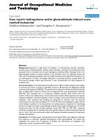
[11–13]. The phosphorylation profile of receptor tyrosine
kinases showed that PDGFRβ kinase exhibited the highest
level of activity and less intense positivity was observed for
EGFR, M-SCFR, Axl and PDGFRα (Fig. 1). Targeted DNA
analysis of the PDGFRB gene and next generation sequencing (NGS) were performed on genomic DNA from peripheral blood samples. We performed Sanger sequencing
of the two PDGFRB regions to detect the presence of the
c.1978C>A (p.Pro660Thr) and c.1681C>T (p.Arg561Cys)
mutations [6] and uncovered a germ-line heterozygous
c.1681C>A missense mutation that had previously been
shown to be an IM causing mutation [14, 15]. To obtain
the complex picture of the genetic background of the case
we performed DNA analysis from peripheral blood with
the Illumina TruSight Cancer panel, which enabled the
sequencing of the hotspots in 94 predisposition cancer genes, according to the standard Illumina protocol
Fig. 1 The relative phosphorylation of kinases in the tumor tissue sample
Page 3 of 7
(Illumina Inc., USA) and identified the heterozygous
Slavic mutation 657del5 in the NBN gene of the NBS.
In the meantime, and based on parental request, the
patient was observed for the next 4 months. He was
doing very well clinically, with a Lansky performance
status of 90% and with respect to his treatment history
with toxicities after chemotherapy; we did not initiate
another chemotherapy regimen but were awaiting the
results of genetic analyses, which have revealed potential
therapeutic targets. Further follow-up confirmed that the
disease continued to progress; several new lesions were
detected within the head and the left orbit, a new one
was detected in the spine, and the spleen lesion had
increased in size.
Due to clear clinical and radiologic progression and
new molecular genetic findings, and with respect to the
history of the disease, we initiated the single agent offlabel treatment with sunitinib 12.5 mg once a day. This
dose corresponded to 2/3 of the recommended adult
dose. An unexpected and dramatic reduction of the
palpable soft tissue and bony lesions on the head was
observed during the 4 weeks of treatment with the single
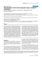
agent sunitinib. An MR scan confirmed the regression of
intracranial and intraorbital lesions as well (Figs. 2, 3
and 4). However, this dosing schedule led to grade 3–4
neutropenia, and the drug was stopped for 4 days. After
only 4 days, we could observe the reactivation of the
skin and soft tissue lesions; therefore, the sunitinib was
given at the same dose every other day. Reactivated
reddish swollen and painful sentinel lesions responded
again to lower doses of sunitinib, but three more weeks
of reduced doses of the single agent sunitinib did not
lead to any further regression of the regressed but still
palpable skin lesions. A low dose of vinblastine was
added to the sunitinib. The starting vinblastine dose was
2 mg/m2; however, based on the further hematological
Mudry et al. BMC Cancer (2017) 17:119
A
Page 4 of 7
B
C
Fig. 2 MRI Frontal view (seq. eFLAIR_long_TR_CLEAR). Two lesions of the left orbit and the skull in the fronto-parietal region (bars). a Before
sunitinib treatment. b Day + 56 of sunitinib. c Day + 156 of sunitinib
toxicity, the dose was tapered down to a 0.4 mg/m2 dose
once weekly.
An unexpected toxicity of sunitinib occurred after 4
months of treatment when accidental hypoglycemia led to
a coma and the patient had to be admitted for glycemia
corrections. Thereafter, the parents were educated on regular feeding before sunitinib administration. Further episodes
of hypoglycemia were not noted. The patient remained on
the treatment paradigm with a marked continuing response
with no disease activity 1 year after the initiation of the
treatment and without any dose limiting toxicities.
Interestingly, the 8 year old sister of the patient, who
had a history of spontaneous regression of subcutaneous
lesions, suffered from the symptomatic re-activation of
the disease when the patient was receiving treatment.
She presented with tumor size of 29 × 24 × 16 mm on
the skull base with night pain. Histopathological and
detailed mutation analyses found the same IM histopathology and the same genotype in the PDGFB and NBN
genes. As with the index case, the sister is doing well on
sunitinib and vinblastine treatment and has exhibited a
rapid response. The nigh pain relieved after 2 weeks on
sunitinib + vinblastine. Initial tumor volume shrinked by
44% after 97 days of combined treatment without any
adverse events requiring reduction of doses. Timeline of
both cases is shown on Additional file 1.
A
B
Discussion and conclusions
Despite the finding that the patient exhibited a partial
response to systemic VAC treatment, the disease continued to progress; moreover, the patient experienced
severe, life threatening dose-limiting toxicities.
Inflammatory myofibroblastic tumors that harbor an
ALK/ROS1 or PDGFRβ kinase fusion are potentially targetable with TKIs due to the presence of a constitutively active
kinase domain that drives cellular proliferation [6, 16]. A
response to the ALK inhibitor crizotinib is reported in
tumors that harbor any of the ALK kinase fusions. Patients
with IMT and ALK negative rearrangements are unlikely to
respond to such targeted treatment.
PDGFRB mutations are reported to be involved in the
pathogenesis of infantile myofibromatosis in a proposed
autosomal dominant pattern with incomplete penetrance
and variable expressivity [7]. The missense PDGFRB
c.1681C>T (R681C) mutation is located in exon 12 and
is predicted to decrease the autoinhibition of the JM
domain (an autoinhibitory domain that masks the catalytic cleft when the receptor is not bound by its ligand)
at baseline, which leads to increased kinase firing and
promotes the formation of myofibromas in tissues with
high PDGFRβ signaling activity. More recently, it was
demonstrated in a cell culture model that the R561C
mutation activates signaling pathways that are normally
C
Fig. 3 MRI Axial view (seq. esT1W_3S_FFE post-contrast). Intracranial lesions of the right temporal and right parieto-occipital regions (bars).
a Before sunitinib treatment. b Day + 56 of sunitinib. c Day + 156 of sunitinib
Mudry et al. BMC Cancer (2017) 17:119
A
Page 5 of 7
B
C
Fig. 4 MRI Sagittal view (seq. esT1W_3S_FFE post-contrast). Frontal and parieto-occipital lesion (bars). a Before sunitinib treatment. b Day + 56 of
sunitinib. c Day + 156 of sunitinib
activated by the stimulated wild-type PDGFRβ receptor
in the absence of PDGF [14]. PDGFR is the immediate
NOTCH3 target gene [17]. If these two signaling pathways are linked and the IM disease-causing mutations in
either PDGFRB or NOTCH3 are demonstrated to be
activating, theoretically, the inhibition of PDGFRB or
NOTCH3 would result in a targeted therapeutic strategy
[7]. Our case report shows the clinical efficacy of such
an approach. Targeted therapy against altered PDGFRβ
with a TKIs inhibitor can overcome tumor growth and
can lead to tumor shrinkage. Compared to the toxicity
of conventional chemotherapy, treatment with sunitinib
was tolerated well except for the occurrence of asymptomatic granulocytopenia and one episode of symptomatic
hypoglycemia. However, the cessation of the drug lead to
increased tumor activity and a decreased drug dose of the
single agent sunitinib led to a stable disease only.
The analysis of tumor tissue or a patient’s samples and
the use of a subsequent results driven treatment provide
a new opportunity for personalized medicine as opposed
to a population based study. Such treatments are supported by new insights into the molecular pathology of
rare diseases, such as IM. A similar strategy would at
least justify the off-label use of new drugs when the individual tumor biology and data about the safety of such
drugs is well defined. TKIs could be an example, as these
drugs are not available to orphan disease patients
because of the absence of appropriate clinical trials. The
careful management and regular observation of the
patient is mandatory, however, in situations where standard approaches are either exploited or ineffective or
absent, the prudent use of targeted agents based on the
mechanism of action might lead to impressive results.
The rapid tumor re-growth that occurred when the
patient was off of the sunitinib during the induction
treatment indicates that metronomic dosing should be
maintained at a lower dose with limited toxicity rather
than being interrupted. The successful use of low dose
vinblastine that is described here, together with the use
of sunitinib at a dose of approximately 1/3 of the usually
recommended dose per kg or m2 in adults, could be at
least in part explained by the fact that targeted agents
could act as biology response modifiers and lower doses
of biological agents and chemotherapy could be nontoxic
and advantageous [18, 19]. This theory is supported by
our observation of the clear disease progression when
sunitinib therapy was interrupted. Regular observations of the patient and preemptive measures such as
the after-feeding dosing of sunitinib should be considered during treatment.
The finding of the Slavic mutation of the NBS was noted
as accidental during NGS sequencing and the relevance
for the disease course is unknown. The toxicity of chemotherapy might be at least in part conditioned by the NBS
mutation As known, the intensity of chemotherapy in
NBS patients must be adapted to individual risk factors
and tolerance. The use of radiomimetics, alkylating agents,
and epipodophyllotoxins should be avoided, and the dose
of methotrexate should be limited [20].
However, the overall duration of such clinically effective treatment remains speculative, especially in patients
with germline mutations. Different approaches that consider cancer to be a chronic disease, such as diabetes,
should be considered in instances in which pathogenic
germline mutations are in place. Should such targeted
agents be maintained for a very long time, e.g., maintenance therapies in childhood acute leukemia, where
other mechanisms of action, not only the cytostatic
effect are in place? [21]. Should some pulses of targeted
agents be considered?
These are only a few of the new questions that arose
by the increased availability of diagnostic methods, such
as NGS and functional proteomics.
The patients with an orphan disease like IM could
benefit from detailed insights into the biology of their
tumor and genome. Such approach is necessary to better understand the molecular pattern of disease and
mechanisms of action of less toxic and effective drugs
except for up to date population-based chemotherapy
regimens. Morover, an unexpected finding of germline
mutation can be important for treatment decisions.
Progressive and resistant incurable infantile myofibromatosis can be successfully treated with the new
approach described herein.
Mudry et al. BMC Cancer (2017) 17:119
Additional file
Additional file 1: Timeline. This file shows timeline of both described
cases. (PDF 466 kb)
Abbreviations
ALK: Anaplastic lymphoma kinase; COG: Children’s oncology group;
EpSSG: European Soft Tissue Sarcoma Study Group; FDG
PET: Fluorodeoxyglucose positron emission tomography; FISH: Fluorescent in
situ hybridization; IHC: Immunohistochemistry; IM: Infantile myofibromatosis;
IMT: Inflammatory myofibroblastic tumor; IVA: Ifosfamide/vincristine/
actinomycine D; MRI: Magnetic resonance imaging; MTD: Maximum tolerated
doses; MTX: Methotrexate; NBS: Nijmegen breakage syndrome; NGS: Next
generation sequencing; PDGFR: Platelet derived growth factor receptor;
PDGFRB: Platelet derived growth factor receptor gene B; PDGFRβ: Platelet
derived growth factor receptor beta; TKI: Tyrosine kinase inhibitor;
VAC: Vincristine/actinomycine D/cyclophosphamide; VBL: Vinblastine
Acknowledgements
The authors thank Drs. Eva Machackova and Lenka Foretova from Masaryk
Memorial Cancer Institute for helpful comments and NGS gene analysis.
Martina Svobodova has substantially contributed to the resolution of
administrative issues of the treatments including insurance coverage.
Funding
This study was supported by projects No. 16-34083A and No. 16-33209A
from the Ministry of Healthcare of the Czech Republic, by project No.
LQ1605 from the National Program of Sustainability II (MEYS CR). The funders
had no role in the study design, data collection and analysis, decision to
publish, or preparation of the manuscript.
Availability of data and materials
The datasets and/or the analyzed current case report are available from the
corresponding author upon reasonable request.
Authors’ contributions
PM performed the review of the literature and wrote the draft of the
manuscript. OS and EM performed the DNA analysis of the PDGRFB gene.
JN and RV designed and performed the phosphoproteomic analysis. JSo
proposed to perform the NGS analysis and participated as clinical geneticist.
KM took care of the patient and participated in the writing of the
manuscript. OR took care of the patient and participated in the writing of
the manuscript. MJ performed the histopathological analysis. AS performed
the radiological evaluation and managed the MRI images. JSt proposed the
study of molecular biology details of the case with a theranostic aim. All of
the authors read and approved the final manuscript.
Competing interests
The authors declare that they have no competing interests.
Consent for publication
Written informed consent for the publication of their clinical details and/or
clinical images was obtained from the parents of the patient. A copy of the
consent form is available for review by the Editor of this journal.
Ethics approval and consent to participate
The study was approved by both the Ethics Committee of the University
Hospital Brno on 9.6.2015 and the Ethics Committee of the School of
Medicine Masaryk University on 23.6.2015, reference number 30/2015. All of
the research described herein was conducted according to the Declaration
of Helsinki. Written informed consent for the tissue and blood analysis and
the off-label treatment of the child with the tyrosine kinase inhibitor was
obtained from parents.
Author details
1
Department of Pediatric Oncology, University Hospital Brno and School of
Medicine, Masaryk University, Cernopolni 9, Brno 613 00, Czech Republic.
2
Central European Institute of Technology, Masaryk University, Kamenice 753/
5, Brno 625 00, Czech Republic. 3Laboratory of Tumor Biology, Department
of Experimental Biology, School of Science, Masaryk University, Kotlarska 2,
Page 6 of 7
Brno 611 37, Czech Republic. 4Division of Medical Genetics, Department of
Biology, University Hospital Brno and School of Medicine, Masaryk University,
Cernopolni 9, Brno 613 00, Czech Republic. 5Department of Pathology,
University Hospital Brno and School of Medicine, Masaryk University,
Cernopolni 9, Brno 613 00, Czech Republic. 6Department of Pediatric
Radiology, University Hospital Brno and School of Medicine, Masaryk
University, Cernopolni 9, Brno 613 00, Czech Republic. 7International Clinical
Research Center, St. Anne’s University Hospital Brno, Pekarska 53, Brno 656
91, Czech Republic.
Received: 2 August 2016 Accepted: 4 February 2017
References
1. Levine E, Fréneaux P, Schleiermacher G, Brisse H, Pannier S, Teissier N, et al.
Risk-adapted therapy for infantile myofibromatosis in children. Pediatr Blood
Cancer. 2012;59:115–20.
2. Johnson K, Notrica DM, Carpentieri D, Jaroszewski D, Henry MM. Successful
treatment of recurrent pediatric inflammatory myofibroblastic tumor in a
single patient with a novel chemotherapeutic regimen containing
celecoxib. J Pediatr Hematol Oncol. 2013;35:414–6.
3. Auriti C, Kieran MW, Deb G, Devito R, Pasquini L, Danhaive O. Remission of
infantile generalized myofibromatosis after interferon alpha therapy.
J Pediatr Hematol Oncol. 2008;30:179–81.
4. Ferrari A, Alaggio R, Meazza C, Chiaravalli S, de Pava MV, Casanova M, et al.
Fibroblastic tumors of intermediate malignancy in childhood. Expert Rev
Anticancer Ther. 2013;13:225–36.
5. Butrynski JE, D’Adamo DR, Hornick JL, Dal Cin P, Antonescu CR, Jhanwar SC,
et al. Crizotinib in ALK -rearranged inflammatory myofibroblastic tumor.
N Engl J Med. 2010;363:1727–33.
6. Lovly CM, Gupta A, Lipson D, Otto G, Brennan T, Chung CT, et al.
Inflammatory myofibroblastic tumors harbor multiple potentially actionable
kinase fusions. Cancer Discov. 2014;4:889–95.
7. Martignetti JA, Tian L, Li D, Ramirez MCM, Camacho-Vanegas O, Camacho
SC, et al. Mutations in PDGFRB cause autosomal-dominant infantile
myofibromatosis. Am J Hum Genet. 2013;92:1001–7.
8. Jo J-C, Hong YS, Kim K-P, Lee J-L, Lee J, Park YS, et al. A prospective
multicenter phase II study of sunitinib in patients with advanced aggressive
fibromatosis. Invest New Drugs. 2014;32:369–76.
9. Skubitz KM, Manivel JC, Clohisy DR, Frolich JW. Response of imatinibresistant extra-abdominal aggressive fibromatosis to sunitinib: case report
and review of the literature on response to tyrosine kinase inhibitors.
Cancer Chemother Pharmacol. 2009;64:635–40.
10. Skoda J, Neradil J, Zitterbart K, Sterba J, Veselska R. EGFR signaling in the
HGG-02 glioblastoma cell line with an unusual loss of EGFR gene copy.
Oncol Rep. 2014;31:480–7.
11. Dewaele B, Floris G, Finalet-Ferreiro J, Fletcher CD, Coindre J-M, Guillou L,
et al. Coactivated platelet-derived growth factor receptor and epidermal
growth factor receptor are potential therapeutic targets in intimal sarcoma.
Cancer Res. 2010;70:7304–14.
12. Ströbel P, Bargou R, Wolff A, Spitzer D, Manegold C, DimitrakopoulouStrauss A, et al. Sunitinib in metastatic thymic carcinomas: laboratory
findings and initial clinical experience. Br J Cancer. 2010;103:196–200.
13. Zhang Y-X, van Oosterwijk JG, Sicinska E, Moss S, Remillard SP,
van Wezel T, et al. Functional profiling of receptor tyrosine kinases
and downstream signaling in human chondrosarcomas identifies
pathways for rational targeted therapy. Clin Cancer Res. 2013;19:
3796–807.
14. Arts FA, Chand D, Pecquet C, Velghe AI, Constantinescu S, Hallberg B, et al.
PDGFRB mutants found in patients with familial infantile myofibromatosis or
overgrowth syndrome are oncogenic and sensitive to imatinib. Oncogene.
2015. doi:10.1038/onc.2015.383.
15. Cheung YH, Gayden T, Campeau PM, LeDuc CA, Russo D, Nguyen V-H, et al.
A recurrent PDGFRB mutation causes familial infantile myofibromatosis. Am
J Hum Genet. 2013;92:996–1000.
16. Davies KD, Doebele RC. Molecular pathways: ROS1 fusion proteins in cancer.
Clin Cancer Res. 2013;19:4040–5.
17. Jin S, Hansson EM, Tikka S, Lanner F, Sahlgren C, Farnebo F, et al. Notch
signaling regulates platelet-derived growth factor receptor-beta expression
in vascular smooth muscle cells. Circ Res. 2008;102:1483–91.
Mudry et al. BMC Cancer (2017) 17:119
Page 7 of 7
18. Reynolds AR. Potential relevance of bell-shaped and u-shaped doseresponses for the therapeutic targeting of angiogenesis in cancer. Doseresponse Publ Int Hormesis Soc. 2009;8:253–84.
19. Reynolds AR, Hart IR, Watson AR, Welti JC, Silva RG, Robinson SD, et al.
Stimulation of tumor growth and angiogenesis by low concentrations of
RGD-mimetic integrin inhibitors. Nat Med. 2009;15:392–400.
20. Chrzanowska K. Nijmegen Breakage Syndrome Treatment & Management.
2016. />Accessed 09 Jan 2016
21. Andre N, Cointe S, Barlogis V, Arnaud L, Lacroix R, Pasquier E, et al.
Maintenance chemotherapy in children with ALL exerts metronomic-like
thrombospondin-1 associated anti-endothelial effect. Oncotarget. 2015;6:
23008–14.
Submit your next manuscript to BioMed Central
and we will help you at every step:
• We accept pre-submission inquiries
• Our selector tool helps you to find the most relevant journal
• We provide round the clock customer support
• Convenient online submission
• Thorough peer review
• Inclusion in PubMed and all major indexing services
• Maximum visibility for your research
Submit your manuscript at
www.biomedcentral.com/submit