Decreased expression of stomatin predicts poor prognosis in HER2-positive breast cancer
Bạn đang xem bản rút gọn của tài liệu. Xem và tải ngay bản đầy đủ của tài liệu tại đây (851.33 KB, 8 trang )
Chen et al. BMC Cancer (2016) 16:697
DOI 10.1186/s12885-016-2681-7
RESEARCH ARTICLE
Open Access
Decreased expression of stomatin predicts
poor prognosis in HER2-positive breast
cancer
Chin-Yau Chen1,2, Chih-Yung Yang1,3, Yen-Chung Chen4, Chia-Wen Shih5, Su-Shun Lo2 and Chi-Hung Lin1*
Abstract
Background: Human epidermal growth factor receptor-2 (HER2) is a transmembrane tyrosine kinase receptor that
is overexpressed in 25 to 30 % of human breast cancers and is preferentially localized in lipid rafts. Stomatin is a
membrane protein that is absent from the erythrocyte plasma membrane in patients with congenital
stomatocytosis and is the major component of the lipid raft.
Results: In a total of 68 clinical cases of HER2-positive breast cancer, the absence of stomatin expression was
associated with a decreased 5-year survival (65 % vs. 93 %, p = 0.005) by survival analysis. For stage I-III HER2-positive
breast cancer, the absence of stomatin expression was associated with a decreased 5-year disease-free survival
(57 % vs. 81 %, p = 0.016) and was an independent prognostic factor by multivariate analysis. Negative stomatin
expression predicts distant metastases in a hazard ratio of 4.0 (95 % confidence interval from 1.3 to 12.5).
Conclusions: These results may suggest that stomatin is a new prognostic indicator for HER2-positive breast
cancer.
Keywords: Breast cancer, Stomatin, HER2, Tumor biomarkers
Abbreviations: AUC, Area under curve; CI, Confidence interval; CMF, Classical CMF chemotherapy, including
cyclophosphamide, methotrexate, and fluorouracil; ER, Estrogen receptor; FISH, Fluorescence in situ hybridization;
HER2, Human epidermal growth factor receptor 2; PR, Progesterone receptor; ROC, Receiver operating curve
Background
Human epidermal growth factor receptor-2 (HER2) is an
important transmembrane tyrosine kinase receptor that
is overexpressed in 25 to 30 % of human breast cancers
[1]. The HER2 receptor is able to promote cell proliferation and is preferentially localized in lipid rafts, which
are special sphingolipid-rich and cholesterol-rich membrane microdomains; these microdomains control activation HER2 by decreasing HER2 homodimerization
and lowering the subsequent spontaneous activation of
the receptor [2]. Trastuzumab (Herceptin®) is a humanized monoclonal antibody that binds to HER2 and
* Correspondence:
Abstract accepted at the 74th Annual Meeting of the Taiwan Surgical
Association, Taipei, Taiwan, Republic of China, March 21–22, 2015.
1
Institute of Microbiology and Immunology, National Yang-Ming University,
155, Sec.2, Li-Nong St, Taipei 11221, Taiwan, Republic of China
Full list of author information is available at the end of the article
inhibits the proliferation and survival of HER2-positive
breast cancers [3].
Stomatin is a membrane protein that is absent from
the erythrocyte plasma membrane in patients with congenital haemolytic anaemia or stomatocytosis [4, 5].
Northern blot analysis has revealed a widespread cellular
distribution of stomatin in reticulocytes, bone marrow,
kidney, brain, gut and heart as well as various cell lines
[6]. Stomatin is the major component of the lipid raft in
the plasma membrane of epithelial cell lines, erythrocytes, and platelet alpha granules [7–11]. Two of the few
well-known functions of stomatin are, firstly, the direct
modulation of the activity of the acid-sensing ion channel and, secondly, the control of glucose transporter type
1 activity [12, 13]. In addition, it has been shown that
hypoxia up-regulates stomatin expression in the cerebral
cortex of rats and alveolar epithelial cells [14, 15]. However, since the discovery of the stomatin in 1982, the
© 2016 Chen et al. Open Access This article is distributed under the terms of the Creative Commons Attribution 4.0
International License ( which permits unrestricted use, distribution, and
reproduction in any medium, provided you give appropriate credit to the original author(s) and the source, provide a link to
the Creative Commons license, and indicate if changes were made. The Creative Commons Public Domain Dedication waiver
( applies to the data made available in this article, unless otherwise stated.
Chen et al. BMC Cancer (2016) 16:697
function of stomatin across a range of different tissues
still remains unknown [4, 6, 16].
Stomatin has been shown to have decreased expression in cancer cells [17]. According to the Swedish Human Protein Atlas project, immunohistochemical
analysis of stomatin protein expression reveals that more
than 75 % of normal breast glandular and myoepithelial
cells are strongly positive for this protein [18]. In contrast, in breast cancer, the expression of stomatin in
these cells was 31 % (7/23) negative, 39 % weak (9/23),
26 % moderate (6/23), and 4 % (1/23) strong positive
when tissue microarrays were analyzed by immunohistochemistry [18].
Although stomatin is expressed in a significant proportion of breast cancers, the relationship between stomatin
expression and breast cancer has not been explored in
detail. Recently reported by Arkhipova and colleagues in
2014, stomatin is down-regulated in non-small cell lung
cancer and is associated with lymph node metastases
[19]. This is the first and the only one study to demonstrate that stomatin has a role in carcinogenesis. In comparison, stomatin-like protein 2, which shows a high
degree of sequence similarity to stomatin, had been reported to be associated with a decreased overall survival
among breast cancer [20], pulmonary squamous carcinoma [21], glioma [22], endometrial adenocarcinoma
[23], laryngeal squamous carcinoma [24], esophageal
squamous carcinoma [25] and colorectal cancer [26]
patients.
Stomatin is the major component of lipid raft where
HER2 is known to be clustered and therefore it seems
likely that stomatin expression may have an impact on
the pathology of HER2-positive breast cancer. In the
present study, the relationship of stomatin expression
and the clinical survival outcome was explored for patients with HER2-positive breast cancer.
Methods
The archival formalin-fixed paraffin-embedded tissue
samples obtained from women diagnosed of infiltrating
ductal carcinoma of female breast from 2001 to 2012. The
women of histologies other than infiltrating ductal carcinoma were excluded. All HER2-positive cases were either
HER2 immunohistochemistry 3+ or 2+ (medium positive)
which was further confirmed by fluorescence in situ
hybridization (FISH) to identify HER2 gene amplification
[27]. There were 5 cases excluded where the HER2 immunohistochemistry results were 2 + but the FISH studies
failed. Tumor grade was defined according to the (Scarff)
Bloom-Richardson (BR) grading system. The results for
ER, PR, and HER2 were obtained from the medical records. Cases where ER and PR were found in more than
5 % of the tumor cells were considered to be positive.
Cancer staging was based on the American Joint
Page 2 of 8
Committee on Cancer (AJCC seventh Edition). All the patients were operated by either one of the two breast surgeons (C-Y Chen and S-S Lo). Chemotherapy was given
according to the institutional guidelines and policy of National Health Insurance Administration in Taiwan.
Anthracycline chemotherapy was unrestricted but taxane
chemotherapy was insurance-paid only for patients whose
cancer was locally advanced or metastatic. For targeted
therapy, palliative trastuzumab therapy was insurancepaid when distant metastasis occurred. In patients without
a distant event, adjuvant trastuzumab was insurance-paid
for patients with positive lymph node status and this policy was only effective after 2010. The duration of trastuzumab therapy was allowed for 1 year at most. In this study,
no patient had ever received targeted therapy other than
trastuzumab. The study was held in the National YangMing University Hospital, located in the north I-Lan
County, Taiwan. The clinical outcomes of the patients
were surveyed until December 31, 2014. Institutional review board approval was obtained before acquisition of
patient health information.
Tissue sections (4 μm thick) were subjected to heatinduced antigen retrieval in the presence of 0.01 M sodium citrate buffer, pH 6.0 for 30 min using a pressure
boiler. Immunohistochemical staining was performed
using a polyclonal primary antibody against stomatin
(Atlas, HPA011419, Uppsala, Sweden) that was diluted
at 1:400; incubation took place for 30 min. Positive control was selected from breast cancer tissue of known
strong positive immunoreactivity for stomatin expression. Negative control experiments were conducted by
replacing the primary antibody with phosphate-buffered
saline.
Immunostaining was scored by the researcher (C-Y
Chen) and the junior pathologist (Y-C Chen), who were
blinded to the patients’ outcome and other clinicopathological parameters. Discordant scores were reevaluated
by the senior pathologist (C-W Shih), and a consensus
score was used for further analysis. Two features, intensity and extent of immunoreactivity, were assessed as described in a previous report by Chang and colleagues
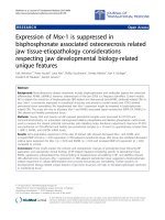
[21]. The intensity of the immunostaining was classified
in four categories: 0, no brown particles in the tumor
cytoplasm or cell membrane; 1, faint brown staining of
cell membrane; 2, weak but definite brown staining of
cell membrane; and 3, deep brown staining of cell membrane together with staining of cytoplasm (Fig. 1). The
percentage of positive cells were determined and classified into four groups: 1, fewer than 25 % positive tumor
cells; 2, 25 to 50 % positive tumor cells; 3, 51 to 75 %
positive tumor cells; and 4, more than 75 % positive
tumor cells. The immunostaining index was the product
of the two scores. We produced time-dependent receiver
operating characteristics (ROC) curves [28, 29] for
Chen et al. BMC Cancer (2016) 16:697
Fig. 1 Immunohistochemical staining of breast cancer tissues. I–IV
Expression of stomatin in breast cancer tissue showing the intensity
score. I) Intensity score 0, no brown particles in the tumor cytoplasm
or cell plasma membrane. II) Intensity score 1, faint brown staining
of cell membrane. III) Intensity score 2, weak but definite brown
staining of cell membrane. IV) Intensity score 3, deep brown staining
of cell membrane with staining of cytoplasm
evaluation of the immunostaining indices. Area under
curve (AUC) at 2- and 5-year survival was 0.765 and
0.543, respectively, suggesting a decrease in the statistical power of stomatin immunoreactivity over time,
which may be explained by the early relapse of the
HER2-positive breast cancer. The optimal cutoff values
were 2 for 5-year-survival ROC and 7 for 2-year-survival
ROC. Therefore, we defined positive expression of stomatin protein as a staining index of 4 or more, while a
staining index from 0 to 3 was indicative of negative stomatin expression.
Statistical analyses were performed using STATA for
Windows 10.0 (StataCorp, College Station, TX). The
Student’s t test and Fisher’s exact test were used for statistical analysis as appropriate. We estimated the survival
curves using the Kaplan-Meier product limit method
[30]. Breast cancer death was defined as death related to
distant metastases. Distant disease-free survival was defined as time to distant metastasis, excluding local or regional recurrence. The log-rank test was used to assess
the association of survival with stomatin expression. Cox
regression analysis was performed to compute hazard ratios and 95 % confidence intervals (CI) and to evaluate
the effects of confounding factors during the multivariate analysis. For all statistical tests, p < 0.05 was considered to be significant. All p values were two-sided.
Page 3 of 8
Results
Using 68 HER2-positive and 58 HER2-negative samples
of infiltrating ductal carcinomas of the female breast,
stomatin protein expression was found to be localized
mainly in the plasma membrane and partially in the
cytosol. For HER2-positive patients, the overall immunohistochemistry staining results showed weak or absent
staining (staining index <4) in 32 cases (47 %) and positive staining (staining index ≥4) in 36 cases (53 %).
There was no statistical difference in patient age, cancer
grade, cancer stage, and expression of estrogen receptor/
progesterone receptor among women of positive stomatin expression compared with those of negative stomatin
expression (Table 1). There was not statistical difference
in types of surgery (mastectomy vs. lumpectomy)
(Table 1). Most women received an anthracycline-based
chemotherapy as first-line adjuvant chemotherapy. For
women of HER2-positive cancers, there were 4 received
CMF chemotherapy (classical CMF: cyclophosphamide,
methotrexate, and fluorouracil) and one woman received
taxane only because of heart disease. There were 5
women of HER2-positive cancer did not receive any adjuvant chemotherapy at all, including 2 women of early
stage and 3 women who refused chemotherapy. There
was not statistical difference in types of chemotherapy
(Table 1). The proportions of patient who had ever received trastuzumab therapy were 42 % (15/36) in
stomatin-positive group and 47 % (15/32) in stomatinnegative group, where there was no statistical difference
in the proportions in receiving trastuzumab (Table 1).
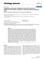
In the 68 women of stage I-IV HER2-positive infiltrating ductal carcinomas, mean follow-up time was
5.0 years. Kaplan-Meier plot (Fig. 2) showed that the 5year breast cancer-specific survival rates were 93 %
(95 % confidence interval = 76 to 98 %) for women of
positive stomatin expression and 65 % for women of
negative stomatin expression (95 % confidence interval
= 38 to 83 %, p = 0.005). Namely, negative stomatin
expression was significantly associated with a lower 5year-survival rate. In comparison, there was no survival
difference in patients with HER2-negative breast cancers.
In women of HER2-negative cancers, the 5-year breast
cancer-specific survival rates were 84 % (95 % confidence interval = 67 to 92 %) for women of positive stomatin expression and 94 % for women of negative
stomatin expression (95 % confidence interval = 67 to
99 %, p = 0.193).
In the follow-up of 65 women of stage I-III HER2positive cancer, there were lung metastases in 5, bone
metastases in 4, liver metastases in 3, lung & bone metastases in 1, lung & liver metastases in 2, distant lymph
nodes metastases in 1, and local recurrence without a
distant metastasis in two women. When the woman of
local recurrence without a distant event was not
Chen et al. BMC Cancer (2016) 16:697
Page 4 of 8
Table 1 Patient age, tumor characteristics, hormone receptor status, and systemic treatment in relation to stomatin expression in
women with HER2-positive breast cancers (n = 68) and HER2-negative breast cancers (n = 58)
Patient
Characteristics
HER2-positive
HER2-negative
Stomatin (+)
(n = 36)
Stomatin (−)
(n = 32)
p values
Stomatin (+)
(n = 38)
Stomatin (−)
(n = 20)
p values
Mean age (year)
53
58
0.109*
54
52
0.659*
I
6
4
0.432†
3
1
0.882†
II
27
22
30
15
III
3
6
5
4
9
5
13
5
Grade
Stage
I
0.420†
II
13
17
12
8
III
13
8
12
7
IV
1
2
1
0
Positive
21
17
27
16
Negative
15
15
11
4
Positive
20
14
25
16
Negative
16
18
13
4
No
17
13
10
4
Yes
19
19
28
16
22
18
32
15
0.857†
Estrogen receptor
0.807†
0.541†
Progesterone receptor
0.466†
0.366†
Hormonal therapy
0.631†
0.751†
Surgery
Mastectomy
0.711†
Lumpectomy
14
13
5
5
No surgery
0
1
1
0
3
2
10
9
20
20
23
8
0.540†
Chemotherapy
None
0.702†
0.333†
Adjuvant/Neoadjuvant
Anthracycline
Anthracycline + Taxane
8
7
4
3
CMF§
3
1
0
0
Taxane
1
0
0
0
Anthracycline
1
0
1
0
Anthracycline + Taxane
0
2
0
0
No
21
17
N/A
N/A
Adjuvant
12
7
Palliative
3
8
Palliative
Trastuzumab therapy
0.175†
N/A
*Student’s t test; †Fisher’s exact test; §CMF: classical CMF, cyclophosphamide, methotrexate, and fluorouracil
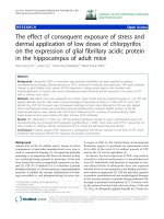
regarded as failure of disease-free status, 5-year distant
disease-free survival were 81 % (95 % confidence interval = 60 to 92 %) for women of positive stomatin expression and 57 % for women of negative stomatin
expression (95 % confidence interval = 33 to 75 %, p =
0.016, Fig. 3). In comparison, there was no difference in
patients with HER2-negative breast cancers. The 5year distant distant disease-free survival rates were
Chen et al. BMC Cancer (2016) 16:697
Page 5 of 8
Fig. 2 Kaplan-Meier method showing 5-year breast cancer-specific survival curves of HER2-positive infiltrating ductal carcinoma of breast comparing
patients of positive and negative stomatin staining. Significant reduction of proportion surviving was noted in patients of stomatin (−) (n = 32),
compared with patients of stomatin (+) (n = 36). p = 0.005
64 % (95 % confidence interval = 46 to 77 %) for
women of positive stomatin expression and 74 % for
women of negative stomatin expression (95 % confidence interval = 46 to 88 %, p = 0.479) for HER2negative patients.
For women of HER2-positive cancers, when either
local recurrences or distant metastases were regarded
as failure, the 5-year disease-free survival were 79 %
(95 % confidence interval = 58 to 90 %) for women of
positive stomatin expression and 59 % for women of
negative stomatin expression (95 % confidence interval = 36 to 77 %, p = 0.037).
Although hormonal receptors expression, cancer stage,
and adjuvant trastuzumab therapy were all known prognostic factors for HER2-positive breast cancer, multivariate analyses revealed stomatin was an independent
factor for cancer metastases (p = 0.017, Table 2) for stage
I-III HER2-positive breast cancers. Negative stomatin
Fig. 3 Kaplan-Meier method showing 5-year disease-free (distant metastases-free) survival curves of HER2-positive infiltrating ductal carcinoma of
breast comparing patients of positive and negative stomatin staining. Significant reduction of proportion surviving was noted in patients of
stomatin (−) (n = 30), compared with patients of stomatin (+) (n = 35). p = 0.016
Chen et al. BMC Cancer (2016) 16:697
Page 6 of 8
Table 2 Multivariate Cox model for distant metastases-free survival in women of HER2-positive infiltrating ductal carcinoma,
stage I-III (n = 65)
Distant metastases
Risk ratio (95 % CIa)
Parameter
‡
Hormonal receptors
p
0.405
ER (+) or PR (+)
1.0 (referent)
ER (−) and PR (−)
1.5 (0.6 to 4.3)
Stage
0.016
I–II
1.0 (referent)
III
3.5 (1.3 to 9.9)
Adjuvant trastuzumab
0.147
Yes
1.0 (referent)
No
3.1 (0.7 to 14.8)
Stomatin expression
0.017
Positive
1.0 (referent)
Negative
4.0 (1.3 to 12.5)
‡
ER estrogen receptor, PR progesterone receptor
a
CI confidence interval
expression predicts distant metastases in a hazard ratio
of 4.0 (95 % confidence interval from 1.3 to 12.5,
Table 2).
Discussion
The present study provides evidence showing a correlation between stomatin protein expression and HER2positive breast cancer prognosis. Compatible with a previous immunohistochemistry study, which showed that
31 % of the breast cancers were negative and 39 % weak
for stomatin staining, the overall immunohistochemistry
staining in our study showed negative staining in 47 %
(32/68) of samples and positive staining in 53 % (36/68)
of samples. Because there was no previous study to define the positivity according the expression level of stomatin, we defined the cutoff point of positive expression
to be a staining index of 4 or more according to a preliminary tome-dependent ROC analysis. It was found
that negative stomatin expression was associated with a
decreased breast cancer-specific survival and diseasefree survival using survival analyses. When distant metastases were defined as cancer recurrence for patients
of stage I-III, stomatin was also an independent prognostic factor using multivariate analysis in this study.
Although stomatin-like protein 2, which is a member of
the stomatin protein family, has been widely reported to be
related to various kinds of cancers, there is only one report
up to the present indicating an association between stomatin expression and carcinogenesis [19]. Arkhipova’s and
colleagues recently reported that down-regulation of stomatin mRNA was correlated with positive lymph node
status in 48 patients [19]. In our study, we initially proposed that lipid-raft localized stomatin might be able to
modulate the activity of the HER2-positive breast cancer.
The results that absence of stomatin expression might predict distant metastases in HER2-positive breast cancer are
comparable to that of Arkhipova’s study for lung cancer
[19].
One reason why previous researchers may have overlooked the relationship between stomatin and carcinogenesis may be because only a subgroup of breast
cancers, namely the HER2-positive cancers, is affected
by stomatin expression. In our study, when the 58 patients with HER2-negative breast cancer were analyzed,
there was no survival difference between the stomatinpositive group and the stomatin-negative group. Although why stomatin down-regulation is associated with
metastases in HER2-positive cancers remains uncertain,
our results may suggest an interaction between HER2
receptor and stomatin in the lipid raft microdomains.
In Taiwan, 90 % of the invasive breast cancers are infiltrating ductal carcinoma and less than 4 % are infiltrating lobular carcinoma, with smaller percentages in other
histology [31]. For reducing bias among different histologies, we chose women of the commonest histological
type, infiltrating ductal carcinoma, as our study subjects.
In our preliminary study for the histologies other than
infiltrating ductal carcinoma, we can not make any conclusion in the influence of stomatin expression to cancer
outcomes because the sample size is too small. Further
large studies are needed to confirm the relationship between stomatin expression and disease outcomes in
other histological types.
Gene expression patterns have distinguished several
subtypes of breast carcinomas [32]. In a study reported
by Prat el al., all molecular subtypes were identified in
the 468 clinical HER2-positive tumors, including HER2enriched (47 %), luminal B (28.2 %), basal-like (14.1 %),
and luminal A (10.7 %) [33]. The molecular subtypes significantly affected survival outcomes [33, 34]. In our
study, although stomatin was an independent factor in
multivariate analysis, the molecular patterns of the
tumors were unknown. Future studies exploring gene
expression profiles are warranted.
By definition, disease-free survival usually includes
local or regional recurrence in addition to distant metastases. However, the impact of local recurrences on survival remains uncertain. Local recurrence itself may be
caused by failed local treatment but not by cancer progression. In this study, we defined disease-free survival
as time to distant recurrence, excluding local or regional
recurrence. Our study provided the valuable results that
negative stomatin expression predicts distant metastases
in a hazard ratio of 4.0 (95 % confidence interval from
1.3 to 12.5) in multivariate analyses.
Chen et al. BMC Cancer (2016) 16:697
In randomized controlled trials, patients receiving adjuvant trastuzumab showed a significant reduction in
mortality and recurrence as compared to those of no adjuvant treatment [35]. The limitation of this study was
that the adjuvant trastuzumab was not used for most
women because of national insurance policy. Only
women had positive lymph nodes were afforded adjuvant
trastuzumab and this policy was effective after 2010. Because the patients who had ever received adjuvant trastuzumab were a minor group (28 %, 19 of 68, Table 1)
and had a short follow-up time, the influence of trastuzumab treatment on stomatin expression could not be
well explored. A clinical trial recruiting more patients is
needed in the future. However, in the multivariate analyses, stomatin expression was an independent prognostic indicator from trastuzumab treatment, with a higher
hazard ratio than that of adjuvant trastuzumab (hazard
ratio 4.0 vs. 3.1). This result may suggest a different
pathway of cancer progression in stomatin compared to
that of HER2 receptor.
Conclusions
Our findings suggest stomatin as one potential prognostic factor that predicts the progression in HER2-positive
breast cancer. Further studies investigating the mechanism whereby stomatin affects HER2-positive breast cancer and how stomatin interacts with HER2 are needed.
Acknowledgements
The authors thank Dr. Pei-Shi Hung in her help with bench works.
Funding
The study was supported by National Science Council Project NSC 99-2314B-010-045, MOST 104-2320-B-010-043-MY3, MOST 105-2319-B010-001, Taiwan,
Republic of China.
Availability of data and materials
The datasets during and/or analysed during the current study are available
from the corresponding author on reasonable request.
Authors’ contributions
C-Y C, S-S L and C-H L analyzed and interpreted the patient data and were
major contributors in writing the manuscript. C-Y C, C-Y Y, Y-C C, C-W S
performed the histological examination. All authors read and approved the
final manuscript.
Competing interests
The authors declare that they have no competing interests.
Consent for publication
Not applicable.
Ethics approval and consent to participate
The Institutional Review Board of National Yang-Ming University Hospital
approved this study. (IRB No.: 2011A011).
Author details
1
Institute of Microbiology and Immunology, National Yang-Ming University,
155, Sec.2, Li-Nong St, Taipei 11221, Taiwan, Republic of China. 2Department
of Surgery, National Yang-Ming University Hospital, Yilan County, Taiwan,
Republic of China. 3Department of Education and Research, Taipei City
Hospital, Taipei, Taiwan, Republic of China. 4Department of Pathology,
National Yang-Ming University Hospital, Yilan County, Taiwan, Republic of
Page 7 of 8
China. 5Department of Pathology, Lotung Poh-Ai Hospital, Yilan County,
Taiwan, Republic of China.
Received: 4 February 2015 Accepted: 3 August 2016
References
1. Slamon DJ, Godolphin W, Jones LA, Holt JA, Wong SG, Keith DE, et al.
Studies of the HER-2/neu proto-oncogene in human breast and ovarian
cancer. Science. 1989;244(4905):707–12.
2. Nagy P, Vereb G, Sebestyen Z, Horvath G, Lockett SJ, Damjanovich S, et al.
Lipid rafts and the local density of ErbB proteins influence the biological
role of homo- and heteroassociations of ErbB2. J Cell Sci. 2002;115(Pt 22):
4251–62.
3. Hudis CA. Trastuzumab—mechanism of action and use in clinical practice.
N Engl J Med. 2007;357(1):39–51.
4. Lapatsina L, Brand J, Poole K, Daumke O, Lewin GR. Stomatin-domain
proteins. Eur J Cell Biol. 2012;91(4):240–5.
5. Stewart GW, Argent AC, Dash BC. Stomatin: a putative cation transport
regulator in the red cell membrane. Biochim Biophys Acta.
1993;1225(1):15–25.
6. Stewart GW, Hepworth-Jones BE, Keen JN, Dash BC, Argent AC, Casimir CM.
Isolation of cDNA coding for an ubiquitous membrane protein deficient in
high Na+, low K+ stomatocytic erythrocytes. Blood. 1992;79(6):1593–601.
7. Snyers L, Umlauf E, Prohaska R. Association of stomatin with lipid-protein
complexes in the plasma membrane and the endocytic compartment. Eur J
Cell Biol. 1999;78(11):802–12.
8. Salzer U, Prohaska R. Stomatin, flotillin-1, and flotillin-2 are major integral
proteins of erythrocyte lipid rafts. Blood. 2001;97(4):1141–3.
9. Mairhofer M, Steiner M, Mosgoeller W, Prohaska R, Salzer U. Stomatin is a
major lipid-raft component of platelet alpha granules. Blood. 2002;100(3):
897–904.
10. Simons K, Ikonen E. Functional rafts in cell membranes. Nature. 1997;
387(6633):569–72.
11. Mrowczynska L, Salzer U, Perutkova S, Iglic A, Hagerstrand H. Echinophilic
proteins stomatin, sorcin, and synexin locate outside gangliosideM1 (GM1)
patches in the erythrocyte membrane. Biochem Biophys Res Commun.
2010;401(3):396–400.
12. Price MP, Thompson RJ, Eshcol JO, Wemmie JA, Benson CJ. Stomatin
modulates gating of acid-sensing ion channels. J Biol Chem. 2004;279(51):
53886–91.
13. Montel-Hagen A, Kinet S, Manel N, Mongellaz C, Prohaska R, Battini JL, et al.
Erythrocyte Glut1 triggers dehydroascorbic acid uptake in mammals unable
to synthesize vitamin C. Cell. 2008;132(6):1039–48.
14. Wang Y, Cao D, Chen J, Liu A, Yu Q, Song X, et al. Distribution of stomatin
expressing in the central nervous system and its up-regulation in cerebral
cortex of rat by hypoxia. J Neurochem. 2011;116(3):374–84.
15. Chen JC, Cai HY, Wang Y, Ma YY, Song LN, Yin LJ, et al. Up-regulation of
stomatin expression by hypoxia and glucocorticoid stabilizes membraneassociated actin in alveolar epithelial cells. J Cell Mol Med.
2013;17(7):863–72.
16. Lande WM, Thiemann PV, Mentzer Jr WC. Missing band 7 membrane
protein in two patients with high Na, low K erythrocytes. J Clin Invest. 1982;
70(6):1273–80.
17. Liang X, Zhao J, Hajivandi M, Wu R, Tao J, Amshey JW, et al. Quantification
of membrane and membrane-bound proteins in normal and malignant
breast cancer cells isolated from the same patient with primary breast
carcinoma. J Proteome Res. 2006;5(10):2632–41.
18. Uhlen M, Oksvold P, Fagerberg L, Lundberg E, Jonasson K, Forsberg M, et al.
Towards a knowledge-based Human Protein Atlas. Nat Biotechnol. 2010;
28(12):1248–50.
19. Arkhipova KA, Sheyderman AN, Laktionov KK, Mochalnikova VV,
Zborovskaya IB. Simultaneous expression of flotillin-1, flotillin-2, stomatin
and caveolin-1 in non-small cell lung cancer and soft tissue sarcomas.
BMC Cancer. 2014;14:100.
20. Cao W, Zhang B, Liu Y, Li H, Zhang S, Fu L, et al. High-level SLP-2 expression
and HER-2/neu protein expression are associated with decreased breast
cancer patient survival. Am J Clin Pathol. 2007;128(3):430–6.
21. Chang D, Ma K, Gong M, Cui Y, Liu ZH, Zhou XG, et al. SLP-2 overexpression
is associated with tumour distant metastasis and poor prognosis in
pulmonary squamous cell carcinoma. Biomarkers. 2010;15(2):104–10.
Chen et al. BMC Cancer (2016) 16:697
Page 8 of 8
22. Song L, Liu L, Wu Z, Lin C, Dai T, Yu C, et al. Knockdown of stomatin-like
protein 2 (STOML2) reduces the invasive ability of glioma cells through
inhibition of the NF-kappaB/MMP-9 pathway. J Pathol. 2012;226(3):534–43.
23. Cui Z, Zhang L, Hua Z, Cao W, Feng W, Liu Z. Stomatin-like protein 2 is
overexpressed and related to cell growth in human endometrial
adenocarcinoma. Oncol Rep. 2007;17(4):829–33.
24. Cao WF, Zhang LY, Liu MB, Tang PZ, Liu ZH, Sun BC. Prognostic significance
of stomatin-like protein 2 overexpression in laryngeal squamous cell
carcinoma: clinical, histologic, and immunohistochemistry analyses with
tissue microarray. Hum Pathol. 2007;38(5):747–52.
25. Zhang L, Ding F, Cao W, Liu Z, Liu W, Yu Z, et al. Stomatin-like protein 2 is
overexpressed in cancer and involved in regulating cell growth and cell
adhesion in human esophageal squamous cell carcinoma. Clin Cancer Res.
2006;12(5):1639–46.
26. Yeoh LC, Loh CK, Gooi BH, Singh M, Gam LH. Hydrophobic protein in
colorectal cancer in relation to tumor stages and grades. World j
gastroenterol: WJG. 2010;16(22):2754–63.
27. Wang S, Saboorian MH, Frenkel E, Hynan L, Gokaslan ST, Ashfaq R.
Laboratory assessment of the status of Her-2/neu protein and oncogene in
breast cancer specimens: comparison of immunohistochemistry assay with
fluorescence in situ hybridisation assays. J Clin Pathol. 2000;53(5):374–81.
28. Heagerty PJ, Lumley T, Pepe MS. Time-dependent ROC curves for censored
survival data and a diagnostic marker. Biometrics. 2000;56(2):337–44.
29. R Core Team R: A Language and Environment for Statistical Computing
[]. Accessed 24 Aug 2016.
30. Kaplan E, Meier P. Nonparametric estimation from incomplete observations.
J Am Stat Assoc. 1958;53:457–81.
31. Taiwan Cancer Registry [ />Accessed 24 Aug 2016.
32. Sorlie T, Perou CM, Tibshirani R, Aas T, Geisler S, Johnsen H, et al. Gene
expression patterns of breast carcinomas distinguish tumor subclasses with
clinical implications. Proc Natl Acad Sci U S A. 2001;98(19):10869–74.
33. Prat A, Carey LA, Adamo B, Vidal M, Tabernero J, Cortes J, Parker JS, Perou
CM, Baselga J. Molecular features and survival outcomes of the intrinsic
subtypes within HER2-positive breast cancer. J Natl Cancer Inst. 2014;106(8):
1–8.
34. Falck AK, Ferno M, Bendahl PO, Ryden L. St Gallen molecular subtypes in
primary breast cancer and matched lymph node metastases–aspects on
distribution and prognosis for patients with luminal A tumours: results from
a prospective randomised trial. BMC Cancer. 2013;13:558.
35. Viani GA, Afonso SL, Stefano EJ, De Fendi LI, Soares FV. Adjuvant
trastuzumab in the treatment of her-2-positive early breast cancer: a metaanalysis of published randomized trials. BMC Cancer. 2007;7:153.
Submit your next manuscript to BioMed Central
and we will help you at every step:
• We accept pre-submission inquiries
• Our selector tool helps you to find the most relevant journal
• We provide round the clock customer support
• Convenient online submission
• Thorough peer review
• Inclusion in PubMed and all major indexing services
• Maximum visibility for your research
Submit your manuscript at
www.biomedcentral.com/submit