Evaluation of CD68 in oral squamous cell carcinoma and their relation with clinicopathological parameters –An immunohistochemical study
Bạn đang xem bản rút gọn của tài liệu. Xem và tải ngay bản đầy đủ của tài liệu tại đây (739.85 KB, 8 trang )
Int.J.Curr.Microbiol.App.Sci (2020) 9(7): 3832-3839
International Journal of Current Microbiology and Applied Sciences
ISSN: 2319-7706 Volume 9 Number 7 (2020)
Journal homepage:
Original Research Article
/>
Evaluation of CD68 in Oral Squamous Cell Carcinoma and their Relation
with Clinicopathological Parameters –An Immunohistochemical Study
Ahlam T. Bdewi1, Ahmed A. Alkadir Mohamed Labib2, Ban F. AL Drobie3,
Bashar H. Abdullah3 and Museedi Omar3
1
2
Department of Oral Diagnosis/ College of Dentistry /Al Anbar University,Iraq
Department of Clinical Oncology/, Clinical Oncologist, Al-Amal Hospital, Baghdad, Iraq
3
Department of Oral Diagnosis/ Oral Pathology, College of Dentistry,
Baghdad University, Iraq
*Corresponding author
ABSTRACT
Keywords
Oral Squamous Cell
Carcinoma, CD68
Immunohistochemical Study
Article Info
Accepted:
22 June 2020
Available Online:
10 July 2020
Macrophages are important cells for the innate immunity. Circulating monocytes are
attracted to tissues by chemotactic factors and become macrophages under the influence of
their microenvironment. Several studies have shown that macrophages are important to the
prognosis of patients with different types of cancer. The aim of this study was to evaluate
the expression of CD68 in OSCC patients and to investigate the possible relationship of
macrophages using CD68 in various histopathological grades, stages and other clinical
parameters of OSCC. preoperative diagnosed specimens (n=30)were processed for
immunohistochemistry to detect CD68 positive cells. Expression of macrophages was
semiquantitatively analyzed. Immunohistochemical study show expression of CD68 in all
specimens. A significant correlation between CD68 infiltration and gender of patients was
found,where it high in females than males. Other clinicopathological findings of OSCC
show non-significant correlation. Immunohistochemical expression of CD68 were
observed in all studying samples of oral squamous cell carcinoma. however, statistically
non-significant correlation was found between the mean expression level of these
infiltrates macrophages with clinicopathological findings of OSCC expect with Gender.
Introduction
Oral squamous cell carcinoma (OSCC)
accounts for about 90% of all oral neoplasms
(1). Despite advanced therapeutic approaches,
the 5 years survival rate of patients with
OSCC remains <50%, mainly due to the
presence of regional lymph node metastasis
(2).
Like other cancer types, OSCC have a
complex tumor microenvironment (TME)
with the presence of several stromal cells that
might be associated to tumor aggressiveness
and the resistance and failure to antineoplastic treatment (4, 5).
Among immune cells, tumor-associated
macrophages (TAMs) are the most abundant
3832
Int.J.Curr.Microbiol.App.Sci (2020) 9(7): 3832-3839
and important stromal cells in the TME (6).
TAMs are considered important players in
tumor progression and are related to
proliferation and survival of tumor cells,
angiogenesis, invasion of surrounding tissues
and metastasis (7).
Tumor microenvironment is a complex
system where tumor cells reprogram stromal
cells for their own benefit. From the ten
hallmarks of cancer described by Hanahan
and Weinberg (2011), these reprogramed cells
contribute to at least seven hallmarks:
sustaining proliferative signaling, evading
growth suppressors, avoiding immune
destruction, deregulating cellular energetics,
resisting cell death, inducing angiogenesis
and activating invasion and metastasis. For
instance ,during oral squamous cell carcinoma
progression, the reprograming of stromal cells
already starts in potentially malignant
disorders, where altered non-neoplastic cells
induce angiogenesis and modifications in
immune cells in the adjacent connective
tissue. When these altered non-neoplastic
cells become malignant cells and invade
adjacent connective tissue shortly after basal
membrane rupture, there is switch to an
immunosuppressive TME, which allows
tumor development.
Macrophages, which derive from circulating
monocytes, play an important role in innate
immunity and adaptive immunity [9-10].
Importantly, CD68 has been widely used as a
pan macrophage marker in over 80 % of
studies. Several reports found that the
presence of CD68 + TAMs was associated
with decreased 5-year survival rates in several
cancer including thyroid, lung, hepatocellular
and esophageal cancers [11.12]. But some
studies
showed
that
no
significant
associations were found between the
percentage of CD68 + TAM and survival time
for OSCC patients.
It had been reported that the presence of
CD68 + TAMs in tumor nest promoted tumor
progression and was related to poor prognosis
in hepatocellular cancer patients [13].
Infiltration of CD68 + macrophages in breast
tumor stroma were positively associated to
tumor size and were an independent
prognostic factor (14).
Here, we focused on the expression of of
CD68 + TAMs in OSCC and analyzed the
correlations of expression of CD68 + TAM
with the clinicopathological parameters.
Materials and Methods
Patients and tissue sample
In this retrospective study, OSCC tissue
specimens from 30 patients were obtained
from the archive department of oral diagnosis,
Collage of Dentistry, Baghdad University.
The study protocol was approved by the
ethical committee. The specimens used in this
study were obtained from tissue samples
collected for routine histopathological
diagnosis, where the diagnosis of OSCC was
based on histologic examination of
hematoxylene and eosin-stained tissue
sections. Specimens were obtained from total
surgical excision of the lesions. The clinical
data (age, gender, tumor location and size)
were obtained from medical records.
Immunohistochemical staining
The formaline –fixed,paraffine –embeded
tissue samples were sliced into 4mm
thickness with a rotation microtome.
Immunohistochemical staining was performed
using peroxidased –labeled streptavidin-biotin
technique. Briefly, tissue sections were
dewaxed in xylole and rehydrate in graded
alcohol and heated in metal slide holder
containing citrate buffer (pH6.0) at 95C for
20 minutes to retrieve antigenicity. Then
3833
Int.J.Curr.Microbiol.App.Sci (2020) 9(7): 3832-3839
sections treated with H2O2 for 10 minutes to
quench endogenous peroxidase activity. Then
treated with protein block to block
nonspecific binding. After washing in 10 mm
PBS, sections were incubated with following
primary antibodies: monoclonal mouse anti
human CD68 (clone KP1 + C68/684;
ab199000; abcam, Cambridge, UK) at
0.5/500, at 37 degree for 1 hour and overnight
incubation at in humidified chamber. After
washing, sections were treated with mouse
and rabbit specific HRP/DAB detection
system (ab236466, abcam, Cambridge, UK).
Finally, the sections were stained with Mayers
hematoxylene and covered. Samples of lymph
node were used as positive control for marker.
Negative control were obtained by omitting
the primary antibodies.
Statistical analysis
The data analyzed using Statistical Package
for Social Sciences (SPSS) version 25. The
data presented as mean, standard deviation
and ranges. Categorical data presented by
frequencies and percentages. Independent ttest and Analysis of Variance (ANOVA) (two
tailed) was used to compare the continuous
variables accordingly. A level of P – value
less than 0.05 was considered significant.
Results and Discussion
Patient Characteristics
The clinico-pathological characteristics of all
of the patients are summarized in Table.1.
Study patients’ age was ranging from 22 to 85
years with a mean of 55.6 years and a
standard deviation of ± 14.7 years. We
noticed that the highest proportion of study
patients was aged > 50 years (63.3%.
(Regarding gender, proportion of males was
higher than females (63.3% versus 36.7%)
with a male to female ratio of 1.7:1
In this study, the lesions were staged IV in
60% of case. Regarding grading, 56.7% of
lesions were well differentiated.
Concerning site of lesion, the most common
site of lesion detected was the tongue
(36.6%).
Immunohistochemical Analysis
To evaluate the CD68 antibodies expression
we considered positive all cells that exhibited
brownish color, regardless of the intensity in
the cytoplasmic or nuclear region. Therefore,
the CD86 expression was examined by two
independent
researchers
using
light
microscope Olympus CH30 (Olympus Japan
Co, Tokyo, Japan). The percentage of
immunopositive cells for marker was
recorded
semi-quantitatively,
in
the
inflammatory infiltrate of the invasion front,
in consecutive fields to the full extent. Thus,
analysis of the percentage of macrophages
was classified as follows: 0 when less than
10%; 1 between 11%-50%; and 2 and when
greater than 50%.
To determine the type of infiltrated immune
cells in OSCC, we performed an
immunohistochemical
analysis
using
antibodies against lineage-specific marker
CD68 for all macrophages. In the current
study, the immunohistochemical analysis
demonstrated that CD68 + cells were observed
in all grades of tumor specimens (Figure1).
The CD68 + macrophages were distributed in
the tumor stroma. There was a non-significant
increased in number of macrophage in poorly
differentiated cases compared with well and
moderate differentiated cases (mean of total
CD68 score 43.3, 36.0 and 37.0 respectively).
However,
statistically
non
signifacnt
correlation was observed between the number
of infiltrating CD68 + cells and the histological
grade of the tumor as seen in table (2).
3834
Int.J.Curr.Microbiol.App.Sci (2020) 9(7): 3832-3839
Table.1 Clinicopathological characterstic of patient with OSCC
Variable
No. (n=30)
Percentage (%)
>50
<50
19
11
63.3
36.6
Male
Female
19
11
63.3
36.6
2
4
6
18
6.7
13.3
20.0
60.0
17
10
3
56.7
33.3
10.0
11
5
5
4
3
2
36.6
16.6
16.6
13.3
10.0
6.7
Age
Gender
Staging
I
II
III
IV
Grading (Differentiation)
Well
Moderate
Poor
Site of lesion
Tongue
Cheek
Jaw
Floor of mouth
Oral cavity
Lip
Table.2 Comparison in mean of total CD68 score percentage according to clinic-pathological
characteristics
Variable
Age (Years)
≤ 50
> 50
Gender
Male
Female
Stage
I
II
III
IV
Grade
Well
Moderate
Poor
Total CD68 score (%)
Mean ± SD
P - Value
39.5 ± 6.9
35.7 ± 5.4
0.098
35.2 ± 5.6
40.4 ± 5.9
0.023
33.8 ± 12.9
34.9 ± 3.7
36.5 ± 6.9
38.1 ± 5.9
0.667
36.0 ± 5.3
37.0 ± 7.0
43.3 ± 6.1
0.168
3835
Int.J.Curr.Microbiol.App.Sci (2020) 9(7): 3832-3839
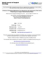
Fig.1 Immunohistochemical staining of OSCC with anti -CD68 antibodies in different
pathological grades (A) grade I, (B) grade II and (C)grade III (original magnification 100x).The
marker shows a cytoplasmic staining. The shape of staining cells include predominately round
cells, but some show spindle shape. Beside macrophage staining, in most cases pale staining of
epithelial cells detectable include tumor cells. Magnified figures (0riginal magnification 400x)
are shown in the bottom right hand corner of all figures
A
B
C
The present study found that, there is
correlation between number of macrophage
and gender. The number of macrophage were
significantaly more increased in females than
males. Although it was not significant, there
is a positive correlation between the number
of cell and the stage of tumor, where the
higher number of positive cell were in stage
IV.
The tumor-associated macrophage is a
predominant cellular component for the tumor
microenvironment in various tumors.
Although previous studies have demonstrated
3836
Int.J.Curr.Microbiol.App.Sci (2020) 9(7): 3832-3839
that TAMs are detected in OSCC [15, 16],
whether these TAMs have the M1 or M2
phenotype is poorly understood. In the present
study, we evaluated surgically dissected
specimens from 30 OSCCs using an
immunohistochemical analysis with the antiCD68 antibody, which is a marker for
macrophages
[17,
18]. The results
demonstrate that many macrophages are
detected in OSCC.
In this study, TAMs were observed in all
cases analyzed, distributed in the tumor
parenchyma, findings also observed by ElRouby (16) and Liu et al., (22), suggesting the
recruitment of these cells towards the tumor
region and the consequent ability to modify
the neoplastic process. In our study, numerous
macrophages were observed along the
invasion front, which indicates that such cells
play an important role in the recognition and
destruction of the tumor. Statically nonsignificant correlation was found with clinical
parameters except gender, where its expressed
increased in number of macrophage in female
more than males. This result disagree with, Li
C. and colleagues in 2002(19) who identified
that tumoral accumulation of macrophages is
associated with stage of invasion. Sica and
colleagues
in
2006
(20)
reported
predominance of the macrophage population
in the peritumoral infiltrate in OSCC. On
other hand, present finding was in the same
line with Kazumasa and colleagues in 2011
(21), who found non significant statistical
difference between infiltrated macrophages
and the pathological grade and stage of
OSCC. In many immunohistochemical
studies, it was observed that the number of
TAMs CD68 positive was associated with
OSCC specimen prognosis. Lu et al., (23)
found that a significantly higher number of
CD68 positive macrophages was observed in
larger tumors, recurrent, with lymph node
metastasis and advanced clinical stages. He et
al., (24) found that CD68 expression were
significantly associated with the presence of
lymph node metastasis. The reasons for these
results were difficult to explain, because
macrophage cells include M1, M2 and
undifferentiated monocytes/macrophages, it is
likely that these mixed cell populations are
functionally heterogeneous regarding the
development and progression of OSCC.
Accumulation of macrophages M1,M2 in
response to tumor cell- derived signals, either
because of tumor selection and evolution or as
part of anti-tumor responses of the host, is
diverted to pro-tumorigenic responses by
stimulate tumor growth and invasion through
enhanced cell proliferation mediated through
the production of TNFa, IL-6, and other
cytokines (21).
In conclusion the results of this study indicate
that macrophages are important cells in the
development and progression of the SCC,
since they were present in all cases analyzed
and the number of macrophage was increased
in poor differentiated cases but statistically
non-significant correlation was found
between the mean expression level of these
infiltrates with clinicopathological findings of
OSCC except with the gender where it
expressed high infiltration in female than
male. however further studies with larger
samples needed to identify their exact
correlation with clinicopathological features
of tumor.
References
1. Perdomo S, Martin Roa G, Brennan P,
Forman D, Sierra MS. Head and neck
cancer burden and preventive measures
in Central and South America. Cancer
Epidemiol. 2016; 44 Suppl 1:S43-s52.
2. Angela C. Chi., et al., Oral Cavity and
Oropharngeal
Squamous
Cell
Carcinoma-An update. A cancer J.2015;
65, 5.
3. Curry JM, Sprandio J, Cognetti D, et al.,
3837
Int.J.Curr.Microbiol.App.Sci (2020) 9(7): 3832-3839
Tumor microenvironment in head and
neck squamous cell carcinoma. Semin
Oncol. 2014; 41 (2):217-234.
4.. Gildener-Leapman N, Ferris RL, Bauman
JE.
Promising
systemic
immunotherapies in head and neck
squamous cell carcinoma. Oral Oncol.
2013;49 (12):1089-1096.
5.. Mantovani A, Allavena P. The interaction
of anticancer therapies with tumor
associated macrophages. J Exp Med.
2015;212(4):435-445.
6. Galdiero MR, Garlanda C, Jaillon S,
Marone G, Mantovani A. Tumor
associated macrophages and neutrophils
in tumor progression. J Cell Physiol.
2013; 228 (7):1404-1412.
7. Ruffell B, Affara NI, Coussens LM.
Differential macrophage programming
in the tumor microenvironment. Trends
Immunol. 2012; 33(3):119-126.
8. Hanahan D, Weinberg RA. Hallmarks of
cancer: the next generation. Cell. 2011;
144 (5):646-674.
9. Shi C, Pamer EG. Monocyte recruitment
during infection and inflammation. Nat
Rev Immunol. 2011;11(11):762–74.
doi:10.1038/ Nri3070.
10. Taylor PR, Martinez-Pomares L, Stacey
M, Lin HH, Brown GD, Gordon S.
Macrophage receptors and immune
recognition. Annu Rev Immunol. 2005;
23:901–44.
doi:10.1146/annurev.immunol.23.
021704.115816.
11. Chen JJW, Lin YC, Yao PL, Yuan A,
Chen HY, Shun CT, et al., Tumorassociated macrophages: the doubleedged sword in cancer progression. J
Clin Oncol. 2005; 23 (5):953–64.
doi:10.1200/Jco. 2005.12.172. 23.
12. Zhu XD, Zhang JB, Zhuang PY, Zhu HG,
Zhang W, Xiong YQ, et al., High
expression of macrophage colonystimulating factor in peritumoral liver
tissue is associated with poor survival
after curative resection of hepatocellular
carcinoma. J Clin Oncol. 2008; 26(16):
2707–16.
doi:10.1200/Jco.2007.15.6521.
13. Zhou J, Ding T, Pan WD, Zhu LY, Li L,
Zheng LM. Increased intratumoral
regulatory T cells are related to
intratumoral macrophages and poor
prognosis in hepatocellular carcinoma
patients.
Int
J
Cancer. 2009;
125(7):1640–8. doi:10.1002/Ijc.24556.
14. Medrek C, Ponten F, Jirstrom K,
Leandersson K. The presence of tumor
associated macrophages in tumor
stroma asaprognostic marker for breast
cancer patients. BMC Cancer. 2012; 12:
306. doi:10. 1186/1471-2407-12-306.
15.. Li, C.; Shintani, S.; Terakado, N.;
Nakashiro,
K.;
Hamakawa,
H.
Infiltration
of
tumor-associated
macrophages in human oral squamous
cell carcinoma. Oncol. Rep. 2002, 9,
1219-1223.
16. El-Rouby, D.H. Association of
macrophages with angiogenesis in oral
verrucous
and
squamous
cell
carcinomas. J. Oral Pathol. Med. 2010,
39, 559-564.
17. Mantovani, A.; Sozzani, S.; Locati, M.;
Allavena, P.; Sica, A. Macrophage
polarization:
Tumor-associated
macrophages as a paradigm for
polarized M2 mononuclear phagocytes.
Trends Immunol. 2002, 23, 549-555.
18.. Lau, S.K.; Chu, P.G.; Weiss, L.M.
CD163: A specific marker of
macrophages in paraffin embedded
tissue samples. Am. J. Clin. Pathol.
2004, 122, 794-801.
19.. Li C, Shintani S, Terakado N, Nakashiro
K, Hamakawa H. Infiltration of tumorassociated macrophages in human oral
squamous cell carcinoma. Oncol Rep
2002; 9(6): 1219-23.
20. Sica A, Schioppa T, Mantovani A,
Allavena
P.
Tumor-associated
3838
Int.J.Curr.Microbiol.App.Sci (2020) 9(7): 3832-3839
macrophages are a distinct M2
polarized population promoting tumor
progression: potential targets of anticancer therapy. Eur J Cancer 2006; 42:
717-27
21. Kazumasa M, Miki H, Jun S, Yoshihiro
O. Infiltration of M2 Tumer –
Associated Macrophages in Oral
Squamous Cell Carcinoma Correlates
With Tumer Malignancy. Cancer J
2011; 3: 3726-39.
22.. Liu SY, Chang LC, Pan LF, Hung YJ,
Lee CH, Shieh YS. Clinicopathologic
significance of tumor cell-lined vessel
and microenvironment in oral squamous
cell carcinoma. Oral Oncol. 2008 Mar;
44(3):
277-85.
PubMed PMID:
17475541.
23. Lu C, Huang CS, Tjiu JW, Chiang CP.
Infiltrating macrophage count: a
significant predictor for the progression
and prognosis of oral squamous cell
carcinomas in Taiwan. Head Neck.
2010 Jan; 32(1): 18-25. PubMed PMID:
19484765.
24. He K, Zhang L, Huang CF, et al.,
CD163+ tumor-associated macrophages
correlated with poor prognosis and
cancer stem cells in oral squamous cell
carcinoma. Biomed Res Int. 2014;
2014. PubMed PMID: 24883329.
How to cite this article:
Ahlam T. Bdewi, Ahmed A. Alkadir Mohamed Labib, Ban F. AL Drobie, Bashar H. Abdullah
and Museedi Omar. 2020. Evaluation of CD68 in Oral Squamous Cell Carcinoma and their
Relation with Clinicopathological Parameters – An Immunohistochemical Study.
Int.J.Curr.Microbiol.App.Sci. 9(07): 3832-3839. doi: />
3839