5-lipoxygenase mediates docosahexaenoyl ethanolamide and N-arachidonoyl-Lalanine-induced reactive oxygen species production and inhibition of proliferation of head and neck squamous cell
Bạn đang xem bản rút gọn của tài liệu. Xem và tải ngay bản đầy đủ của tài liệu tại đây (1.58 MB, 14 trang )
Park et al. BMC Cancer (2016) 16:458
DOI 10.1186/s12885-016-2499-3
RESEARCH ARTICLE
Open Access
5-lipoxygenase mediates docosahexaenoyl
ethanolamide and N-arachidonoyl-Lalanine-induced reactive oxygen species
production and inhibition of proliferation
of head and neck squamous cell carcinoma
cells
Seok-Woo Park1†, J. Hun Hah1,2,3†, Sang-Mi Oh1, Woo-Jin Jeong4 and Myung-Whun Sung1,2,3,5*
Abstract
Background: Endocannabinoids have recently drawn attention as promising anti-cancer agents. We previously
observed that anandamide (AEA), one of the representative endocannabinoids, effectively inhibited the proliferation
of head and neck squamous cell carcinoma (HNSCC) cell lines in a receptor-independent manner. In this study,
using HNSCC cell lines, we examined the anti-cancer effects and the mechanisms of action of docosahexaenoyl
ethanolamide (DHEA) and N-arachidonoyl-L-alanine (NALA), which are polyunsaturated fatty acid (PUFA)-based
ethanolamides like AEA.
Methods and Results: DHEA and NALA were found to effectively inhibit HNSCC cell proliferation. These
anti-proliferative effects seemed to be mediated in a cannabinoid receptor-independent manner, since the
antagonist of cannabinoid receptor-1 (CB1) and vanilloid receptor-1 (VR1), two endocannabinoid receptors, did not
reverse the ability of DHEA and NALA to induce cell death. Instead, we observed an increase in reactive oxygen
species (ROS) production and a decrease of phosphorylated Akt as a result of DHEA and NALA treatment.
Antioxidants efficiently reversed the inhibition of cell proliferation and the decrease of phosphorylated Akt induced
by DHEA and NALA; inhibition of 5-lipoxygenase (5-LO), which is expected to be involved in DHEA- and
NALA-degradation pathway, also partially blocked the ability of DHEA and NALA to inhibit cell proliferation and
phosphorylated Akt. Interestingly, ROS production as a result of DHEA and NALA treatment was decreased by
inhibition of 5-LO.
Conclusions: From these findings, we suggest that ROS production induced by the 5-LO pathway mediates the
anti-cancer effects of DHEA and NALA on HNSCC cells. Finally, our findings suggest the possibility of a new
cancer-specific therapeutic strategy, which utilizes 5-LO activity rather than inhibiting it.
Keywords: Endocannabinoid, DHEA, NALA, 5-lipoxygenase, ROS, Head and neck cancer
* Correspondence:
†
Equal contributors
1
Cancer Research Institute, Seoul National University College of Medicine,
Seoul, South Korea
2
Department of Otorhinolaryngology-Head and Neck Surgery, Seoul National
University Hospital, Seoul, South Korea
Full list of author information is available at the end of the article
© 2016 The Author(s). Open Access This article is distributed under the terms of the Creative Commons Attribution 4.0
International License ( which permits unrestricted use, distribution, and
reproduction in any medium, provided you give appropriate credit to the original author(s) and the source, provide a link to
the Creative Commons license, and indicate if changes were made. The Creative Commons Public Domain Dedication waiver
( applies to the data made available in this article, unless otherwise stated.
Park et al. BMC Cancer (2016) 16:458
Background
Endocannabinoids are endogenously-produced cannabinoids that are involved in a variety of physiological processes (including pain-sensation and memory) through
the activation of cannabinoid receptors [1]. Endocannabinoids recently gained attention because cannabis
began to be clinically used [2]. More interestingly, these
endogenous molecules have been reported to exert cytostatic, apoptotic, and anti-angiogenic effects in different
cancer cell lines and cancer xenografts [3–5].
Although the mechanistic actions of endocannabinoids
have been revealed in several cancer cell types, the exact
mechanisms underlying their anti-cancer action are still
unclear. This may be because of the complexity and variety of the signaling pathways that endocannabinoids induce, which seem to involve both receptor-dependent
and receptor-independent pathways [6, 7]. Evidence suggests that endocannabinoids might suppress cancer cell
viability through the activation of classic cannabinoid receptors such as cannabinoid receptor-1/2 (CB1 and
CB2) and vanilloid receptor-1 (VR1). However, increased
production of ceramide and reactive oxygen species
(ROS), and activation of caspase, PPARs, p38, and JNK
signaling are reported to be related to the anti-cancer
action of endocannbinoids [8–12]. New putative receptors for endocannabinoids, such as GPR55, have been recently identified, and there is a possibility that these
receptors contribute to off-target endocannabinoid effects in order to suppress cancer cell viability [13].
Since cyclooxygenase-2 (COX-2), the enzyme that produces prostanoids from arachidonic acid (AA), is well
known to be associated to cell viability in several types
of cancer [14], COX-2 has been studied as a useful
therapeutic target for the treatment of various cancers
[14, 15]. 5-Lipoxygenase (5-LO), the other enzyme
involved in AA metabolism, was reported to be overexpressed in some cancer cells [16]. Similar to COX-2, 5LO is expected to be a promising target for molecular
targeted cancer therapy because 5-LO has been identified as being related to carcinogenesis due to its ability
to promote cell proliferation and angiogenesis [17–19].
Previously, several groups observed that the cancer
cell-killing effects of anandamide (AEA) were mediated
through prostamides produced by COX-2 in some types
of cancer [20]. These findings are important for molecular targeted cancer therapy, since COX-2 has been found
to be highly expressed in many cancer cells. However,
we expected that targeting 5-LO, may be another potential therapeutic strategy. In this study, using head and
neck squamous cell carcinoma (HNSCC) cancer cells,
we investigated the precise role of AA-catabolizing
enzymes in regulating the receptor-independent anticancer effects of several endocannabinoids that are similar to AA in chemical structure. Since both 5-LO and
Page 2 of 14
COX-2 are associated with AA metabolism, we hypothesized that 5-LO might be also be related to the catabolism of some endocannabinoids, including DHEA, EPEA
and NALA, all of which are similar in structure to AA.
Although we have already analyzed and observed (especially through the induction of angiogenesis) the carcinogenic role of 5-LO in head and neck cancer cells
[17], here, we further investigated the possibility of targeting 5-LO as a possible cancer treatment.
Methods
Cell culture
SNU-1041, SNU-1066 and SNU-1076 cells (human
HNSCC cell lines) were obtained from the Korean Cell
Line Bank (Seoul National University, Seoul, Korea),
while PCI-1 (human HNSCC cell lines) was obtained
from the Pittsburgh Cancer Institute (University of
7Pittsburgh, PA) [17]. HOK 16B is an immortalized cell
from pharyngeal mucosa (a gift from Dr. Jeffrey N.
Myers in M.D. Anderson Cancer Center, University of
Texas) [21]. Cells were maintained at 37 °C in a humidified, 5 % CO2, 95 % air atmosphere and routinely subcultured using trypsin-EDTA.
Reagents
Endocannabinoids - docosahexaenoyl ethanolamide
(DHEA), eicosapentaenoyl Ethanolamide (EPEA) and Narachidonoyl-L-alanine (NALA), antagonists of CB1 and
VR1 (AM251, cay10448), antioxidants (NAC and GSH),
and inhibitors of 5-LO (AA861, zileuton and ebselen)
were obtained from Cayman Chemical (Ann Arbor, MI).
Cell proliferation assay
Cells were seeded in culture plates and incubated for the
specific time at 37 °C prior to treatment with specific
drugs for the indicated time. After treatment, Cell
Counting Kit-8 (Dojindo Lab., Tokyo, Japan) was used to
measure cell proliferation according to the manufacturer’s instructions.
Measurement of apoptosis by Annexin-V staining assay
Apoptosis of SNU-1041 and SNU-1076 by DHEA and
NALA was assessed using an Annexin-V staining kit
(Koma Biotech, Seoul, Korea). After exposure to 20 μM
of DHEA or NALA for 60 h, cells were harvested and
washed with cold PBS and re-suspended in binding buffer containing fluorescein isothiocyanate (FITC)-conjugated annexin V protein and propidium iodide. Annexin
V binding and PI staining were determined by flow cytometric analysis (Becton Dickinson, San Jose, CA, USA).
Apoptotic cells were defined as PI-negative and annexin
V-positive.
Park et al. BMC Cancer (2016) 16:458
Plasmids expressing FAAH and 5-LO
Using each cDNA, we established pcDNA3.1 expressing
vectors (pcDNA3.1-lacZ, -FAAH and -5LO). Cells were
transfected with 0.5-1 μg of plasmids by electroporation
using Microporator MP-100 (NanoEnTek Inc., Seoul,
South Korea), following the protocol provided by the
manufacturer. Then, cells were seeded in culture plates
and incubated for an additional 36 h before another
treatment of AEA.
Transfection of siRNA
Individual siRNAs against COX-2 (D-004557-04), 5-LO
(L-004530-00) and non-targeting control (D-001210-01)
were obtained from Dharmacon RNA Technologies
(Lafayette, CO). The best conditions of siRNAs application (used doses and treatment time) were established
beforehand by western blotting and EIA [17]. Cells were
transfected with 200 nM of siRNA by electroporation
using Microporator MP-100 (NanoEnTek Inc., Seoul,
South Korea), following the protocol provided by the
manufacturer. Then, cells were seeded in culture plates
and incubated for an additional 48 h before another
treatment of tested drugs (like DHEA).
Quantification of PGE2 and LTB4 production
The amount of the desired factor released by the cells
was determined using PGE2 or LTB4 enzyme immunoassay kits (EIA) (Cayman Chemical, Ann Arbor, MI) according to the manufacturer’s instructions.
Cell co-culture with transwell system
SNU-1041 cells were transfected with 200 nM of siRNA
against 5-LO or non-targeting control and placed at
once in the lower side of a transwell (NUNC Company,
Denmark) chamber partitioned by a polycarbonate
membrane (8.0 μm pore size, Corning Incorporated,
Costar). Then SNU-1041 cells (with no transfection)
were seeded in the upper side and co-cultured for 48 h.
Subsequently, cells were treated with 30 μM of DHEA
or NALA for additional 48 h. Both cells (in upper and
lower side) were separately applied to the cell proliferation assay (at a total of 96 h).
Measurements of production of reactive oxygen species
(ROS)
The generation of ROS was measured by using the
DCFH2-DA assay [22]. Intracellular ROS production was
determined directly in cell monolayers in black 96-well
flat-bottom microtiter plates using a Fluoroskan Ascent
FL microplate reader (Labsystems, Sweden). Cells in
complete medium were incubated with the indicated
drugs for 18 h. To measure the production of ROS, cells
were treated with 5 μM DCFH2-DA at 37 °C for 30 min,
and the fluorescence of DCF was measured at 530 nm
Page 3 of 14
after excitation at 485 nm (DCFH2-DA, after deacetylation to DCFH2, is oxidized intracellularly to its fluorescent derivative DCF). Assays were performed in
modified Hank’s buffered salt solution (HBSS).
Western blot analysis
Denatured protein lysates were resolved by 4–12 %
NuPAGE gels (Invitrogen, Carlsbad, CA) and transferred
to nitrocellulose membranes (Schleicher & Schuell,
Dachen, Germany). The membranes were incubated
with anti-5-LO (BD, Franklin Lakes, NJ); anti-p-Akt
(Ser473), anti-pan-Akt (Cell signaling, Danvers, MA); or
monoclonal anti-β-actin (Santa Cruz Biotechnology,
Santa Cruz, CA) for 2 h at room temperature or overnight at 4 °C. Membranes were then washed (4 times)
with TBS-T and incubated with horseradish peroxidaseconjugated secondary antibody (Pierce, Rockford, IL) for
1 h. Immunoreactive proteins were visualized by developing them with Lumi-light western blotting substrate
(Roche Diagnostics GmbH, Mannheim, Germany),
followed by exposure in a LAS-3000 (Fuji Film Co.,
Tokyo, Japan) according to the manufacturer’s instructions. This was followed by quantitation of specific
bands with the Multi Gauge software (Fuji Film Co.,
Tokyo, Japan).
Statistical analysis
Data are presented as the mean ± standard deviation
(SD) of at least triplicates, or as a representative of 3
separate experiments. Significance was determined between treated and untreated groups by two-sided Student’s t-test. P values <0.05 were considered statistically
significant.
Results
DHEA and NALA effectively inhibit the proliferation of
HNSCC cell lines
DHEA and NALA effectively inhibited cell viability in
the HNSCC cell lines we tested, but EPEA only had a
weak inhibitory effect on cancer cell proliferation
(Fig. 1a). Non-cancerous cell lines (HOK16B and fibroblasts) were not affected by DHEA and NALA at the
tested doses (10-30 μM) (Fig. 1a). DHEA and NALA effectively induced the cell death in the HNSCC cell lines
(Fig. 1b). CB1 is expressed only in SNU-1066 and no expression of CB2 is observed in all the cells tested, while
VR1 expression is observed in all cells (in our own
study) [23]. We also found that the anti-cancer effect of
DHEA and NALA was not reversed by antagonists of
the endocannabinoid receptors CB1 and VR1 (AM251
and cay10448) (Fig. 1c). From these observations, we assumed that the anti-cancer effect induced by DHEA and
NALA was mediated through a receptor-independent
action. The cell lines SNU-1041 and SNU-1076 were
Park et al. BMC Cancer (2016) 16:458
Fig. 1 (See legend on next page.)
Page 4 of 14
Park et al. BMC Cancer (2016) 16:458
Page 5 of 14
(See figure on previous page.)
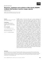
Fig. 1 DHEA and NALA effectively inhibit cell proliferation and induce cell death in HNSCC cell lines. a Cells were treated with 20 μM of DHEA,
EPEA and NALA. At 72 h, cells were subjected to cell proliferation assay. b SNU-1041 and SNU-1076 were treated with 20 μM of DHEA and NALA.
At 60 h, cells were subjected to Annexin-V staining assay. c SNU-1041 and SNU-1076 were treated with DHEA (20 μM) and NALA (20 μM) plus
AM251 (2 μM) or cay10448 (2 μM). At 72 h, cells were subjected to cell proliferation assay. Results are expressed as a percentage relative to
control (% of control). P values were based on comparison with control (*P < 0.001, **P < 0.05) or DHEA/NALA-treated group (#P < 0.05)
chosen for further analysis of the cancer-killing effect of
DHEA and NALA.
The anti-cancer action of DHEA and NALA occurs at an
intracellular location
FAAH is known to catabolize polyunsaturated fatty acidbased endocannabinoids (like AEA) to polyunsaturated
fatty acid and ethanolamide [24]. To verify the possibility
that DHEA and NALA affected cell viability through a
receptor-independent action that occurred after intracellular transport, cells were transfected with plasmids expressing fatty acid amide hydrolase (FAAH). The activity
of transfected FAAH was confirmed by using arachidonoyl p-nitroaniline-based assay (Additional file 1: Figure
S1). We observed that the growth-inhibitory action of
DHEA and NALA was completely blocked (Fig. 2).
These observations suggested that DHEA and NALA
might have anti-cancer effect through intracellular
localization by a receptor-independent mechanism in
HNSCC cell lines. The used cells in this study had little
FAAH activity (data not shown).
Anti-cancer effect of DHEA and NALA was reversed by
inhibition of 5-LO, but not by inhibition of COX-2
AEA, which is structurally similar to AA, has been reported to have an anti-cancer effect when it is catabolized by COX-2 [20]. Therefore, we hypothesized that
the mechanism by which DHEA and NALA inhibited
cell proliferation might also be a result of their catabolism by COX-2. However, we found that inhibition of
COX-2 had no effect on the ability of DHEA and NALA
to inhibit cell proliferation of HNSCC (Additional file 2:
Figure S2). Next, we tried to investigate if 5-LO might
regulate the ability of DHEA and NALA to inhibit cell
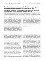
Fig. 2 The anti-cancer action of DHEA and NALA occurs at an intracellular location. Plasmids (1 μg) expressing LacZ and FAAH were transfected
into (a) SNU-1041 and (b) SNU-1076 (LacZ expressing plasmid was used for controls). Thirthy-six hours later cells were treated with the indicated
concentrations (μM) of DHEA or NALA. At additional 48 h, cells were subjected to cell proliferation assay. Results are expressed as a percentage
relative to control (% of control). P values are based on a comparison with DHEA-treated group and NALA-treated group in LacZ
(*P < 0.001, #P < 0.005)
Park et al. BMC Cancer (2016) 16:458
Fig. 3 (See legend on next page.)
Page 6 of 14
Park et al. BMC Cancer (2016) 16:458
Page 7 of 14
(See figure on previous page.)
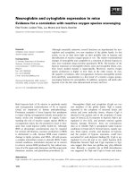
Fig. 3 Anti-cancer effect of DHEA and NALA was reversed by inhibition of 5-LO, but not by inhibition of COX-2. (a) SNU-1041 and (b) SNU-1076 were
treated with DHEA or NALA (20 μM) plus AA861 (5 μM) or zileuton (5 μM) or Ebselen (5 μM). At 72 h, cells were subjected to cell proliferation assay
(Left). The siRNA of 5-LO was transfected at 200 nM doses (the si-NC was used for negative control of siRNA). Forty-eight hours later cells were treated
with DHEA or NALA (20 μM). At additional 48 h, cells were subjected to cell proliferation assay (Right). Results are expressed as a percentage relative to
control (% of control). P values were based on comparison with DHEA-treated group and NALA-treated group (*P < 0.005, #P < 0.01). c 5-LO siRNA was
transfected into SNU-1076 cells. At 48 h, total cell lysates were prepared and the expression of 5-LO was determined by western blotting (upper). Data
are presented as a representative of 3 separate experiments. At 48 h, cells were treated with 20 μM of arachidonic acid. After an additional 2 h, cultured
media were collected and applied to LTB4 EIA (lower). The inhibitory effect of 5-LO siRNA was compared with that of 5-LO inhibitors – AA861 and
zileuton. Results are expressed as a percentage relative to the control (% of control)
proliferation. The high expression and activity of 5-LO
in HNSCC cells were already measured in our previous
study [17]. Cells were treated with 5-LO inhibitors
(AA861, zileuton, and ebselen) and 5-LO siRNAs together with DHEA or NALA before cell proliferation
was measured. We were able to demonstrate that 5-LO
mediated the growth-inhibitory actions of DHEA and
NALA in SNU-1041 (Fig. 3a) as well as in SNU-1076
(Fig. 3b). The inhibition of 5-LO activity by its inhibitors
and by its siRNA was confirmed by using an leukotriene
B4 (LTB4) EIA (Fig. 3c).
The anti-cancer effects of DHEA and NALA are not mediated by any products generated by the 5-LO pathway
Because of the structural similarity between AA and
DHEA/NALA, we could detect weak LTB4-like products
synthesized by 5-LO from DHEA and NALA using an
LTB4 EIA kit (Fig. 4a). However, when cells transfected
with siRNAs of negative control (NC) or 5-LO were cocultured with cells in upper side (with basic condition)
and treated with DHEA and NALA, we observed that
cell viability was partially reversed only in 5-LO siRNAtransfected cells (Fig. 4b).
Together with Fig. 5, this finding confirms that DHEA-/
NALA-induced ROS might play a role in the anti-cancer
effect of DHEA and NALA on HNSCC cells. In addition,
we found that 5-LO siRNAs blocked the increase of
DHEA/NALA-induced ROS production in SNU-1041
and SNU-1076 (Fig. 6b).
5-LO-induced ROS mediates the decrease of
phosphorylated Akt by DHEA and NALA
It was already known that Akt activity is important in
maintaining the cell viability of several cancer cells, including HNSCC cells [25, 26]. To identify the role of increased ROS in the ability of DHEA/NALA to affect the
phosphorylated form of Akt in HNSCC cells, we treated
SNU-1041 with DHEA/NALA and the antioxidants
NAC. DHEA and NALA decreased the phosphorylated
form of Akt and the antioxidants reversed DHEA/
NALA-inhibited p-Akt in SNU-1041 (Fig. 7a). In
addition, we found that 5-LO inhibition by siRNAs reversed the decrease of DHEA/NALA-inhibited p-Akt in
SNU-1041 (Fig. 7b).
DHEA and NALA increase ROS production
Exogenous transfection of plasmids expressing 5-LO promotes the anti-cancer action of DHEA and NALA in
HNSCC cells
In our own study, we observed that AEA increased
intracellular oxidative stress, including lipid peroxidation
[23]. Since DHEA and NALA are very similar to AEA,
we assumed that DHEA and NALA might affect cell viability by increasing intracellular ROS production. As expected, we observed an increase in ROS production as a
result of DHEA and NALA treatment in SNU-1041
(Fig. 5a) and SNU-1076 (Fig. 5b). These data suggest
that ROS production induced by DHEA and NALA
seems to be involved in mediating the anti-cancer effects
of DHEA and NALA in HNSCC cells.
Finally, we investigated the effect of enhanced 5-LO activity on the anti-cancer action of DHEA and NALA in
SNU-1041. Transfecting cells with plasmids expressing
5-LO, we observed that the growth-inhibitory activity of
DHEA and NALA significantly improved with increasing
5-LO expression (Fig. 8a). The expression of transfected
5-LO was verified by western blotting (Fig. 8b). In
addition, ROS production in the presence of DHEA or
NALA increased proportionally with expression of 5LO, which was more prominently than in the presence
of AA (the basic substrate of 5-LO pathway) (Fig. 8c).
5-LO inhibition as well as antioxidant treatment partially
reversed DHEA- and NALA-inhibited cell proliferation
Discussion
Since psychotropic side effects by cannabis are reported
to be mediated by classic cannabinoid receptors [1],
there might be some concern about the idea of adopting
endocannabinoids as a cancer treatment. However, it has
been also reported that the cell-killing effect of several
endocannabinoids is mediated by cannabinoid receptor-
Next, to identify the role of increased ROS in the ability
of DHEA and NALA to inhibit cell proliferation, we
treated SNU-1041 with DHEA/NALA and the antioxidants NAC and GSH. The antioxidants partially reversed
DHEA-/NALA-inhibited cell proliferation (Fig. 6a).
Park et al. BMC Cancer (2016) 16:458
Page 8 of 14
Fig. 4 The anti-cancer effects of DHEA and NALA are not mediated by any products generated by the 5-LO pathway. a SNU-1041 and SNU-1076
were treated with AA, AEA, DHA, DHEA and NALA (20 μM). At 4 h, cells were subjected to the LTB4 EIA. Results are expressed as a percentage
relative to control (% of control). b SNU-1041 cells were transfected with 200 nM of siRNA against 5-LO or si-NC and placed at once in the lower
side of a transwell chamber. Then SNU-1041 cells (with no transfection) were seeded in the upper part and co-cultured for 48 h. Subsequently,
cells were treated with 30 μM of DHEA or NALA for additional 48 h. Both cells (in upper and lower side) were separately applied to the cell
proliferation assay. Results are expressed as a percentage relative to control (% of control). P values were based on comparison with control
(*P < 0.01, **P < 0.05) or DHEA/NALA-treated group (#P < 0.005, ##P < 0.05)
independent mechanisms [6, 7, 23]. In addition to classic
receptors like CB1 and CB2, GPR55 and GPR35 were recently reported as putative receptors of endocannabinoids [13, 27]. Given these observations, it might be
possible to find a way to avoid the psychotropic side effects of endocannabinoids and use them as
chemotherapeutic agents. In our study, we hoped to find
a CB receptor-independent effect of the endocannabinoids in order to develop them as new cancer therapeutics without psychotropic side effects.
Although DHEA was reported to activate classic cannabinoid receptors [6], the anti-cancer action of DHEA
Park et al. BMC Cancer (2016) 16:458
Page 9 of 14
Fig. 5 DHEA and NALA increase ROS production. a SNU-1041 and (b) SNU-1076 were treated with the indicated concentrations (μM) of DHEA
and NALA. At 18 h, cells were subjected to the DCFH2-DA assay to measure the change of ROS level. Results are expressed as a percentage
relative to control (% of control). P values were based on comparison with control (*P < 0.001, **P < 0.005, #P < 0.01)
seemed to be mediated by receptor-independent pathways in our study, since antagonists of cannabinoid receptors had no effect on it. Our observation of the
perfect reversal of the anti-cancer effect of DHEA and
NALA by transfecting FAAH into HNSCC cells confirms that DHEA and NALA can be degraded by FAAH.
The fact that COX-2 and 5-LO are highly expressed in
cancer cells than in non-cancerous cells suggests that
they might be useful molecular targets for cancer therapy [18, 28]. Their inhibition has been shown to have efficient suppressive effects on cancer cell viability in
several types of cancer, such as colon cancer [14, 19]. In
our previous study using HNSCC cells, we observed little anti-proliferative effect by inhibiting COX-2 and 5LO directly [29]. However, in this study, we observed
that COX-2 and/or 5-LO activity might be able to
promote the cell-killing action induced by some endocannabinoids. This observation suggests that COX-2
and/or 5-LO might be used as specific targets for cancer
therapy in ways other than simply inhibiting their activities. Indeed, we identified that DHEA and NALA were
able to kill HNSCC cells through 5-LO-mediated ROS
production in a receptor-independent manner, even
though HNSCC cells might have expression of their receptors such as CB1 and/or VR1.
Until now, it has not been reported that endocannabinoids like DHEA and NALA might be the substrates for
5-LO, even though various polyunsaturated fatty acid
(PUFAs) like DHA are known to be degraded by 5-LO
[30]. We could efficiently detect LTB4-like products generated from DHA and AEA by 5-LO, but could only detect low levels of the products from DHEA and NALA
(Fig. 4a). Since SNU-1041 and SNU-1076 have little
FAAH activity, we assumed that we could detect LTB4like products directly generated from DHEA and NALA,
not those from DHA and AA converted by FAAH.
In cell co-culture experiment, we observed that inhibition of cell viability by DHEA and NALA treatment was
partially reversed in 5-LO siRNA-transfected cells of
lower side (Fig. 4c and d). It means that the anti-cancer
effects of DHEA and NALA are not mediated by LTB4like products generated by the 5-LO pathway but mediated by other mechanisms such as ROS production,
which should be induced through the processes of oxygenation and peroxidation by 5-LO. If any end-products
of 5-LO released to culture medium showed cell killing
Park et al. BMC Cancer (2016) 16:458
Page 10 of 14
Fig. 6 5-LO inhibition as well as antioxidant treatment partially reversed DHEA- and NALA-inhibited cell proliferation. a Cells were treated with
20 μM of DHEA and NALA plus NAC (1 mM) or GSH (2 mM). At 72 h, cells were subjected to cell proliferation assay. Results are expressed as a
percentage relative to control (% of control). P values were based on comparison with DHEA-treated group and NALA-treated group (*P < 0.001,
#
P < 0.01). b Cells were transfected at 200 nM doses of 5-LO siRNA (the siNC was used for negative control of siRNA). Forty-eight hours later cells
were treated with DHEA or NALA (20 μM). At additional 18 h, cells were subjected to the DCFH2-DA assay to measure the change of ROS level.
Results are expressed as a percentage relative to control (% of control). P values were based on comparison with DHEA-treated group and
NALA-treated group in siNC (*P < 0.01, #P < 0.05)
action, 5-LO siRNA-transfected cells in lower chamber
should have been killed as well.
Other studies also observed the increase of intracellular
oxidative stress during AA metabolism, independently of
produced eicosanoids [31, 32]. Furthermore, 5-LO activating protein (FLAP) and leukotriene C4 (LTC4) synthase
are included in the membrane associated proteins in the
eicosanoid and glutathione metabolism (MAPEG) superfamily related with glutathione-dependent catalysis [33].
FLAP and LTC4 synthase might cause glutathione depletion (which leads to increased ROS) in the conversion of
AA to leukotrienes by 5-LO [34].